Embed Size (px)
Citation preview
© Copyright 2013 Elsevier, Ltd. All rights reserved.
1 Pain
CHAPTER CONTENTS
Definition of pain 3
Perception and modulation of pain 3
Peripheral nociceptive system. . . . . . . . . . . . . . 4Afferent nociceptive system . . . . . . . . . . . . . . . 4Pain modulation systems . . . . . . . . . . . . . . . . 5
Referred pain 6
Introduction . . . . . . . . . . . . . . . . . . . . . . . 6Possible mechanisms . . . . . . . . . . . . . . . . . . 6Clinical consequences . . . . . . . . . . . . . . . . . . 7Rules of referred pain . . . . . . . . . . . . . . . . . . 7Dermatomes . . . . . . . . . . . . . . . . . . . . . . . 9Discrepancies between dermatomes and myotomes . . . . . . . . . . . . . . . . . . . . . . . 14Referred pain in visceral diseases. . . . . . . . . . . 15Referred pain is felt deeply and distally in the dermatome. . . . . . . . . . . . . . . . . . . . . . . 15Segmentally referred pain does not cross the midline . . . . . . . . . . . . . . . . . . . . . . . 16Dura mater an ‘exception’ to segmental reference . . . . . . . . . . . . . . . . . . . . . . . . 16Referred tenderness . . . . . . . . . . . . . . . . . . 16Factors determining reference of pain . . . . . . . . 18
Definition of pain
Pain is the presenting symptom in almost every orthopaedic patient. A complaint of pain is always indicative of some variety or degree of dysfunction1 and results from a combination of physical and psychological causes, although sometimes one or the other predominates. All pain must be regarded as real. Pain entirely devoid of somatic cause is labelled ‘psychogenic pain’:
although no peripheral tissue damage exists, the pain is just as distressing as somatic pain2 (see Section 16).
The taxonomy committee of the International Association for the Study of Pain defined pain as: ‘an unpleasant sensory and emotional experience associated with actual or potential tissue damage, or described in terms of such damage’.3 Pain is thus not a ‘primary sensation’ in the sense that smell, taste, touch, vision and hearing are, but is an ‘emotional state’, like sorrow, love or hate. The consequence is that it is extremely difficult to explain one’s pain to another person. This is reflected in the numerous words that patients use to describe intensity and quality of pain: twinge, ache, distress, discomfort, soreness, cramp, suffering, misery, agony, torment, anguish.4 The fact that pain is always a subjective experience provides the first difficulty in its use in diagnosis. The language used is not always easy to understand, and the examiner usually needs a high level of competence and understanding to translate patients’ subjective descriptions into more objective and useful statements.
However, unlike the other affective states, pain is always felt in some particular part of the body. Having said this, the localization of the pain very often lacks precision, and it is often experienced at some distance from its source – ‘referred pain’. This constitutes the second problem in using the symptom of pain as a diagnostic aid.
Perception and modulation of pain
The intensity of pain does not depend only on the intensity of irritation of the peripheral nociceptive system (receptors and their afferents). Centripetal transmission of peripheral nociceptive stimulation is subject to varying degrees of facilitatory and inhibitory modulation at different synapses during its course to the cerebral cortex. An important modulation site, of major concern to the orthopaedic physician, is the gateway synapse in the basal spinal nucleus, but there are also
General Principles
4
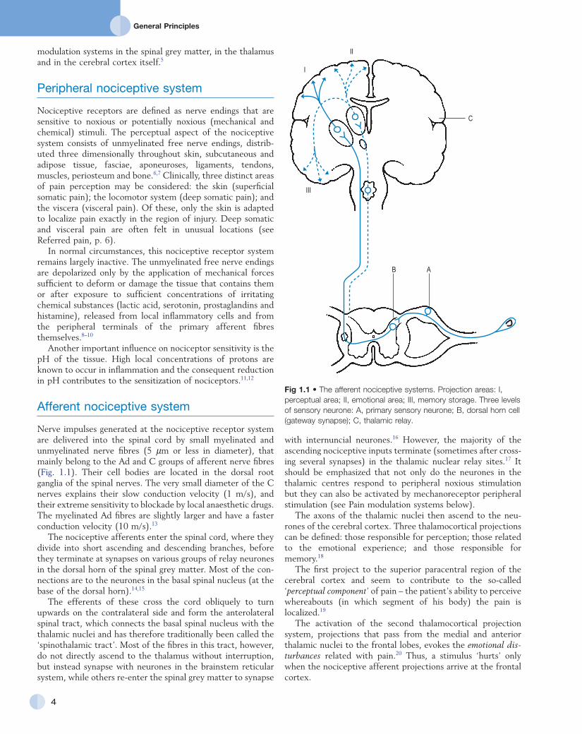
with internuncial neurones.16 However, the majority of the ascending nociceptive inputs terminate (sometimes after crossing several synapses) in the thalamic nuclear relay sites.17 It should be emphasized that not only do the neurones in the thalamic centres respond to peripheral noxious stimulation but they can also be activated by mechanoreceptor peripheral stimulation (see Pain modulation systems below).
The axons of the thalamic nuclei then ascend to the neurones of the cerebral cortex. Three thalamocortical projections can be defined: those responsible for perception; those related to the emotional experience; and those responsible for memory.18
The first project to the superior paracentral region of the cerebral cortex and seem to contribute to the socalled ‘perceptual component’ of pain – the patient’s ability to perceive whereabouts (in which segment of his body) the pain is localized.19
The activation of the second thalamocortical projection system, projections that pass from the medial and anterior thalamic nuclei to the frontal lobes, evokes the emotional dis-turbances related with pain.20 Thus, a stimulus ‘hurts’ only when the nociceptive afferent projections arrive at the frontal cortex.
modulation systems in the spinal grey matter, in the thalamus and in the cerebral cortex itself.5
Peripheral nociceptive system
Nociceptive receptors are defined as nerve endings that are sensitive to noxious or potentially noxious (mechanical and chemical) stimuli. The perceptual aspect of the nociceptive system consists of unmyelinated free nerve endings, distributed three dimensionally throughout skin, subcutaneous and adipose tissue, fasciae, aponeuroses, ligaments, tendons, muscles, periosteum and bone.6,7 Clinically, three distinct areas of pain perception may be considered: the skin (superficial somatic pain); the locomotor system (deep somatic pain); and the viscera (visceral pain). Of these, only the skin is adapted to localize pain exactly in the region of injury. Deep somatic and visceral pain are often felt in unusual locations (see Referred pain, p. 6).
In normal circumstances, this nociceptive receptor system remains largely inactive. The unmyelinated free nerve endings are depolarized only by the application of mechanical forces sufficient to deform or damage the tissue that contains them or after exposure to sufficient concentrations of irritating chemical substances (lactic acid, serotonin, prostaglandins and histamine), released from local inflammatory cells and from the peripheral terminals of the primary afferent fibres themselves.8–10
Another important influence on nociceptor sensitivity is the pH of the tissue. High local concentrations of protons are known to occur in inflammation and the consequent reduction in pH contributes to the sensitization of nociceptors.11,12
Afferent nociceptive system
Nerve impulses generated at the nociceptive receptor system are delivered into the spinal cord by small myelinated and unmyelinated nerve fibres (5 µm or less in diameter), that mainly belong to the Ad and C groups of afferent nerve fibres (Fig. 1.1). Their cell bodies are located in the dorsal root ganglia of the spinal nerves. The very small diameter of the C nerves explains their slow conduction velocity (1 m/s), and their extreme sensitivity to blockade by local anaesthetic drugs. The myelinated Ad fibres are slightly larger and have a faster conduction velocity (10 m/s).13
The nociceptive afferents enter the spinal cord, where they divide into short ascending and descending branches, before they terminate at synapses on various groups of relay neurones in the dorsal horn of the spinal grey matter. Most of the connections are to the neurones in the basal spinal nucleus (at the base of the dorsal horn).14,15
The efferents of these cross the cord obliquely to turn upwards on the contralateral side and form the anterolateral spinal tract, which connects the basal spinal nucleus with the thalamic nuclei and has therefore traditionally been called the ‘spinothalamic tract’. Most of the fibres in this tract, however, do not directly ascend to the thalamus without interruption, but instead synapse with neurones in the brainstem reticular system, while others reenter the spinal grey matter to synapse
Fig 1.1 • The afferent nociceptive systems. Projection areas: I, perceptual area; II, emotional area; III, memory storage. Three levels of sensory neurone: A, primary sensory neurone; B, dorsal horn cell (gateway synapse); C, thalamic relay.
B A
C
II
I
III
C H A P T E R 1Pain
5
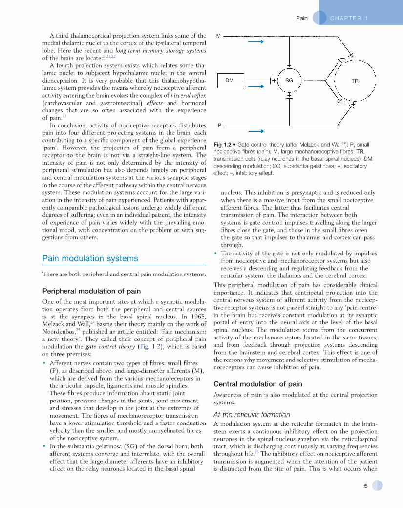
nucleus. This inhibition is presynaptic and is reduced only when there is a massive input from the small nociceptive afferent fibres. The latter thus facilitates central transmission of pain. The interaction between both systems is gate control: impulses travelling along the larger fibres close the gate, and those in the small fibres open the gate so that impulses to thalamus and cortex can pass through.
• The activity of the gate is not only modulated by impulses from nociceptive and mechanoreceptor systems but also receives a descending and regulating feedback from the reticular system, the thalamus and the cerebral cortex.
This peripheral modulation of pain has considerable clinical importance. It indicates that centripetal projection into the central nervous system of afferent activity from the nociceptive receptor systems is not passed straight to any ‘pain centre’ in the brain but receives constant modulation at its synaptic portal of entry into the neural axis at the level of the basal spinal nucleus. The modulation stems from the concurrent activity of the mechanoreceptors located in the same tissues, and from feedback through projection systems descending from the brainstem and cerebral cortex. This effect is one of the reasons why movement and selective stimulation of mechanoreceptors can cause inhibition of pain.
Central modulation of painAwareness of pain is also modulated at the central projection systems.
At the reticular formationA modulation system at the reticular formation in the brainstem exerts a continuous inhibitory effect on the projection neurones in the spinal nucleus ganglion via the reticulospinal tract, which is discharging continuously at varying frequencies throughout life.26 The inhibitory effect on nociceptive afferent transmission is augmented when the attention of the patient is distracted from the site of pain. This is what occurs when
A third thalamocortical projection system links some of the medial thalamic nuclei to the cortex of the ipsilateral temporal lobe. Here the recent and long-term memory storage systems of the brain are located.21,22
A fourth projection system exists which relates some thalamic nuclei to subjacent hypothalamic nuclei in the ventral diencephalon. It is very probable that this thalamohypothalamic system provides the means whereby nociceptive afferent activity entering the brain evokes the complex of visceral reflex (cardiovascular and gastrointestinal) effects and hormonal changes that are so often associated with the experience of pain.23
In conclusion, activity of nociceptive receptors distributes pain into four different projecting systems in the brain, each contributing to a specific component of the global experience ‘pain’. However, the projection of pain from a peripheral receptor to the brain is not via a straightline system. The intensity of pain is not only determined by the intensity of peripheral stimulation but also depends largely on peripheral and central modulation systems at the various synaptic stages in the course of the afferent pathway within the central nervous system. These modulation systems account for the large variation in the intensity of pain experienced. Patients with apparently comparable pathological lesions undergo widely different degrees of suffering; even in an individual patient, the intensity of experience of pain varies widely with the prevailing emotional mood, with concentration on the problem or with suggestions from others.
Pain modulation systems
There are both peripheral and central pain modulation systems.
Peripheral modulation of painOne of the most important sites at which a synaptic modulation operates from both the peripheral and central sources is at the synapses in the basal spinal nucleus. In 1965, Melzack and Wall,24 basing their theory mainly on the work of Noordenbos,25 published an article entitled: ‘Pain mechanism: a new theory’. They called their concept of peripheral pain modulation the gate control theory (Fig. 1.2), which is based on three premises:
• Afferent nerves contain two types of fibres: small fibres (P), as described above, and largediameter afferents (M), which are derived from the various mechanoreceptors in the articular capsule, ligaments and muscle spindles. These fibres produce information about static joint position, pressure changes in the joints, joint movement and stresses that develop in the joint at the extremes of movement. The fibres of mechanoreceptor transmission have a lower stimulation threshold and a faster conduction velocity than the smaller and mostly unmyelinated fibres of the nociceptive system.
• In the substantia gelatinosa (SG) of the dorsal horn, both afferent systems converge and interrelate, with the overall effect that the largediameter afferents have an inhibitory effect on the relay neurones located in the basal spinal
Fig 1.2 • Gate control theory (after Melzack and Wall24): P, small nociceptive fibres (pain); M, large mechanoreceptive fibres; TR, transmission cells (relay neurones in the basal spinal nucleus); DM, descending modulation; SG, substantia gelatinosa; +, excitatory effect; –, inhibitory effect.
M
P
DM TRSG
General Principles
6
another painful site elsewhere in the body is stimulated (counterirritation), when the patient concentrates on work or other activities or when hypnosis is induced.27 The inhibitory effect of this reticular system also increases when the blood concentration of catecholamines is very high, as can be the case in states of great emotional tension.28 Also some drugs (chlorpromazine, diazepam and morphine) may selectively increase the activity of the reticular neurones that operate this inhibitory system.29
Inhibitory reticular activity is depressed and pain is enhanced when attention is concentrated on the painful site, or following the administration of barbiturates, caffeine or theophylline.30
The cerebral cortexThe cerebral cortex, especially the sectors located in the frontal and paracentral regions, in turn regulates the activity of the reticular formation. Reticular activity is increased, and perception of pain thus inhibited during rest and sleep and after the ingestion of alcohol. Conversely, depression of reticular activity is seen during increasing cortical activity, for example with anxiety, uncertainty and fear.
Referred pain
Introduction
When the skin is pricked with a pin, the patient can exactly pinpoint the injury. This ability to localize the pain is limited to skin and does not apply when the source of the pain is in deep tissue. Deep somatic pain and visceral pain are often felt far from their point of source. In consequence, the examiner needs to know the patterns of pain reference so as not to be misled about where to search for the seat of the trouble. Diagnosis of orthopaedic lesions often rests entirely on history and clinical examination and is therefore almost impossible if the rules and conditions relevant to referred pain are not clearly understood.
Those who originally studied pain reference soon noted that although it appeared erroneous and anarchic, some rules of presentation did exist. For instance, pain from specific structures is always referred to the same parts of the body: colic from a ureteral stone to groin and testicle, diaphragmatic disorders typically to the shoulder, angina pectoris to one or both arms, and the pain caused by arthritis of a hip very often to the ipsilateral knee. Also pain is, in the main, referred distally and its localization depends in a certain way on the severity of the lesion.
In 1905, Sir Henry Head described referred pain in the abdominal wall caused by a visceral disease.31 Using the dermatological appearances in herpes zoster, he constructed schemes of segmental innervation of the skin.32 He also described dermatomic zones that became painful in the event of provocation of a related visceral structure. His theory of pain reference was built on the concept of the segmental organization of the human body and its nociceptive system. Further experiments in this sphere were conducted by Sir Thomas Lewis in 1936.33 In 1938 and 1939, Kellgren published
the results of a systematic examination of the phenomena of referred pain, demonstrating segmental radiation and failure to cross the midline.34,35
His experiments were confirmed by others.36–38 Later, the concept of segmental reference of pain was refined39,40 and exact borders of the different dermatomes mapped out.41–44
Possible mechanisms
The fact that referred pain is an error in perception was first pointed out by John Hunter in 1835 (cited by Cyriax).45 It was obvious that if pain is felt elsewhere than at its true site, the nociceptive mechanism is reacting inappropriately. However, since there seems to be logical consistency in the way the errors are made (pain from specific lesions is always referred to the same areas), there must also be a logical explanation for ‘failures’. (If a machine always makes the same mistake, a structural or functional disorder must exist.) The basis for the inadequacy must therefore be sought in a miscalculation in the pain mechanism. Theoretically, the defect can lie anywhere along the afferent pathway, from the peripheral receptors to the synapses in the spinal cord and the reticular area and projection zones in the sensory cortex.
During the last century, numerous investigators have studied referred pain. Two main hypotheses have been put forward:
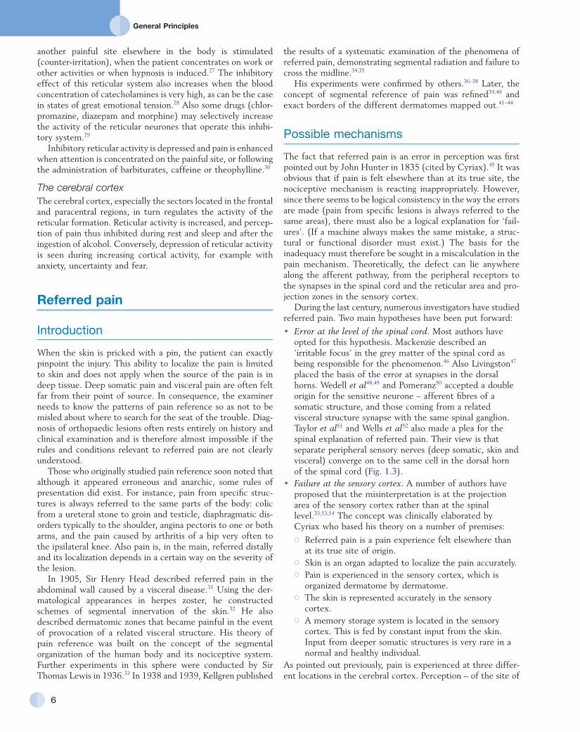
• Error at the level of the spinal cord. Most authors have opted for this hypothesis. Mackenzie described an ‘irritable focus’ in the grey matter of the spinal cord as being responsible for the phenomenon.46 Also Livingston47 placed the basis of the error at synapses in the dorsal horns. Wedell et al48,49 and Pomeranz50 accepted a double origin for the sensitive neurone – afferent fibres of a somatic structure, and those coming from a related visceral structure synapse with the same spinal ganglion. Taylor et al51 and Wells et al52 also made a plea for the spinal explanation of referred pain. Their view is that separate peripheral sensory nerves (deep somatic, skin and visceral) converge on to the same cell in the dorsal horn of the spinal cord (Fig. 1.3).
• Failure at the sensory cortex. A number of authors have proposed that the misinterpretation is at the projection area of the sensory cortex rather than at the spinal level.33,53,54 The concept was clinically elaborated by Cyriax who based his theory on a number of premises: Referred pain is a pain experience felt elsewhere than
at its true site of origin. Skin is an organ adapted to localize the pain accurately. Pain is experienced in the sensory cortex, which is
organized dermatome by dermatome. The skin is represented accurately in the sensory
cortex. A memory storage system is located in the sensory
cortex. This is fed by constant input from the skin. Input from deeper somatic structures is very rare in a normal and healthy individual.
As pointed out previously, pain is experienced at three different locations in the cerebral cortex. Perception – of the site of
C H A P T E R 1Pain
7
correct what is seen (provided the observer knows the correction formula) and so locate the object accurately. The same applies to referred pain. The examiner must constantly ask if the localization of the pain is also the exact localization of the disorder and, if the answer is negative, what corrections must be made to arrive at the exact localization.
Before this discussion of referred pain is continued, it should be stressed that root pain reference does not necessarily mean that a nerve is involved. The false idea that wide radiation of pain is evidence of involvement of nerves is still strongly held by some and is usually the most important obstacle to a logical understanding of referred pain. To approach the problem of referred pain with an open mind, the reader must constantly remember that referred pain is an error of perception. Although the nerve supply to these peripheral structures is distributed on a segmental basis, it does not indicate that referred pain ‘runs down’ a somatic nerve. For instance, pain at the anterior aspect of the leg does not necessarily mean that a nerve structure (L3, femoral nerve or peripheral branches of the femoral nerve) is involved. Although inflammation of the dural sleeve of the L3 nerve root does of course lead to pain extending in the L3 dermatome, the same pain can be provoked by a lesion in any other tissue belonging to the L3 segment (e.g. hip joint or psoas bursa). There will not be any difference in the nature and extent of the pain. The only distinction between pain as the result of a compressed and inflamed nerve root and pain originating from trauma to other structures is the appearance of paraesthesia (see Pressure on nerves, Ch. 2).
Rules of referred pain (Box 1.1)
The first rule – reference of pain within the borders of the skin area that belongs to the same segment as the tissue lesion that causes pain – follows directly from the premise that the nociceptive mechanism is organized on a segmental basis. Nociceptors, afferent fibres and the sensory cortex are all arranged segmentally. Afferents from skin, deep somatic structures and visceral organs from the same segment relay on the same dorsal neurones and project to the same area in the sensory cortex. Pain reference is therefore confined to, and remains within, the borders of the cutaneous area (or dermatome) that belongs
pain – is located in the superior paracentral cortex. The frontal lobes evoke the emotional disturbances related to pain, and the memory store is in the temporal lobes.
The ability to localize pain in the region of injury is limited to skin and does not apply when the source of the pain is in deep tissue. In due course, a certain pain memory is built up in the temporal lobes, and achieves a high degree of anatomical precision. The efficacy of the longterm memory storage system is not simply a function of the intensity of the painful experience but also relates to the length of time a painful experience lasts or to the frequency with which it is repeated.55,56 Since the frequency of painful stimuli coming from the skin is much higher than the frequency of stimuli coming from deeper structures, it will be obvious that pain memory will centre around painful experiences from the skin. When the same cortical cells receive a painful message arriving from a deepseated structure, the memory will interpret it on the basis of past experience; in that the sensory cortex is arranged segmentally, the pain will be ascribed to the correct segment but the system will fail to localize it accurately at the site of the lesion. The brain therefore ‘places’ it in the tissue it has a reference for – the skin. Pain is thus felt under the surface area connected with the particular cells that belong to the same segment as the tissue from which the nociceptive afferents originate. The pain is felt deep to the skin of the relevant dermatome and not accurately in the skin.
Clinical consequences
The concept of referred pain is extremely important to the orthopaedic physician, who has to deal daily with the problem. If the principles of erroneous localization by the cortex are clearly understood, the examiner can turn a misleading phenomenon to diagnostic advantage. In the Cyriax concept, referred pain obeys certain rules. The inadequacy in the sensory cortex is structural and therefore can easily be accommodated. To a certain degree, referred pain can be compared with the refraction of light when it falls on a water surface. The observer does not see objects under the water surface at their exact localization. However, since the error of perception is structural and obeys particular physical rules and laws, it is easy to
Fig 1.3 • Separate peripheral sensory nerves converge onto the same cell in the dorsal horn of the spinal cord.
Visceral pain
Superficial somatic pain
Deep somatic pain
Sympathetic ganglion
Sympathetic nerve
General Principles
8
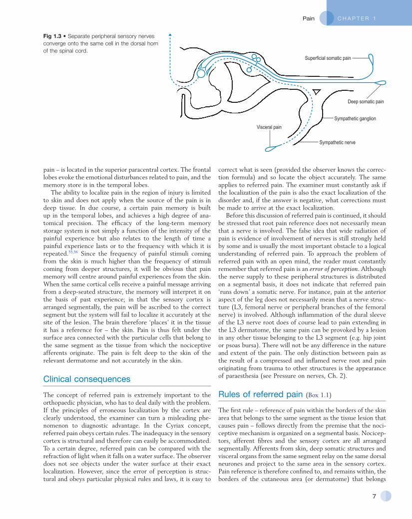
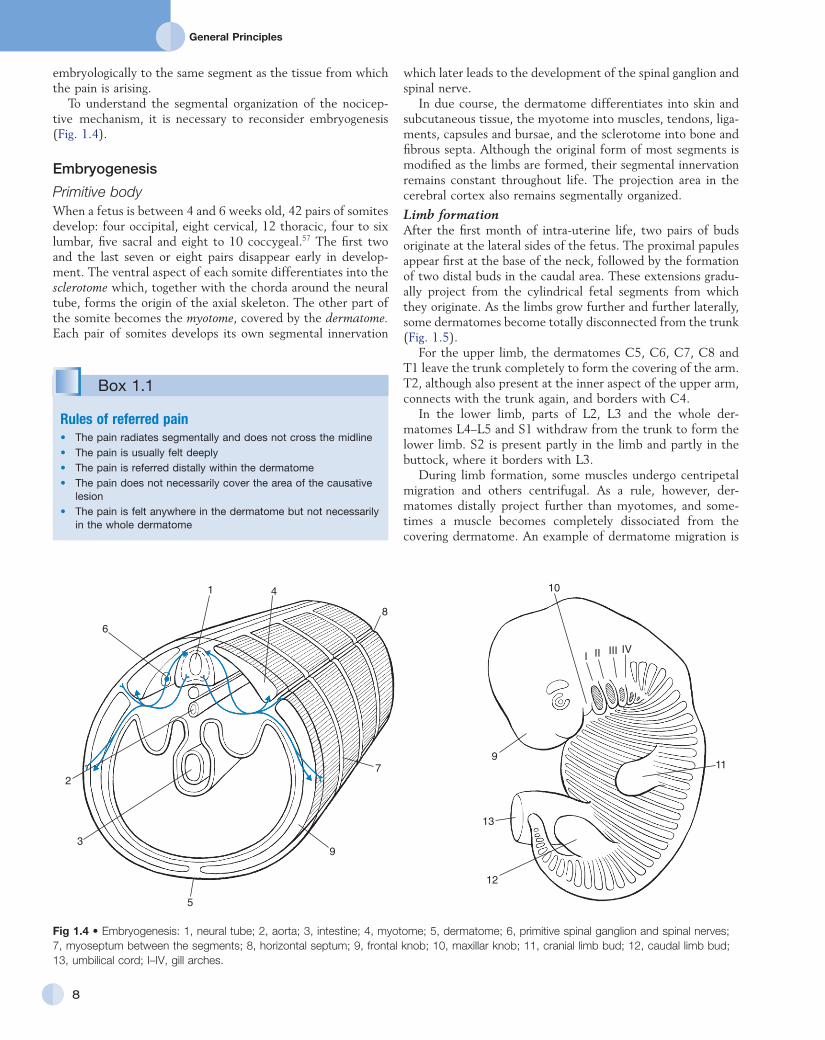
Fig 1.4 • Embryogenesis: 1, neural tube; 2, aorta; 3, intestine; 4, myotome; 5, dermatome; 6, primitive spinal ganglion and spinal nerves; 7, myoseptum between the segments; 8, horizontal septum; 9, frontal knob; 10, maxillar knob; 11, cranial limb bud; 12, caudal limb bud; 13, umbilical cord; I–IV, gill arches.
I II III IV
10
9
13
12
11
1 4
8
6
2
3
5
9
7
Box 1.1
Rules of referred pain• The pain radiates segmentally and does not cross the midline• The pain is usually felt deeply• The pain is referred distally within the dermatome• The pain does not necessarily cover the area of the causative
lesion• The pain is felt anywhere in the dermatome but not necessarily
in the whole dermatome
embryologically to the same segment as the tissue from which the pain is arising.
To understand the segmental organization of the nociceptive mechanism, it is necessary to reconsider embryogenesis (Fig. 1.4).
Embryogenesis
Primitive bodyWhen a fetus is between 4 and 6 weeks old, 42 pairs of somites develop: four occipital, eight cervical, 12 thoracic, four to six lumbar, five sacral and eight to 10 coccygeal.57 The first two and the last seven or eight pairs disappear early in development. The ventral aspect of each somite differentiates into the sclerotome which, together with the chorda around the neural tube, forms the origin of the axial skeleton. The other part of the somite becomes the myotome, covered by the dermatome. Each pair of somites develops its own segmental innervation
which later leads to the development of the spinal ganglion and spinal nerve.
In due course, the dermatome differentiates into skin and subcutaneous tissue, the myotome into muscles, tendons, ligaments, capsules and bursae, and the sclerotome into bone and fibrous septa. Although the original form of most segments is modified as the limbs are formed, their segmental innervation remains constant throughout life. The projection area in the cerebral cortex also remains segmentally organized.
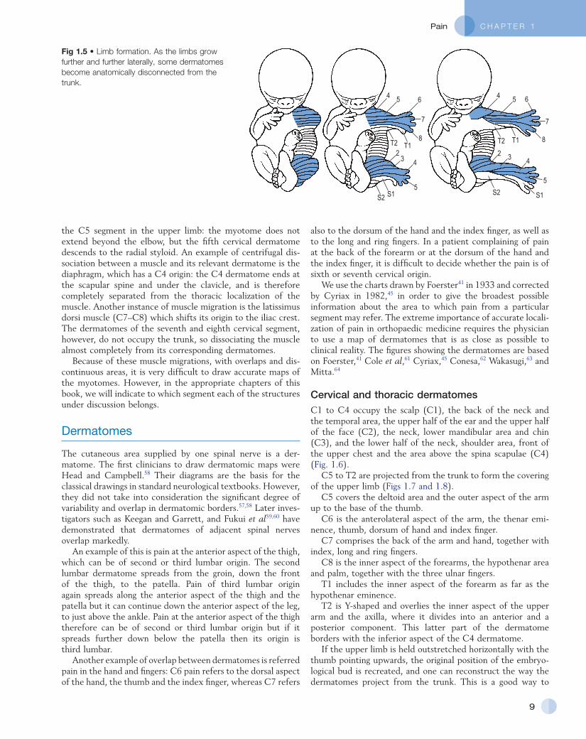
Limb formationAfter the first month of intrauterine life, two pairs of buds originate at the lateral sides of the fetus. The proximal papules appear first at the base of the neck, followed by the formation of two distal buds in the caudal area. These extensions gradually project from the cylindrical fetal segments from which they originate. As the limbs grow further and further laterally, some dermatomes become totally disconnected from the trunk (Fig. 1.5).
For the upper limb, the dermatomes C5, C6, C7, C8 and T1 leave the trunk completely to form the covering of the arm. T2, although also present at the inner aspect of the upper arm, connects with the trunk again, and borders with C4.
In the lower limb, parts of L2, L3 and the whole dermatomes L4–L5 and S1 withdraw from the trunk to form the lower limb. S2 is present partly in the limb and partly in the buttock, where it borders with L3.
During limb formation, some muscles undergo centripetal migration and others centrifugal. As a rule, however, dermatomes distally project further than myotomes, and sometimes a muscle becomes completely dissociated from the covering dermatome. An example of dermatome migration is
C H A P T E R 1Pain
9
also to the dorsum of the hand and the index finger, as well as to the long and ring fingers. In a patient complaining of pain at the back of the forearm or at the dorsum of the hand and the index finger, it is difficult to decide whether the pain is of sixth or seventh cervical origin.
We use the charts drawn by Foerster41 in 1933 and corrected by Cyriax in 1982,45 in order to give the broadest possible information about the area to which pain from a particular segment may refer. The extreme importance of accurate localization of pain in orthopaedic medicine requires the physician to use a map of dermatomes that is as close as possible to clinical reality. The figures showing the dermatomes are based on Foerster,41 Cole et al,61 Cyriax,45 Conesa,62 Wakasugi,63 and Mitta.64
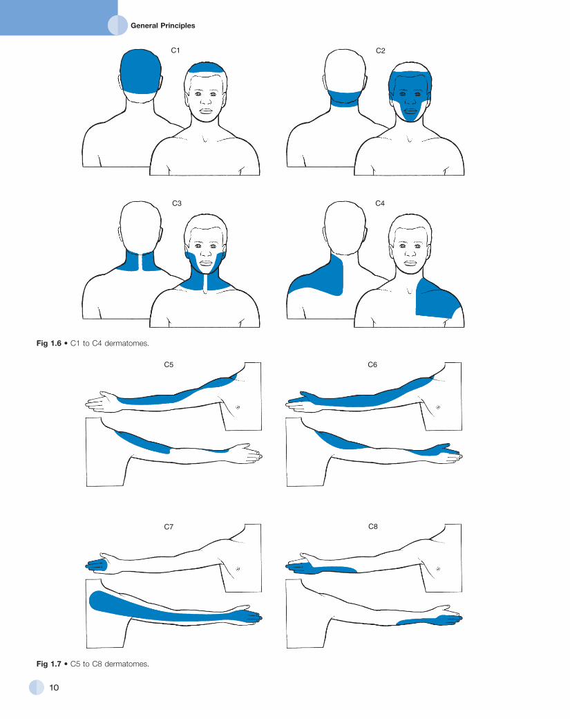
Cervical and thoracic dermatomesC1 to C4 occupy the scalp (C1), the back of the neck and the temporal area, the upper half of the ear and the upper half of the face (C2), the neck, lower mandibular area and chin (C3), and the lower half of the neck, shoulder area, front of the upper chest and the area above the spina scapulae (C4) (Fig. 1.6).
C5 to T2 are projected from the trunk to form the covering of the upper limb (Figs 1.7 and 1.8).
C5 covers the deltoid area and the outer aspect of the arm up to the base of the thumb.
C6 is the anterolateral aspect of the arm, the thenar eminence, thumb, dorsum of hand and index finger.
C7 comprises the back of the arm and hand, together with index, long and ring fingers.
C8 is the inner aspect of the forearms, the hypothenar area and palm, together with the three ulnar fingers.
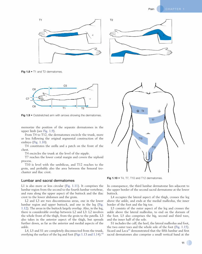
T1 includes the inner aspect of the forearm as far as the hypothenar eminence.
T2 is Yshaped and overlies the inner aspect of the upper arm and the axilla, where it divides into an anterior and a posterior component. This latter part of the dermatome borders with the inferior aspect of the C4 dermatome.
If the upper limb is held outstretched horizontally with the thumb pointing upwards, the original position of the embryological bud is recreated, and one can reconstruct the way the dermatomes project from the trunk. This is a good way to
the C5 segment in the upper limb: the myotome does not extend beyond the elbow, but the fifth cervical dermatome descends to the radial styloid. An example of centrifugal dissociation between a muscle and its relevant dermatome is the diaphragm, which has a C4 origin: the C4 dermatome ends at the scapular spine and under the clavicle, and is therefore completely separated from the thoracic localization of the muscle. Another instance of muscle migration is the latissimus dorsi muscle (C7–C8) which shifts its origin to the iliac crest. The dermatomes of the seventh and eighth cervical segment, however, do not occupy the trunk, so dissociating the muscle almost completely from its corresponding dermatomes.
Because of these muscle migrations, with overlaps and discontinuous areas, it is very difficult to draw accurate maps of the myotomes. However, in the appropriate chapters of this book, we will indicate to which segment each of the structures under discussion belongs.
Dermatomes
The cutaneous area supplied by one spinal nerve is a dermatome. The first clinicians to draw dermatomic maps were Head and Campbell.58 Their diagrams are the basis for the classical drawings in standard neurological textbooks. However, they did not take into consideration the significant degree of variability and overlap in dermatomic borders.57,58 Later investigators such as Keegan and Garrett, and Fukui et al59,60 have demonstrated that dermatomes of adjacent spinal nerves overlap markedly.
An example of this is pain at the anterior aspect of the thigh, which can be of second or third lumbar origin. The second lumbar dermatome spreads from the groin, down the front of the thigh, to the patella. Pain of third lumbar origin again spreads along the anterior aspect of the thigh and the patella but it can continue down the anterior aspect of the leg, to just above the ankle. Pain at the anterior aspect of the thigh therefore can be of second or third lumbar origin but if it spreads further down below the patella then its origin is third lumbar.
Another example of overlap between dermatomes is referred pain in the hand and fingers: C6 pain refers to the dorsal aspect of the hand, the thumb and the index finger, whereas C7 refers
Fig 1.5 • Limb formation. As the limbs grow further and further laterally, some dermatomes become anatomically disconnected from the trunk.
4 5 6
7
8T1T2
23 4
5S1S2
4 5 6
7
8T1T2
2 3 4
5
S1S2
General Principles
10
Fig 1.6 • C1 to C4 dermatomes.
C1 C2
C3 C4
Fig 1.7 • C5 to C8 dermatomes.
C5 C6
C7 C8
C H A P T E R 1Pain
11
Fig 1.8 • T1 and T2 dermatomes.
T1 T2
Fig 1.9 • Outstretched arm with arrows showing the dermatomes.
T1 T2
C5C6
C7
C8
Fig 1.10 • T4, T7, T10 and T12 dermatomes.
T4
T7
T10
T12
memorize the position of the separate dermatomes in the upper limb (see Fig. 1.9).
From T4 to T12, the dermatomes encircle the trunk, more or less following the original segmental construction of the embryo (Fig. 1.10).
T4 constitutes the axilla and a patch on the front of the chest.
T4 encircles the trunk at the level of the nipple.T7 reaches the lower costal margin and covers the xiphoid
process.T10 is level with the umbilicus, and T12 reaches to the
groin, and probably also the area between the femoral trochanter and iliac crest.
Lumbar and sacral dermatomesL1 is also more or less circular (Fig. 1.11). It comprises the lumbar region from the second to the fourth lumbar vertebrae, and runs along the upper aspect of the buttock and the iliac crest to the lower abdomen and the groin.
L2 and L3 are two discontinuous areas, one in the lower lumbar region and upper buttock, and one in the leg (Fig. 1.12). The areas in the buttock largely overlap. Also, in the leg, there is considerable overlap between L2 and L3: L2 involves the whole front of the thigh, from the groin to the patella. L3 also takes in the anterior aspect of the thigh, but spreads further down, as far as the anterior and medial aspects of the ankle.
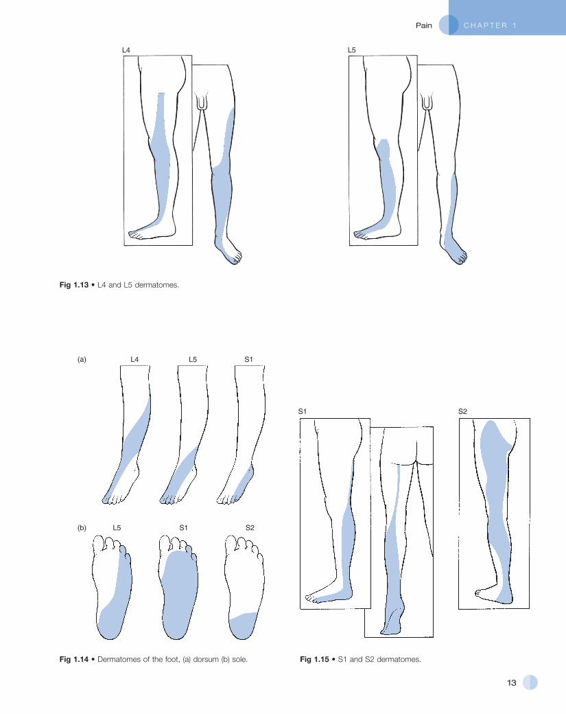
L4, L5 and S1 are completely disconnected from the trunk, overlying the surface of the leg and foot (Figs 1.13 and 1.14).64
In consequence, the third lumbar dermatome lies adjacent to the upper border of the second sacral dermatome at the lower buttock.
L4 occupies the lateral aspect of the thigh, crosses the leg above the ankle, and ends at the medial malleolus, the inner border of the foot and the big toe.
L5 consists of the outer aspect of the leg and crosses the ankle above the lateral malleolus, to end on the dorsum of the foot. L5 also comprises the big, second and third toes, and the inner half of the sole.
S1 includes the calf, the heel, the lateral malleolus and foot, the two outer toes and the whole sole of the foot (Fig. 1.15). Sicard and Leca65 demonstrated that the fifth lumbar and first sacral dermatomes also comprise a small vertical band at the

General Principles
12
Fig 1.12 • L2 and L3 dermatomes.
L2 L3
Fig 1.11 • L1 dermatome.
L1 posterior aspect of the thigh, which could account for the thigh pain commonly described by patients suffering from L5 or S1 sciatica.
S2 is large and comprises the plantar aspect of the heel, the calf, the back of the whole thigh and the lower buttock. In the buttock, it borders with the lumbar part of L3 (Fig. 1.15).
S3 is a narrow zone at the inner side of the thigh, where it borders with L2 anteriorly and S2 posteriorly (Fig. 1.16). The tip ends just proximal to the knee. The upper extent reaches the inguinal ligament where it adjoins the 12th thoracic and first and second lumbar dermatomes. It follows that the groin is a confluence of dermatomes, and pain, apart from that of local origin, may be referred from a 12th thoracic, a first or second lumbar, or a third sacral origin. The groin is also a common site for extrasegmental dural pain reference.
S4 comprises the saddle area, anus, perineum, and scrotum and penis or labia and vagina.
S5 is the coccyx.As in the upper limb, the original position of the distal
embryological bud can be reconstructed by abducting the thigh to 90°, and by lateral rotation until the big toe points upwards. This position demonstrates the way the dermatomes were projected from the trunk and also constitutes a good way of memorizing the position of the various dermatomes in the lower limb (Fig. 1.17). Proximally to distally and then turning proximally again, the following are encountered: L2 at the
C H A P T E R 1Pain
13
Fig 1.13 • L4 and L5 dermatomes.
L4 L5
Fig 1.14 • Dermatomes of the foot, (a) dorsum (b) sole.
L4 L5 S1(a)
(b) L5 S1 S2
Fig 1.15 • S1 and S2 dermatomes.
S1 S2

General Principles
14
Fig 1.16 • S3 to S5 dermatomes.
S3 S4
S3
S4
S5
S3–S5
Fig 1.17 • Outstretched and rotated leg, with arrows showing the dermatomes.
L2
L3
S1S2
L4L5
anterior thigh, L3 at thigh and leg, L4 at the lateral aspect of the leg, anterior aspect of ankle and inner border of foot up to the big toe, L5 at the dorsum of the foot and the three inner toes, S1 at the lateral aspect of the foot, outer malleolus and calf, S2 at the posterior aspect of the leg; turning back to the trunk in the gluteal area, the boundary zone in the perineum, between leg and trunk is comprised of S3 and S4.
Discrepancies between dermatomes and myotomes
We have mentioned already that, as the outcome of embryological development, dermatomes do not always precisely cover the underlying myotomes. Cyriax described eight areas in the human body where the skin and the structure it covers have completely different embryological derivations (Cyriax45). These are the head, scapular and pectoral region, hand, intrathoracic and intraabdominal region, buttock and scrotum.
HeadThe skull, head and face are derived from the two remaining occipital somites, originally situated at the back of the neck. During development, a pair of frontal knobs and two mandibular arches form and fold forwards to create the skeleton and soft tissues around the buccal cavity. The skin of the head and face, however, are formed from the upper two cervical segments.
Scapular regionThe growth of the protuberances that are to become the upper limbs draws some segments out from the cylindrical cervical and thoracic structures. At the same time, the scapula and its muscles, together with the latissimus dorsi (C5–C7) move centripetally between the skin of the thorax (circularly arranged thoracic dermatomes) and the underlying ribs and intercostal muscles (circularly arranged thoracic sclerotomes and myotomes). Therefore, pain in the scapular area can have both scapular (cervical) and thoracic origins.
Pectoral regionDuring the growth of the upper limb bud, the same phenomenon as occurs in the scapular region takes place at the pectoral region. The pectoral muscles, derived from cervical segments (C6–C7) move centripetally between the thoracic dermatomes and their myotomes.
The handThe thenar muscles form part of the eight cervical and first thoracic myotomes, but the skin is formed from the fifth and sixth cervical dermatomes. The interosseus muscles are C8 and T1, but the skin of the dorsum of the hand, except at the ulnar border which is also C8, is derived from the C6 and C7 segments.
Intrathoracic regionIt is obvious that there are numerous discrepancies in origin between the thoracic cage and its content. The diaphragm, for instance, is derived from the third and fourth cervical segments
C H A P T E R 1Pain
15
abdominal pain, a list of the segmental derivations of the viscera, based on the work of Cyriax45 (see his p. 30), William and Warwick,67 Lindsay et al68 and Guyton (cited by Van Cranenburgh69 is given in Box 1.2).
Referred pain is felt deeply and distally in the dermatome
An important difference between local pain and referred pain is that in the latter the pain is felt deeply and vaguely. The
and thereafter descends. Hence a lesion of the diaphragm may cause pain felt in the neck and at the upper scapular and pectoral region, even though it lies at the lower thoracic level. The heart is derived from C8 to T4. Therefore myocardial pain may radiate to the chest, the shoulder and the inner aspect of the arm, as far as the ulnar border of the hand. It is presumed that a small part of the myocardium, probably the auricles, has a third cervical origin, which could explain the wellknown clinical fact that the pain of angina often radiates to the neck anteriorly. The oesophagus is T4–T6 and the lungs have a T2–T5 origin.
Intra-abdominal regionThe abdominal wall has a more or less circular construction, from T7 at the xiphoid process, over T10 at the umbilicus, to L1 at the iliac crest, inguinal ligament and groin. In the abdominal wall, the dermatomes exactly overlie the myotomes.
Most of the intraabdominal content also has a mid and lower thoracic origin. The embryological derivation of the stomach and duodenum (T6–T10), liver (T7–T9 right), gall bladder (T6–T10 right),66 pancreas (T7–T8) and small intestine (T9–T10), fit very well with their actual localization in the abdominal cavity, and therefore pain derived from these organs approximates with their surface representation. Structures of lower thoracic, lumbar or even sacral origin, however, show a more complicated pattern of referred pain. The kidney and ureter, for instance, have a T11–L1 derivation and, although they are localized high up in the abdomen, referred pain can reach the inguinal fossa and the groin (T12–L1). The colonic flexure is from L2 to L3, which allows the pain not only to radiate to the lower back but also to the front of the thigh. The sigmoid colon and rectum have S3–S5 origin. Hence, in diseases of the sigmoid and the rectum, pain can be felt in the iliac fossa, perineum, penis, vulva and inner aspect of the thigh.
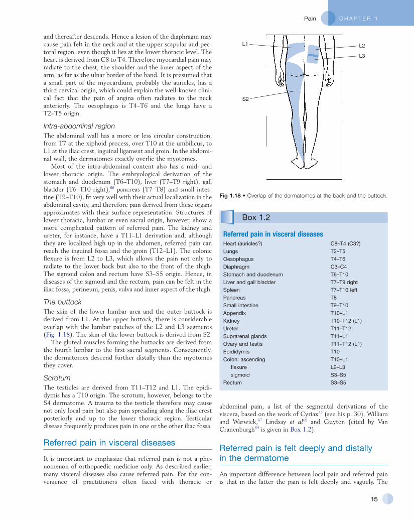
The buttockThe skin of the lower lumbar area and the outer buttock is derived from L1. At the upper buttock, there is considerable overlap with the lumbar patches of the L2 and L3 segments (Fig. 1.18). The skin of the lower buttock is derived from S2.
The gluteal muscles forming the buttocks are derived from the fourth lumbar to the first sacral segments. Consequently, the dermatomes descend further distally than the myotomes they cover.
ScrotumThe testicles are derived from T11–T12 and L1. The epididymis has a T10 origin. The scrotum, however, belongs to the S4 dermatome. A trauma to the testicle therefore may cause not only local pain but also pain spreading along the iliac crest posteriorly and up to the lower thoracic region. Testicular disease frequently produces pain in one or the other iliac fossa.
Referred pain in visceral diseases
It is important to emphasize that referred pain is not a phenomenon of orthopaedic medicine only. As described earlier, many visceral diseases also cause referred pain. For the convenience of practitioners often faced with thoracic or
Fig 1.18 • Overlap of the dermatomes at the back and the buttock.
L1
S2
L3
L2
Box 1.2
Referred pain in visceral diseasesHeart (auricles?) C8–T4 (C3?)Lungs T2–T5Oesophagus T4–T6Diaphragm C3–C4Stomach and duodenum T6–T10Liver and gall bladder T7–T9 rightSpleen T7–T10 leftPancreas T8Small intestine T9–T10Appendix T10–L1Kidney T10–T12 (L1)Ureter T11–T12Suprarenal glands T11–L1Ovary and testis T11–T12 (L1)Epididymis T10Colon: ascending T10–L1 flexure L2–L3 sigmoid S3–S5
Rectum S3–S5

General Principles
16
patient thus does not point to a localized and precise area, but outlines an approximate one and tends to describe it as ‘deep’.
That pain – with some exceptions – is always referred distally, remains a purely empirical clinical observation and has hitherto not been explained on neurophysiological grounds. The fact that pain that arises from the proximal part of a segment can be felt distally in the dermatome but a distal lesion is not referred proximally within this same segment, remains an inconsistency that is hard to rationalize. However, this clinical observation is very important to the clinician confronted with referred pain, for the lesion must never be sought in a structure that is localized distally of the painful area. Pain at the distal aspect of the L3 dermatome (knee and lower limb) can have a proximal origin (spine, hip), but pain only in the hip or the groin, cannot be caused by a lesion at the knee or the thigh.
Segmentally referred pain does not cross the midline
The segments in the body are arranged in pairs, each of which has its own segmental innervation and its own projection area in the cerebrum. Hence it is obvious that the cerebral cortex will easily differentiate between a leftside or a rightside pain, and no one will question that a C5 pain on the left side has a leftside origin and vice versa. The fact that pain stemming from a unilateral structure does not cross the midline, becomes important in the interpretation of more or less centrally localized pain, for instance in aches in the neck or back. It is evident that a lesion of a unilateral facet joint will not cause pain radiating all over the lower back. Only centrally localized structures (vertebral body, longitudinal ligaments, intra and supraspinal ligaments and dura mater) can theoretically be responsible for a bilaterally radiating pain at both sides of the midline. A pain felt centrally or bilaterally must originate from a central structure, or from two bilateral structures (two facet joints or two sacroiliac joints) but it can never be the result of a lesion in a unilateral structure.
Dura mater an ‘exception’ to segmental reference
Pain originating from the dura mater has a rather peculiar behaviour. First, in that the dura is a midline structure, it is innervated from both sides, so that pain refers bilaterally. Second, pain stemming from the dura has a very broad reference and seems to cover several consecutive dermatomes. For instance, pressure of a lumbar disc on the dura at the L5 level can cause pain in the back which radiates to the abdomen and groins, down to the anterior and posterior aspect of both thighs and legs, and upwards to the back of the lower chest. This type of pain reference is inexplicable in terms of segments. We therefore call it ‘extrasegmental reference’ of pain. Because in orthopaedic medicine, the dura is the exclusive source of this type of pain radiation, it is also called dural reference. Pain of this nature can be very difficult to interpret if it is felt in a part or parts of the possible reference area. As in
purely segmental reference, dural reference can be to only a part of the respective dermatomes. Thus, instead of the broad radiation to the whole back, both glutei and both legs, dural pain sometimes only affects a small part, for instance one groin, or one buttock or the whole posterior aspect of the thigh. The differential diagnosis from a more segmental pain, for instance as the result of a nerve root compression, is then difficult.
A common clinical finding in a cervical disc protrusion that impacts on the dura is unilateral interscapular pain or pain in the trapezius or pectoral area. In the latter instance, suspicion of angina pectoris may then easily arise. Also the removal of an appendix for dural pain of lumbar origin referred extrasegmentally into the iliac fossa and groin is not at all exceptional.
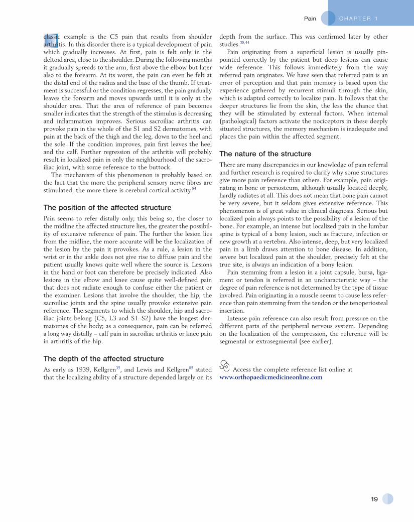
A possible explanation for the misleading pain reference of the dura may lie in its multisegmental origin, which is reflected in the great overlap between the fibres of the consecutive sinuvertebral nerves innervating its anterior aspect.70–73 More recent researchers describe division of the nerves into ascending and descending branches which ramify variously, to give off longitudinally and transversely orientated branches.74,75 In a recent study that used the very sensitive acetylcholinesterase method, more ramifications between the nerve branches were demonstrated (Fig. 1.19).76 Ascending branches up to four segments cranial to the level of entry into the dural nerve plexus and also descending branches extending up to four segments caudally were observed. In addition, many vertical and horizontal interconnections between the various ascending and descending branches were seen. The conclusion is that dural nerves may spread over eight segments and that a great overlap exists between adjacent and contralateral dural nerves.
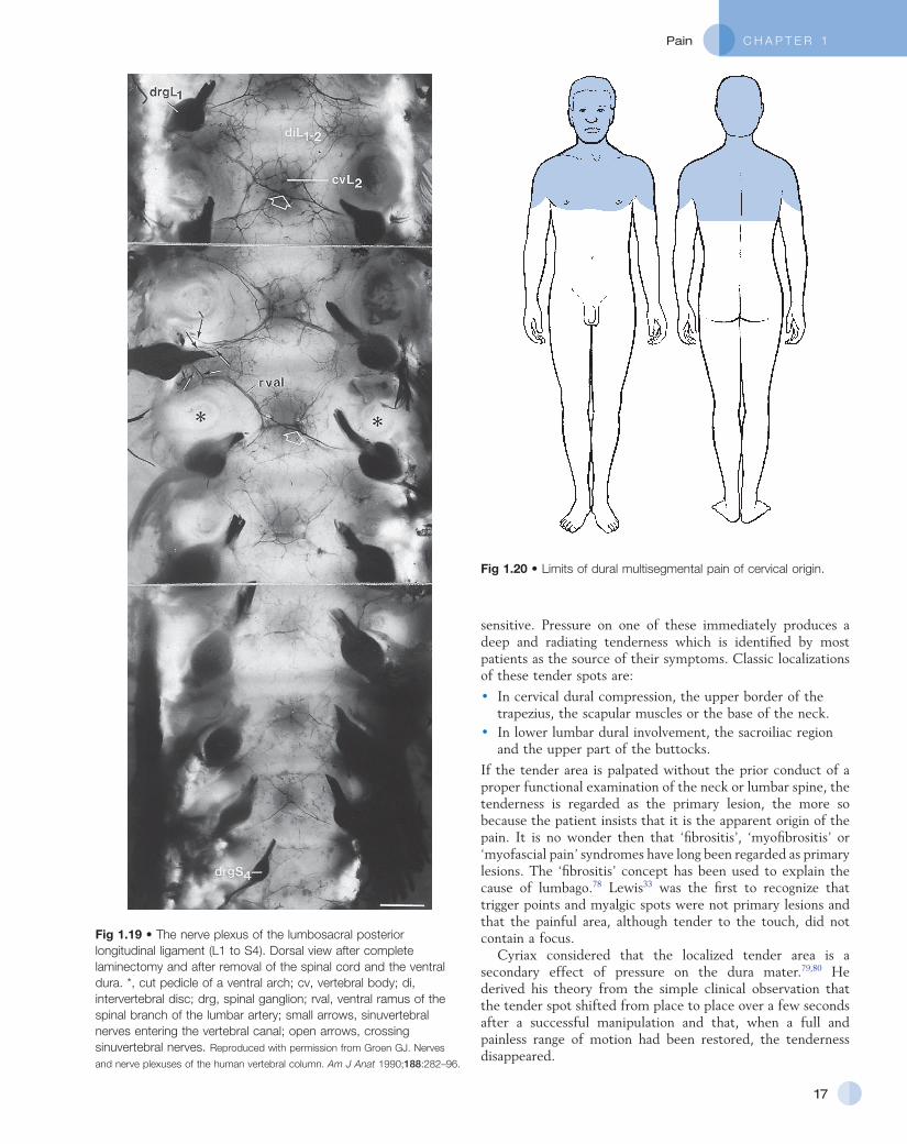
These findings may form an anatomical explanation for the clinical observations of Cyriax on the limits of extrasegmental reference of dural pain.77 Upwards, pain originating in the lower cervical part of the dura may spread to the occiput, skull and forehead (Fig. 1.20). Downwards it can descend to T7 which corresponds with the lower angles of the scapulae. Anteriorly the pain can occupy the whole pectoral area. Extrasegmental pain does not extend beyond the upper half of the arms.
A middle thoracic disc lesion can cause dural extrasegmental pain that may radiate to the base of the neck, and downwards to the whole abdomen and to the upper lumbar region (Fig. 1.21).
Dural extrasegmental reference from a low lumbar level may reach the lower thorax posteriorly, the lower abdomen deep to the umbilicus, the groins, the buttocks, and the sacrum and coccyx (Fig. 1.22). Unlike the cervical dura – which does not radiate far into the arms – extrasegmental reference from a lumbar origin may also involve the legs, both the anterior and the posterior aspects, and descend to the ankles.
Referred tenderness
There seems to be more to referred pain than just mislocation by the patient. Within or near the area of referred pain, it is often possible to find small trigger points which are exquisitely
C H A P T E R 1Pain
17
sensitive. Pressure on one of these immediately produces a deep and radiating tenderness which is identified by most patients as the source of their symptoms. Classic localizations of these tender spots are:
• In cervical dural compression, the upper border of the trapezius, the scapular muscles or the base of the neck.
• In lower lumbar dural involvement, the sacroiliac region and the upper part of the buttocks.
If the tender area is palpated without the prior conduct of a proper functional examination of the neck or lumbar spine, the tenderness is regarded as the primary lesion, the more so because the patient insists that it is the apparent origin of the pain. It is no wonder then that ‘fibrositis’, ‘myofibrositis’ or ‘myofascial pain’ syndromes have long been regarded as primary lesions. The ‘fibrositis’ concept has been used to explain the cause of lumbago.78 Lewis33 was the first to recognize that trigger points and myalgic spots were not primary lesions and that the painful area, although tender to the touch, did not contain a focus.
Cyriax considered that the localized tender area is a secondary effect of pressure on the dura mater.79,80 He derived his theory from the simple clinical observation that the tender spot shifted from place to place over a few seconds after a successful manipulation and that, when a full and painless range of motion had been restored, the tenderness disappeared.
Fig 1.19 • The nerve plexus of the lumbosacral posterior longitudinal ligament (L1 to S4). Dorsal view after complete laminectomy and after removal of the spinal cord and the ventral dura. *, cut pedicle of a ventral arch; cv, vertebral body; di, intervertebral disc; drg, spinal ganglion; rval, ventral ramus of the spinal branch of the lumbar artery; small arrows, sinuvertebral nerves entering the vertebral canal; open arrows, crossing sinuvertebral nerves. Reproduced with permission from Groen GJ. Nerves
and nerve plexuses of the human vertebral column. Am J Anat 1990;188:282–96.
Fig 1.20 • Limits of dural multisegmental pain of cervical origin.

General Principles
18
Fig 1.21 • Limits of dural multisegmental pain of thoracic origin. Fig 1.22 • Limits of multisegmental pain of lumbar origin.
However, referred tenderness has also been demonstrated in muscular and fibrous tissue lesions,81 in visceral lesions such as ischaemic heart attacks82 and in pathological viscera. The phenomena are not completely understood as yet but it is reasonable to assume that trigger points are caused by summation mechanisms which can be understood in terms of gatecontrol mechanisms.79
Summation – the excitatory effect of converging inputs – is an important pain mechanism. Pain may be triggered by two sources of nerve impulses: a major one from the lesion; and a minor one from normal skin which add together. If one source is removed, it becomes more difficult for the other to trigger the feeling of pain.83
Factors determining reference of pain
Referred pain is a faulty perception of the origin of a pain. In orthopaedic medicine it is common to see marked differences in the extent of reference between segments, between distinct affected tissues and between different degrees of the same condition.
The degree of reference – that is the distance between the localization of the perception and the site of the lesion – depends on four different factors (Box 1.3). Some can be explained on the basis of the known pathways, others remain unexplained and result purely from clinical observations.
Box 1.3
Factors favouring reference of painStrength of the stimulus:
• The stronger the stimulus, the more reference of painPosition of the affected tissue:
• The more central the lesion, the more reference of pain• The more distal the lesion, the less reference of pain
Depth of the affected structure:
• More reference from deep-seated structures• Less reference from superficial structures
Nature of the affected tissue:
• Little reference: bone and periosteum• More reference: muscle• Much reference: capsule, ligament, bursa, tendon, dura, dural
sleeve and perineurium
The strength of the stimulusThe greater the stimulus, the more extensive the reference of pain. In other words, intense stimulation radiates the pain widely and slight irritation localizes the pain closer to its origin.
This phenomenon is used in orthopaedic medicine to estimate the degree of irritation or to evaluate treatment. A
C H A P T E R 1Pain
19
depth from the surface. This was confirmed later by other studies.38,44
Pain originating from a superficial lesion is usually pinpointed correctly by the patient but deep lesions can cause wide reference. This follows immediately from the way referred pain originates. We have seen that referred pain is an error of perception and that pain memory is based upon the experience gathered by recurrent stimuli through the skin, which is adapted correctly to localize pain. It follows that the deeper structures lie from the skin, the less the chance that they will be stimulated by external factors. When internal (pathological) factors activate the nociceptors in these deeply situated structures, the memory mechanism is inadequate and places the pain within the affected segment.
The nature of the structureThere are many discrepancies in our knowledge of pain referral and further research is required to clarify why some structures give more pain reference than others. For example, pain originating in bone or periosteum, although usually located deeply, hardly radiates at all. This does not mean that bone pain cannot be very severe, but it seldom gives extensive reference. This phenomenon is of great value in clinical diagnosis. Serious but localized pain always points to the possibility of a lesion of the bone. For example, an intense but localized pain in the lumbar spine is typical of a bony lesion, such as fracture, infection or new growth at a vertebra. Also intense, deep, but very localized pain in a limb draws attention to bone disease. In addition, severe but localized pain at the shoulder, precisely felt at the true site, is always an indication of a bony lesion.
Pain stemming from a lesion in a joint capsule, bursa, ligament or tendon is referred in an uncharacteristic way – the degree of pain reference is not determined by the type of tissue involved. Pain originating in a muscle seems to cause less reference than pain stemming from the tendon or the tenoperiosteal insertion.
Intense pain reference can also result from pressure on the different parts of the peripheral nervous system. Depending on the localization of the compression, the reference will be segmental or extrasegmental (see earlier).
classic example is the C5 pain that results from shoulder arthritis. In this disorder there is a typical development of pain which gradually increases. At first, pain is felt only in the deltoid area, close to the shoulder. During the following months it gradually spreads to the arm, first above the elbow but later also to the forearm. At its worst, the pain can even be felt at the distal end of the radius and the base of the thumb. If treatment is successful or the condition regresses, the pain gradually leaves the forearm and moves upwards until it is only at the shoulder area. That the area of reference of pain becomes smaller indicates that the strength of the stimulus is decreasing and inflammation improves. Serious sacroiliac arthritis can provoke pain in the whole of the S1 and S2 dermatomes, with pain at the back of the thigh and the leg, down to the heel and the sole. If the condition improves, pain first leaves the heel and the calf. Further regression of the arthritis will probably result in localized pain in only the neighbourhood of the sacroiliac joint, with some reference to the buttock.
The mechanism of this phenomenon is probably based on the fact that the more the peripheral sensory nerve fibres are stimulated, the more there is cerebral cortical activity.84
The position of the affected structurePain seems to refer distally only; this being so, the closer to the midline the affected structure lies, the greater the possibility of extensive reference of pain. The further the lesion lies from the midline, the more accurate will be the localization of the lesion by the pain it provokes. As a rule, a lesion in the wrist or in the ankle does not give rise to diffuse pain and the patient usually knows quite well where the source is. Lesions in the hand or foot can therefore be precisely indicated. Also lesions in the elbow and knee cause quite welldefined pain that does not radiate enough to confuse either the patient or the examiner. Lesions that involve the shoulder, the hip, the sacroiliac joints and the spine usually provoke extensive pain reference. The segments to which the shoulder, hip and sacroiliac joints belong (C5, L3 and S1–S2) have the longest dermatomes of the body; as a consequence, pain can be referred a long way distally – calf pain in sacroiliac arthritis or knee pain in arthritis of the hip.
The depth of the affected structureAs early as 1939, Kellgren35, and Lewis and Kellgren85 stated that the localizing ability of a structure depended largely on its
Access the complete reference list online at www.orthopaedicmedicineonline.com
C H A P T E R 1Pain
19.e1© Copyright 2013 Elsevier, Ltd. All rights reserved.
References
1. Bonica JJ, Albe Fessard D. Advances in Pain Research and Therapy. New York: Raven Press; 1976.
2. Elton D, Stuart GV, Burrows GD. Selfesteem and chronic pain. J Psychosom Res 1978;22:25.
3. Merskey H. Pain terms: a list with definitions and notes on usage. Recommended by the IASP Subcommittee on Taxonomy. Pain 1979;6:249.
4. Melzack R, Torgerson WS. On the language of pain. Anaesthesiology 1971;35:50.
5. Wyke BD. Neurological mechanisms in the experience of pain. Acupunct Electrother Res J 1979;4:27.
6. Ralston HJ, Miller MR, Kasahara M. Nerve endings in human fasciae, tendons, ligaments, periosteum and joint synovial membrane. Anat Rec 1960;136:137.
7. Besson JM, Guilbaud G, Abdelmoumene M, Chaouch A. Physiologie de la nociception. J Physiol (Paris) 1982;78:7–107.
8. Iggo A. The case for ‘pain’ receptors, In: Janzen R, Keidel WD, Herz A, Steichele C, editors. Pain: Basic Principles, Pharmacology, Therapy. Stuttgart: Thieme; 1972. p. 60.
9. Van Hees J, Gybels JM. Pain related to single afferent C fibres from human skin. Brain Res 1972;48:397.
10. Sarkin LS, Wallace MS. Acute pain mechanisms. Surg Clin North Am 1999;79(2):213–29.
11. Dray A. Inflammatory mediators of pain. Br J Anaesth 1995;75:125–31.
12. Reeh PW, Steen KH. Tissue acidosis in nociception and pain. In: Kunasawa T, Kruger C, Ulisumara K, editors. Progress in Brain Research, vol. 13. Amsterdam: Elsevier Science; 1996. p. 143–51.
13. Wall PD, McMahon SB. Microneuronography and its relation to perceived sensation. Pain 1985;21:209.
14. Nathan PW. The gatecontrol theory of pain: a critical review. Brain 1976;99:123.
15. Price DD, Dubner R. Neurons that subserve the the sensorydiscriminative aspects of pain. Pain 1977;3:307.
16. Wyke BD. The neurology of low back pain. In: Jayson MIV, editor. The Lumbar Spine and Back Pain. 2nd ed. Bath: Pitman Medical; 1980. p. 265–339.
17. Yaksh TL. Spinal Afferent Processing. New York: Plenum; 1986.
18. Melzack R, Casey KL. Sensory, motivational and central control determinants of pain: a new conceptual model. In: Kenshalo D, editor. The Skin Senses. Springfield: Thomas; 1968. p. 923–93.
19. Hand PJ, Morrison AR. Thalamocortical projections from the ventrobasal complex to somatic sensory area I and II. Exp Neurol 1970;27:291.
20. Desijaru T, Purpura DP. Organisation of specific–nonspecific thalamic internuclear synaptic pathways. Brain Res 1970;21:169.
21. Penfield W. The role of the temporal cortex in recall of past experience and interpretation of the present. In: Wolstenholme GEW, O’Connor CM, editors. The Neurological Basis of Behaviour. London: Churchill; 1958. p. 149.
22. Newcombe F. Memory. In: Critchley M, O’Leary JL, Jennett B, editors. Scientific Foundations of Neurology. London: Heinemann; 1972. p. 205.
23. Black P. Physiological Correlates of Emotion. New York: Academic Press; 1970.
24. Melzack R, Wall PD. Pain mechanism: a new theory. Science 1965;150:971–9.
25. Noordenbos W. Pain. Problems Pertaining to the Transmission of Nerve Impulses Which Give Rise to Pain. Amsterdam: Elsevier; 1959.
26. Mayer DJ, Price D. Central nervous system of analgesia. Pain 1976;2:379.
27. Tan SY. Cognitive and behavioural methods for pain control: a selective review. Pain 1982;12:201–28.
28. Langen D. Psychosomatic aspects in the treatment of pain. In: Janzen R, Keidel WD, Herz A, Steichele C, editors. Pain: Basic Principles, Pharmacology, Therapy. Stuttgart: Thieme; 1972. p. 164.
29. Chapman CR, Feather BW. Effects of diazepam on human pain tolerance and pain sensitivity. Psychosom Med 1973;35:330.
30. Fields HL, Henricher MH. Anatomy and physiology of a nociceptive modulatory system. Phil Trans Roy Soc B 1985;308:361–79.
31. Head H. The afferent nervous system from a new aspect. Brain 1905;28:99.
32. Head H, Campbell AW. The pathology of herpes zoster and its bearing on sensory location. Brain 1900;23:353–523.
33. Lewis T. Pain. New York: MacMillan; 1942.34. Kellgren JH. Observations of referred pain
arising from muscle. Clin Sci 1938;3:175.35. Kellgren JH. On the distribution of pain
from deep somatic structures. Clin Sci 1939;4:35.
36. Inman VT, Saunders JB de CM. Referred pain from skeletal structures. J Nerv Ment Dis 1944;99:660.
37. Travell J, Berry C, Bigelow N. Effects of referred somatic pain on structures in the reference zone. Fed Proc 1944;3:49.
38. McCall IW, Park WM, O’Brien JP. Induced pain referred from posterior elements in normal subjects. Spine 1979;4:441.
39. Hansen K, Schliack H. Segmental Innervation. Stuttgart: Thieme; 1962.
40. Kunert W. Wirbelsäule and Innere Medizin. Stuttgart: F. Enke; 1975.
41. Foerster O. Dermatomes in man. Brain 1933;56:1.
42. Lewis T, Kellgren JH. Observations relating to referred pain. Visceromotor reflexes and other associated phenomena. Clin Sci 1939;4:47.
43. Keegan JJ, Garett FD. The segmental distribution of the cutaneous nerves in the limbs of man. Anat Rec 1948;102:409.
44. Hockaday JM, Whitty CWM. Patterns of referred pain in the normal subject. Brain 1967;90(3):481–96.
45. Cyriax JH. Textbook of Orthopaedic Medicine, vol. 1. 8th ed. London: Baillière Tindall; 1982. p. 22, 35.
46. MacKenzie J. Krankheitszeichen und ihre Auslegung 3. Translated by J Müller, Würzburg: Kabitsch; 1917.
47. Livingston WK. Pain Mechanisms. New York: Macmillan; 1944.
48. Wedell G, Sinclair DG, Feindel WH. Anatomical basis for alterations in quality of pain sensibility. J Neurophys 1948;11:99.
49. Wedell G. Referred pain in relation to the mechanism of common sensibility. Proc Roy Soc Med 1957;50:581.
50. Pomeranz B, Wall PD, Weber WV. Cord cells responding to fine myelinated afferents from viscera, muscle and skin. J Physiol 1968;199:511–32.
51. Taylor DCM, Pierau FRK, Mizutain M. Possible bases for referred pain. In: Holden AV, Winslow W, editors. The Neurobiology of Pain. Manchester: Manchester University Press; 1984. p. 143.
52. Wells PE, Frampton V, Bowsher D. Pain Management by Physiotherapy. 2nd ed. Oxford: ButterworthHeinemann; 1994.
53. Cyriax JH. Massage, Manipulation and Local Anaesthesia. London: Hamilton; 1941.
54. Ruch TC. Visceral sensation and referred pain. In: Fulton JF, editor. Howell’s Textbook of Physiology. Philadelphia: WB Saunders; 1946.
55. Merskey H, Spear FG. Pain: Psychological and Psychiatric Aspects. London: Baillière Tindall and Cassell; 1967.
56. Neurological aspects of the diagnosis and treatment of facial pain. In: Cohen B, Kramer I, editors. Scientific Foundations of Dentistry. London: Heinemann; 1974. p. 278.
57. Patten BM. Human Embryology. New York: McGrawHill; 1968.
58. Head H, Campbell AW. The pathology of herpes zoster and its bearing on sensory location. Brain 1900;23:353–523.
59. Keegan JJ, Garrett ED. The segmental distribution of the cutaneous nerves in the limbs of man. Anat Rec 1949;101:409.
60. Fukui S, Ohseto K, et al. Distribution of referred pain from the zygapophyseal joints and dorsal rami. Clin J Pain 1997;13(4):303–7.
61. Cole JP, Lesswing AL, Cole JR. Analysis of lumbosacral dermatomes in man. Clin Orthop 1968;61:241.
62. Conesa SH, Argote ML. A Visual Aid to the Examination of Nerve Roots. London: Baillière Tindall; 1976.
63. Wakasugi B. Dermatomes of the body and the extremeties. Surg Treatment 1982;132:270.
64. Mitta H. Study on dermatomes by means of selective lumbar spinal nerve block. Spine 1993;18:1782–6.
General Principles
19.e2© Copyright 2013 Elsevier, Ltd. All rights reserved.
65. Sicard A, Leca A. Place de rachiotomie dans le traitement chirurgical des sciatiques. Press Med 1954;62:1737.
66. Doran FSA. The sites to which pain is referred from the common bile duct in man and its implication for the theory of referred pain. Br J Surg 1967;54:599–606.
67. Williams PL, Warwick R. Gray’s Anatomy. Edinburgh: Churchill Livingstone; 1980.
68. Lindsay KW, Bone I, Callander R. Neurology and Neurosurgery Illustrated. 2nd ed. Edinburgh: Churchill Livingstone; 1991.
69. Van Cranenburgh B. Segmentale verschijnselen. Utrecht: Bohn, Scheltema and Holkema; 1985.
70. Pedersen HE, Conrad FJ, Blunck MD, Gartner E. The anatomy of lumbosacral posterior rami and meningeal branches of spinal nerves (sinuvertebral nerves). J Bone Joint Surg 1956;38A(2):377–91.
71. Stillwell DL. The nerve supply of the vertebral column and its associated structures in the monkey. Anat Rec 1956;125(2):139–62.
72. Kimmel DL. Innervation of spinal dura mater, and dura mater of the posterior cranial fossa. Neurology 1961;11:800–9.
73. Edghar MA, Nundy S. Innervation of the spinal dura mater. J Neurol Neurosurg Psychiatr 1966;29:530–4.
74. Jackson HC, Winkelmann RK, Bickel WH. Nerve endings in the human lumbar spinal column and related structures. J Bone Joint Surg 1966;48A:1272–81.
75. Edgar MA, Ghadially JA. Innervation of the lumbar spine. Clin Orthop Rel Res 1976;115:35–41.
76. Groen GJ, Baljet B, Drukker J. The innervation of the spinal dura mater: anatomy and clinical implications. Acta Neurochir 1988;92:39–46.
77. Cyriax JH. Dural pain. Lancet 1978;1:919–21.
78. Gowers W. Lumbago. BMJ 1904;i:117, 79. Travell JG, Simons DG. Myofascial Pain
and Dysfunction. Baltimore: Williams and Wilkins; 1983.
80. Cyriax JH. Fibrositis. BMJ 1948;ii:251. 81. Simons DG. Muscle pain syndromes, part
II. Am J Phys Med 1976;55:15–42. 82. Kennard MA, Haugen FP. The relation of
subcutaneous focal sensitivity to referred pain of cardiac origin. Anaesthesiology 1955;16:297–311.
83. Melzack R, Wall P. The Challenge of Pain. London: Penguin; 1991.
84. Woolsey CN, Marshall WH, Bard P. Observations on cortical somatic sensory mechanism of cat and monkey. J Neurophysiol 1941;4:1.
85. Lewis T, Kellgren JH. Observations relating referred pain, visceromotor reflexes and other associated phenomena. Clin Sci 1939;4:47.