Embed Size (px)
Citation preview
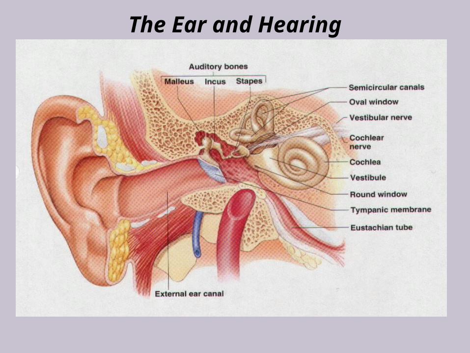
The Ear and Hearing
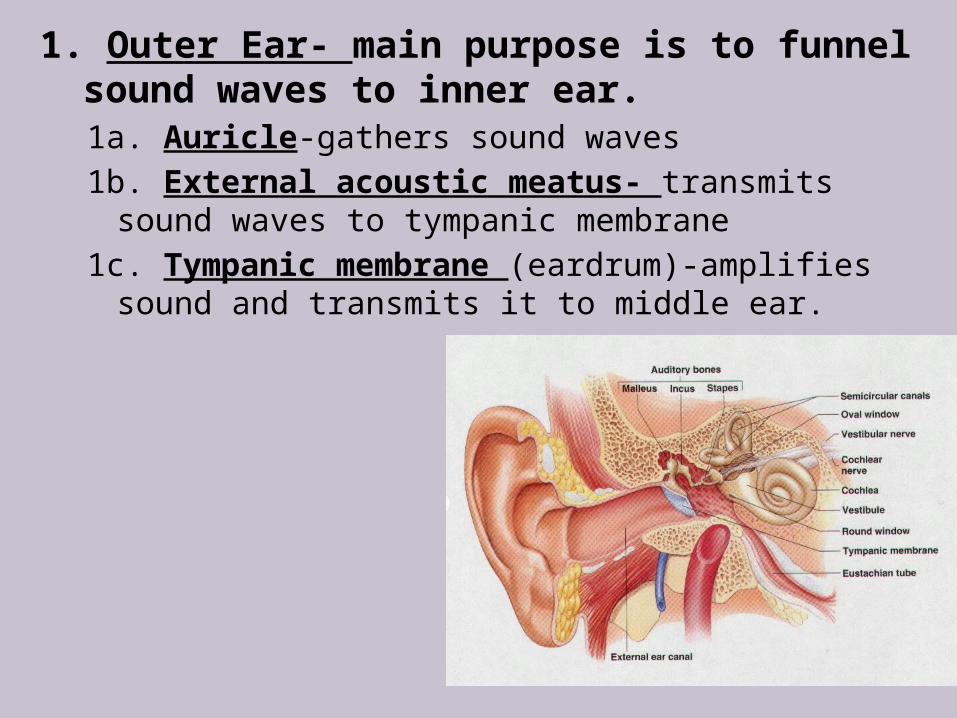
1. Outer Ear- main purpose is to funnel sound waves to inner ear.1a. Auricle-gathers sound waves 1b. External acoustic meatus- transmits sound
waves to tympanic membrane1c. Tympanic membrane (eardrum)-amplifies
sound and transmits it to middle ear.
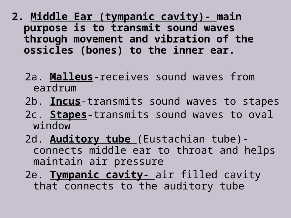
2. Middle Ear (tympanic cavity)- main purpose is to transmit sound waves through movement and vibration of the ossicles (bones) to the inner ear.
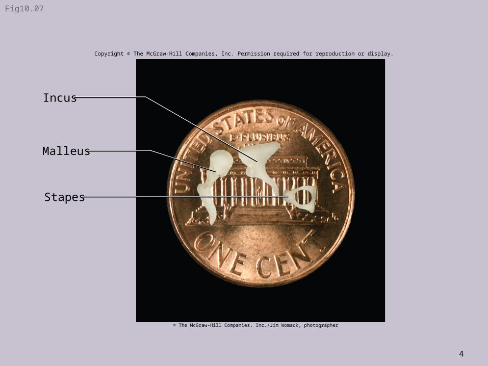
2a. Malleus-receives sound waves from eardrum
2b. Incus-transmits sound waves to stapes2c. Stapes-transmits sound waves to oval
window2d. Auditory tube (Eustachian tube)-
connects middle ear to throat and helps maintain air pressure
2e. Tympanic cavity- air filled cavity that connects to the auditory tube
4
Fig10.07
Stapes
Incus
Malleus
Copyright © The McGraw-Hill Companies, Inc. Permission required for reproduction or display.
© The McGraw-Hill Companies, Inc./Jim Womack, photographer
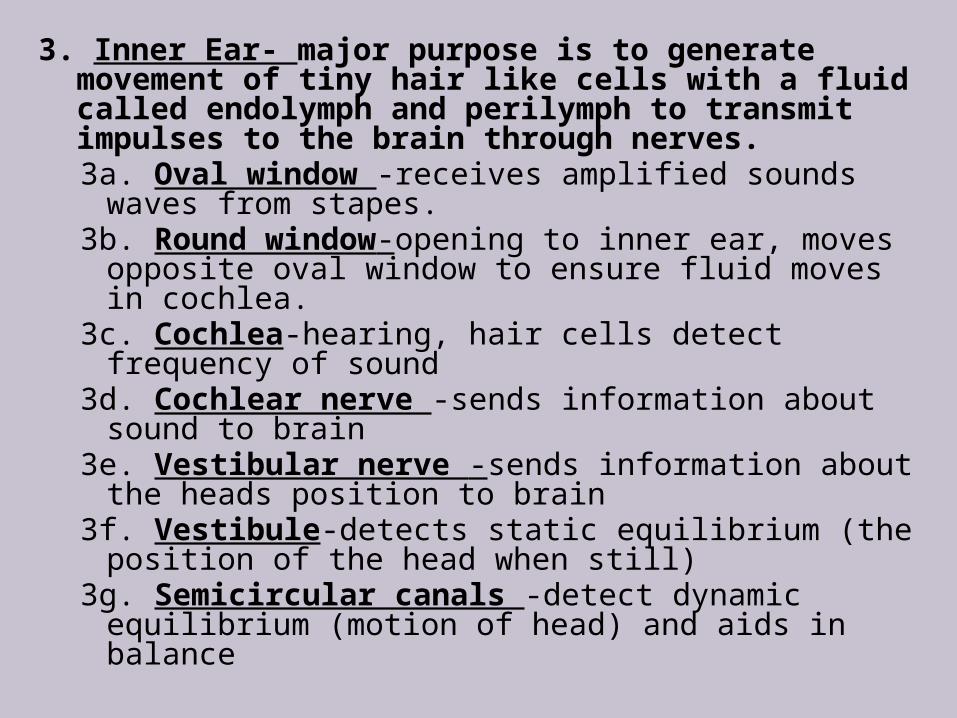
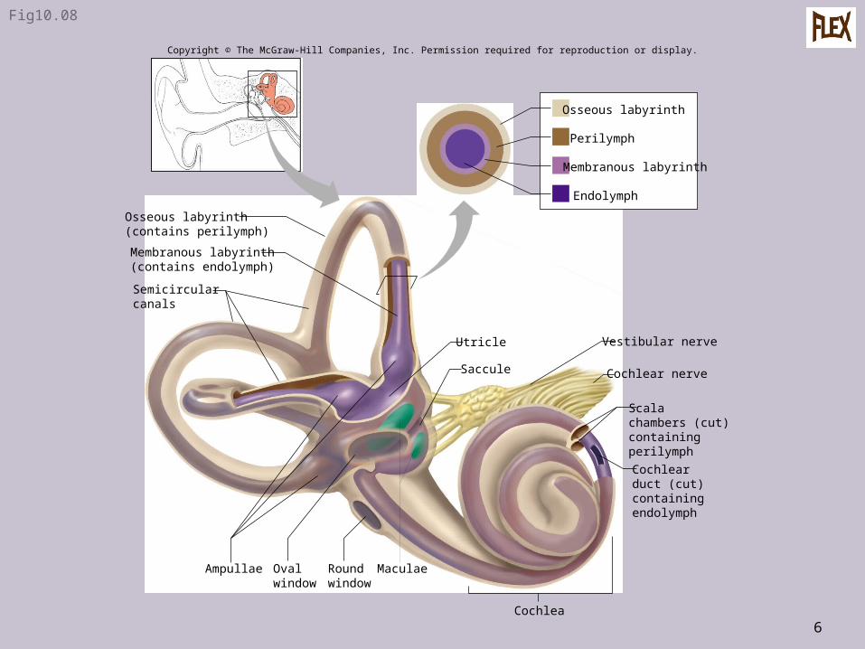
3. Inner Ear- major purpose is to generate movement of tiny hair like cells with a fluid called endolymph and perilymph to transmit impulses to the brain through nerves.3a. Oval window -receives amplified sounds
waves from stapes.3b. Round window-opening to inner ear, moves
opposite oval window to ensure fluid moves in cochlea.
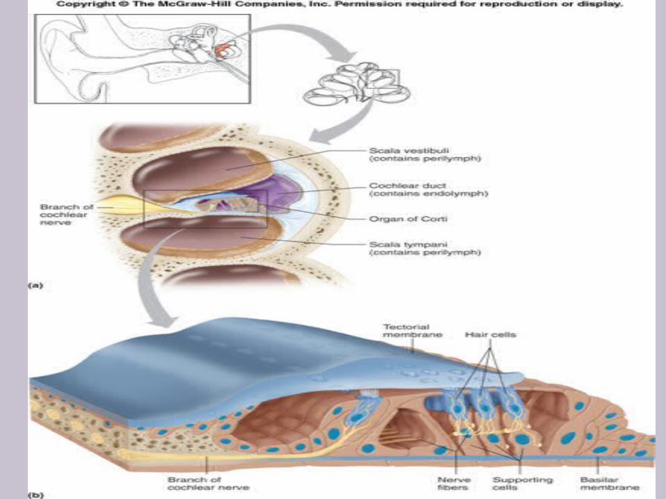
3c. Cochlea-hearing, hair cells detect frequency of sound
3d. Cochlear nerve -sends information about sound to brain
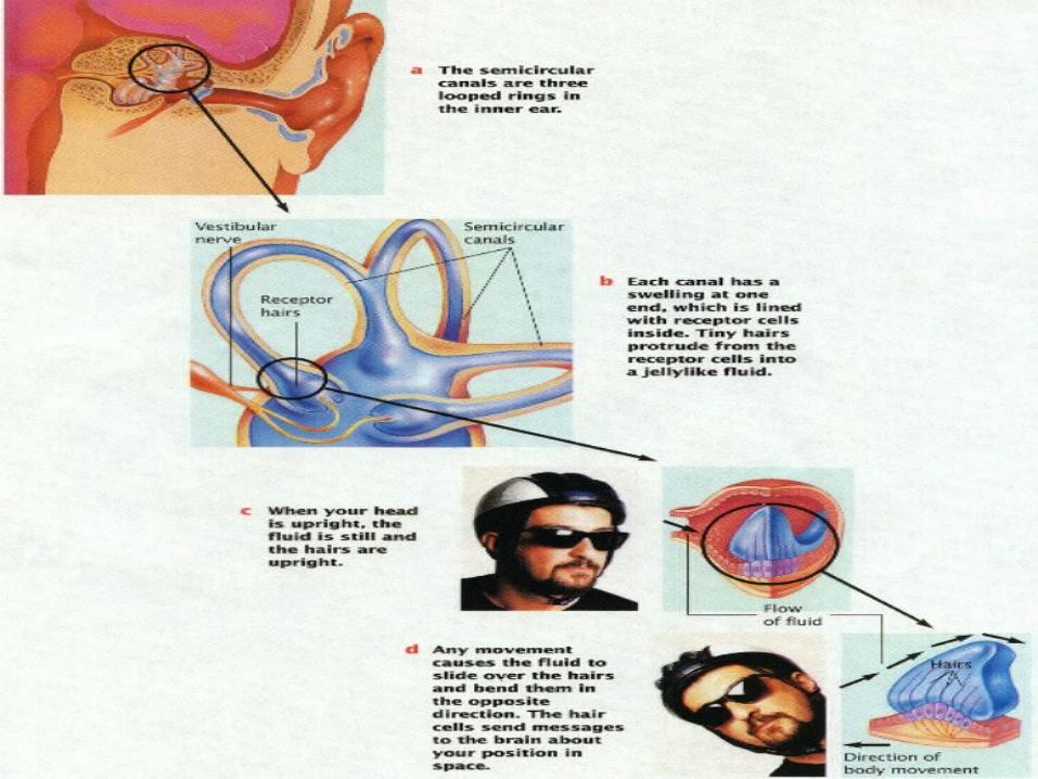
3e. Vestibular nerve -sends information about the heads position to brain
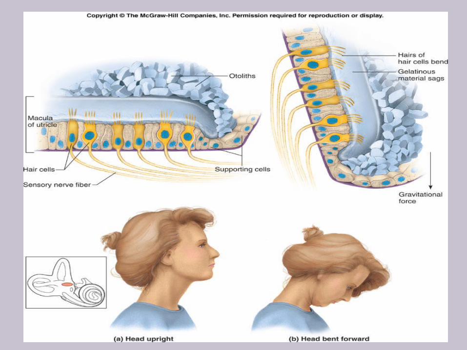
3f. Vestibule-detects static equilibrium (the position of the head when still)
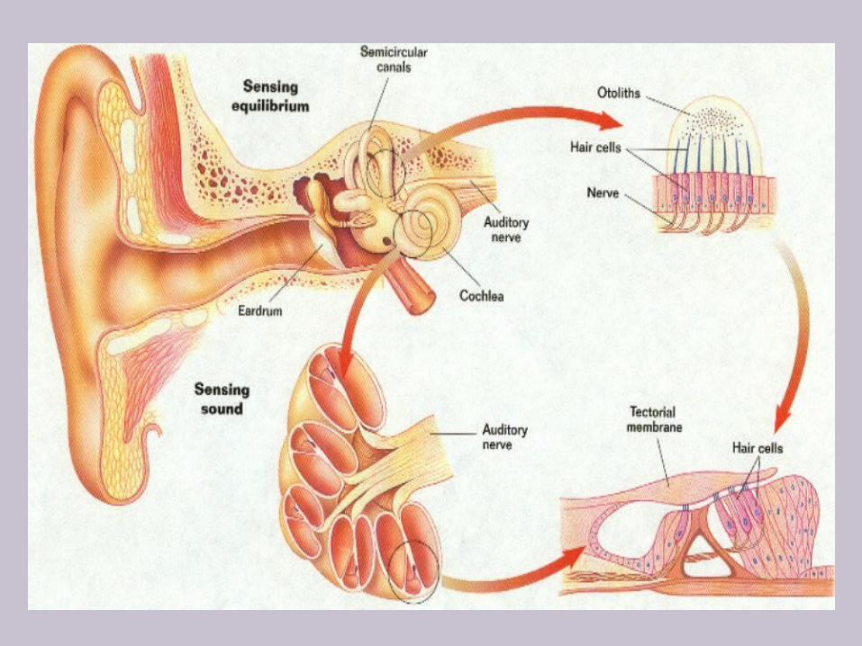
3g. Semicircular canals -detect dynamic equilibrium (motion of head) and aids in balance
6
Fig10.08
Semicircularcanals
Cochlear nerve
Maculae
Utricle Vestibular nerve
Membranous labyrinth(contains endolymph)
Osseous labyrinth(contains perilymph)
Cochlea
Cochlearduct (cut)containingendolymph
Scalachambers (cut)containingperilymph
Roundwindow
Saccule
Ovalwindow
Ampullae
Endolymph
Perilymph
Membranous labyrinth
Osseous labyrinth
Copyright © The McGraw-Hill Companies, Inc. Permission required for reproduction or display.
Video Links
How Hearing Works Conductive Hearing Loss Sensorineural Hearing Loss Cochlear Implants
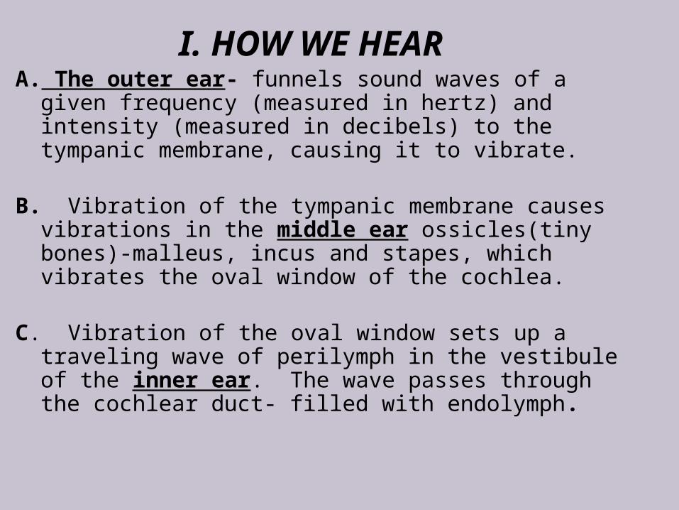
I. HOW WE HEARA. The outer ear- funnels sound waves of a
given frequency (measured in hertz) and intensity (measured in decibels) to the tympanic membrane, causing it to vibrate.
B. Vibration of the tympanic membrane causes
vibrations in the middle ear ossicles(tiny bones)-malleus, incus and stapes, which vibrates the oval window of the cochlea.
C. Vibration of the oval window sets up a
traveling wave of perilymph in the vestibule of the inner ear. The wave passes through the cochlear duct- filled with endolymph.
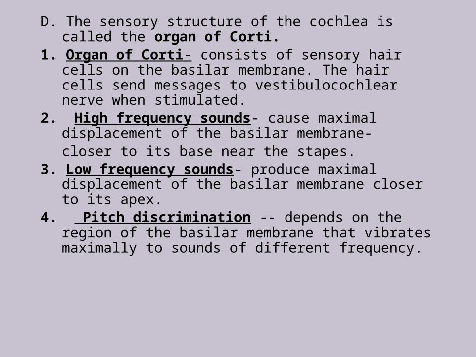
D. The sensory structure of the cochlea is called the organ of Corti.
1. Organ of Corti- consists of sensory hair cells on the basilar membrane. The hair cells send messages to vestibulocochlear nerve when stimulated.
2. High frequency sounds- cause maximal displacement of the basilar membrane-closer to its base near the stapes.
3. Low frequency sounds- produce maximal displacement of the basilar membrane closer to its apex.
4. Pitch discrimination -- depends on the region of the basilar membrane that vibrates maximally to sounds of different frequency.
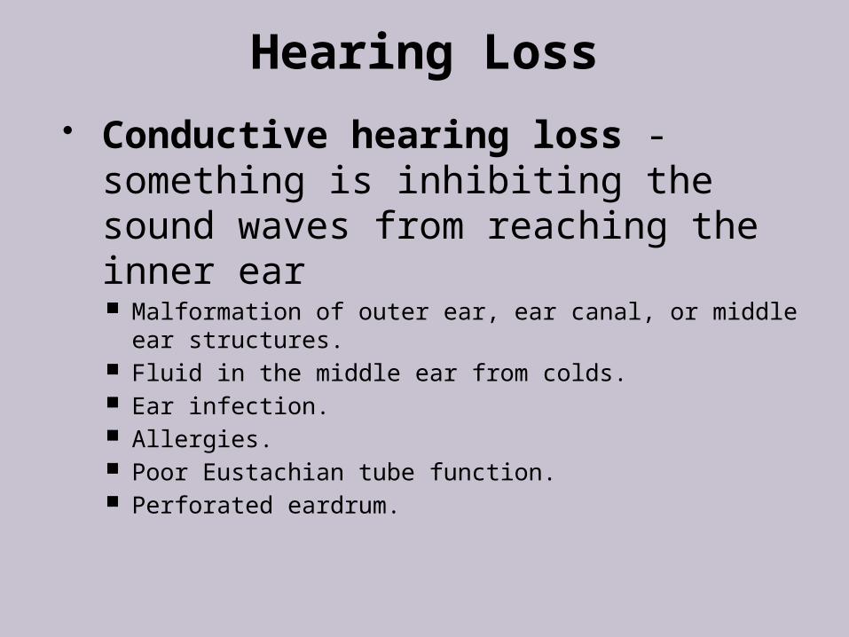
Hearing Loss Conductive hearing loss -
something is inhibiting the sound waves from reaching the inner ear Malformation of outer ear, ear canal, or middle ear
structures. Fluid in the middle ear from colds. Ear infection. Allergies. Poor Eustachian tube function. Perforated eardrum.
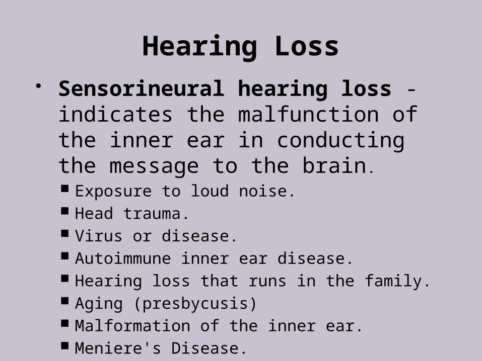
Hearing Loss Sensorineural hearing loss -
indicates the malfunction of the inner ear in conducting the message to the brain. Exposure to loud noise. Head trauma. Virus or disease. Autoimmune inner ear disease. Hearing loss that runs in the family. Aging (presbycusis) Malformation of the inner ear. Meniere's Disease.
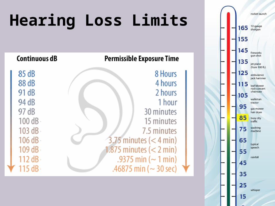
Hearing Loss Limits
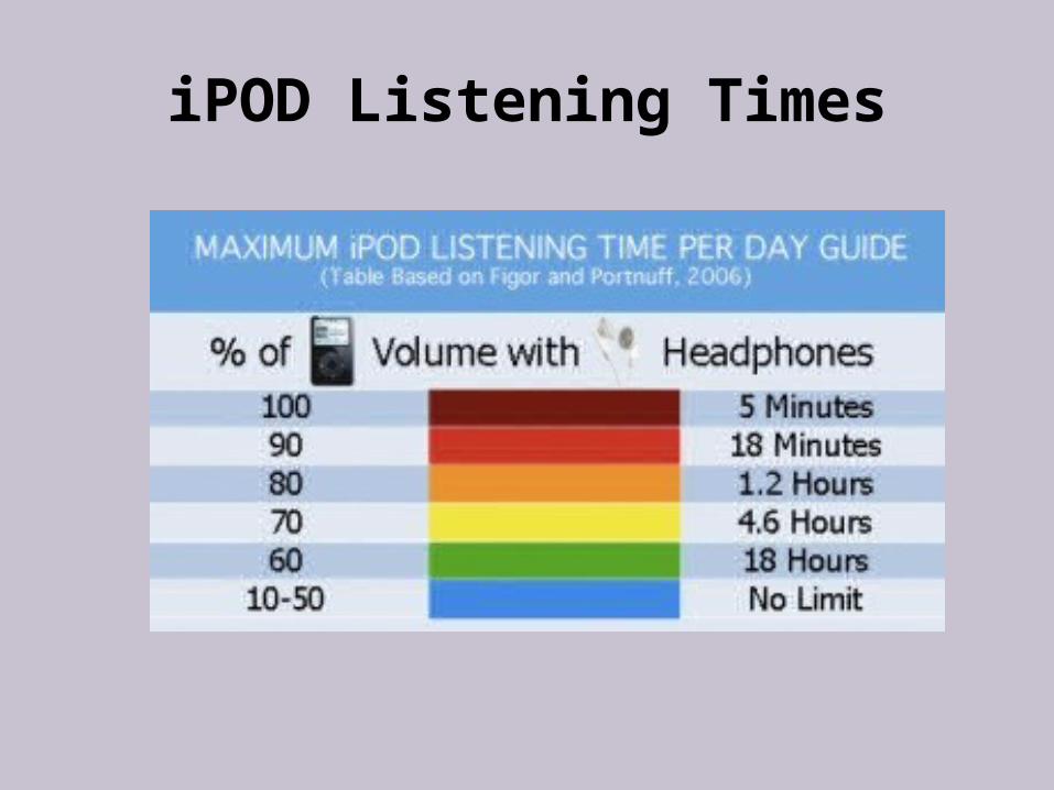
iPOD Listening Times
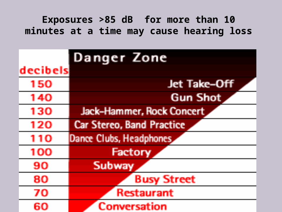
Exposures >85 dB for more than 10 minutes at a time may cause hearing loss


![17.2 Sound Waves: In Halliday and Resnick: Longitudinal waves are sound waves! Chapter 17: [Sound] Waves-(II) Sound waves propagate in gases. Can they](https://img.pdfslide.us/doc/110x75/56649eb25503460f94bb9375/172-sound-waves-in-halliday-and-resnick-longitudinal-waves-are-sound-waves.jpg)