Embed Size (px)
Citation preview
1
NCLEX RN Preparation Program
Respiratory Disorders
Module 5, Part 3 of 3
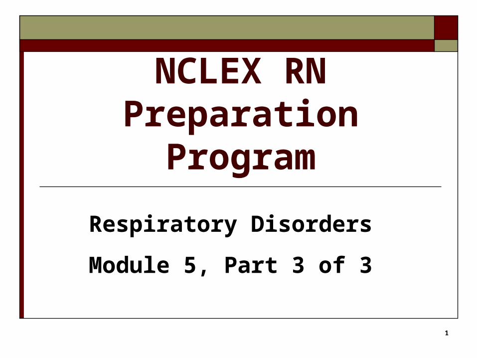
2
Chronic Airflow Limitation
Emphysema
+
Chronic Bronchitis
=
COPD
Chronic Obstructive Pulmonary Disease
Photo Source: National Heart, Lung and Blood Institute (NHLBI http://www.nhlbi.nih.gov/health/dci/Diseases/Copd/Copd_WhatIs.html
3
Emphysema
Loss of lung elasticity
Hyperinflation
Air trapped in lungs
Alveoli over-stretched bullae
4
Chronic Bronchitis
Recurrent inflammation Vasodilation, Congestion, Edema, Spasm
Excessive thick mucus blocks air flow Hypoxemia, CO2 retained
5
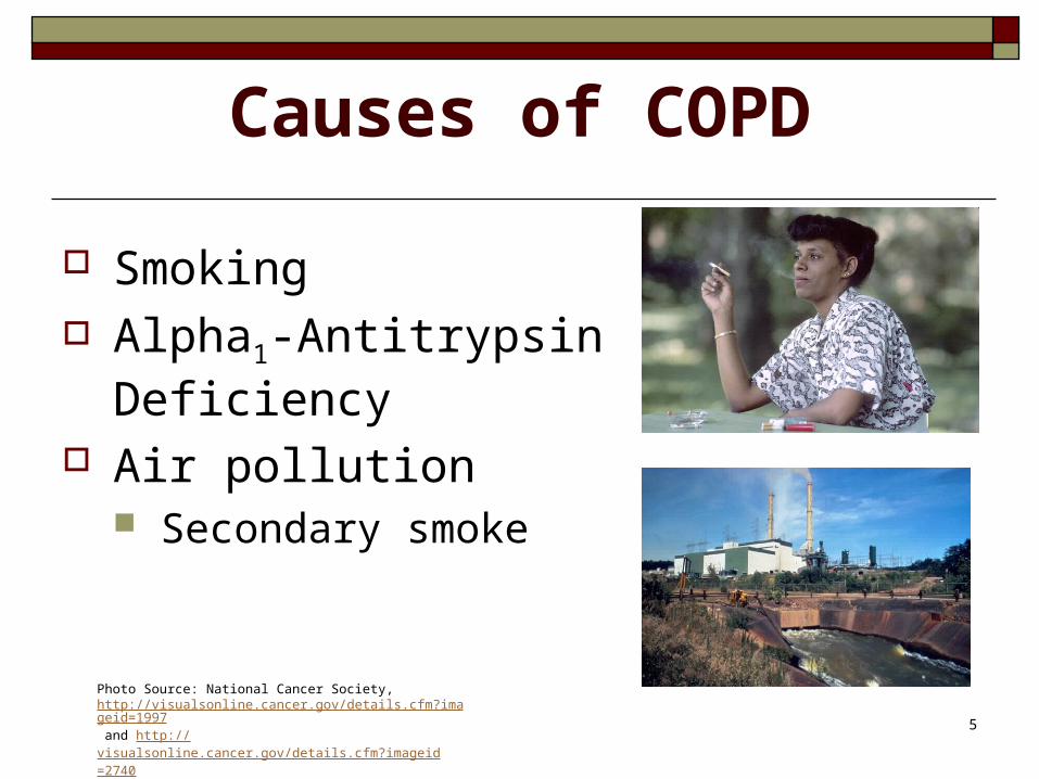
Causes of COPD
Smoking Alpha1-Antitrypsin
Deficiency Air pollution
Secondary smoke
Photo Source: National Cancer Society, http://visualsonline.cancer.gov/details.cfm?imageid=1997 and http://visualsonline.cancer.gov/details.cfm?imageid=2740
6
Signs of COPD
General Breathing Sputum Sounds Skin Finger tips

7
Assess
* LOC* Airway status and breathing* Pulses* RR, depth* BP, Heart Rate* SpO 2 level on room air* Color, temperature & capillary refill
8
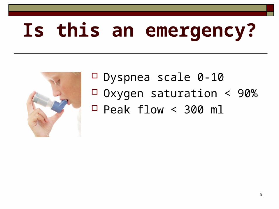
Is this an emergency?
Dyspnea scale 0-10 Oxygen saturation < 90% Peak flow < 300 ml
9
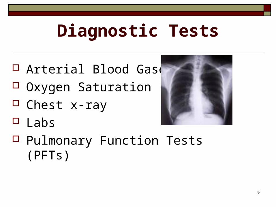
Diagnostic Tests
Arterial Blood Gases Oxygen Saturation Chest x-ray Labs Pulmonary Function Tests (PFTs)
10
Goal: Patent Airway
Position Secretions Mucolytics Expectoration Hydration Humidifier
11
Teach Effective Breathing Diaphragm Pursed-lips Controlled cough Orthopneic position
http://emphysemafoundation.org/pulhthex.jsp
12
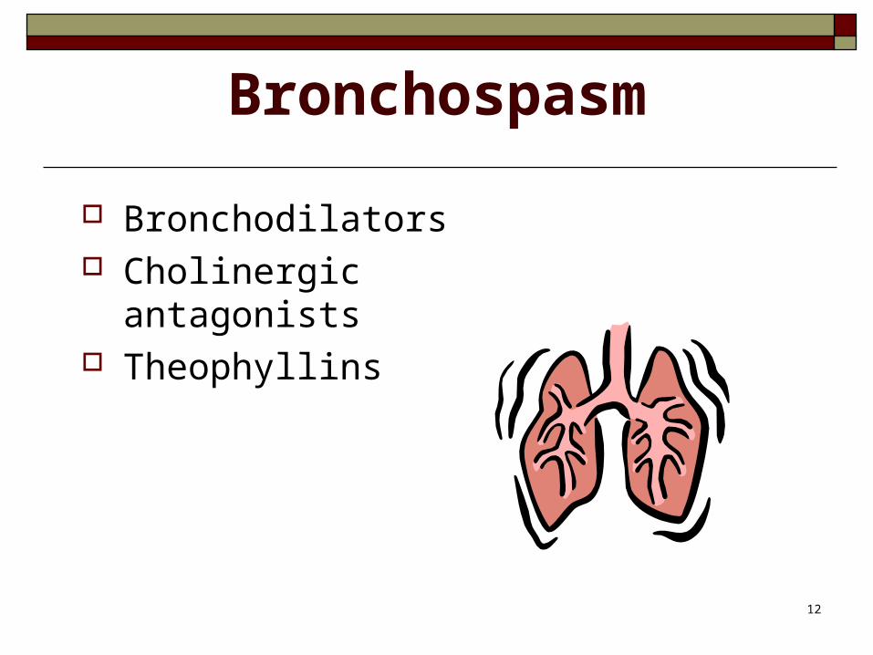
Bronchospasm
Bronchodilators Cholinergic antagonists Theophyllins
13
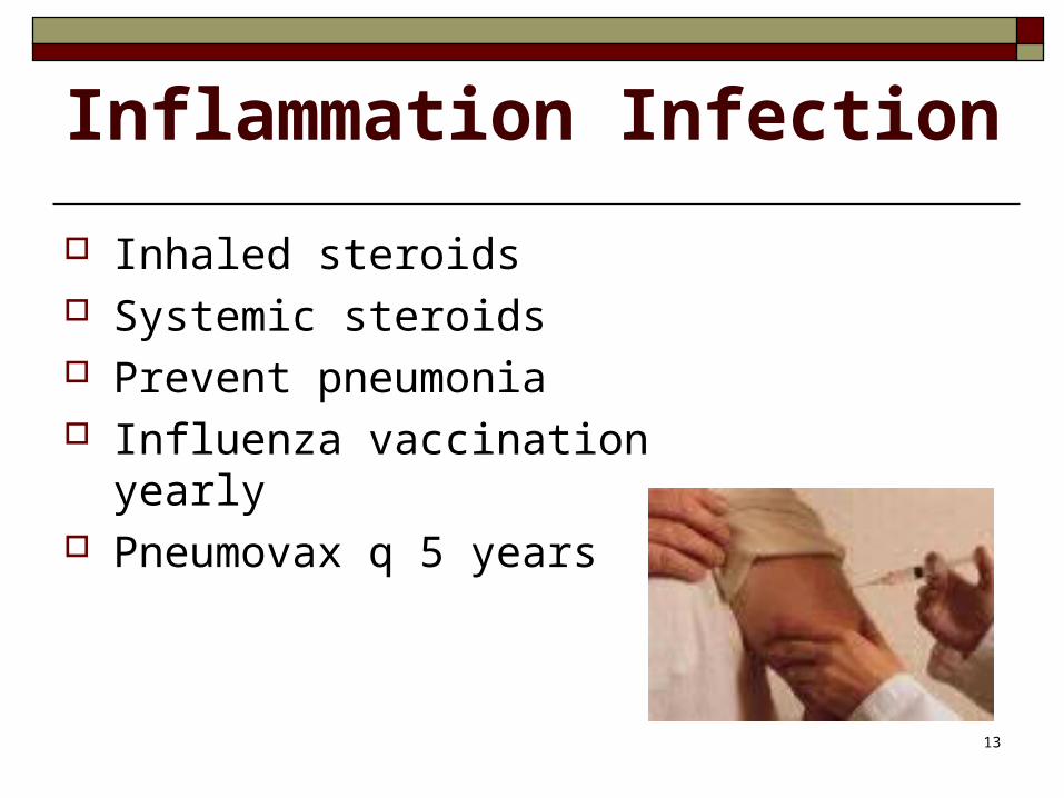
Inflammation Infection
Inhaled steroids Systemic steroids Prevent pneumonia Influenza vaccination yearly Pneumovax q 5 years
14
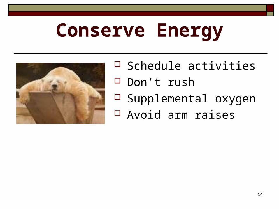
Conserve Energy
Schedule activities Don’t rush Supplemental oxygen Avoid arm raises
15
Mealtime Strategies
Rest 4-6 small meals Bronchodilator ac Easy chewing Supplements Avoid gas-producing foods
16
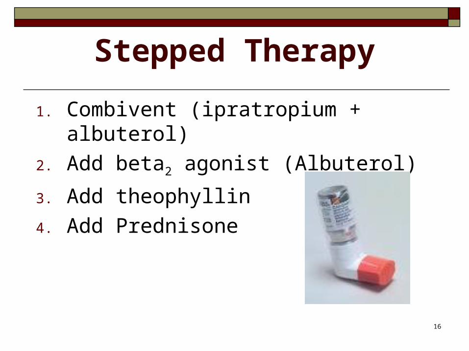
Stepped Therapy
1. Combivent (ipratropium + albuterol)
2. Add beta2 agonist (Albuterol)
3. Add theophyllin
4. Add Prednisone
17
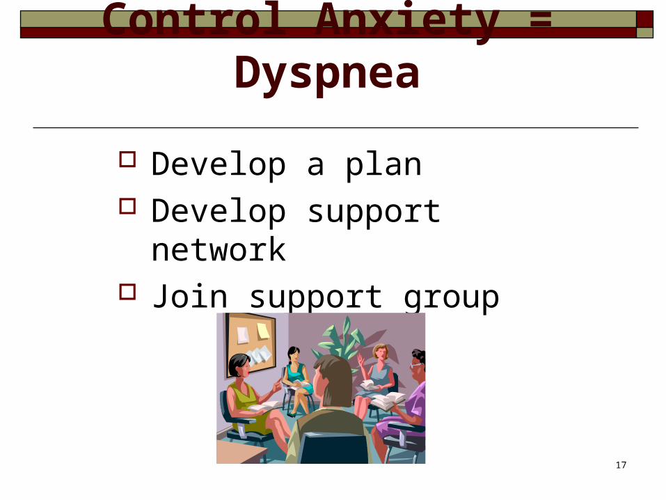
Control Anxiety = Dyspnea
Develop a plan Develop support network Join support group
18
Complementary/Alternatives
Ask about non-prescribed methods used
Teach relaxation techniques
19
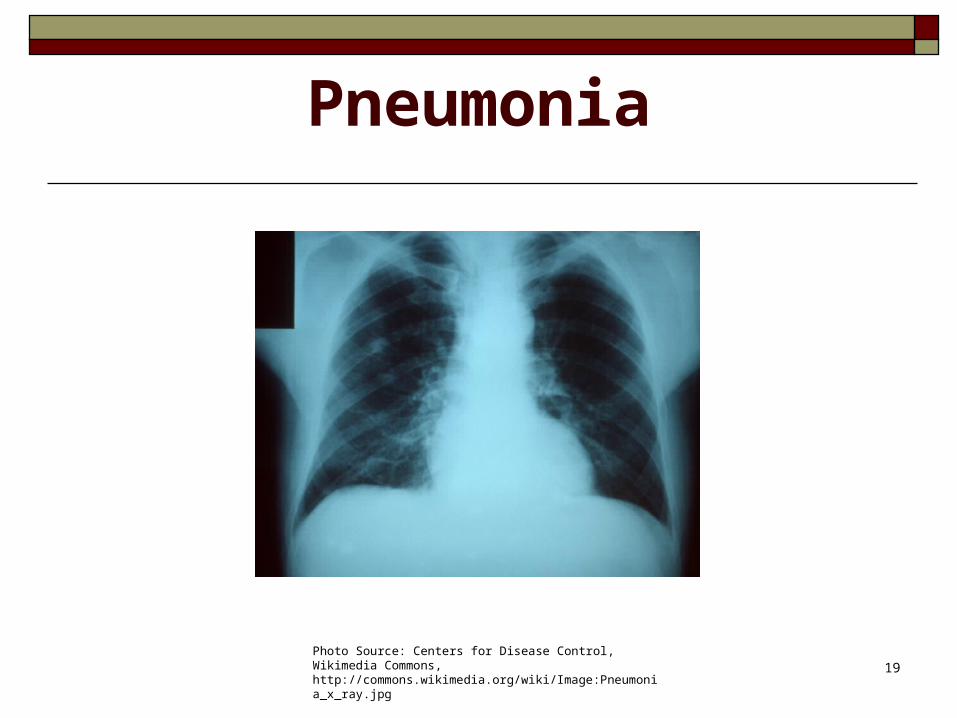
Pneumonia
Photo Source: Centers for Disease Control, Wikimedia Commons, http://commons.wikimedia.org/wiki/Image:Pneumonia_x_ray.jpg
20
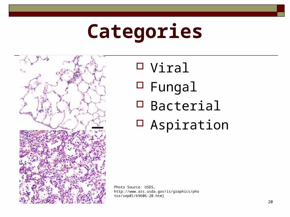
Categories
Viral Fungal Bacterial Aspiration
Photo Source: USDS, http://www.ars.usda.gov/is/graphics/photos/sep01/k9606-20.htm]
21
Classification
Causative agent (Streptococcus pneumoniae)
Anatomic location of the infection (lobar pneumonia)
By where it was acquired (community vs. hospital/nosocomial)
22
Major Organisms
Community-acquired: Streptococcus pneumoniae (gram +) Staphylococcus aureus (gram +)
Nosocomial: Staphylococcus aureus (gram +) MRSA
23
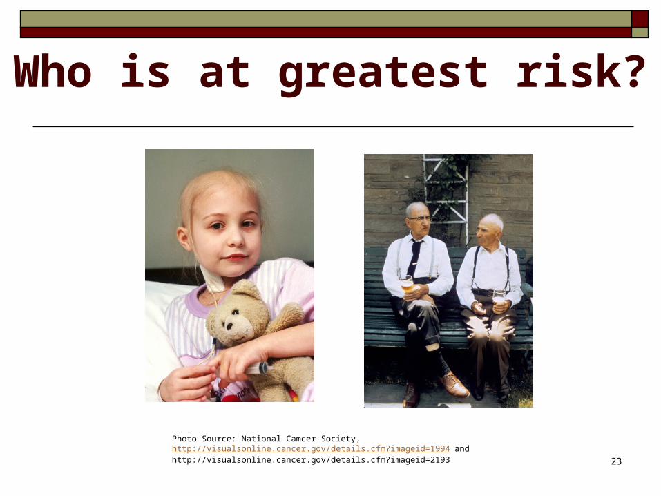
Who is at greatest risk?
Photo Source: National Camcer Society, http://visualsonline.cancer.gov/details.cfm?imageid=1994 and http://visualsonline.cancer.gov/details.cfm?imageid=2193
24
Community Prevention
PneumovaxWash handsDon’t smokeWear mask: dusty, moldy areasAvoid crowdsEat healthy dietExercise
25
Nosocomial Prevention
Prevent aspiration - How? Prevent cross-contamination Vaccinate inpatients Education Mouth care??
26
Signs & Symptoms
Fever, chills Dyspnea, RR, shallow breathing Coughing, crackles, wheezing Pleuritic pain Anorexia Hypoxemia Sputum: purulent, blood-tinged, rusty
27
Diagnosis
Sputum C&S Leukocytosis ABG’s Blood C&S Chest x-ray Oxygen saturation
28
Goal: Improve Gas Exchange
Oxygen Antibiotics Rest Incentive spirometry Raise head of bed No smoking
29
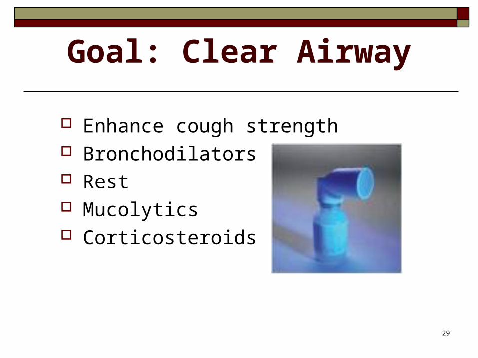
Goal: Clear Airway
Enhance cough strength Bronchodilators Rest Mucolytics Corticosteroids
30
Goal: Control Pain and Fever
Pain Fever control Adequate volume
31
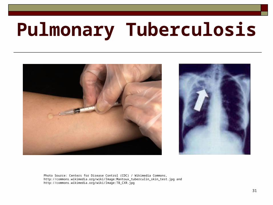
Pulmonary Tuberculosis
Photo Source: Centers for Disease Control (CDC) / Wikimedia Commons, http://commons.wikimedia.org/wiki/Image:Mantoux_tuberculin_skin_test.jpg and http://commons.wikimedia.org/wiki/Image:TB_CXR.jpg
32
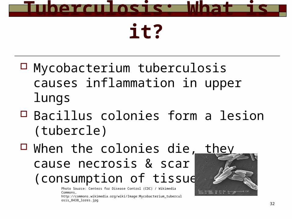
Tuberculosis: What is it?
Mycobacterium tuberculosis causes inflammation in upper lungs
Bacillus colonies form a lesion (tubercle) When the colonies die, they cause
necrosis & scar tissue (consumption of tissue)
Photo Source: Centers for Disease Control (CDC) / Wikimedia Commons, http://commons.wikimedia.org/wiki/Image:Mycobacterium_tuberculosis_8438_lores.jpg
33
How do I know I have it?
Cough that will not go away Feeling tired all the time Weight loss Loss of appetite Fever Coughing up blood Night sweats
34
Diagnosis
Initial Screening – skin test Positive if >10mm induration
Chest x-ray Sputum for AFB
35
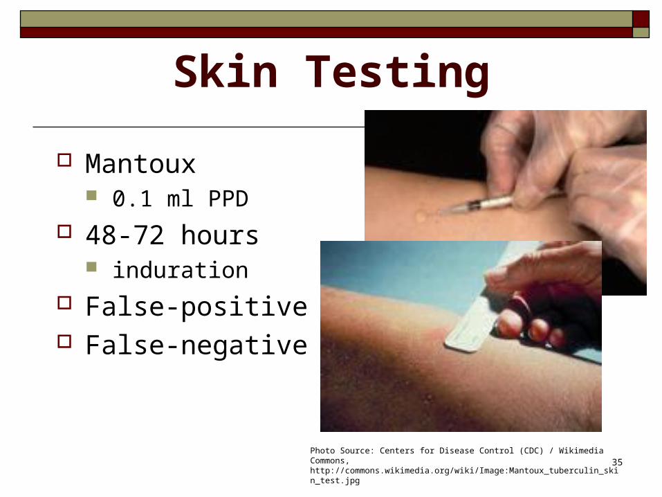
Skin Testing
Mantoux 0.1 ml PPD
48-72 hours induration
False-positive False-negative
Photo Source: Centers for Disease Control (CDC) / Wikimedia Commons, http://commons.wikimedia.org/wiki/Image:Mantoux_tuberculin_skin_test.jpg
36
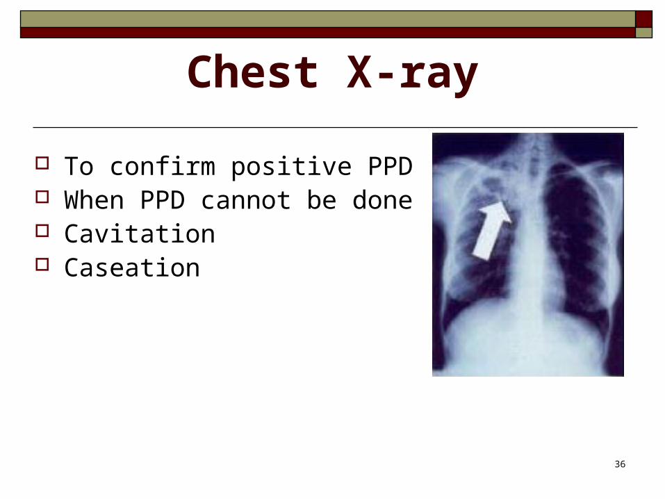
Chest X-ray
To confirm positive PPD When PPD cannot be done Cavitation Caseation
37
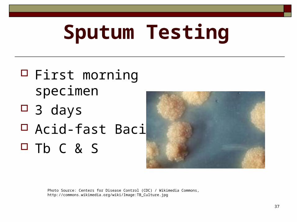
Sputum Testing
First morning specimen 3 days Acid-fast Bacilli Tb C & S
Photo Source: Centers for Disease Control (CDC) / Wikimedia Commons, http://commons.wikimedia.org/wiki/Image:TB_Culture.jpg
38
How is it treated?
Initial Therapy may include: Isoniazid (INH) Rifampin Pyrazinamide (PZA) Ethambutol or Streptomycin
After two months: Isoniazid Rifampin
39
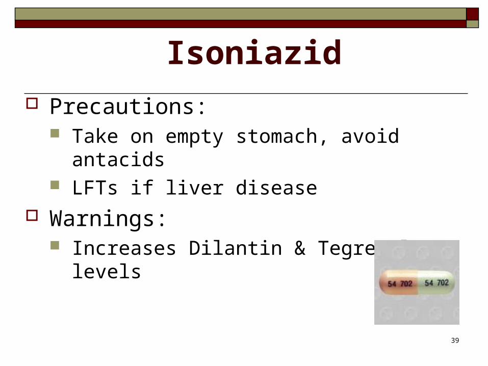
Isoniazid
Precautions: Take on empty stomach, avoid antacids LFTs if liver disease
Warnings: Increases Dilantin & Tegretol levels
40
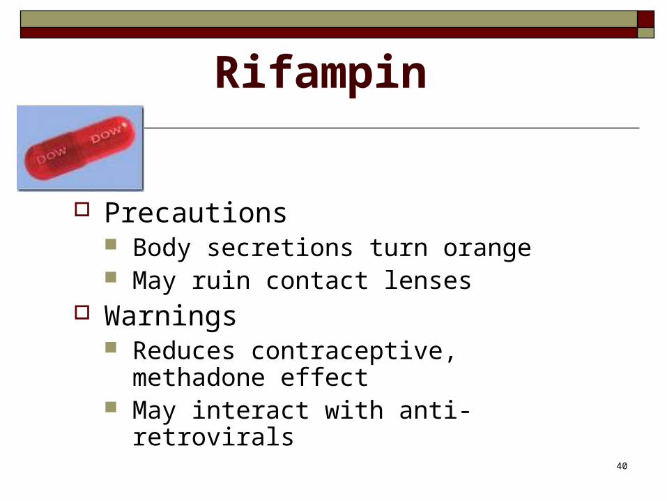
Rifampin
Precautions Body secretions turn orange May ruin contact lenses
Warnings Reduces contraceptive, methadone
effect May interact with anti-retrovirals
41
Rifapentine
Precautions Probably discolors body secretions
Warnings Decreased potency diabetes meds,
barbs, antibiotics, contraceptives
42
Ethambutol Precautions
Decreased visual acuity Decreased red-green color discrimination
Warnings Optic toxicity is dose related Increased toxicity with renal insufficiency
43
Pyrazinamide
Precautions Hepatotoxicity Nausea/vomiting Polyarthralgias Hyper-uricemia Transient rash Photo-sensitive dermatitis
44
Hospitalization
Isolate all patients with active pulmonary TB in negative-pressure rooms with high-volume air replacement and circulation
Continue isolation until combined drug treatment has been administered for 2 weeks, and three consecutive sputum smears have tested negative.
45
Transplant Recipients
Immune suppressed Donor organ with latent TB Reactivate pt’s latent infection Diagnosis difficult
Decreased PPD reaction
46
HIV positive
Increased risk: Why? Interactions with protease inhibitors Decreased CD4 cell count anergy
(impaired or absent ability to react to common antigens administered through skin) PPD testing early in HIV infection Use control to rule out anergy
?? INH prophylaxis
47
Drug Toxicity
Hx of liver disease Consuming alcohol daily Baseline + repeat LFTs Watch!
Dark urine Light stools Fatigue
48
Drug Resistance
Multi-drug resistant TB (MDRTB) Second-line drugs Increased time of treatment
49
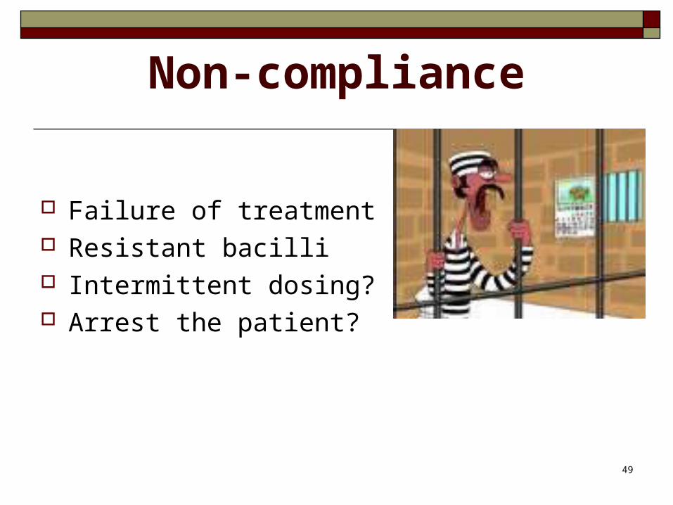
Non-compliance
Failure of treatment Resistant bacilli Intermittent dosing? Arrest the patient?
50
Patient/Family Teaching
Prevention Phone contact Test entire family Use precautions Follow-up sputum cultures Diet
51
Acute Respiratory Failure
Dyspnea, tachycardia Progressive respiratory distress Breath sounds Mental status
52
ARDS
Aspiration Sepsis Drowning Trauma

53
Diagnostic Tests
pO2 < 60 mmHg
pCO2 > 50 mmHg
O2 saturation < 90% Chest x-ray – increasing
infiltrates to “white out”
54
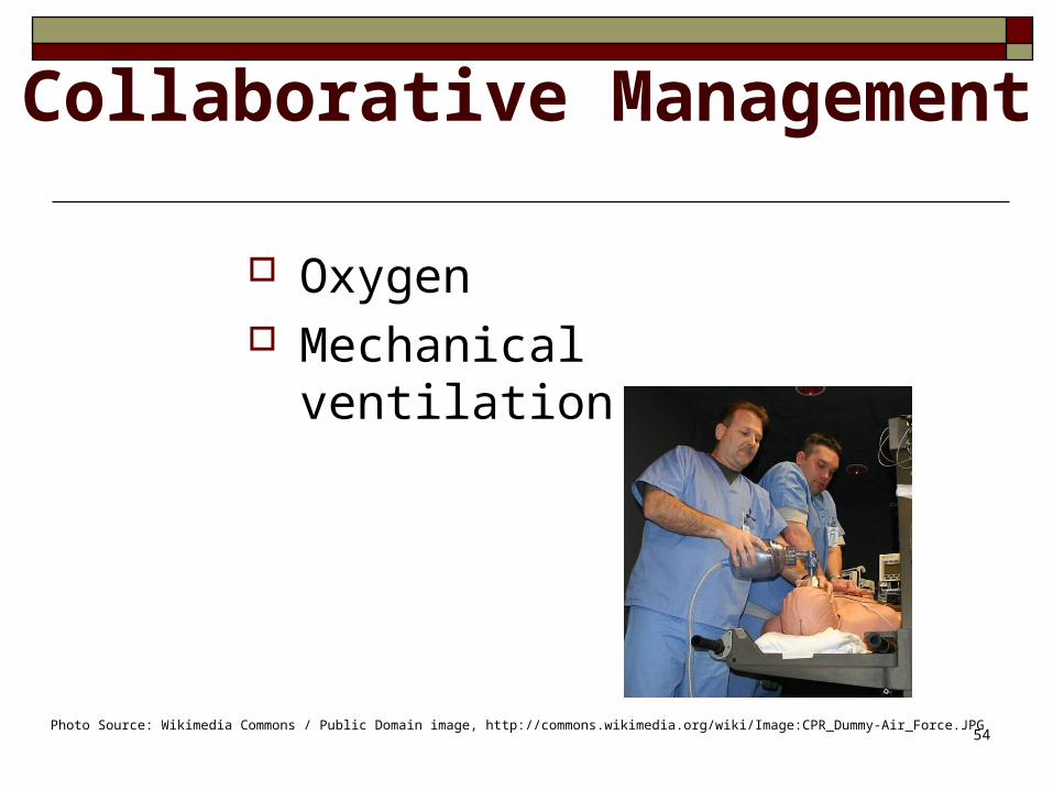
Collaborative Management
Oxygen Mechanical ventilation
Photo Source: Wikimedia Commons / Public Domain image, http://commons.wikimedia.org/wiki/Image:CPR_Dummy-Air_Force.JPG
55
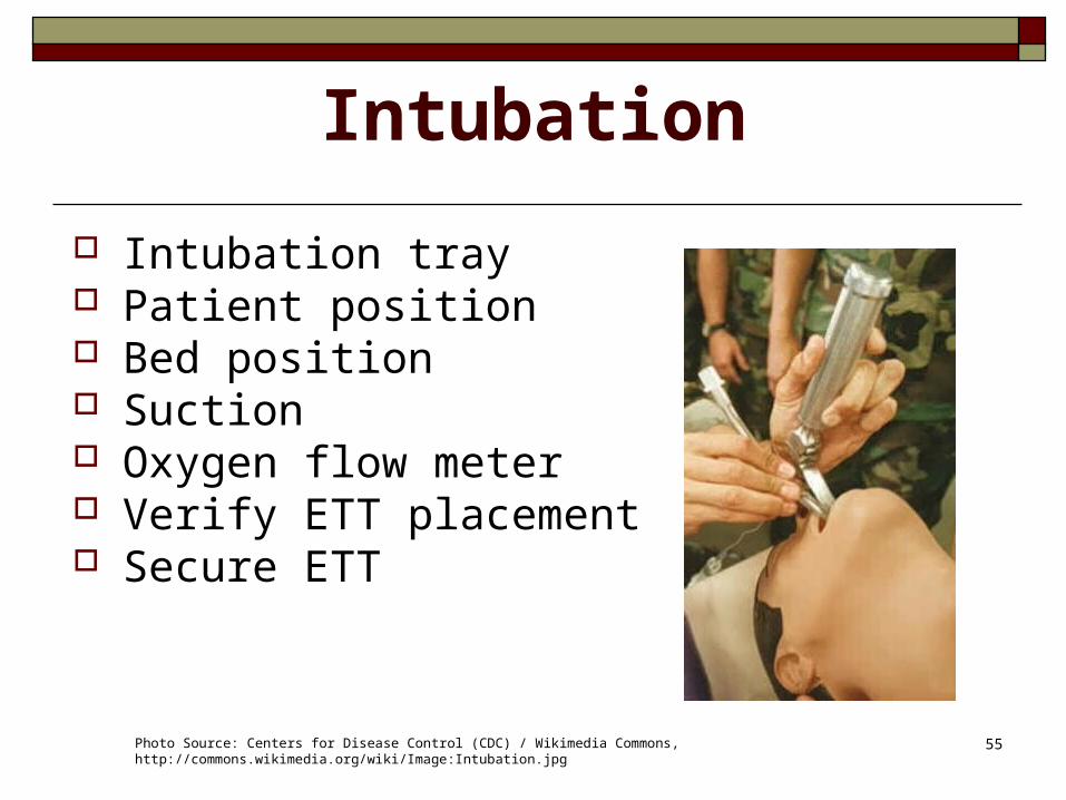
Intubation
Intubation tray Patient position Bed position Suction Oxygen flow meter Verify ETT placement Secure ETT
Photo Source: Centers for Disease Control (CDC) / Wikimedia Commons, http://commons.wikimedia.org/wiki/Image:Intubation.jpg
56
Mechanical Ventilation
FiO2 100%
Tidal volume (Vt) 6-7 ml/kg Rate 20-28/minute
57
Ventilators
Negative pressure Pressure cycled Time cycled Volume cycled
58
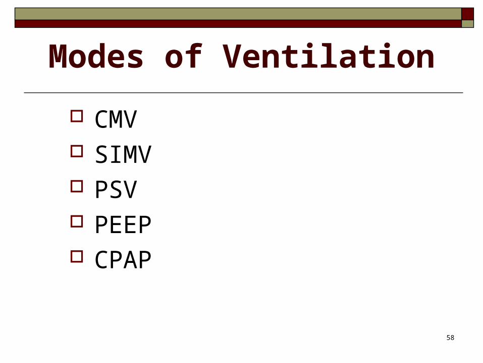
Modes of Ventilation
CMV SIMV PSV PEEP CPAP
59
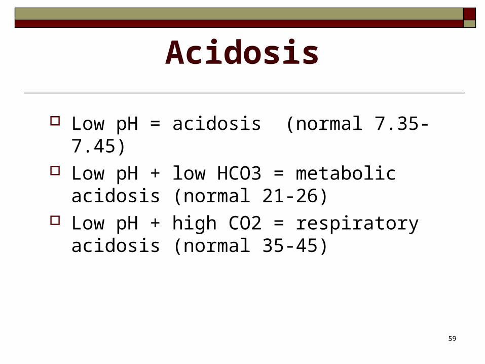
Acidosis
Low pH = acidosis (normal 7.35-7.45) Low pH + low HCO3 = metabolic
acidosis (normal 21-26) Low pH + high CO2 = respiratory
acidosis (normal 35-45)
60
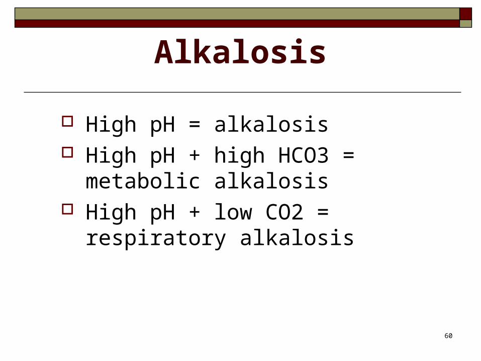
Alkalosis
High pH = alkalosis High pH + high HCO3 = metabolic
alkalosis High pH + low CO2 = respiratory
alkalosis
61
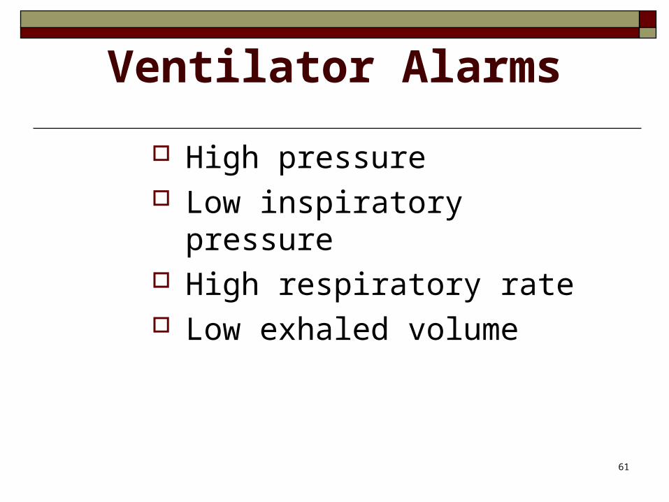
Ventilator Alarms
High pressure Low inspiratory pressure High respiratory rate Low exhaled volume
62
Monitor Physiological Response
Breath sounds Breathing pattern Skin color Secretions Oxygen saturation ABGs, daily chest x-ray
63
Monitor Psychological Response
Anxiety Communication Anticipate questions/needs
64
Manage the Ventilator
Correct settings? Alarms on? Maintain humidity Monitor inline temperature ETT placement, cuff Tubing adjustments
65
Prevent Complications
Barotrauma Stress ulcers Infection: Ventilator-assisted
pneumonia (VAP) Ventilator dependence Pressure necrosis
66
Weaning from Ventilator
Awake, rested Muscle strength Heart rhythm Breath sounds ABGs Pulmonary function tests
67
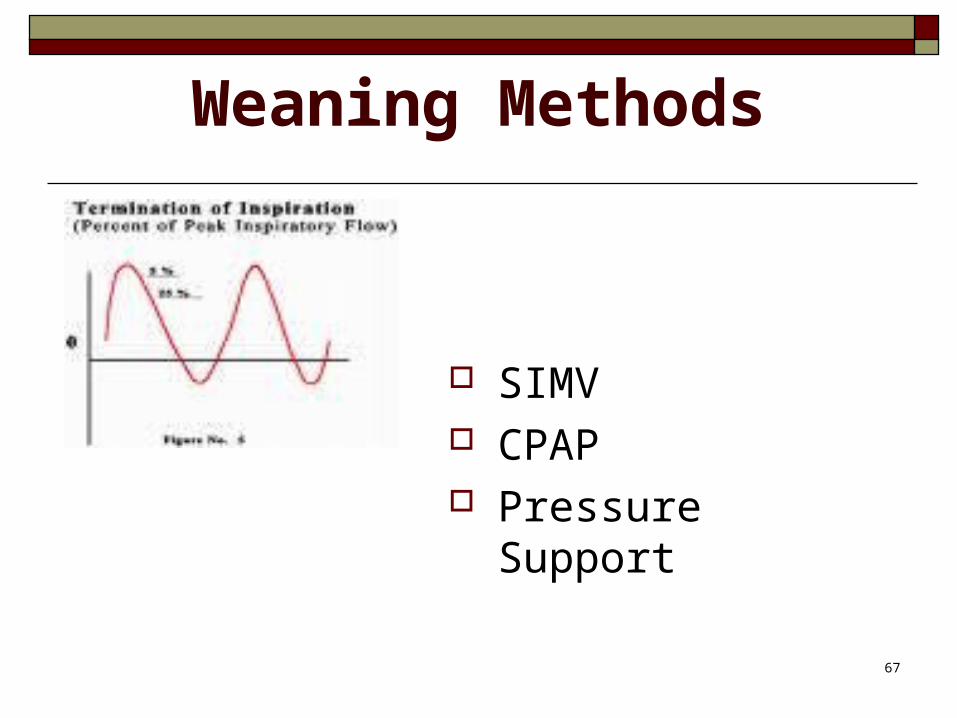
Weaning Methods
SIMV CPAP Pressure Support
68
Extubation
Explain procedure Prepare: nasal cannula, towel, Chux Hyper-oxygenate Suction Deflate pilot balloon Suction Pull tube No talking!
69
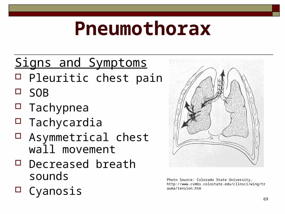
Pneumothorax
Signs and Symptoms Pleuritic chest pain SOB Tachypnea Tachycardia Asymmetrical chest wall
movement Decreased breath sounds Cyanosis Photo Source: Colorado State University,
http://www.cvmbs.colostate.edu/clinsci/wing/trauma/tension.htm
70
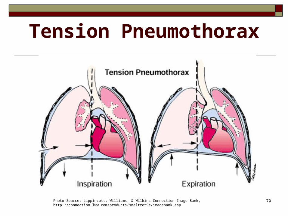
Tension Pneumothorax
Photo Source: Lippincott, Williams, & Wilkins Connection Image Bank, http://connection.lww.com/products/smeltzer9e/imagebank.asp
71
Tension Pneumothorax
Signs and Symptoms Tracheal deviation Distended neck veins Hypotension Compensatory tachycardia
& tachypnea Decreased cardiac outputMust be treated promptly
72
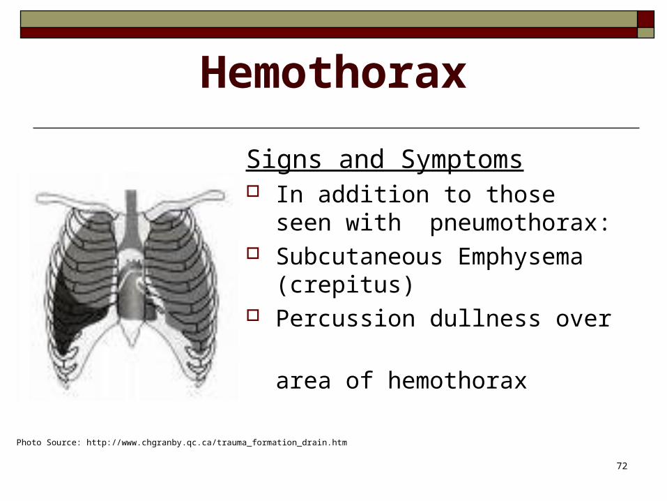
Hemothorax
Signs and Symptoms In addition to those seen with
pneumothorax: Subcutaneous Emphysema
(crepitus) Percussion dullness over
area of hemothorax
Photo Source: http://www.chgranby.qc.ca/trauma_formation_drain.htm
73
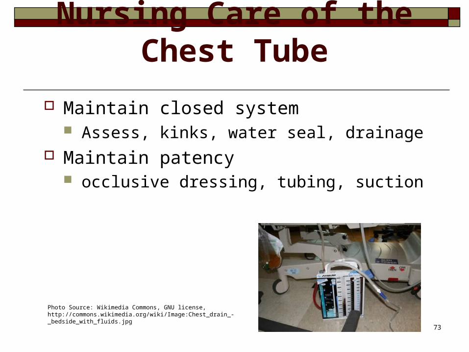
Nursing Care of the Chest Tube
Maintain closed system Assess, kinks, water seal, drainage
Maintain patency occlusive dressing, tubing, suction
Photo Source: Wikimedia Commons, GNU license, http://commons.wikimedia.org/wiki/Image:Chest_drain_-_bedside_with_fluids.jpg
74
Nursing Care of the Patient
Oxygen Vital signs Chest wall movement, trachea, neck
veins Position Watch for distress
75
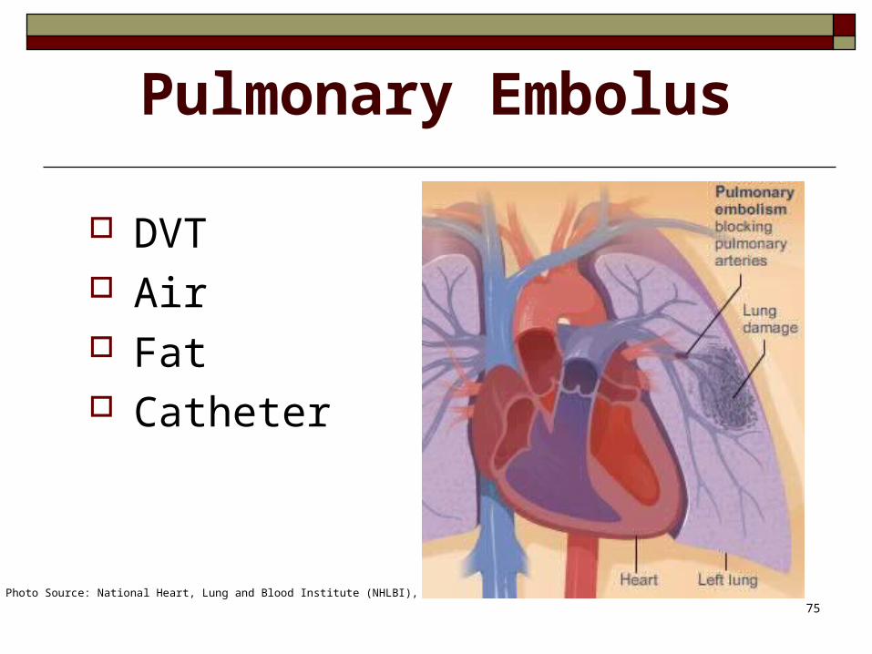
Pulmonary Embolus
DVT Air Fat Catheter
Photo Source: National Heart, Lung and Blood Institute (NHLBI),
76
Signs/Symptoms
Classic triad Common: dyspnea, tachypnea,
pleuritic pain Pleuritic chest pain + dyspnea +
predisposing factor
77
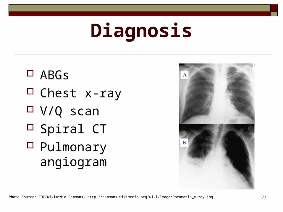
Diagnosis
ABGs Chest x-ray V/Q scan Spiral CT Pulmonary angiogram
Photo Source: CDC/Wikimedia Commons, http://commons.wikimedia.org/wiki/Image:Pneumonia_x-ray.jpg
78
Prevention
Early ambulation Hydration Anti-embolic stockings Sequential pumps Avoid lower extremity punctures Aspirate clotted IVs SQ heparin or LMWH
79
Emergency Measures
Oxygen HOB up Support Stat ABGs, chest x-ray Prepare for code blue
80
Collaborative Management
Continue oxygen Bed rest Heparin drip Coumadin Thrombolytics? Embolectomy, umbrella filter
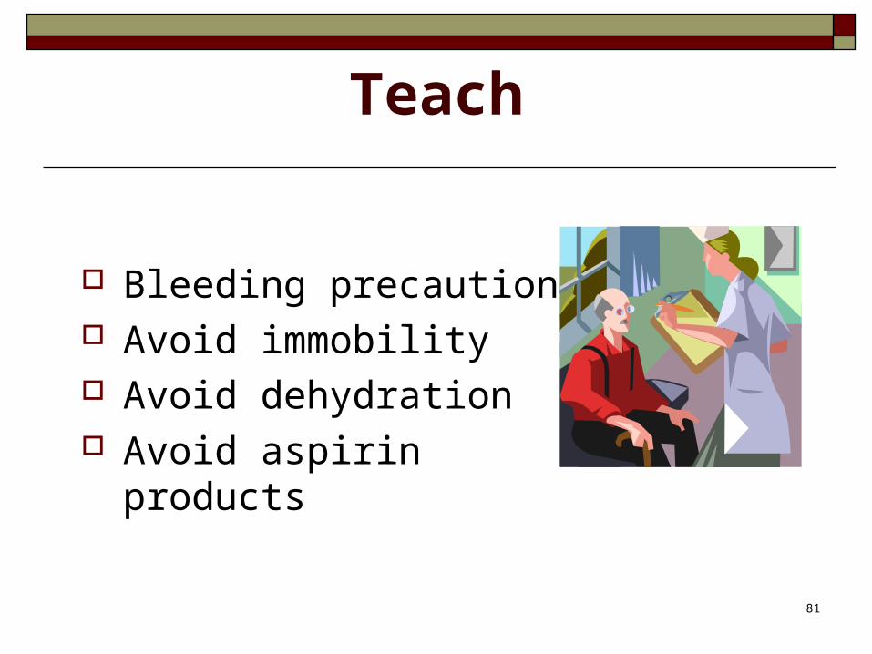
81
Teach
Bleeding precautions Avoid immobility Avoid dehydration Avoid aspirin products
82
Photo Acknowledgement:Unless noted otherwise, all photos and clip art contained in this module were obtained from the 2003 Microsoft Office Clip Art Gallery.