Embed Size (px)
Citation preview
1
Synthesis of silver nanoparticles from Anagalis arvensis and their biomedical applications
Mohib Shah1*, Natasha Anwar2, Samreen Saleem3, Iqbal Munir5, Niaz Ali Shah4, Baseerat Fida1
Abbas Khan2, Anwar Hussain1 and Faiz Ul Amin5.
1Department of Botany, Abdul Wali Khan University Mardan, 23200, Pakistan
2Department of Chemistry, Abdul Wali Khan University Mardan, 23200, Pakistan
3Department of Botany, Women University Swabi, Pakistan
4Tsinghua University, Beijing, China.Postcode: 100084
5Institute of Biotechnology and Genetic Engineering (IBGE), The University of Agriculture
Peshawar, Pakistan.
* Corresponding author
Mohib Shah, Prof. Ph.D.
Department of Botany,
Abdul Wali Khan University, Mardan 23200, Pakistan
Tel #: +92-937-830056 Mobile #: +92-314-9047303
E-mail: [email protected]
Preprints (www.preprints.org) | NOT PEER-REVIEWED | Posted: 16 September 2018 doi:10.20944/preprints201809.0282.v1
© 2018 by the author(s). Distributed under a Creative Commons CC BY license.
2
Abstract
Background. Nanotechnology is promising field for generating new applications. A green
synthesis of nanoparticles through biological methods using plant extract have a reliable and
ecofriendly approach to improve our global environment.
Methods. Silver nanoparticles (AgNPs) were synthesized using aqueous extract of Anagalis
arvensis L and silver nitrate and were physicochemically characterized.
Results. The stability of AgNPs toward acidity, alkalinity, salinity and temperature showed that
they remained stable at room temperature for more than two months. The SEM and TEM
analysis of the AgNPs showed that they have a uniform spherical shape with an average size in
the range of 40–78 nm. Further 1-Dibhenyl-2-Picrylhydrazl radical in Anagalis arvensis
L.mediated AgNPs showed a maximum activity of 98% at concentration of 200μg/mL.
Hydrogen peroxide scavenging assay in Anagalis arvensis L. mediated AgNPs showed a
maximum activity of 85% at concentration of 200μg/mL. Reducing power of Anagalis arvensis
L.Ag NPs exhibited a higher activity of 330 μg/mL at concentration of 200 μg/mL. These NPs
have cytotoxic effects against brine shrimp (Artemia salina) nauplii with a value of 53% LD
178.04μg/mL.
Conclusion. The AgNPs synthesized using Anagalis arvensis L. extract demonstrate a broad
range of applications.
Keywords: Silver Nanoparticles, Green Synthesis, Anti diabetic, Cytotoxic.
Preprints (www.preprints.org) | NOT PEER-REVIEWED | Posted: 16 September 2018 doi:10.20944/preprints201809.0282.v1
3
1. Introduction
In the last two decades, nanoparticle (NPs) research has become one of the most important areas
in modern materials science. Nanotechnology deals with exploitation of unique properties of tiny
particles at molecular levels i.e. millionth of millimeter. Scientists from different disciplines
including chemists, engineers, material scientists, physicists as well as biologists are involved in
this emerging field of research. Generally, nanotechnology works with materials, devices and
other structures with at least one dimension sized from 1 to 100 nm (Gardea-Torresdey et al.
2003). The term "nano-technology" was used by Norio Taniguichi in 1974 to designate
semiconductor processes involving control on the order of a nanometer. From the mid of 1980s,
progress in nanometer scale science and technology was exploded. The term “nano” is derived
from Greek origin and means dwarf or extremely small. This term was widely adapted by almost
all walks of people. This technology is being applied in almost every field including
biomedicine, mechanics, energy science, materials development, magnetics, optics, electronics
and information technology. It is multidisciplinary field concerning research and advancement of
technology in various fields of science like physics, chemistry, material science and
biotechnology (Taniguchi 1974).
Nanotechnology concerns with the progress of experimental processes for the synthesis of NPs
of different sizes, shapes and controlled disparity (Bhatt & Tripathi 2011). Ultrafine particle or
NPs are between 1 to 100 nm in size (Meshot et al. 2012). The NPs have greater surface area per
volume than larger particles and exerting a stronger effect on the surrounding environment and
reaction with other substances. Consequently, nanotechnology can make harmless substances
assume hazardous characteristics. A reduction in the size of nano-sized particles increases the
Preprints (www.preprints.org) | NOT PEER-REVIEWED | Posted: 16 September 2018 doi:10.20944/preprints201809.0282.v1
4
particle surface area. Additional chemical molecules may attach to this surface, enhancing the
reactivity and increasing toxic effects.
The NPs research is currently an area of scientific interest due to a wide variety of potential
application in bio-medicinal, optical and electronic fields. In the recent few decades the field of
metal NPs has a wonderful growth. Fe NPs were synthesized for the first time from Fe(CO)5 in
organic solvents and similar approach was used to make Co NPs. Amorphous Fe NPs may be
prepared at low temperature and the carbonyl is decomposed chemically. But great research has
been done on silver and gold NPs due to their unique properties (e.g., size and shape depending
optical, electrical and magnetic properties) which can be incorporated into antimicrobial
applications, biosensor materials, composite fibers, cryogenic superconducting materials,
cosmetic products and electronic components. Metal NPs can be synthesized using different
physical and chemical methods (Mohanpuria et al. 2008; Yu 2007) i.e. citrate reduction
method(Turkevich et al. 1951), two phase synthesis and one phase synthesis in organic
solvents(Shem et al. 2009). Among the noble metal NPs, silver NPs are a playful product from
the field of nanotechnology which has gained boundless interests because of their distinctive
properties such as chemical stability, good conductivity, catalytic and most important
antibacterial, anti-viral, antifungal and anti-inflammatory activities which can be incorporated
into composite fibers, cryogenic superconducting materials, cosmetic products, food industry and
electronic components(Ahmad et al. 2003; Klaus-Joerger et al. 2001). They have also lot of
application on biomedical side; being added to wound dressings, topical creams, antiseptic
sprays and fabrics.Silver functions as an antiseptic and displays a broad biocidal effect against
microorganisms through the disruption of their unicellular membrane thus disturbing their
enzymatic activities. In our present study AgNPs were synthesized by green way because it play
Preprints (www.preprints.org) | NOT PEER-REVIEWED | Posted: 16 September 2018 doi:10.20944/preprints201809.0282.v1
5
an important role in our daily life and have wide-ranging applications such as bimolecular
detection, catalysis, biosensors and medicine; it is been acknowledged to have strong inhibitory
and bactericidal effects along with the anti-fungal, anti-inflammatory and anti-angiogenesis
activities (El-Chaghaby & Ahmad 2011). To the best of our knowledge, this is the first report of
silver NPs synthesis using an aqueous extract of Anagalis arvensis Lor scarlet pimpernel
commonly known as blue-scarlet pimpernel (Khoshkholgh-Pahlaviani et al. 2013).
2. Results
Optimization of AgNO3/Plant ratios for Anagalis arvensis L. stabilized Ag NPs
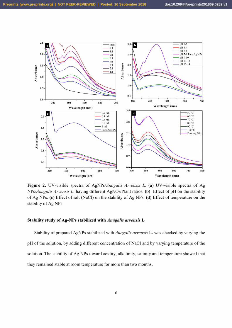
UV-Visible spectra of AgNPs stabilized with Anagalis arvensis L. showed a strong
absorbance band near 420nm. Resonance plasmon band in the region of 400nm to 500nm was an
indication of the presence of Ag NPs (Ahmad et al. 2011). Optimization study was performed by
carrying out a number of reactions while varying the amounts of salt to plant ratio. The UV-Vis
spectra for such experiments are shown in Fig. 2a. The best optimized ratio, having good SPR in
characteristic region of AgNPs, selected for further studies was 2:1.
Preprints (www.preprints.org) | NOT PEER-REVIEWED | Posted: 16 September 2018 doi:10.20944/preprints201809.0282.v1
6
Figure 2. UV-visible spectra of AgNPs/Anagalis Arvensis L. (a) UV-visible spectra of Ag NPs/Anagalis Arvensis L. having different AgNO3/Plant ratios. (b) Effect of pH on the stability of Ag NPs. (c) Effect of salt (NaCl) on the stability of Ag NPs. (d) Effect of temperature on the stability of Ag NPs.
Stability study of Ag-NPs stabilized with Anagalis arvensis L
Stability of prepared AgNPs stabilized with Anagalis arvensis L. was checked by varying the
pH of the solution, by adding different concentration of NaCl and by varying temperature of the
solution. The stability of Ag NPs toward acidity, alkalinity, salinity and temperature showed that
they remained stable at room temperature for more than two months.
Preprints (www.preprints.org) | NOT PEER-REVIEWED | Posted: 16 September 2018 doi:10.20944/preprints201809.0282.v1
7
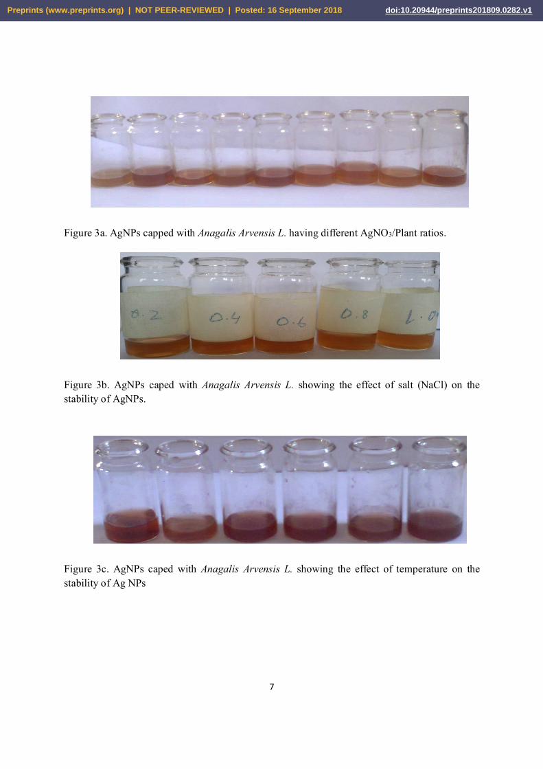
Figure 3a. AgNPs capped with Anagalis Arvensis L. having different AgNO3/Plant ratios.
Figure 3b. AgNPs caped with Anagalis Arvensis L. showing the effect of salt (NaCl) on the stability of AgNPs.
Figure 3c. AgNPs caped with Anagalis Arvensis L. showing the effect of temperature on the stability of Ag NPs
Preprints (www.preprints.org) | NOT PEER-REVIEWED | Posted: 16 September 2018 doi:10.20944/preprints201809.0282.v1
8
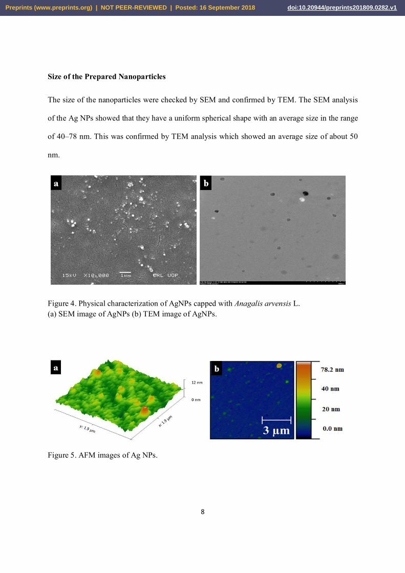
Size of the Prepared Nanoparticles
The size of the nanoparticles were checked by SEM and confirmed by TEM. The SEM analysis
of the Ag NPs showed that they have a uniform spherical shape with an average size in the range
of 40–78 nm. This was confirmed by TEM analysis which showed an average size of about 50
nm.
Figure 4. Physical characterization of AgNPs capped with Anagalis arvensis L. (a) SEM image of AgNPs (b) TEM image of AgNPs.
Figure 5. AFM images of Ag NPs.
Preprints (www.preprints.org) | NOT PEER-REVIEWED | Posted: 16 September 2018 doi:10.20944/preprints201809.0282.v1
9
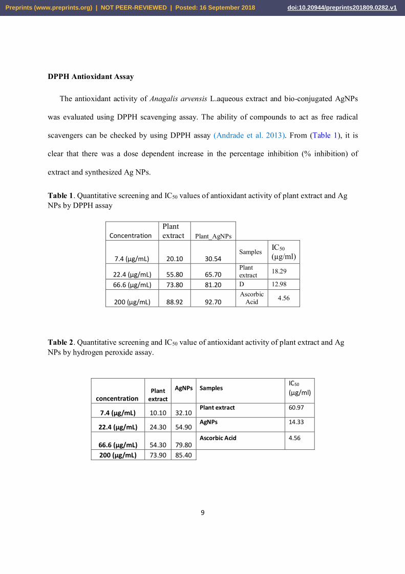
DPPH Antioxidant Assay
The antioxidant activity of Anagalis arvensis L.aqueous extract and bio-conjugated AgNPs
was evaluated using DPPH scavenging assay. The ability of compounds to act as free radical
scavengers can be checked by using DPPH assay (Andrade et al. 2013). From (Table 1), it is
clear that there was a dose dependent increase in the percentage inhibition (% inhibition) of
extract and synthesized Ag NPs.
Table 1. Quantitative screening and IC50 values of antioxidant activity of plant extract and Ag NPs by DPPH assay
Table 2. Quantitative screening and IC50 value of antioxidant activity of plant extract and Ag NPs by hydrogen peroxide assay.
concentration Plant
extract AgNPs Samples
IC50
(µg/ml)
7.4 (µg/mL) 10.10 32.10 Plant extract 60.97
22.4 (µg/mL) 24.30 54.90 AgNPs 14.33
66.6 (µg/mL) 54.30 79.80 Ascorbic Acid 4.56
200 (µg/mL) 73.90 85.40
Concentration Plant extract Plant_AgNPs
7.4 (µg/mL) 20.10 30.54 Samples IC50
(µg/ml)
22.4 (µg/mL) 55.80 65.70 Plant extract 18.29
66.6 (µg/mL) 73.80 81.20 D 12.98
200 (µg/mL) 88.92 92.70 Ascorbic
Acid 4.56
Preprints (www.preprints.org) | NOT PEER-REVIEWED | Posted: 16 September 2018 doi:10.20944/preprints201809.0282.v1
10
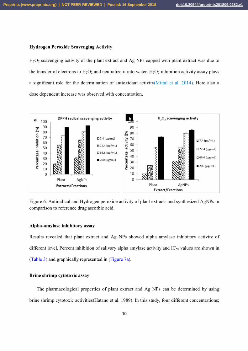
Hydrogen Peroxide Scavenging Activity
H2O2 scavenging activity of the plant extract and Ag NPs capped with plant extract was due to
the transfer of electrons to H2O2 and neutralize it into water. H2O2 inhibition activity assay plays
a significant role for the determination of antioxidant activity(Mittal et al. 2014). Here also a
dose dependent increase was observed with concentration.
Figure 6. Antiradical and Hydrogen peroxide activity of plant extracts and synthesized AgNPs in comparison to reference drug ascorbic acid.
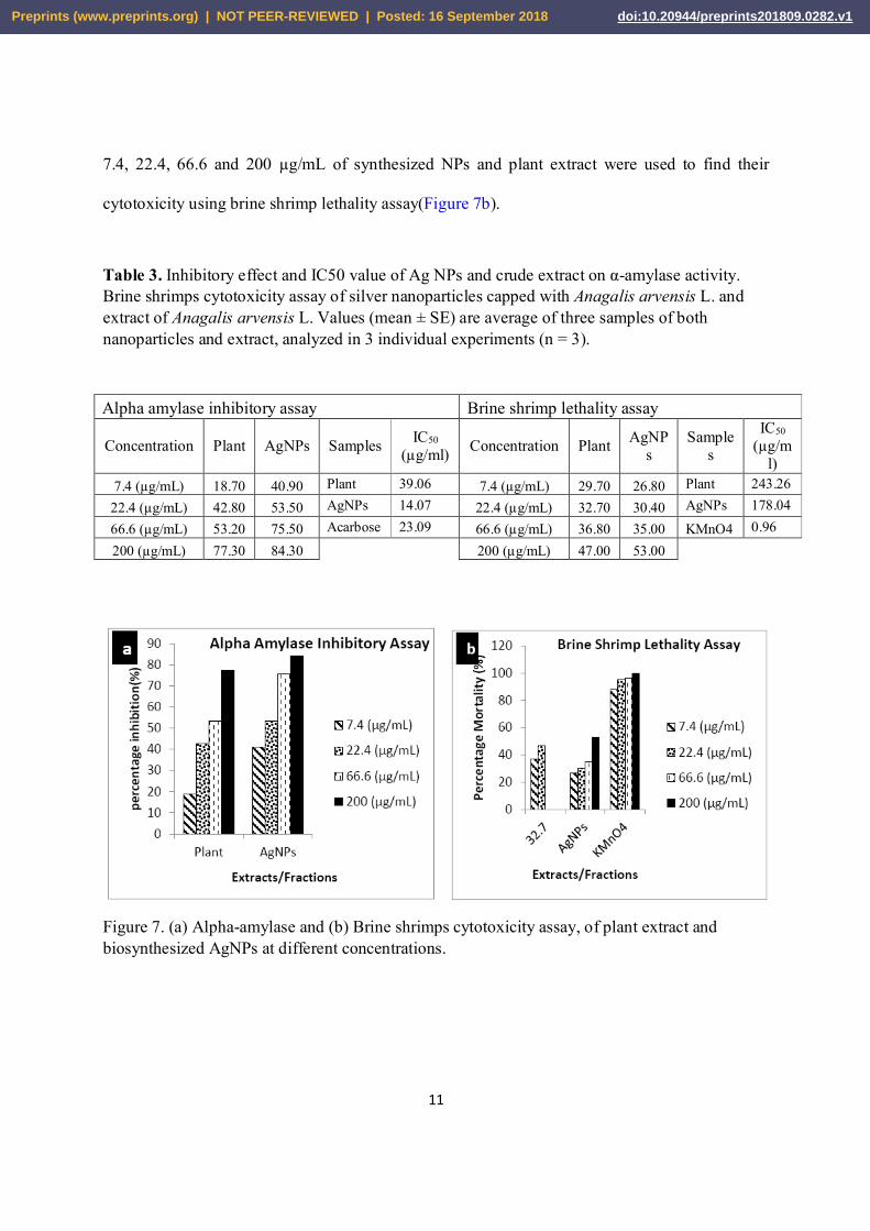
Alpha-amylase inhibitory assay
Results revealed that plant extract and Ag NPs showed alpha amylase inhibitory activity of
different level. Percent inhibition of salivary alpha amylase activity and IC50 values are shown in
(Table 3) and graphically represented in (Figure 7a).
Brine shrimp cytotoxic assay
The pharmacological properties of plant extract and Ag NPs can be determined by using
brine shrimp cytotoxic activities(Hatano et al. 1989). In this study, four different concentrations;
Preprints (www.preprints.org) | NOT PEER-REVIEWED | Posted: 16 September 2018 doi:10.20944/preprints201809.0282.v1
11
7.4, 22.4, 66.6 and 200 µg/mL of synthesized NPs and plant extract were used to find their
cytotoxicity using brine shrimp lethality assay(Figure 7b).
Table 3. Inhibitory effect and IC50 value of Ag NPs and crude extract on α-amylase activity. Brine shrimps cytotoxicity assay of silver nanoparticles capped with Anagalis arvensis L. and extract of Anagalis arvensis L. Values (mean ± SE) are average of three samples of both nanoparticles and extract, analyzed in 3 individual experiments (n = 3).
Alpha amylase inhibitory assay Brine shrimp lethality assay
Concentration Plant AgNPs Samples IC50
(µg/ml) Concentration Plant AgNPs
Samples
IC50
(µg/ml)
7.4 (µg/mL) 18.70 40.90 Plant 39.06 7.4 (µg/mL) 29.70 26.80 Plant 243.26
22.4 (µg/mL) 42.80 53.50 AgNPs 14.07 22.4 (µg/mL) 32.70 30.40 AgNPs 178.04
66.6 (µg/mL) 53.20 75.50 Acarbose 23.09 66.6 (µg/mL) 36.80 35.00 KMnO4 0.96
200 (µg/mL) 77.30 84.30 200 (µg/mL) 47.00 53.00
Figure 7. (a) Alpha-amylase and (b) Brine shrimps cytotoxicity assay, of plant extract and biosynthesized AgNPs at different concentrations.
Preprints (www.preprints.org) | NOT PEER-REVIEWED | Posted: 16 September 2018 doi:10.20944/preprints201809.0282.v1
12
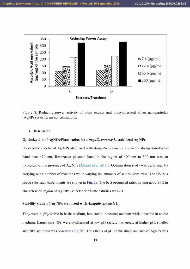
Reducing power
The ability of extract and Ag NPs to reduce Fe3+ to Fe2+ (reducing effect) was determined
according to the previously described method (Subramanian et al. 2013). The reducing capacity
of both plant extract and Ag NPs gives an indication of its potential antioxidant activity (Meir et
al. 1995). Ascorbic acid was used for comparison. Compounds with reducing power indicate that
they are electron donors and can reduce the oxidized intermediates of lipid peroxidation
processes; so that they can act as primary and secondary antioxidants (SivaKumar et al.
2015).The results were expressed as μg/mL of ascorbic acid, plant extract and NPs (Table 4).
Table 4. Reducing power assay and IC50 value of plant extract and biosynthesized Ag NPs
Concentration Plant extract AgNPs
7.4(µg/mL) 111.43 121.40 Samples IC50
(µg/ml)
22.4(µg/mL) 147.92 156.80 plant 60.97
66.6(µg/mL) 215.00 222.80 AgNPs 14.33
200 (µg/mL) 322.68 330.30 Ascorbic
Acid 4.56
Preprints (www.preprints.org) | NOT PEER-REVIEWED | Posted: 16 September 2018 doi:10.20944/preprints201809.0282.v1
13
Figure 8. Reducing power activity of plant extract and biosynthesized silver nanoparticles (AgNPs) at different concentrations.
3. Discussion
Optimization of AgNO3/Plant ratios for Anagalis arvensisL. stabilized Ag NPs
UV-Visible spectra of Ag NPs stabilized with Anagalis arvensis L.showed a strong absorbance
band near 420 nm. Resonance plasmon band in the region of 400 nm to 500 nm was an
indication of the presence of Ag NPs (Ahmad et al. 2011). Optimization study was performed by
carrying out a number of reactions while varying the amounts of salt to plant ratio. The UV-Vis
spectra for such experiments are shown in Fig. 2a. The best optimized ratio, having good SPR in
characteristic region of Ag NPs, selected for further studies was 2:1.
Stability study of Ag-NPs stabilized with Anagalis arvensis L.
They were highly stable in basic medium, less stable in neutral medium while unstable in acidic
medium. Larger size NPs were synthesized at low pH (acidic), whereas, at higher pH, smaller
size NPs synthesis was observed (Fig.2b). The effects of pH on the shape and size of AgNPs was
Preprints (www.preprints.org) | NOT PEER-REVIEWED | Posted: 16 September 2018 doi:10.20944/preprints201809.0282.v1
14
studied and it was found that NPs aggregation seems to surpass the nucleation process in acidic
conditions. At high pH, though, the great numbers of nuclei formation, instead of aggregation led
to the synthesis of more of NPs with smaller diameter. At high pH there was high concentration
of hydroxyl ions on the surface of NPs, repulsive forces dominated and particles aggregation is
reduced. The optimum pH was found to be 13-14 (Iravani & Zolfaghari 2013).
The stability of AgNPs capped with Anagalis arvensis L.decreases with increase in concentration
of NaCl (Fig.2c). It means that stability of AgNPs is inversely proportional to the concentration
of the salt used. Adding more concentration of the salt will cause aggregation of AgNPs due to
Cl-1 ions because halide salts are known to affect the properties of AgNPs. They are used to
induce aggregation by increasing the ionic strength of the solutions. So adding more
amount/concentration of NaCl to NPs can decrease its stability and more aggregation can occur
(Naz et al. 2013). The effect can be attributed to the variation of electrostatic interactions
(attraction/repulsion) of Ag+ ions with those of NaCl in aqueous media at constant pH. The
variation of ionic strength of salt may also affect the micro-environment around the ions and
hence the stability of AgNPs.
While studying temperature effect it was observed that as the temperature increases the
absorbance decreases as shown in (Fig.2d). With the optimized ratio which was 2:1 at room
temperature, peak arises at 430 nm that indicates monodispersed fine particles and reduction in
size of the NPs. Whereas at 50°C to 90°C the absorbance is decreased and asymmetry with broad
peak width was observed and at which start off the aggregation of NPs due to denaturation of
proteins capping at high temperature. Room temperature was an optimized condition for heating
at which maximum synthesis of Ag NPs occur (Amin et al. 2012).
Preprints (www.preprints.org) | NOT PEER-REVIEWED | Posted: 16 September 2018 doi:10.20944/preprints201809.0282.v1
15
Size of the Prepared Nanoparticles.
SEM images show that the NPs were nearly spherical in shape (Andrade et al.
2013)(Fig.4a).which was confirmed by TEM image (Fig.4b) . The major sizes of AgNPs
prepared from Anagalis arvensis L. leaves extracts were 26, 33 and 39 nm, respectively. The
synthesized AgNPs were capped by different plant constituents (polysaccharides, protein,
polyphenolic and flavonoidal compounds) that prevented their aggregation. Inherent capping
offers the additional advantage of the stability in the green chemical synthesis (Morones et al.
2005). Further AFM images showed the detailed size and morphology of Ag NPs(Logeswari et
al. 2015). The area analyzed was 1.9 μm × 1.9 μm. The particle size of the Ag NPs was found to
be 26 nm, 30 nm, 22 nm and 15nm (Fig.5).
DPPH Antioxidant Assay
The recorded value (% inhibition) for the lowest concentration (7.4 μg/mL) of the aqueous
extract was 20.10 and this value was increased to 55.80 when the concentration was increased to
22.4 μg/mL. However, for AgNPs the percent inhibition values recorded were 30.54 for the
concentration of 7.4 μg/mL and 65.70 for the concentration of 22.4 μg/mL. These values indicate
that the synthesized bio-conjugated AgNPs have better antiradical potential than the extracts
alone. The lower value of IC50 indicates a higher antioxidant activity. In DPPHradical
scavenging, the highest activity was observed in AgNPs(200 μg/mL). The IC50 values of both
extract and synthesized AgNPs are reported in (Table 1). It can be concluded from the data that
synthesized AgNPs displayed better antioxidant activity in comparison to the Anagalis arvensis
L.extract (Figure 6a)(Parveen et al. 2016).
Preprints (www.preprints.org) | NOT PEER-REVIEWED | Posted: 16 September 2018 doi:10.20944/preprints201809.0282.v1
16
Hydrogen Peroxide Scavenging Activity
Anagalis arvensis L. showed minimum activity of 10.10 at concentration of 7.4 µg/mL and
maximum activity of 73.90 at a concentration of 200 µg/mL followed by Ag NPs capped with
plant extract showing a minimum activity of 32.10 at concentration of 7.4µg/mL and maximum
activity of 85.40 at a concentration of 200 µg/mL (Figure 6b). The IC50 values of both extract
and synthesized AgNPs are reported in (Table 2). It can be concluded from the data that
synthesized AgNPs displayed better antioxidant activity compared to Anagalis arvensis L.
extract(Parveen et al. 2016).
Alpha-amylase inhibitory assay
Percent inhibition of salivary alpha amylase enzyme by Anagalis arvensis L. extract (at
concentration ranging from 7.4 µg/mL to 200µg/mL) was found to be 18.70to 77.50with IC50
value of 39.06 µg/mL. This inhibition by AgNPs (at concentration ranging from 7.4 µg/mL to
200µg/mL) was found to be 40.90to 84.30with IC50 value of 14.06 µg/mL. AgNPs synthesized
from Anagalis arvensis L.demonstrated high inhibitory activity against α-amylase than
acarbose(Rehana et al. 2017).
Brine shrimp cytotoxic assay
IC50 value of extract was found up to 243µg/mL (Table 3). The lowered IC50 value suggested the
cytotoxic nature of AgNPs compared to plant extract. This enhanced cytotoxicity of NPs having
IC50 of 178.04 µg/mL to brine shrimp revealing the presence of toxic constituents. The cytotoxic
effect of NPs shows that they can be used as alternative source of anticancer drugs (Ghareed et
al. 2014). These days much attention is being given metallic NPs and their anticancer activity
(Ruby et al. 2012).
Preprints (www.preprints.org) | NOT PEER-REVIEWED | Posted: 16 September 2018 doi:10.20944/preprints201809.0282.v1
17
Reducing power
There was dose dependent increase in the reducing power of plant extract as well as Ag NPs.The
reducing capacity of the plant extract for the concentration of 200 µg/mLwas 322.68 µg/mLand
for Ag NPs this value was 330 µg/mL.The reducing power of ascorbic acid was found to be
significantly higher than those of plant extract and Ag NPs. This reducing capacity was due to
polyphenols flavonoids. The reducing power increased with increasing the phenolic content of
the extract, so that’s why there was dose dependent increase in the reducing power with
concentration (Figure 8).
4. Materials and Methods
Silver nitrate (AgNO3), hydrochloric acid (HCl) and sodium hydroxide (NaOH) were
purchased from Merck. Deionized distilled water (ddH2O) was used for all reactions, solution
preparations and analysis purpose and was obtained from Millipore Milli-Q system (Millipore,
Bedford, MA, USA). Perkin-Elmer spectrophotometer (Lamda 25) was used initially for
optimization study of plant/salt ratio for study of better samples. FT-IR spectra were recorded on
IR Affinity.1 Shimidzo (Japan). Particle morphology and average size were investigated by using
SEM(FEI Quanta FEG 200; FEI Company, Hillsboro,OR) with high and low vacuum. For
optimization of reaction samples, pH of the medium was also set using pH meter Milwaukee,
MW802, pH/EC/TDS meter.Atomic force microscopy (AFM) analysis for synthesized NPs was
done by using the instrument NOVA NT–MDT SOLVER NEXT, Russia. TEM analysis was
performed by using HR-3100 advanced 0 kV TEM from Hitachi (Japan).
Preprints (www.preprints.org) | NOT PEER-REVIEWED | Posted: 16 September 2018 doi:10.20944/preprints201809.0282.v1
18
Collection of plant material
In the present study Anagalis arvensis L. was freshly collected from various localities of
district Swabi (Khyber Pakhtoonkhwa, Pakistan). The plant was collected in March, April and
June. Samples were brought to the laboratory in polythene bags and were clean thoroughly with
fresh water to remove the debris and other micro flora and fauna. The plant samples were
identified with the available literature of flora of Pakistan . It is considered one of the precious
assets in the medicinal plant market. Owing its importance in the market it was highly desired to
use it for green synthesis.
Preparation of crude extracts of Anagalis arvensis L.
Plant leaf extract of Anagalis arvensis L. was prepared by taking 5 g of the leaves and
properly washed in distilled water. They were then cut into fine pieces and taken in a 250
mL Erlenmeyer flask with 100 mL of sterile distilled water. It was boiled and filtered. The
extract was stored at 4 °C and used within a week.
Synthesis of Ag NPs stabilized with Anagalis arvensis L.
Anagalis arvensis L. was also used to further explore the green synthesis and stability of
AgNPs. 100mL of the prepared solution of plant was taken in a volumetric flask. 1mM AgNO3
solution was also prepared for the reaction in a separate vial. Both solutions were taken in a vial
with different salt/plant molar ratios, such as 1:1, 2:1….10:1 etc., for different reactions. The
reaction mixtures were kept on stirring for 4 hours at room temperature. The change in
colorgives a primary indication of NPs formation. A particular absorption plasmon band in the
region of 400 to 500 nm showed the presence of Ag NPs formation. The AgNPs were isolated by
Preprints (www.preprints.org) | NOT PEER-REVIEWED | Posted: 16 September 2018 doi:10.20944/preprints201809.0282.v1
19
centrifugation at 14000 rpm. The solid AgNPs were collected for FT-IR , SEM, TEM and AFM
studies.
Biological activities
To see the potential biomedical use of these green synthesized/stabilized noble metal NPs,
DPPH antioxidant assay, hydrogen peroxide (H2O2) scavenging assay, reducing power,brine
shrimp cytotoxic assay and alpha-amylase inhibitory assay were also investigated.
DPPH Antioxidant Assay
The scavenging activity of DPPH free radical of plant extracts were achieved by using
method described by (Kulisic et al. 2004)and adapted by (Obied et al. 2005).Aliquots of 200 µL
of each test sample (200 µg/0.5mL, 66.6/0.5mL, 33.3/0.5mL, 7.4/0.5mL) µg/mL of all extracts
were stirred with 2800 µL of DPPH 3.2 mg/100 mL solution in test tube.After 1hr of incubation
in dark at room temperature, absorbance of each reaction mixture was taken at 517nm by using
UV-visible spectrophotometer. In negative control 200 µL of respective solvent(methanol) and
2800 µL of DPPH solution were used and in blank methanol were used. In positive control,
Ascorbic acid is used as same concentrations of plant sample. Each experiment was repeated in
triplicates. The DPPH scavenging activity was calculated by using given formula and graphical
method was used to calculate the IC50 value.
Percentage inhibition = [(Ac−As)/Ac] ×100
Whereas, Ac is negative control absorbance and As is absorbance of sample. Same method
was used to determine the antioxidant activity of Ag NPs stabilized with Anagalis arvensis
L.extract.
Preprints (www.preprints.org) | NOT PEER-REVIEWED | Posted: 16 September 2018 doi:10.20944/preprints201809.0282.v1
20
Hydrogen peroxide scavenging assay
The capacity of the plant extract and silver nanoparticles capped with Anagalis arvensis L. to
scavenge H2O2 was determined according to the method of (Ruch et al. 1989).A solution of 2
mM H2O2 was prepared in 50 mM phosphate buffer (pH 7.4). An aliquot of 100 µL of the
sample was transferred to an Eppendorf tube to have final concentration in reaction mixture of
200, 66.6, 22.2 and 7.4 µg/mL and the volume was made up to 400 µl with 50 mM phosphate
buffer (pH 7.4). After addition of 600 µL of H2O2 solution, tubes were vortexed and absorbance
of H2O2 at 230 nm was determined after 10 minutes, against a blank. Ascorbic acid was used as
positive control. The percentage H2O2 scavenging ability of samples (extract/fraction) was then
calculated by using the following equation:
H2O2scavenging activity = [1-As/Ac] × 100
Whereas, As isabsorbance of sample, Ac isabsorbance of control. All experiments were
performed in triplicate.
Brine shrimp cytotoxic assay
The toxicity of Anagalis arvensis L. extract and Ag NPs capped with Anagalis arvensis L.
were determined by brine shrimp cytotoxicity assay as reported by (Hossain et al. 2012).2.5 mL
of plant extract and Ag NPs solution was added to 2.5 mL of sea water containing 10 nauplii.
Prepared plant extract and Ag NPs in different concentrations can be checked by implementing
the assured volume of the plant extract and Ag NPs in vials have brine solution (Meyer et al.
1982). Selective ten brine shrimp nauplii were added into every sample vial and artificial sea
water is added to adjust the final volume of every vial. Each vial consists of the 0.5mL of plant
extract and Ag NPs and 4.5mL of manufactured sea water and 10 nauplii of brine
shrimp(Hossain et al. 2012).Total volume of test solution is 5mL in each vial. The
Preprints (www.preprints.org) | NOT PEER-REVIEWED | Posted: 16 September 2018 doi:10.20944/preprints201809.0282.v1
21
experimentswere performed in triplicate. All sample vials were kept uncovered under light
source for next 24hrs in incubator at room temperature. After 24 hrs, the number of alive shrimps
were counted and noted carefully by using magnifying glass.
Alpha amylase inhibition assay
Alpha-amylase inhibitory activity of extract and Ag NPs were performed according to the
previously reported method (Anjana et al. 2011)with modestmodification. In a test tube, reaction
mixture containing 350μL phosphate buffer (50mM, pH= 6.8), 70μL alpha-amylase (10U/mL)
[SRL] and 140μL of varying concentrations(7.4,22.2,66.6,200 µg/mL) of Anagalis arvensis L.
plant extract and Ag NPs capped with Anagalis arvensis L.was pre-incubated at 37C for 10 min.
Then 140μL soluble starch (0.05%) [HiMedia] was added as a substrate and incubated further at
370oC for 15 min. The reaction was stopped by adding 140μL 1N HCl, followed by addition of
700μL iodine reagent (5mM I2 and 5mM KI, stored in amber colored bottle). The absorbance
was read at 620nm using UV−visible spectrophotometer. Each experiment was performed in
triplicates, along with appropriate blanks. Acarbose at various concentrations
(7.4,22.2,66.6,200μg/mL) was included as a standard. Negative control without extracts was set
up in parallel. The result is expressed as percentage inhibition, which was calculated as,
% Inhibition = Atest–Anegative control/Atest X 100
Whereas, A is absorbance and the result were also expressed as IC50 value.
Reducing power
The reducing power of was evaluated according to procedure reported by (Oyaizu 1986). The
reducing potential of the plant extract sample and Ag NPs was determined by using 200 µL
aliquot of extract prepared in DMSO mixed with 500 µL of 2 M phosphate buffer at pH 6.6 and
Preprints (www.preprints.org) | NOT PEER-REVIEWED | Posted: 16 September 2018 doi:10.20944/preprints201809.0282.v1
22
500 µL of 1% potassium ferricyanide [K3Fe(CN)6]. The mixture was incubated at 50°C for 20
minutes. A volume of 500 µL of 10% trichloroacetic acid was added to the mixture, which was
then centrifuged at 3000 rpm for 10 minutes. The upper layer of the mixture (500 µL) was mixed
with equal volume of distilled water and 100 µL of 0.1% ferric chloride (FeCl3). Absorbance was
measured at 700 nm. Blank was prepared by adding 200 µL of DMSO instead of the extract and
Ag NPs. The reducing power of each sample was expressed as ascorbic acid equivalent. All
experiments were performed in triplicate.
5. Conclusion
This study demonstrated an ecofriendly, rapid, green approach to synthesis AgNPs by using
Anagalis Arvensis L. as reducing agent which supplies a simple, cost-effective and ecological
method for the synthesis of AgNPs. The AgNPs so-prepared exhibited effective antioxidant,
antidiabetic and cytotoxic activities, suggesting AgNPs might be useful as a silver dressing for
wounds or as an alternative material.The significances of this study demonstrate a broad range of
applications of synthesized NPs.
Author Contributions
Conceptualization, Mohib Shah, Natasha Anwar and Iqbal Munir; Data curation, Iqbal Munir
and Faiz Ul Amin; Formal analysis, Natasha Anwar; Funding acquisition, Anwar Hussain;
Investigation, Niaz Ali Shah; Methodology, Samreen Saleem and Niaz Ali Shah; Resources,
Abbas Khan; Supervision, Mohib Shah; Validation, Baseerat Fida; Visualization, Anwar
Hussain; Writing – original draft, Mohib Shah; Writing – review & editing, Faiz Ul Amin.
Preprints (www.preprints.org) | NOT PEER-REVIEWED | Posted: 16 September 2018 doi:10.20944/preprints201809.0282.v1
23
Refrences.
Ahmad A, Mukherjee P, Senapati S, Mandal D, Khan MI, Kumar R, and Sastry M. 2003.
Extracellular biosynthesis of silver nanoparticles using the fungus Fusarium oxysporum.
Colloids and Surfaces B: Biointerfaces 28:313-318.
Ahmad N, Sharma S, Singh V, Shamsi S, Fatma A, and Mehta B. 2011. Biosynthesis of silver
nanoparticles from Desmodium triflorum: a novel approach towards weed utilization.
Biotechnology Research International 2011.
Amin M, Anwar F, Janjua MRSA, Iqbal MA, and Rashid U. 2012. Green synthesis of silver
nanoparticles through reduction with Solanum xanthocarpum L. berry extract:
characterization, antimicrobial and urease inhibitory activities against Helicobacter
pylori. International Journal of Molecular Sciences 13:9923-9941.
Andrade JEd, Machado R, Macêdo MA, and Cunha FGC. 2013. AFM and XRD characterization
of silver nanoparticles films deposited on the surface of DGEBA epoxy resin by ion
sputtering. Polímeros 23:19-23.
Anjana R, Pradeepa R, Deepa M, Datta M, Sudha V, Unnikrishnan R, Bhansali A, Joshi S, Joshi
P, and Yajnik C. 2011. Prevalence of diabetes and prediabetes (impaired fasting glucose
and/or impaired glucose tolerance) in urban and rural India: Phase I results of the Indian
Council of Medical Research–INdia DIABetes (ICMR–INDIAB) study. Diabetologia
54:3022-3027.
Bhatt I, and Tripathi BN. 2011. Interaction of engineered nanoparticles with various components
of the environment and possible strategies for their risk assessment. Chemosphere
82:308-317.
Preprints (www.preprints.org) | NOT PEER-REVIEWED | Posted: 16 September 2018 doi:10.20944/preprints201809.0282.v1
24
El-Chaghaby GA, and Ahmad AF. 2011. Biosynthesis of silver nanoparticles using Pistacia
lentiscus leaves extract and investigation of their antimicrobial effect. Oriental Journal of
Chemistry 27:929.
Gardea-Torresdey JL, Gomez E, Peralta-Videa JR, Parsons JG, Troiani H, and Jose-Yacaman M.
2003. Alfalfa sprouts: a natural source for the synthesis of silver nanoparticles. Langmuir
19:1357-1361.
Ghareed M, Shoeb HA, Madkour HMF, Refaey L, Mohamed MA-M, and Saad AM. 2014.
Antioxidant and cytotoxic activities of Tectona grandis linn leaves. International Journal
of Phytopharmacology 5:143-157.
Hatano T, Edamatsu R, Hiramatsu M, Mori A, Fujita Y, Yasuhara T, Yoshida T, and Okuda T.
1989. Effects of the interaction of tannins with co-existing substances. VI.: effects of
tannins and related polyphenols on superoxide anion radical, and on 1, 1-Diphenyl-2-
picrylhydrazyl radical. Chemical and Pharmaceutical Bulletin 37:2016-2021.
Hossain S, Kader G, Nikkon F, and Yeasmin T. 2012. Cytotoxicity of the rhizome of medicinal
plants. Asian Pacific journal of tropical biomedicine 2:125-127.
Iravani S, and Zolfaghari B. 2013. Green synthesis of silver nanoparticles using Pinus eldarica
bark extract. BioMed research international 2013.
Khoshkholgh-Pahlaviani MRM, Massiha AR, Issazadeh K, Bidarigh S, Giahi M, and Ramtin M.
2013. Evaluation of Antifungal Activity of Methanol Extract of Acacia (Anagalis
Arvensis) Leaves and Nystatin against Candida Albicans in Vitro. Zahedan J Res Med
Sci 15.
Preprints (www.preprints.org) | NOT PEER-REVIEWED | Posted: 16 September 2018 doi:10.20944/preprints201809.0282.v1
25
Klaus-Joerger T, Joerger R, Olsson E, and Granqvist C-G. 2001. Bacteria as workers in the
living factory: metal-accumulating bacteria and their potential for materials science.
TRENDS in Biotechnology 19:15-20.
Kulisic T, Radonic A, Katalinic V, and Milos M. 2004. Use of different methods for testing
antioxidative activity of oregano essential oil. Food chemistry 85:633-640.
Logeswari P, Silambarasan S, and Abraham J. 2015. Synthesis of silver nanoparticles using
plants extract and analysis of their antimicrobial property. Journal of Saudi Chemical
Society 19:311-317.
Meir S, Kanner J, Akiri B, and Philosoph-Hadas S. 1995. Determination and involvement of
aqueous reducing compounds in oxidative defense systems of various senescing leaves.
Journal of Agricultural and Food Chemistry 43:1813-1819.
Meshot ER, Verploegen E, Bedewy M, Tawfick S, Woll AR, Green KS, Hromalik M, Koerner
LJ, Philipp HT, and Tate MW. 2012. High-speed in situ X-ray scattering of carbon
nanotube film nucleation and self-organization. Acs Nano 6:5091-5101.
Meyer B, Ferrigni N, Putnam J, Jacobsen L, Nichols Dj, and McLaughlin JL. 1982. Brine
shrimp: a convenient general bioassay for active plant constituents. Planta medica 45:31-
34.
Mittal AK, Bhaumik J, Kumar S, and Banerjee UC. 2014. Biosynthesis of silver nanoparticles:
elucidation of prospective mechanism and therapeutic potential. Journal of Colloid and
Interface Science 415:39-47.
Mohanpuria P, Rana NK, and Yadav SK. 2008. Biosynthesis of nanoparticles: technological
concepts and future applications. Journal of Nanoparticle Research 10:507-517.
Preprints (www.preprints.org) | NOT PEER-REVIEWED | Posted: 16 September 2018 doi:10.20944/preprints201809.0282.v1
26
Morones JR, Elechiguerra JL, Camacho A, Holt K, Kouri JB, Ramírez JT, and Yacaman MJ.
2005. The bactericidal effect of silver nanoparticles. Nanotechnology 16:2346.
Naz SS, Islam NU, Shah MR, Alam SS, Iqbal Z, Bertino M, Franzel L, and Ahmed A. 2013.
Enhanced biocidal activity of Au nanoparticles synthesized in one pot using 2, 4-
dihydroxybenzene carbodithioic acid as a reducing and stabilizing agent. Journal of
nanobiotechnology 11:13.
Obied HK, Allen MS, Bedgood DR, Prenzler PD, Robards K, and Stockmann R. 2005.
Bioactivity and analysis of biophenols recovered from olive mill waste. Journal of
Agricultural and Food Chemistry 53:823-837.
Oyaizu M. 1986. Studies on products of browning reaction. The Japanese Journal of Nutrition
and Dietetics 44:307-315.
Parveen M, Ahmad F, Malla AM, and Azaz S. 2016. Microwave-assisted green synthesis of
silver nanoparticles from Fraxinus excelsior leaf extract and its antioxidant assay. Applied
Nanoscience 6:267-276.
Rehana D, Mahendiran D, Kumar RS, and Rahiman AK. 2017. In vitro antioxidant and
antidiabetic activities of zinc oxide nanoparticles synthesized using different plant
extracts. Bioprocess and Biosystems Engineering:1-15.
Ruby K, Chauhan R, Sharma S, and Dwivedi J. 2012. Polypharmacological activities of
Bergenia species. Int J pharm pharmaceutical sci 1:100-109.
Ruch RJ, Cheng S-j, and Klaunig JE. 1989. Prevention of cytotoxicity and inhibition of
intercellular communication by antioxidant catechins isolated from Chinese green tea.
Carcinogenesis 10:1003-1008.
Preprints (www.preprints.org) | NOT PEER-REVIEWED | Posted: 16 September 2018 doi:10.20944/preprints201809.0282.v1
27
Shem PM, Sardar R, and Shumaker-Parry JS. 2009. One-step synthesis of phosphine-stabilized
gold nanoparticles using the mild reducing agent 9-BBN. Langmuir 25:13279-13283.
SivaKumar T, Rathimeena T, Thangapandian V, and Shankar T. 2015. Silver nanoparticles
synthesis of Mentha arvensis extracts and evaluation of antioxidant properties. J Biosci
Bioeng 1:22-28.
Subramanian R, Subbramaniyan P, and Raj V. 2013. Antioxidant activity of the stem bark of
Shorea roxburghii and its silver reducing power. SpringerPlus 2:28.
Taniguchi N. 1974. On the basic concept of nano-technology. Proc Intl Conf Prod London, 1974:
British Society of Precision Engineering.
Turkevich J, Stevenson PC, and Hillier J. 1951. A study of the nucleation and growth processes
in the synthesis of colloidal gold. Discussions of the Faraday Society 11:55-75.
Yu D-G. 2007. Formation of colloidal silver nanoparticles stabilized by Na+–poly (γ-glutamic
acid)–silver nitrate complex via chemical reduction process. Colloids and Surfaces B:
Biointerfaces 59:171-178.
Preprints (www.preprints.org) | NOT PEER-REVIEWED | Posted: 16 September 2018 doi:10.20944/preprints201809.0282.v1