Embed Size (px)
Citation preview
Thermal Biophysics of Membranes. Thomas HeimburgCopyright © 2007 WILEY-VCH Verlag GmbH & Co. KGaA, WeinheimISBN: 978-3-527-40471-1
1
1Membranes—An Introduction
In the second half of the 19th century it became evident that an osmotic bar-rier separates the inside and the outside of cells (Nägeli and Cramer, 1855;de Vries, 1871, 1884; Pfeffer, 1877). Plant cell protoplasts were permeable towater but not to larger macromolecules like sucrose (de Vries, 1871). Pfefferwas the first to study the osmotic pressure within cells and formulated theidea that the protoplasm of cells is surrounded by a thin layer, which he calledthe plasma membrane. In fact, Pfeffer proposed that this membrane does notonly cover the outer surface of cells but also separates all aqueous environ-ments of different composition from each other. One may therefore considerPfeffer as the father of membrane theory. The developments in biology andbotany coincided with a rapid development in the theory of thermodynam-ics of solutions. In particular, based on Pfeffer’s work van’t Hoff found theformal analogy of concentrations of solutes in water and the partial pressuresof ideal gases (van’t Hoff, 1887). Ostwald formulated descriptions for the os-motic pressure across semipermeable walls and the related electrical proper-ties (Ostwald, 1887, 1890).1
1.1Overton (1895)
Charles Ernest Overton is a very important figure in the development of a pic-ture of cell membranes. He investigated the osmotic properties of cells and no-ticed in the late 19th century that the permeation of molecules through mem-branes is related to their partition coefficient between water and oil (Overton,1895). Overton’s findings led to the hypothesis that the thin membranes sur-rounding cells have the properties of oil. In his book on anesthesia (Over-ton, 1901. Jena, Germany. English translation: Studies of Narcosis, Chapmanand Hall, 1991, R. Lipnick, Ed., 1991) he called the layers surrounding cells“lipoids” made from lipids and cholesterol. The properties of lipids are de-scribed in detail in Chapter 3 and theory of anesthesia is treated in Chapter 19.
1) The history of biomembrane research is nicely reviewed in Ling(2001).
2 1 Membranes—An Introduction
1.2Langmuir (1917) and Gorter and Grendel (1925)
Langmuir (1917) developed an apparatus in which molecular layers of lipidswere spread at the air–water interface. With this monolayer trough (see Sec-tion 6.7 and Fig. 6.14) the lateral pressure of the monolayer films could be mea-sured. Langmuir proposed that in the molecular film the polar head groupswere directed toward the water whereas the hydrophobic hydrocarbons arepointed toward the air phase.
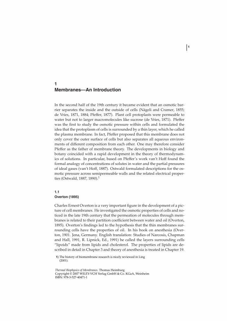
Gorter and Grendel (1925) experimentally investigated the surface area oflipids. For this purpose they extracted the lipids from red blood cells of man,dog, rabbit, sheep, guinea pig, and goat in acetone. The lipids were spreadon a water surface and the area was measured using a Langmuir film balance.From the same blood preparations they measured the surface area of the redblood cells from the microscopic images. They found that the surface area ofthe monofilms was within error exactly two times that of the cells. They con-cluded that cell membranes are made of two opposing thin molecular layers,and they proposed that this double layer is constructed such that two lipidlayers form a bilayer with the polar head groups pointing toward the aqueousenvironment (Fig. 1.1). This is the picture of the lipid membrane we know to-day. As Robertson (1959) noted later, the attractive simplicity of Gorter’s andGrendel’s pictures is also its greatest weakness since it fails to account for themanifold of functions attributed to cell membranes.
Fig. 1.1 The cell membrane according to Gorter and Grendel (1925).They proposed the lipid bilayer structure.
1.3Danielli and Davson (1935)
The earliest molecular model for the biomembrane structure including pro-teins was the model from Danielli and Davson (1935). They took into accountthat the layers surrounding cells had a significant content of proteins adsorbed
1.3 Danielli and Davson (1935) 3
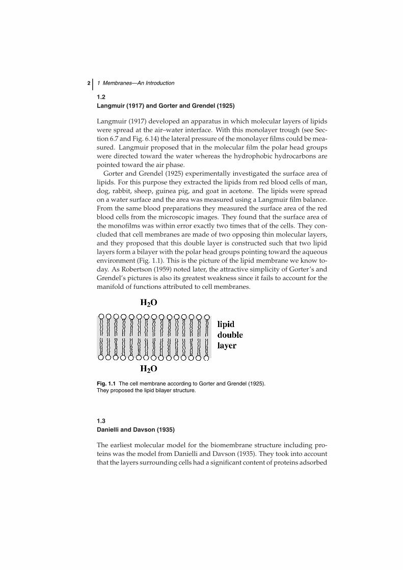
Fig. 1.2 Danielli and Davson (1935) pro-posed a membrane model including proteins.They used their model to interpret the ob-servation of different membrane permeabili-ties of ions and hydrocarbons. In particular,they assumed that the membrane has both alipophilic and a hydrophilic character. Water-
containing regions in the membrane give riseto ion transport depending on water contentof the membrane and its charge; lipophilicparts are responsible for the transport ofwater-insoluble molecules. Figure adaptedfrom Danielli and Davson (1935) with permis-sion.
to the layers. It was known that phospholipids have an amphiphilic nature.Furthermore, proteins investigated were mostly water soluble but neverthe-less often adsorbed to membranes. Jim Danielli and Hugh Davson thus pro-posed a model of the cell membrane consisting of a lipid bilayer, with whicha protein layer is tightly associated (Fig. 1.2, left). As in earlier membranestudies (e.g., by Overton) they were in particular interested in the permeationproperties of membranes. In a theoretical paper they made the following con-sideration.
• Proteins are adsorbed to the lipophilic layers surrounding cells. The pro-teins possess hydrophobic interiors and a water-containing outer layer.
• The lipid layer possesses amphiphilic or charged head groups. This im-plies that the lipid membrane also contains some water.
• The water-containing regions of protein layers adsorped on lipid layersare permeable for charged solutes, e.g., ions.
• Divalent cations as calcium form complexes with lipids or proteins thatreduce their interaction with water. Therefore membranes containingcalcium are less permeable for ions.
• Hydrophobic molecules such as ether penetrate the membranes throughtheir lipophilic lipid part.
4 1 Membranes—An Introduction
They included some theoretical considerations about the different dependen-cies of the permeabilities of membranes to ions and hydrophobic molecules asa function of temperature.
Danielli and Davson concluded that the permeabilities of membranesfor solutes are explainable within the concepts of the physical chemistry ofthe hydrophilic and lipophilic regions of the cell membranes and that noparticular chemical reactions including the solutes are needed to explainthe transport properties.
Unfortunately, this very sober view is nowadays not in the focus of muchof the biochemical membrane research due to the emphasis of the localizedfunction of ion- and solute-specific transport channel proteins. In the chapteron permeability (Chapter 17) we will return to the quite realistic physical viewof Danielli and Davson.
Danielli and Davson did not exclude the possibility that the proteins mayspan the membrane such that a “mosaic” of protein-rich and lipid-rich regionsis formed. However, they did restrain themselves from speculating about sucha structure due to the lack of experimental evidence. The term “mosaic mem-brane” was later used again by Singer and Nicolson (1972).
1.4Robertson (1958)
So far most evidence about the structure of cell membranes was indirect. Theresolution of light microscopy is restricted to the regime above 200 nm, whichis not sufficient for revealing the bimolecular structure of the biological mem-brane that is between 5 and 10 nm thick. This changed with the progressesin electron microscopy. In 1959, J. David Robertson wrote a review in which
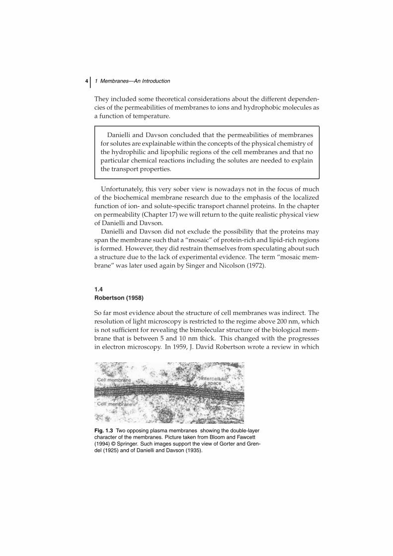
Fig. 1.3 Two opposing plasma membranes showing the double-layercharacter of the membranes. Picture taken from Bloom and Fawcett(1994) © Springer. Such images support the view of Gorter and Gren-del (1925) and of Danielli and Davson (1935).
1.5 The Fluid Mosaic Model of Singer and Nicolson (1972) 5
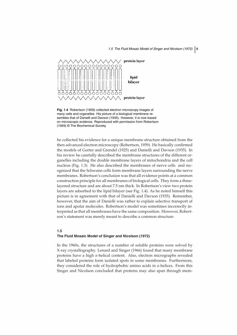
Fig. 1.4 Robertson (1959) collected electron microscopy images ofmany cells and organelles. His picture of a biological membrane re-sembles that of Danielli and Davson (1935). However, it is now basedon microscopic evidence. Reproduced with permission from Robertson(1959) © The Biochemical Society.
he collected his evidence for a unique membrane structure obtained from thethen advanced electron microscopy (Robertson, 1959). He basically confirmedthe models of Gorter and Grendel (1925) and Danielli and Davson (1935). Inhis review he carefully described the membrane structures of the different or-ganelles including the double membrane layers of mitochondria and the cellnucleus (Fig. 1.3). He also described the membranes of nerve cells and rec-ognized that the Schwann cells form membrane layers surrounding the nervemembranes. Robertson’s conclusion was that all evidence points at a commonconstruction principle for all membranes of biological cells. They form a three-layered structure and are about 7.5 nm thick. In Robertson’s view two proteinlayers are adsorbed to the lipid bilayer (see Fig. 1.4). As he noted himself thispicture is in agreement with that of Danielli and Davson (1935). Remember,however, that the aim of Danielli was rather to explain selective transport ofions and apolar molecules. Robertson’s model was sometimes incorrectly in-terpreted as that all membranes have the same composition. However, Robert-son’s statement was merely meant to describe a common structure.
1.5The Fluid Mosaic Model of Singer and Nicolson (1972)
In the 1960s, the structures of a number of soluble proteins were solved byX-ray crystallography. Lenard and Singer (1966) found that many membraneproteins have a high α-helical content. Also, electron micrographs revealedthat labeled proteins form isolated spots in some membranes. Furthermore,they considered the role of hydrophobic amino acids in α-helices. From thisSinger and Nicolson concluded that proteins may also span through mem-
6 1 Membranes—An Introduction
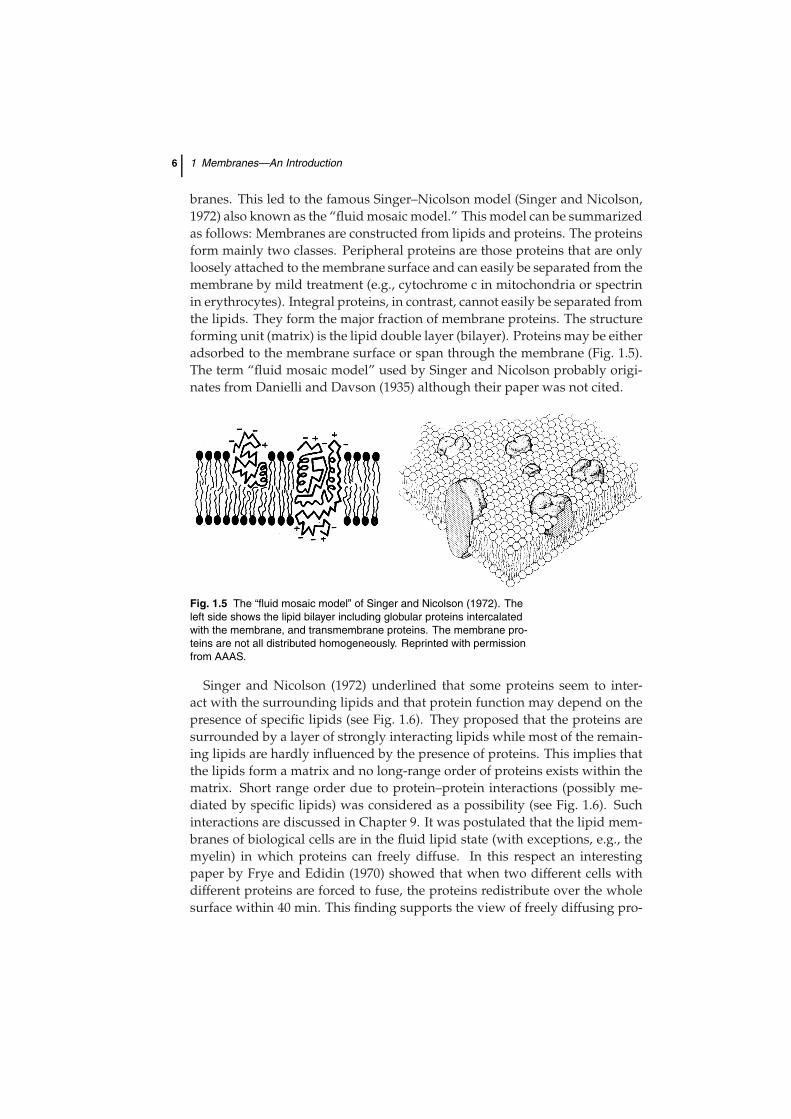
branes. This led to the famous Singer–Nicolson model (Singer and Nicolson,1972) also known as the “fluid mosaic model.” This model can be summarizedas follows: Membranes are constructed from lipids and proteins. The proteinsform mainly two classes. Peripheral proteins are those proteins that are onlyloosely attached to the membrane surface and can easily be separated from themembrane by mild treatment (e.g., cytochrome c in mitochondria or spectrinin erythrocytes). Integral proteins, in contrast, cannot easily be separated fromthe lipids. They form the major fraction of membrane proteins. The structureforming unit (matrix) is the lipid double layer (bilayer). Proteins may be eitheradsorbed to the membrane surface or span through the membrane (Fig. 1.5).The term “fluid mosaic model” used by Singer and Nicolson probably origi-nates from Danielli and Davson (1935) although their paper was not cited.
Fig. 1.5 The “fluid mosaic model” of Singer and Nicolson (1972). Theleft side shows the lipid bilayer including globular proteins intercalatedwith the membrane, and transmembrane proteins. The membrane pro-teins are not all distributed homogeneously. Reprinted with permissionfrom AAAS.
Singer and Nicolson (1972) underlined that some proteins seem to inter-act with the surrounding lipids and that protein function may depend on thepresence of specific lipids (see Fig. 1.6). They proposed that the proteins aresurrounded by a layer of strongly interacting lipids while most of the remain-ing lipids are hardly influenced by the presence of proteins. This implies thatthe lipids form a matrix and no long-range order of proteins exists within thematrix. Short range order due to protein–protein interactions (possibly me-diated by specific lipids) was considered as a possibility (see Fig. 1.6). Suchinteractions are discussed in Chapter 9. It was postulated that the lipid mem-branes of biological cells are in the fluid lipid state (with exceptions, e.g., themyelin) in which proteins can freely diffuse. In this respect an interestingpaper by Frye and Edidin (1970) showed that when two different cells withdifferent proteins are forced to fuse, the proteins redistribute over the wholesurface within 40 min. This finding supports the view of freely diffusing pro-
1.6 The Mattress Model by Mouritsen and Bloom (1984) 7
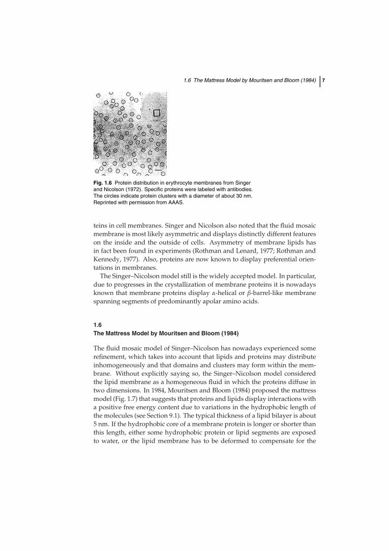
Fig. 1.6 Protein distribution in erythrocyte membranes from Singerand Nicolson (1972). Specific proteins were labeled with antibodies.The circles indicate protein clusters with a diameter of about 30 nm.Reprinted with permission from AAAS.
teins in cell membranes. Singer and Nicolson also noted that the fluid mosaicmembrane is most likely asymmetric and displays distinctly different featureson the inside and the outside of cells. Asymmetry of membrane lipids hasin fact been found in experiments (Rothman and Lenard, 1977; Rothman andKennedy, 1977). Also, proteins are now known to display preferential orien-tations in membranes.
The Singer–Nicolson model still is the widely accepted model. In particular,due to progresses in the crystallization of membrane proteins it is nowadaysknown that membrane proteins display α-helical or β-barrel-like membranespanning segments of predominantly apolar amino acids.
1.6The Mattress Model by Mouritsen and Bloom (1984)
The fluid mosaic model of Singer–Nicolson has nowadays experienced somerefinement, which takes into account that lipids and proteins may distributeinhomogeneously and that domains and clusters may form within the mem-brane. Without explicitly saying so, the Singer–Nicolson model consideredthe lipid membrane as a homogeneous fluid in which the proteins diffuse intwo dimensions. In 1984, Mouritsen and Bloom (1984) proposed the mattressmodel (Fig. 1.7) that suggests that proteins and lipids display interactions witha positive free energy content due to variations in the hydrophobic length ofthe molecules (see Section 9.1). The typical thickness of a lipid bilayer is about5 nm. If the hydrophobic core of a membrane protein is longer or shorter thanthis length, either some hydrophobic protein or lipid segments are exposedto water, or the lipid membrane has to be deformed to compensate for the
8 1 Membranes—An Introduction
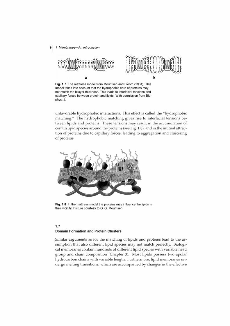
Fig. 1.7 The mattress model from Mouritsen and Bloom (1984). Thismodel takes into account that the hydrophobic core of proteins maynot match the bilayer thickness. This leads to interfacial tensions andcapillary forces between protein and lipids. With permission from Bio-phys. J.
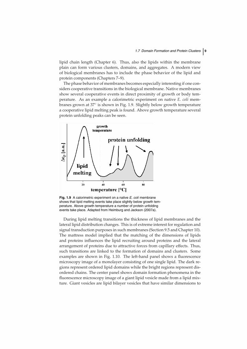
unfavorable hydrophobic interactions. This effect is called the “hydrophobicmatching.” The hydrophobic matching gives rise to interfacial tensions be-tween lipids and proteins. These tensions may result in the accumulation ofcertain lipid species around the proteins (see Fig. 1.8), and in the mutual attrac-tion of proteins due to capillary forces, leading to aggregation and clusteringof proteins.
Fig. 1.8 In the mattress model the proteins may influence the lipids intheir vicinity. Picture courtesy to O. G. Mouritsen.
1.7Domain Formation and Protein Clusters
Similar arguments as for the matching of lipids and proteins lead to the as-sumption that also different lipid species may not match perfectly. Biologi-cal membranes contain hundreds of different lipid species with variable headgroup and chain composition (Chapter 3). Most lipids possess two apolarhydrocarbon chains with variable length. Furthermore, lipid membranes un-dergo melting transitions, which are accompanied by changes in the effective
1.7 Domain Formation and Protein Clusters 9
lipid chain length (Chapter 6). Thus, also the lipids within the membraneplain can form various clusters, domains, and aggregates. A modern viewof biological membranes has to include the phase behavior of the lipid andprotein components (Chapters 7–9).
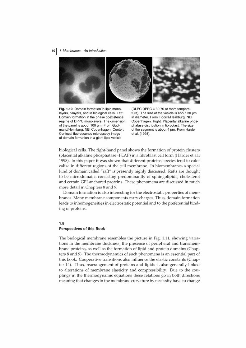
The phase behavior of membranes becomes especially interesting if one con-siders cooperative transitions in the biological membrane. Native membranesshow several cooperative events in direct proximity of growth or body tem-perature. As an example a calorimetric experiment on native E. coli mem-branes grown at 37◦ is shown in Fig. 1.9. Slightly below growth temperaturea cooperative lipid melting peak is found. Above growth temperature severalprotein unfolding peaks can be seen.
Fig. 1.9 A calorimetric experiment on a native E. coli membraneshows that lipid melting events take place slightly below growth tem-perature. Above growth temperature a number of protein unfoldingevents take place. Adapted from Heimburg and Jackson (2007a).
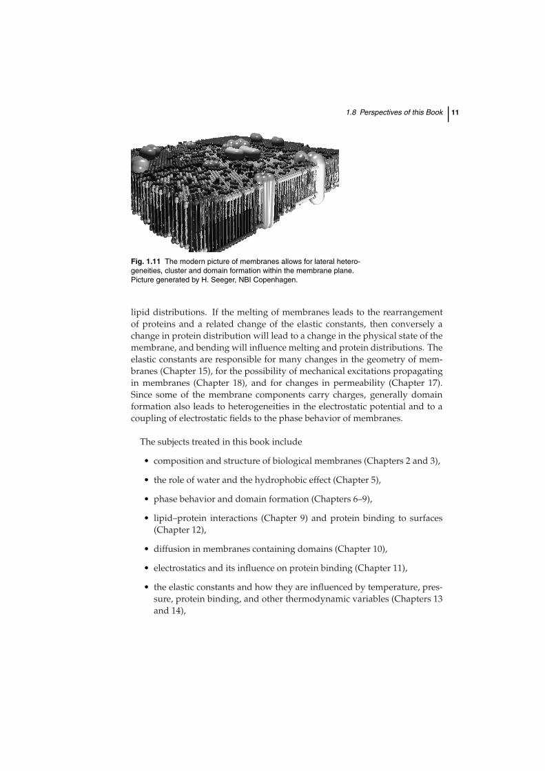
During lipid melting transitions the thickness of lipid membranes and thelateral lipid distribution changes. This is of extreme interest for regulation andsignal transduction purposes in such membranes (Section 9.5 and Chapter 10).The mattress model implied that the matching of the dimensions of lipidsand proteins influences the lipid recruiting around proteins and the lateralarrangement of proteins due to attractive forces from capillary effects. Thus,such transitions are linked to the formation of domains and clusters. Someexamples are shown in Fig. 1.10. The left-hand panel shows a fluorescencemicroscopy image of a monolayer consisting of one single lipid. The dark re-gions represent ordered lipid domains while the bright regions represent dis-ordered chains. The center panel shows domain formation phenomena in thefluorescence microscopy image of a giant lipid vesicle made from a lipid mix-ture. Giant vesicles are lipid bilayer vesicles that have similar dimensions to
10 1 Membranes—An Introduction
Fig. 1.10 Domain formation in lipid mono-layers, bilayers, and in biological cells. Left:Domain formation in the phase coexistenceregime of DPPC monolayers. The dimensionof the panel is about 100 µm. From Gud-mand/Heimburg, NBI Copenhagen. Center:Confocal fluorescence microscopy imageof domain formation in a giant lipid vesicle
(DLPC:DPPC = 30:70 at room tempera-ture). The size of the vesicle is about 30 µmin diameter. From Fidorra/Heimburg, NBICopenhagen. Right: Placental alkaline phos-phatase distribution in fibroblast. The sizeof the segment is about 4 µm. From Harderet al. (1998).
biological cells. The right-hand panel shows the formation of protein clusters(placental alkaline phosphatase=PLAP) in a fibroblast cell form (Harder et al.,1998). In this paper it was shown that different proteins species tend to colo-calize in different regions of the cell membrane. In biomembranes a specialkind of domain called “raft” is presently highly discussed. Rafts are thoughtto be microdomains consisting predominantly of sphingolipids, cholesteroland certain GPI-anchored proteins. These phenomena are discussed in muchmore detail in Chapters 8 and 9.
Domain formation is also interesting for the electrostatic properties of mem-branes. Many membrane components carry charges. Thus, domain formationleads to inhomogeneities in electrostatic potential and to the preferential bind-ing of proteins.
1.8Perspectives of this Book
The biological membrane resembles the picture in Fig. 1.11, showing varia-tions in the membrane thickness, the presence of peripheral and transmem-brane proteins, as well as the formation of lipid and protein domains (Chap-ters 8 and 9). The thermodynamics of such phenomena is an essential part ofthis book. Cooperative transitions also influence the elastic constants (Chap-ter 14). Thus, rearrangement of proteins and lipids is also generally linkedto alterations of membrane elasticity and compressibility. Due to the cou-plings in the thermodynamic equations these relations go in both directionsmeaning that changes in the membrane curvature by necessity have to change
1.8 Perspectives of this Book 11
Fig. 1.11 The modern picture of membranes allows for lateral hetero-geneities, cluster and domain formation within the membrane plane.Picture generated by H. Seeger, NBI Copenhagen.
lipid distributions. If the melting of membranes leads to the rearrangementof proteins and a related change of the elastic constants, then conversely achange in protein distribution will lead to a change in the physical state of themembrane, and bending will influence melting and protein distributions. Theelastic constants are responsible for many changes in the geometry of mem-branes (Chapter 15), for the possibility of mechanical excitations propagatingin membranes (Chapter 18), and for changes in permeability (Chapter 17).Since some of the membrane components carry charges, generally domainformation also leads to heterogeneities in the electrostatic potential and to acoupling of electrostatic fields to the phase behavior of membranes.
The subjects treated in this book include
• composition and structure of biological membranes (Chapters 2 and 3),
• the role of water and the hydrophobic effect (Chapter 5),
• phase behavior and domain formation (Chapters 6–9),
• lipid–protein interactions (Chapter 9) and protein binding to surfaces(Chapter 12),
• diffusion in membranes containing domains (Chapter 10),
• electrostatics and its influence on protein binding (Chapter 11),
• the elastic constants and how they are influenced by temperature, pres-sure, protein binding, and other thermodynamic variables (Chapters 13and 14),
12 1 Membranes—An Introduction
• changes in membrane geometry due to changes in the elastic constants(Chapter 15),
• relaxation phenomena (Chapter 16),
• some considerations on the permeability of membranes for ions andlarger molecules and how it is related to the thermodynamics of themembrane (Chapter 17).
• the propagation of density pulses and a related thermodynamic theoryfor the propagation of nerve pulses (Chapter 18),
• a thermodynamic theory for anesthesia (Chapter 19),
The function of the biological membrane cannot be understood without con-sideration of its thermodynamics. It is a multicomponent system that sensi-tively responds to changes in temperature, pressure, and the chemical poten-tials of its components. Therefore, this book also contains a basic introductioninto thermodynamics (Chapter 4). The purpose of this book is to describe theconcepts of thermodynamic couplings of seemingly independent properties ofmembranes. It will be shown that all of the above phenomena are intimatelyrelated and fit into a coherent thermodynamic picture.
1.9 Summary: Key Ideas of Chapter 1 13
1.9Summary: Key Ideas of Chapter 1
• Biomembranes mainly consist of lipids and proteins. They are macro-scopic ensembles.
• Lipids are amphiphilic molecules with polar head groups and apolarchains. Lipids spontaneously form bilayers in water.
• Proteins may be peripherally adsorbed to the lipid bilayer surface, orthey may be integral proteins spanning through the bilayer core.
• The thickness of biological membranes is on the order of about 5–8 nm.
• Due to differences in the size of the different molecules in membranescapillary forces can exist that influence the distribution of molecules inthe membranes.
• As a consequence, proteins and lipids are not homogeneously dis-tributed within membranes but form domains, clusters, and aggregates.
• The lateral distribution of the molecules is altered when the thermody-namic variables of the system change.
• Biological membranes can undergo order transitions.
• These order transitions are coupled to changes in the elastic constants ofthe membranes.