Embed Size (px)
Citation preview

1
Figure 49.0 Bat locating a moth
2
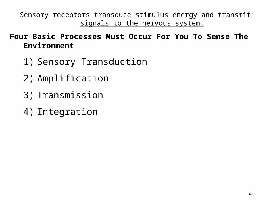
Sensory receptors transduce stimulus energy and transmit signals to the nervous system.
Four Basic Processes Must Occur For You To Sense The Environment
1) Sensory Transduction
2) Amplification
3) Transmission
4) Integration

3
Figure 49.2 Sensory transduction by a taste receptor

4
Umm, Umm, Good. . . Taste Receptors
1. Taste buds are not as localized on the tongue like we once thought.
2. But a sweet, bitter, sour, salty, molecule binds to its particular receptor.
3. Signal is amplified and sets off a Signal Transduction Pathway
4. Potassium ion channels will be closed by the action of second messengers
5. Sodium moves into the cell causing depolarization.
6. This causes calcium ion uptake into the receptor (like at the end of the presynaptic neuron)
7. Neurotransmitters are released that stimulate a sensory neuron and subsequent action potential.

5
Figure 49.3 Sensory receptors in human skin

6
Figure 49.4 Mechanoreception by a hair cell

7
Figure 49.6 Specialized electromagnetic receptors: Rattle snake with infrared receptors, beluga whale pod
Infrared receptors: sense infrared radiation given off by prey (mouse or jogger’s ankle)

8
Figure 49.7 Eye cups and orientation behavior of a planarian
Side with no screening pigment
Side with no screening pigment

9
Figure 49.8 Compound eyes
(a)
Image Forming
Found in insects and crustaceans
Consists of lots of ommatidia
Each ommatidia has its own lens to focus light
Differences in light intensity produce a mosaic image.
Detects movement

10
Figure 49.8x1 SEM of compound eye

11
Figure 49.8x2 Insect vision: A black-eyed Susan (Rudbeckia hirta) as humans see it and in ultraviolet light as visible to an insect

12
Figure 49.9 Structure of the Vertebrate Eye
Forms the corneaForms the iris
No photoreceptors: “blind spot”
Highest Conc. Of Cones
13
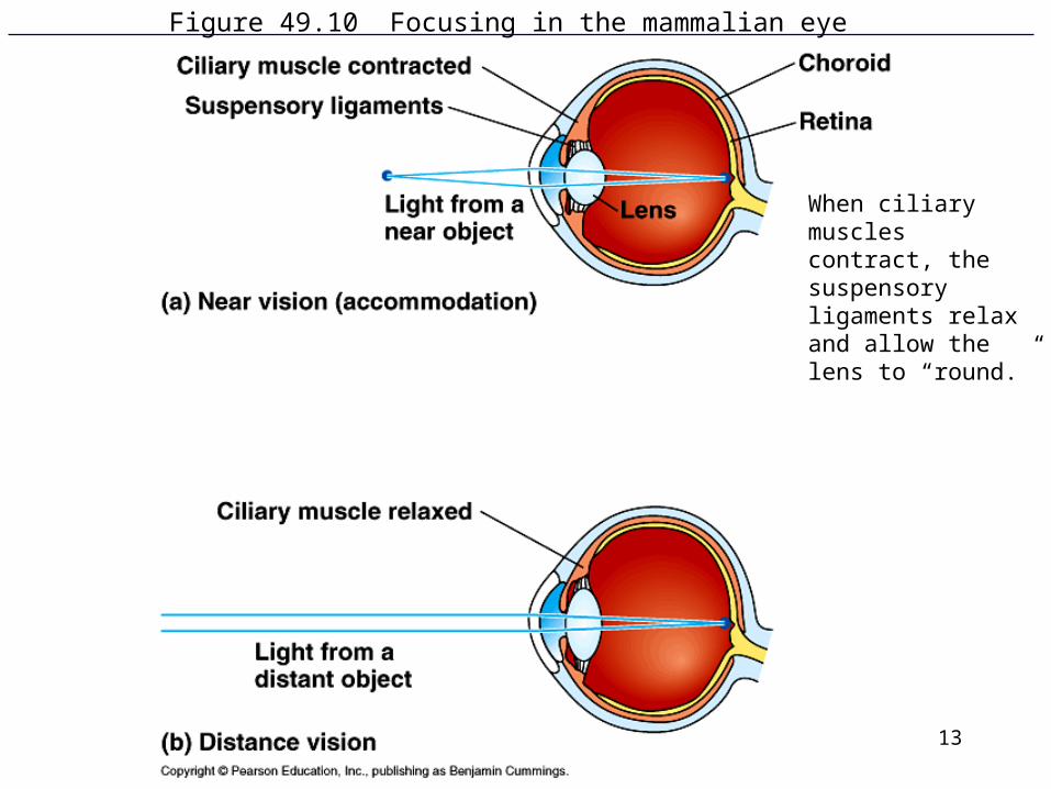
Figure 49.10 Focusing in the mammalian eye
When ciliary muscles contract, the suspensory ligaments relax and allow the lens to “round.”

14
The Biochemistry of Sight
1. Rods are responsible for noncolor dim light; cones are responsible for color vision in bright light.
2. Rod photoreceptor: made up of outer and inner segments, cell body, synaptic region where the nerve impulse is passed to a retinal nerve cell.
3. Outer Segment: made up of stacks of discs containing rhodopsin molecules. The rhodopsin contains a molecule called retinal and opsin that can actually exist in two forms.
4. Light hits retinal; retinal changes shape, separates from opsin and the opsin now has a different shape. The retinal changes color from purple to yellow.
This is “bleaching” of the rhodopsin

15
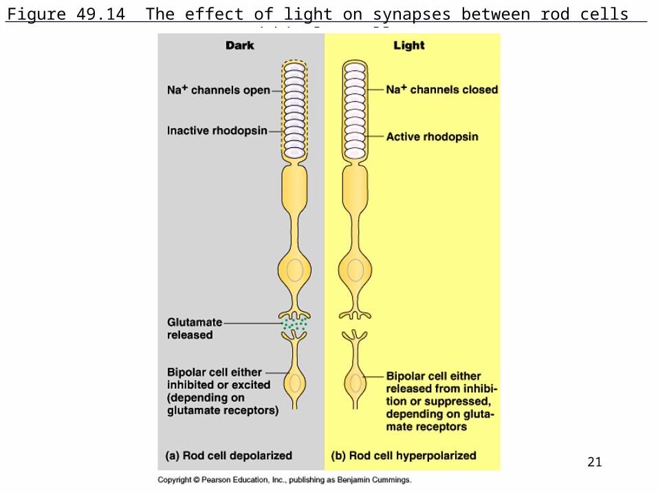
The Biochemistry of Sight (cont’d)
5. This change in the rhodopsin activates a G protein (related to cGMP) called transducin.
6. Transducin then activates an enzyme that breaks down cGMP.
7. The low levels of cGMP cause sodium ion channels to close which hyperpolarizes the rod cell and changes the amount of neurotransmitter it releases (it decreases)
8. In the dark, there is plenty of cGMP and it is bound to sodium channels, keeping them open so it already has a relatively depolarized resting potential. Sodium ions are continually entering the outer segment of the cell. The neurotransmitter, glutamate is released.
9. Glutamate stimulates the bipolar cells in the retina.

16
Biochemistry of Vision (cont’d)
10. Glutamate binds to receptors on the bipolar cells and stimulates some and inhibits others depending on the receptor type.
11. Once again, when light flashes, you’ll have a decrease in cGMP, the sodium channels close and hyperpolarization occurs which decreases glutamate levels and this excites or inhibits bipolar cells depending on the receptor type.
17
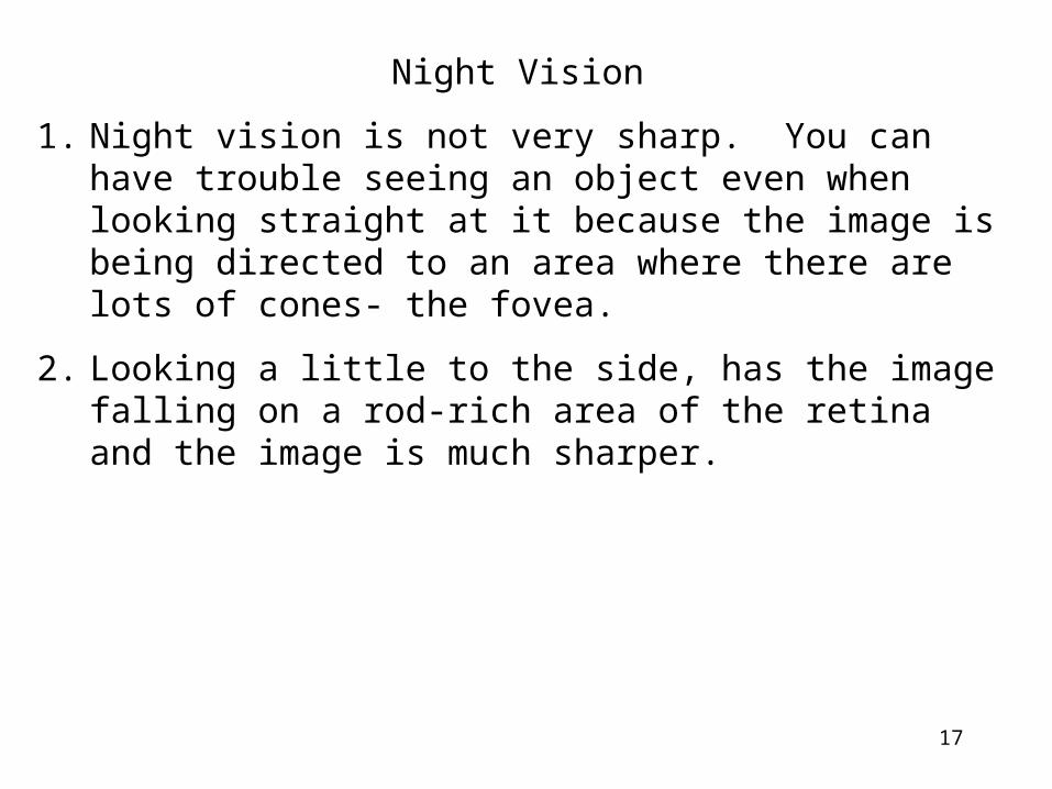
Night Vision
1. Night vision is not very sharp. You can have trouble seeing an object even when looking straight at it because the image is being directed to an area where there are lots of cones- the fovea.
2. Looking a little to the side, has the image falling on a rod-rich area of the retina and the image is much sharper.

18
Figure 49.11 Photoreceptors in the vertebrate retina
Rhodopsin: a transmembrane protein

19
Figure 49.12 Effect of light on retinal

20
Figure 49.13 From light reception to receptor potential: A rod cell’s signal-transduction pathway
21
Figure 49.14 The effect of light on synapses between rod cells and bipolar cells

22
Color Vision
1. 3 kinds of cone cells, each with slightly different types of opsin molecules.
2. Each cone type absorbs different wavelengths: blue, red and green
3. Intermediate wavelengths of light excite different classes of these cones in different proportions.

23
Figure 49.15 The vertebrate retina
24
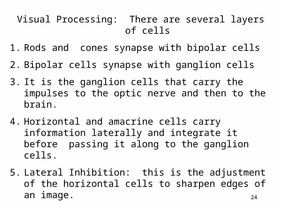
Visual Processing: There are several layers of cells
1. Rods and cones synapse with bipolar cells
2. Bipolar cells synapse with ganglion cells
3. It is the ganglion cells that carry the impulses to the optic nerve and then to the brain.
4. Horizontal and amacrine cells carry information laterally and integrate it before passing it along to the ganglion cells.
5. Lateral Inhibition: this is the adjustment of the horizontal cells to sharpen edges of an image.
25
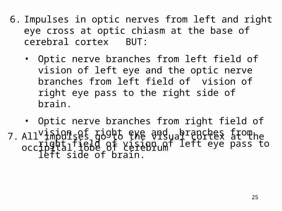
6. Impulses in optic nerves from left and right eye cross at optic chiasm at the base of cerebral cortex BUT:
• Optic nerve branches from left field of vision of left eye and the optic nerve branches from left field of vision of right eye pass to the right side of brain.
• Optic nerve branches from right field of vision of right eye and branches from right field of vision of left eye pass to left side of brain.
7. All impulses go to the visual cortex at the occipital lobe of cerebrum
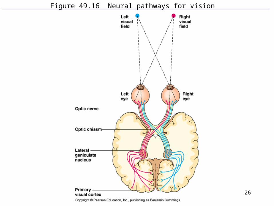
26
Figure 49.16 Neural pathways for vision

27
Figure 49.15x Photoreceptor cells

28
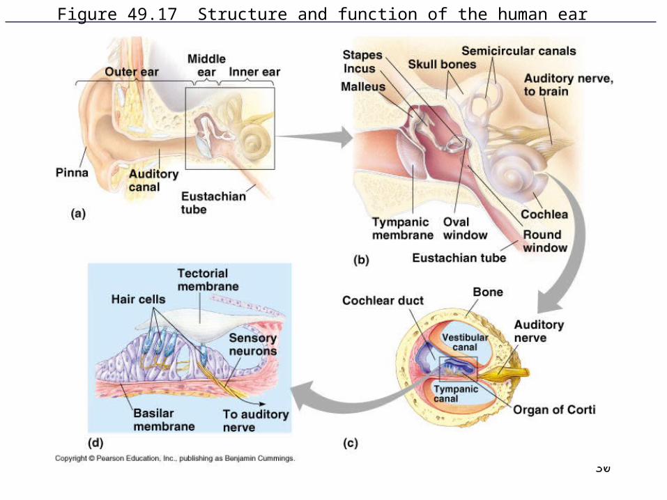
Figure 49.17 Structure and function of the human ear
29
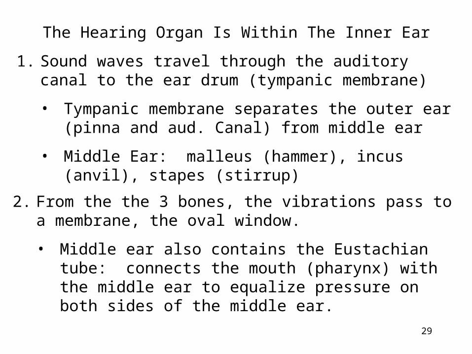
The Hearing Organ Is Within The Inner Ear
1. Sound waves travel through the auditory canal to the ear drum (tympanic membrane)
• Tympanic membrane separates the outer ear (pinna and aud. Canal) from middle ear
• Middle Ear: malleus (hammer), incus (anvil), stapes (stirrup)
2. From the the 3 bones, the vibrations pass to a membrane, the oval window.
• Middle ear also contains the Eustachian tube: connects the mouth (pharynx) with the middle ear to equalize pressure on both sides of the middle ear.
30
Figure 49.17 Structure and function of the human ear

31
3. Inner ear consists:
•Cochlea
•Fluids
• Organ of Corti
4. And the process is:
a) Sound waves vibrate tympanic membrane
b) Vibes are transferred through the bones of middle ear to oval window
c) Oval window contacts the cochlea which contains 3 canals
i. Vestibular canal and tympanic canal which both contain fluid called perilymph
ii. Cochlear duct which contains endolymph

32
Figure 49.17 Structure and function of the human ear

33
c. (cont’d)
iii. Floor of the organ of Corti is the actual “hearing” structure. Components are:
1) Basilar Membrane which forms the floor of the organ of Corti upon which various cell types are attached.
2) Hair Cells which will pick up the vibrations from the cochlear fluid and move against the:
3) Tectorial Membrane
d. Vibes are carried into fluid of vestibular canal and into the tympanic canal.

34
Figure 49.17 Structure and function of the human ear

35
d. Basilar Membrane picks up vibrations, transfers them to hair cells which brush against the tectorial membrane causing depolarization of hair cells, neurotransmitter release and an action potential in the sensory neuron going to the auditory nerve.

36
Figure 49.17 Structure and function of the human ear
37
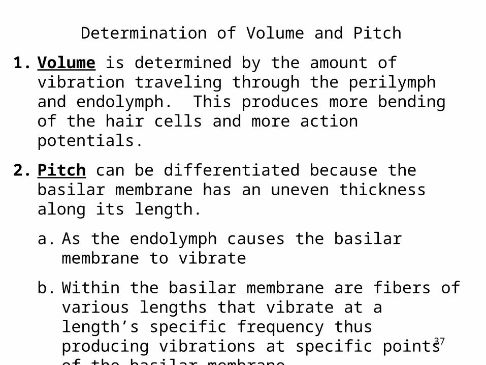
Determination of Volume and Pitch
1. Volume is determined by the amount of vibration traveling through the perilymph and endolymph. This produces more bending of the hair cells and more action potentials.
2. Pitch can be differentiated because the basilar membrane has an uneven thickness along its length.
a. As the endolymph causes the basilar membrane to vibrate
b. Within the basilar membrane are fibers of various lengths that vibrate at a length’s specific frequency thus producing vibrations at specific points of the basilar membrane.
c. At the specific point of basilar membrane are specific hair cells that get vibrated and stimulate specific sensory cells which send messages to specific auditory areas of the cerebral cortex.

38
Figure 49.18 How the cochlea distinguishes pitch

39
Perception of Balance
1. 3 semicircular canals are oriented in various planes and each has an ampulla at its base.
2. Within the ampulla is a gelatinous material called the cupula with its projecting hair cells.
3. When the head rotates the endolymph in the canals stimulates hair cells. If head’s rotation is at constant speed, the firing of the hair cells is minimalized. But if you are rotating and then stop suddenly, the endolymph “sloshes” around, stimulates hair cells with no time for adjustment and you may feel dizzy.
4. Saccule and Utricle with their accompanying hair cells inform us of head’s position of up, down or sideways. Otoliths rest on hair cells and will shift in response to movement, bending hair cells.
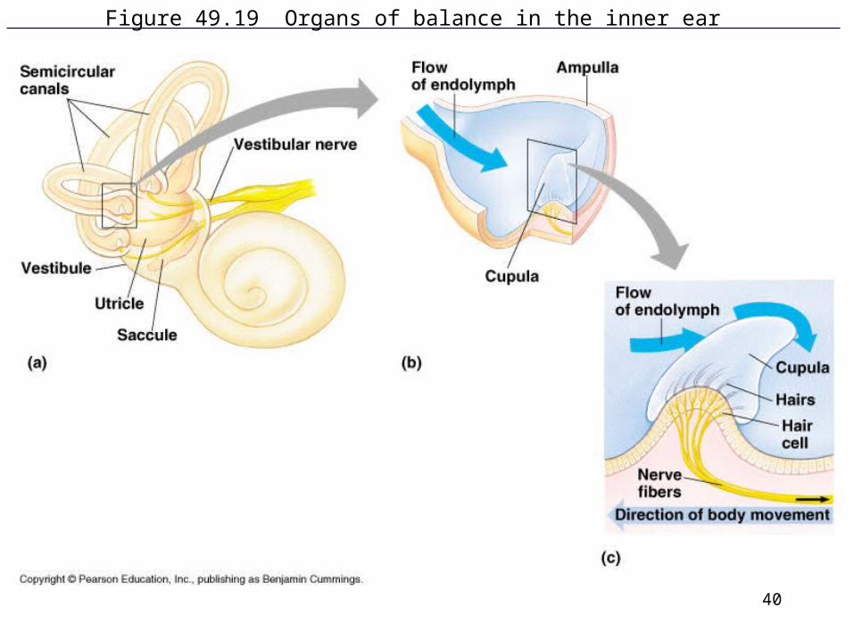
40
Figure 49.19 Organs of balance in the inner ear

41
Figure 49.20 The lateral line system in a fish
•Fish possess an inner ear containing a utricle, saccule and semicircular canals.
•There is no eardrum so vibrations from water travel through skeleton of head to the inner ears which stimulate otoliths and hair cells.
•Swim bladder, which is filled with air, vibrates with sound and transfers vibes to inner ear. Swim bladders are found in most bony fish (not sharks)
•Lateral Line System is made up of receptors (neuromasts) in sensory pits that detect water current via the stimulation of hair cells.

42
Figure 49.21 The statocyst of an invertebrate
Hair cells or cilia surround a chamber containing statoliths (grains of sand). Statoliths are present in jellies’ bells, crayfish antennae.
43
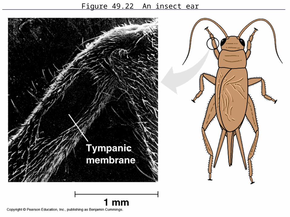
Figure 49.22 An insect ear

44
Figure 49.x2 Salmon follow their noses home

45
Figure 49.23 The mechanism of taste in a blowfly

46
Figure 49.23x Sensillae (hairs) on the foot of an insect

47
Figure 49.24 Olfaction in humans

48
Figure 49.25 The cost of transport

49
Figure 49.x3 Swimming

50
Figure 49.x4 Locomotion on land

51
Figure 49.x5 Flying
52
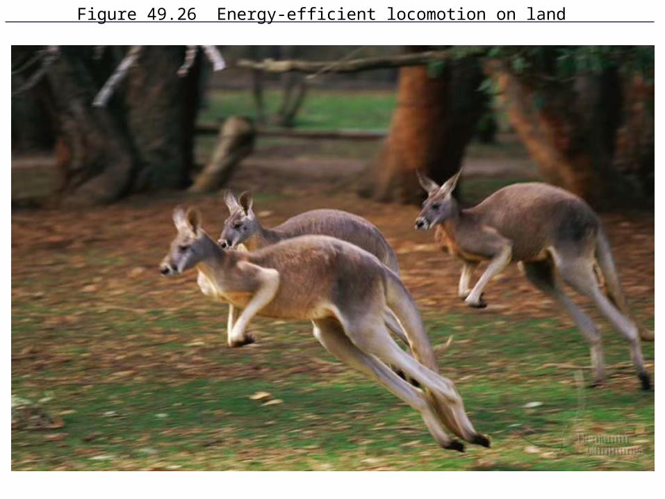
Figure 49.26 Energy-efficient locomotion on land

53
Figure 49.27 Peristaltic locomotion in an earthworm

54
Figure 49.28a The human skeleton

55
Figure 49.28b The human skeleton
56
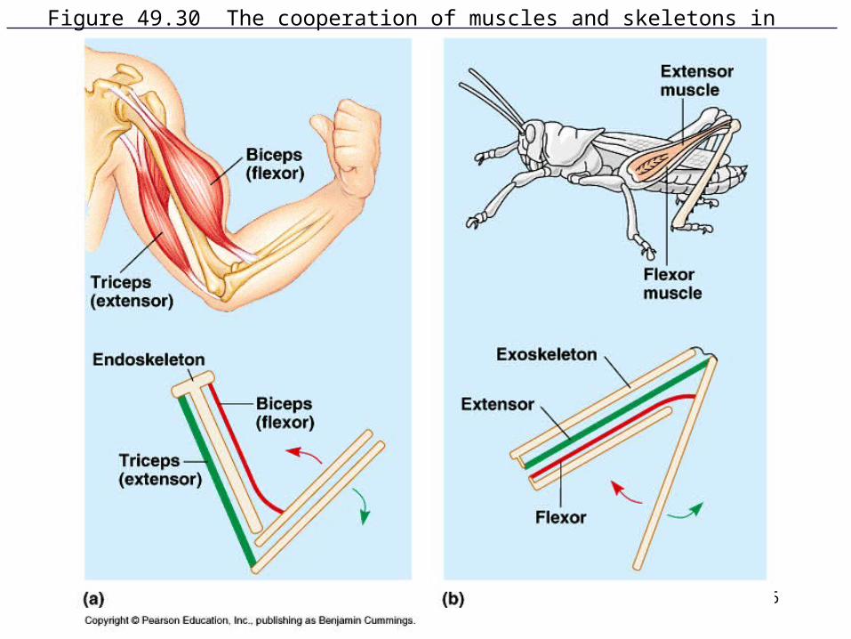
Figure 49.30 The cooperation of muscles and skeletons in movement

57
Figure 49.31 The structure of skeletal muscle
Myofibrils are made up of:
1) actin or thin filaments
2) myosin or thick filaments
I Band: where only the actin filaments are located.
A Band: where the overlap of actin and myosin filaments is located.
58
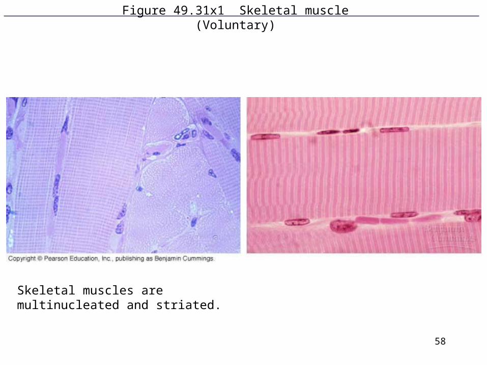
Figure 49.31x1 Skeletal muscle(Voluntary)
Skeletal muscles are multinucleated and striated.
59
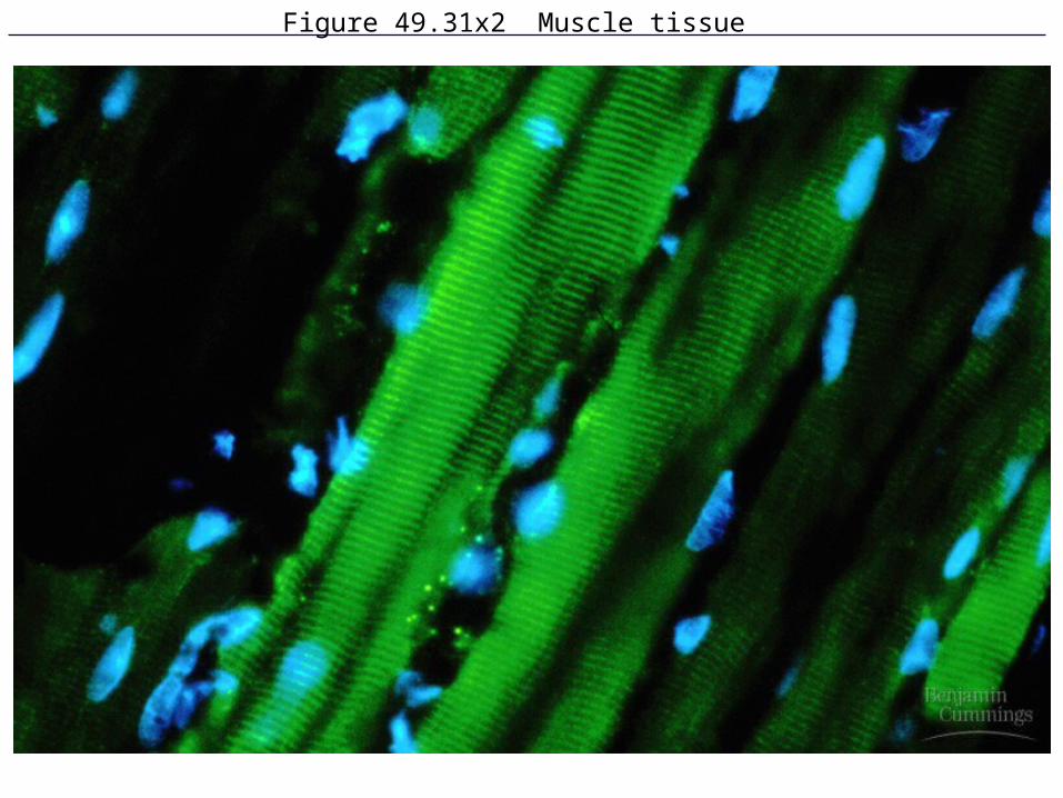
Figure 49.31x2 Muscle tissue

60
Figure 49.32 The sliding-filament model of muscle contraction
Sarcomere shortens during muscle contraction or distance from Z line to Z line decreases.

61
Figure 49.33 One hypothesis for how myosin-actin interactions generate the force for muscle contraction (Layer 1)
1. ATP is bound to myosin head
2. We call this the low energy position or the state when no contraction is occurring.
3. ATP has to be hydrolyzed for contraction to proceed: ATP ADP + Pi
62
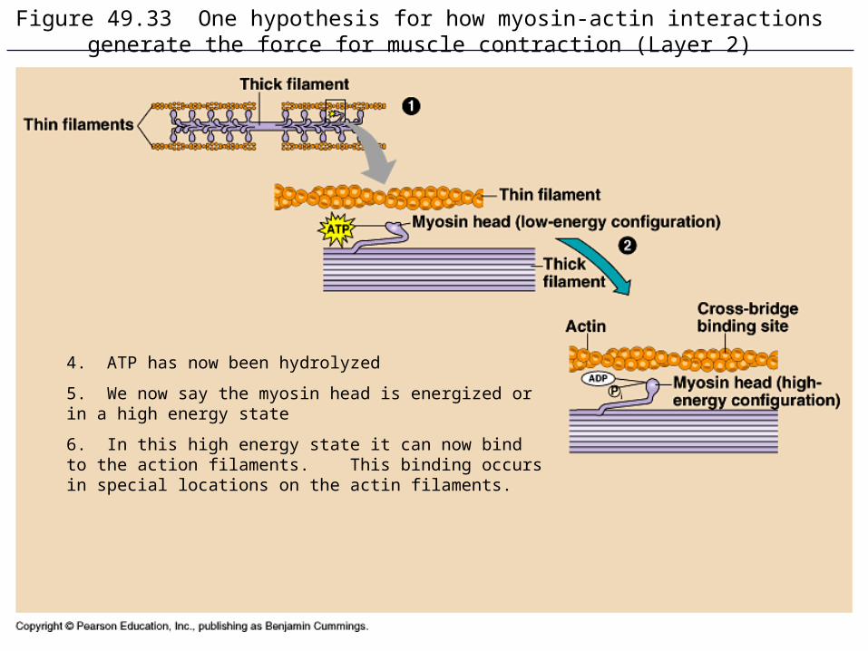
Figure 49.33 One hypothesis for how myosin-actin interactions generate the force for muscle contraction (Layer 2)
4. ATP has now been hydrolyzed
5. We now say the myosin head is energized or in a high energy state
6. In this high energy state it can now bind to the action filaments. This binding occurs in special locations on the actin filaments.

63
Figure 49.33 One hypothesis for how myosin-actin interactions generate the force for muscle contraction (Layer 3)

64
Figure 49.33 One hypothesis for how myosin-actin interactions generate the force for muscle contraction (Layer 4)

65
Figure 49.34 Hypothetical mechanism for the control of muscle contraction
Muscle Contraction Animation Another Animation

66
The Interaction of Actin and Myosin
1. Prior to contraction, the myosin binding sites are covered by a protein called tropomyosin.
2. Upon nerve impulse stimulation (by acetylcholine) of a muscle cell, an action potential travels through the muscle cell via the Transverse or T-tubules to the sarcoplasmic reticulum. Calcium ions are released from the sarcoplasmic reticulum.
3. Action potential travels through Transverse or T-tubules through the muscle (not muscle cell). Recall that a muscle is made up of thousands of muscle cells.
4. Calcium ions will bind to a protein called troponin
5. The Ca-troponin complex will pull or move the tropomyosin off of the binding sites for myosin.
6. Now the myosin binds to action and slides it forward, thus muscle contraction occurs.

67
Figure 49.35 The roles of the muscle fiber’s sarcoplasmic reticulum and T tubules in contraction

68
Figure 49.36 Review of skeletal muscle contraction

69
Motor Units
Definition: a motor unit is a motor neuron and all the muscle cells (fibers) it innervates.
• If one neuron innervates lots of muscle fibers you have “gross” control (a large motor unit) but if one neuron innervates very few fibers you have very precise control of small group of fibers (a small motor unit).
•As you are required to lift more and more weight, your body recruits more motor units and therefore more muscle fibers.

70
Figure 49.38 Motor units in a vertebrate muscle

71
Fast and Slow Twitch Muscle Fibers
Fast Twitch Fibers
•Anaerobic, so few mitochondria
•Lots of sarcoplasmic reticulum to remove calcium for fast contractions
Slow Twitch Fibers
•Aerobic, so lots of mitochondria
•Few sarcoplasmic reticulum so calcium remains in muscle fibers for sustained contractions.
•Contain myoglobin producing the “dark” meat.

72
Other Types of Muscle Tissue
Cardiac Muscle
•It’s striated (contains actin and myosin)
•Contains intercalated disks: located between cardiac cells to pass on the electrical signals from one cell to all others throughout the heart
•Does not require a motor neuron for stimulation. The plasma membrane can generate its own depolarizations, producing an action potential and therefore cardiac muscle cell contraction
•Action potential lasts longer
73
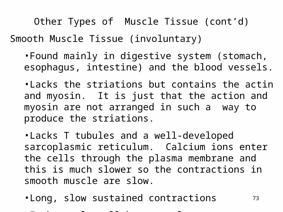
Other Types of Muscle Tissue (cont’d)
Smooth Muscle Tissue (involuntary)
•Found mainly in digestive system (stomach, esophagus, intestine) and the blood vessels.
•Lacks the striations but contains the actin and myosin. It is just that the action and myosin are not arranged in such a way to produce the striations.
•Lacks T tubules and a well-developed sarcoplasmic reticulum. Calcium ions enter the cells through the plasma membrane and this is much slower so the contractions in smooth muscle are slow.
•Long, slow sustained contractions
•Each muscle cell has a nucleus

74
Invertebrate Muscle Tissue
Vertebrates and invertebrates have similar skeletal muscle cells and smooth muscle cells.
Insect Flight Muscles: these muscles can contract faster than the action potentials can arrive
•One action potential can generate 50 “wing beats.”

75
Figure 49.38x Motor units in a vertebrate muscle

76
Figure 49.29 Posture helps support large land vertebrates, such as bears, deer, moose, and cheetahs
77
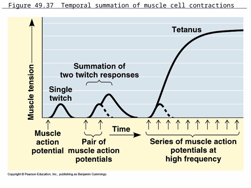
Figure 49.37 Temporal summation of muscle cell contractions

78
Figure 49.5 Chemoreceptors in an insect: Female silk moth Bombyx mori releasing pheromones;
SEM of male Bombyx mori antenna
79
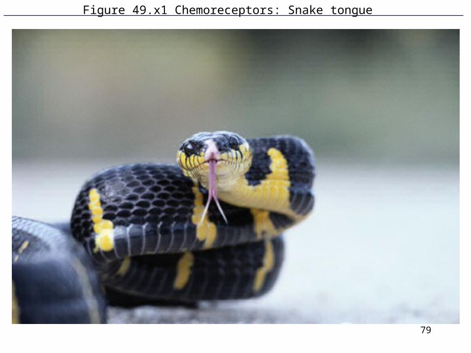
Figure 49.x1 Chemoreceptors: Snake tongue

80
Figure 49.6bx Beluga whale pod
![Allure~ MD Tropical warehouse moth (cocoa moth) [Ephestia cautel/a], Tobacco moth [Ephestia elute/la], Raisin moth [Cadra figulilel/a] 3. APPLICATION: Place dispensers every 20 to](https://img.pdfslide.us/doc/110x75/5fc80c76c4631870720794eb/allure-md-tropical-warehouse-moth-cocoa-moth-ephestia-cautela-tobacco-moth.jpg)