Embed Size (px)
Citation preview
1
Effects of CoCr metal wear debris generated from metal-on-metal hip implants and Co 1
ions on human monocyte-like U937 cells 2
Olga M Posadaa1
, Rothwelle J. Tateb and M. Helen Grant
ac 3
aBiomedical Engineering Department, University of Strathclyde, Wolfson Centre,Glasgow 4
G4 0NW, UK. [email protected], 5
cCorresponding author: [email protected]. TEL: +44 (0)141 548 3438. 6
bStrathclyde Institute for Pharmacy & Biomedical Sciences, University of Strathclyde, 7
Glasgow G4 0RE, UK. [email protected] 8
9
Abstract 10
Hip resurfacing with cobalt-chromium (CoCr) alloy was developed as a surgical alternative to 11
total hip replacement. However, the biological effects of nanoparticles generated by wear at 12
the metal-on-metal articulating surfaces has limited the success of such implants. The aim of 13
this study was to investigate the effects of the combined exposure to CoCr nanoparticles and 14
cobalt ions released from a resurfacing implant on monocytes (U937 cells) and whether these 15
resulted in morphology changes, proliferation alterations, toxicity and cytokine release. The 16
interaction between prior exposure to Co ions and the cellular response to nanoparticulate 17
debris was determined to simulate the situation in patients with metal-on-metal implants 18
receiving a second implant. Effects on U937 cells were mainly seen after 120h of treatment. 19
Prior exposure to Co ions increased the toxic effects induced by the debris, and by Co ions 20
themselves, suggesting the potential for interaction in vivo. Increased TNF-α secretion by 21
resting cells exposed to nanoparticles could contribute to osteolysis processes in vivo, while 22
increased IFN-γ production by activated cells could represent cellular protection against 23
1 Present address: LICAMM laboratories, University of Leeds, Leeds LS2 9JT, UK.
2
tissue damage. Data suggest that interactions between Co ions and CoCr nanoparticles would 24
occur in vivo, and could threaten the survival of a CoCr metal implant. 25
26
Keywords: metal wear debris; hip replacements; nanoparticles; metal ions; cobalt-chrome 27
alloy; monocytes 28
29
1. Introduction 30
The most common cause of failure of total hip arthroplasty is aseptic loosening of the implant 31
initiated by adverse tissue response to prostheses wear particles (Luo et al., 2005). Current 32
evidence indicates that the size of wear particles generated by CoCr alloy metal-on-metal 33
(MoM) articulations is in the nanometre size range (Hosman et al., 2010). The large surface 34
area enhances release of metal ions, predominantly Co and Cr ions, into the circulation 35
(Lucarelli et al., 2004). Wear particles from articular surfaces are phagocytosed mainly by 36
macrophages. When particles are phagocytosed in sufficient amounts, the macrophages enter 37
an active state of metabolism, releasing an array of cytokines, chemokines, and growth 38
factors inducing inflammation, which accelerates osteoclast formation and bone resorption 39
resulting in periprosthetic osteolysis (Germain et al., 2003; Yagil-Kelmer et al., 2004). 40
41
Circulating physiological levels of Co and Cr are normally <0.25μg/l (0.005μM) (Andrews et 42
al., 2011). Elevated levels of Co and Cr ions occur in both the hip synovial fluid and in 43
peripheral blood after MoM hip replacement, and there is concern about the toxicity and 44
biological effects of such ions both locally and systemically (Bisseling et al., 2011; 45
Friesenbichler et al., 2012; Penny et al., 2013). 46
47
3
Co corrodes faster than Cr under physiological conditions (Xia et al., 2011) and, in contrast 48
to Cr, Co ions tend to remain mobile, which is reflected in the higher levels measured in 49
blood, allowing the ions to reach and enter remote organs (Afolaranmi et al., 2012). Elevated 50
Co concentrations in patients with MoM implants are a concern, since increased Co levels in 51
blood have also been reported to be associated with neurological (hand tremor, 52
incoordination, cognitive decline, depression, vertigo, hearing loss, and visual changes) 53
(Oldenburg et al., 2009; Tower, 2010), cardiac (myocardiopathy) (Dadda et al., 1994; 54
Seghizzi et al., 1994; Gilbert et al., 2013) and endocrine (aberrant oestrogen signalling, 55
altered the production or circulation of sex hormones, and altered thyroid metabolism) 56
(Keegan et al., 2007; Oldenburg et al., 2009) symptoms. 57
58
In addition to the above, data from the seventh annual report of the National Joint Registry 59
for England and Wales showed high failure rates for MoM hip prostheses 60
(http://www.njrcentre.org.uk/njrcentre/portals/0/njr%207th%20annual%20report%202010.pd61
f), which led to the market recall of the DePuy ASRTM
, both the Resurfacing and XL Systems 62
in August 2010 (DePuy International Ltd, Leeds, UK) (MDA/2010/069). Following this, the 63
Medicines and Healthcare products Regulatory Agency (MHRA) safety alert in September 64
2010 drew attention to the long term biological safety of all types of MoM hip implants. In 65
this document (MDA/2010/069; http://www.mhra.gov.uk/home/groups/dts-66
bs/documents/medicaldevicealert/con093791.pdf) the MHRA explained the details behind the 67
safety alert and included four situations in which measurements of blood metal ions in 68
patients were recommended: 1) in patients who have symptoms associated with loose MoM 69
bearings; 2) in patients showing radiological features associated with adverse outcomes 70
including component position or small component size; 3) if the patient or surgeon are 71
concerned regarding the MoM bearing; and 4) if there is concern about patients with higher 72
4
than expected rates of failure. The MHRA have suggested that combined whole blood Co and 73
Cr levels of greater than 7ppb (7µg/l or 0.1µM of the combined ions) are associated with 74
significant soft-tissue reactions and failed MoM hips. 75
76
Aseptic loosening usually leads to revision surgery where the implant is removed and 77
replaced with an alternative bearing (Maezawa et al., 2009; Naal et al., 2011; Sehatzadeh et 78
al., 2012). At the time of revision these patients may have high circulating metal ion 79
concentrations (particularly cobalt), and these may alter the response of the patient to the new 80
device. To investigate the effects of the metal ions already present in these patients in terms 81
of the biological response to the new device, cells were pre-treated in vitro with 0.1µM Co in 82
the present study for 4 days before being treated with the metal wear debris. Continued 83
exposure to Co ion release from an existing implant may also influence the responses to wear 84
debris and for this reason the combined effect of exposure to Co ions and wear debris was 85
also investigated. The concentration of Co ions used in the study was chosen to reflect the 86
maximum circulating concentration recommended in patients with MoM implants by MHRA 87
in 2010 (MDA/2010/069; http://www.mhra.gov.uk/home/groups/dts-88
bs/documents/medicaldevicealert/con093791.pdf). 89
90
U937 cells are a human macrophage-like cell line derived from human leukemic monocyte 91
lymphoma (Yagil-Kelmer et al., 2004). This cell line has been used previously as the cell 92
culture model to study the biological effects of different kinds of particles and ions, and it has 93
been demonstrated that U937 cells have comparable responses to polyethylene particles 94
(Matthews et al., 2001) and metal ions (Wang et al., 1996) as do primary macrophages in 95
terms of cytokine release. 96
97
5
The aim of this study was to find out if exposure to CoCr nanoparticles released from a 98
resurfacing implant could activate monocytes, and whether this resulted in cytotoxicity and 99
cytokine release. The interactions between prior exposure to Co ions and the nanoparticulate 100
debris, and between combined exposure to wear debris and Co ions, were determined in order 101
to simulate the in vivo situation in patients with MoM implants. 102
103
2. Methods 104
2.1. Preparation of wear debris 105
CoCr wear debris was a gift from DePuy International (Leeds, UK). A high-carbon (≥ 0.2%) 106
content CoCr alloy (ISO 5832-12: Co Balance, Cr 26.0–30.0%, Mo 5.0–7.0%, Ni 1.0% max., 107
Si 1.0% max., Mn 1.0% max., Fe 0.75% max., C 0.35% max., N 0.25% max.) hip resurfacing 108
implant was worn on a multi-station hip joint simulator using the following protocol. The 109
wear debris was produced over 250000 cycles using distilled water as the lubricating fluid. 110
The use of only distilled water (instead of the more usual bovine serum (25% v/v) in distilled 111
water) resulted in a more rapid and aggressive wear regime which produced a greater volume 112
of wear debris of similar morphology and size for testing purposes to that produced under 113
similar conditions in 25 per cent serum but in a more conducive time-frame (personal 114
communication, Dr C. Hardaker, DePuy International, Leeds, UK). Wear debris produced by 115
hip simulator under different conditions has previously been shown to be of similar size and 116
morphology (Brown et al., 2007). 117
118
Once produced, the wear debris was centrifuged at 3500g for 20 minutes. The majority of the 119
water was then aspirated. The remaining suspension was heat-treated (180°C for 5h, 60kPa) 120
in a vacuum oven to eliminate the remaining water and destroy any endotoxin. The dry debris 121
was then suspended in sterile phosphate buffered saline (PBS; Invitrogen; Paisley, UK). The 122
6
sterility of the treated wear debris was tested as described by Akbar et al. (2012) by exposing 123
dendritic cells (isolated from bone marrow of male BALB/c (Harlan, UK) mouse femurs and 124
tibias (Lutz et al., 1999)) to the debris for 24h, in vitro, and then assessing the expression of 125
surface activation markers via flow cytometry. The debris was found not to increase the 126
surface expression of CD40, CD86, or MHC II on these cells, and, therefore, the suspended 127
debris was deemed sterile and endotoxin-free (data not shown). 128
129
Heat treated wear debris was imaged with a Field Emission Scanning Electron Microscope 130
(FE-SEM) (Hitachi SU-6600, Hitachi; Germany) at magnifications of 100-1000x. Energy 131
Dispersive X-ray Spectroscopy (EDS) was used for quantitative analysis of elemental 132
composition. Hitachi TM-1000 and EDSwift-TM software was used to obtain the images and 133
chemical spectra of the wear debris. Metal ion release from the debris was determined by 134
incubating 156.25μg debris/cm2
for 24h at 37oC in an atmosphere of 5% (v/v) CO2 in air in 135
1ml of complete RPMI-1640 medium in the presence of 10% (v/v) foetal bovine serum (FBS) 136
as described below. The medium was collected and stored at -80oC until ICP-MS analysis 137
using an Agilent 7700x octopole collision system in helium gas mode using scandium as 138
internal standard. Quantification was based on the maximum signal for a particular isotope, 139
and five readings were taken, with the result taken as the mean value. 140
141
2.2. Cell culture 142
U937 (Human leukemic monocyte lymphoma cell line; European Collection of Cell Cultures) 143
cells were cultured in RPMI-1640 medium supplemented with 10% (v/v) FBS, 50U/ml 144
penicillin and 50µg/ml streptomycin. These cells were routinely split every three days at a 145
ratio of 1:10 from a starting passage number of 4 and for no more than 20 passages. This was 146
achieved by taking 3ml of cell suspension into a fresh 75cm2 culture flask containing 27ml of 147
7
fresh complete RPMI-1640 medium. Cells were activated by incubating them with 10nM 148
phorbol 12-myristate 13-acetate (PMA, Sigma-Aldrich; Dorset, UK) for 72h (Lucarelli et al., 149
2004). Activation was confirmed by monitoring cell morphology, adhesion, and aggregation 150
on a Zeiss Imager.Z1 microscope using wet lenses (20X) every 24h. For cobalt pre-treatment 151
of cells, Co2+
solutions were freshly prepared using cobalt chloride (CoCl2) (Alfa Aesar; 152
Lancashire, UK) and diluted to 0.1µM in growth medium under sterile conditions. Resting 153
and activated cells were incubated with 0.1µM Co2+
for 72h. 154
155
2.3. Cell number and viability 156
In order to assess cell viability, U937 cells were exposed to metal debris and Co2+
in resting 157
and activated states for 24 and 120h. For both resting and activated U937 cells, the cells were 158
cultured (1x10⁴ cells/well) in 96-well plates with 156.25μg debris/cm
2 (5mg 159
debris/1x106cells), 0.1µM of Co
2+, or the combination of 156.25μg debris/cm
2 plus 0.1µM of 160
Co2+
in complete RPMI-1640 for 24 and 120h at 37°C under 5% (v/v) CO2. Culture medium 161
was not changed during these incubation periods. At each culture endpoint, cells were 162
washed twice with PBS to remove the debris layer before cell viability was assessed by the 163
neutral red (NR) and 3-(4,5-dimethylthiazol-2-yl)-2,5-diphenyltetrazolium bromide (MTT) 164
assays, as described by Repetto et al. (2008) and Mosmann (1983), respectively. The cells 165
were incubated with NR for 3h and MTT for 4h, and absorbance of the both NR and MTT 166
assay samples were then measured at 540nm using a Bio-Rad Model 450 microplate reader 167
(Bio-Rad, Hertfordshire, UK). 168
169
Viability was also monitored microscopically by double staining with Acridine Orange (AO; 170
Sigma-Aldrich; Dorset, UK) and Propidium Iodide (PI; Life Technologies; Paisley, UK) as 171
previously described by Bank (1988). Cells were viewed using a Carl Zeiss Axio Imager 172
8
microscope under a 40X water immersion lens with a numeric aperture of 0.80. Fluorescence 173
was excited using a mercury lamp and emission recorded using a fluorescein isothiocyanate 174
(FITC)/Rhodamine filter block (485/515-530nm; 546/580-563nm) for AO and PI. Cells 175
fluorescing green were scored as viable, and those fluorescing red as nonviable. 176
177
2.4. Cell proliferation 178
Resting and activated cells were cultured (1x10⁴cells/well) in 96-well plates with 156.25μg 179
debris/cm2, 0.1µM of Co
2+, or the combination of 156.25μg debris/cm
2 plus 0.1µM of Co
2+ in 180
complete RPMI-1640 for 24h at 37°C under 5% (v/v) CO2. U937 cell proliferation was then 181
determined using a BrdU Cell Proliferation Immunoassay kit (kit number QIA58, Merck 182
Chemicals; Nottingham, UK), as suggested by the manufacturer. Briefly, the BrdU label was 183
diluted 1:2000 into fresh complete RPMI-1640 medium. To label the DNA, 20μl of this 184
working solution were added to cells in 96-well plates during the final 4 hours of the 185
experiment. Plates were then centrifuged (300xg for 10min) and the supernatant aspirated. 186
Cells were then fixed, permeabilized, and the DNA denatured. Plates were incubated for 187
30min at room temperature and the fixative/denaturing solution was then aspirated. 100μl of 188
anti-BrdU antibody diluted in antibody dilution buffer were added to each well and incubated 189
with the cells for 1h at room temperature. Cells were washed 3 times with 1X wash buffer, 190
before 100μl of peroxidase goat anti-mouse IgG HRP conjugate diluted in conjugate diluent 191
were incubated with the cells for 30min at room temperature. Plates were then flooded with 192
dH2O, emptied, and incubated with 100μl of substrate solution in the dark at room 193
temperature for 15min. Following the addition of 100μl of stop solution the absorbance was 194
measured using a Thermo Scientific Multiskan Ascent spectrophotometer plate reader at dual 195
wavelengths of 450-540nm. 196
197
9
2.5. Cytokine production: ELISA 198
Cytokine levels were determined by collecting the supernatants from the cell cultures at each 199
end point. The concentrations of tumour necrosis factor-alpha (TNF-α), interferon-γ (IFNγ) 200
and interleukin-6 (IL-6) in the culture media were determined from aliquots of cell-free 201
isolates using Ready-Set-Go! ELISA kits (eBioscience; Hatfield, UK) in accordance with the 202
manufacturer’s instructions. Each of the kits had a sensitivity level of 4pg/ml, and linear 203
standard curves were generated between 0-500pg/ml for TNFα and IFNγ, and 0-200pg/ml for 204
IL-6. The presence of metal ions in the medium did not influence the measurement of the 205
cytokines. 206
207
2.6. Statistics 208
Statistical analyses were carried out by a one-way analysis of variance (ANOVA), followed 209
by a Dunnett’s multiple comparison test, and a two-sample t-test. Significance was assigned 210
where p values were found to be <0.05. 211
212
3. Results 213
3.1. Characterisation of wear debris and release of metal ions 214
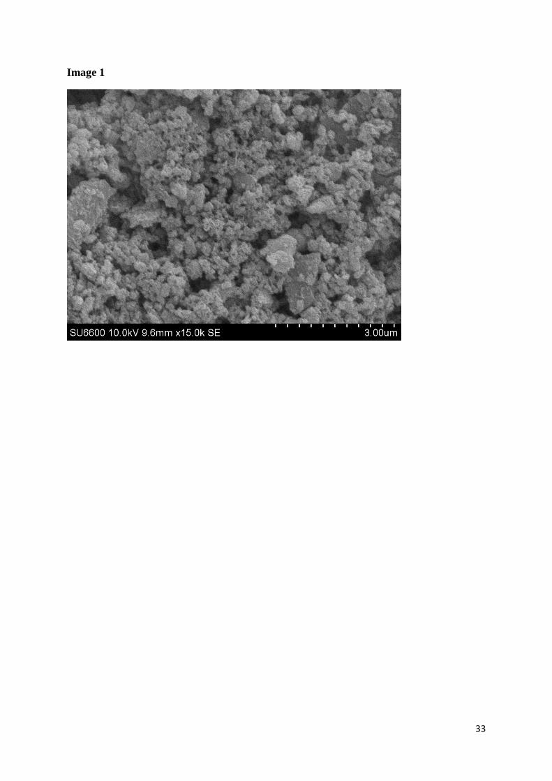
Wear particles generated by the hip simulator process have been reported with a mean size of 215
50nm (Brown et al, 2007). SEM images show irregular shapes and sizes varying from the 216
nano to the micro scale (from 150nm to 6.5µm). The larger irregular shaped particles suggest 217
that the debris aggregates (Image 1), and this has been reported previously by Akbar and co-218
workers (2012). Doorn et al (1998) isolated particles from MoM retrieval tissues that varied 219
in size (51-116nm particles to micrometre sized aggregates) and shape. Moreover, metal 220
particles (0.1-3 microns in size) have also been found in tissues post-mortem (Brown et al., 221
2013). EDS analysis indicated that the wear debris is primarily composed of Co and Cr 222
10
(which is in agreement with the alloy composition (Singh and Dahotre, 2007)). Analysis of 223
25 different particles indicated a mean composition of 59.57 per cent Co, 40.43 per cent Cr 224
and a small amount of Mo which was below the limit of quantification. The CoCr wear debris 225
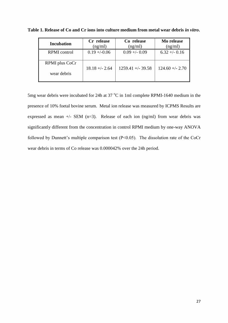
released metal ions into culture medium as shown on Table 1. Co was the predominant ion 226
released after 24h incubation. The percentage dissolution of the debris over the 24h period 227
was negligible, 0.000042%, although the cobalt concentration released was 1259.41+/- 39.58 228
ng/ml. 229
230
3.2. Cell viability 231
In this study, the 156.25μg debris/cm2 (5mg/1x10
6cells) concentration was chosen to mimic 232
the local metallosis environment surrounding an implant. The viability of the cells was 233
assessed after exposure to Co ions, to wear debris, and to a combination of the wear debris 234
and Co ions. Some of the cultures from each type of exposure had been pre-treated with Co 235
ions for 3 days prior to the experiment. Changes in cell number were measured by both NR 236
and MTT assays. The reduction of MTT is influenced by both reductase enzyme activities 237
and the supply of NADH/NADPH (the redox status) in the cells and is therefore, a measure 238
of the metabolic activity and redox status of the cells. In contrast, the NR assay is not 239
dependent on these factors. The latter may, however, be altered by changes in the number of 240
lysosomes in the cells. Resting and activated U937 cell numbers were assessed after 24 and 241
120h of treatment with the wear debris and/or ions. Figure 2 and Figure 3 summarise the 242
120h results, there being no significant changes in cell numbers for either resting or activated 243
U937 cells after 24h of exposure to the different treatments (data not shown). 244
245
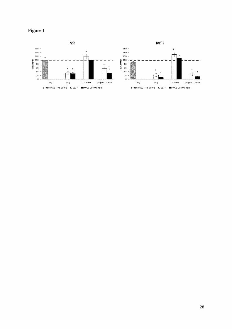
After 120h there was a significant decrease in the absorbances measured by NR and MTT 246
assays respectively in both resting and activated cells, where the metal debris was present. In 247
11
the resting cells, exposure to Co ions for 120h caused an increase in the cell number 248
compared to controls, which was apparent when measured by both MTT and NR (Figure 2). 249
Additionally, cells exposed only for 3 days to Co and then the Co ions kept for 120h in 250
culture medium (PreCo U937+no debris) did not show any differences when compared to 251
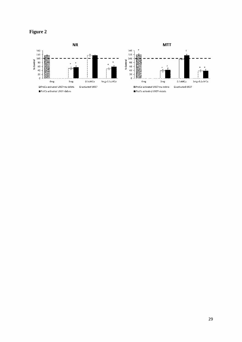
untreated cells. The effect observed on resting cells was not as marked after activation of the 252
cells by PMA (Figure 3), although increased absorbances in activated cells were detected 253
where pre-treatment with Co ions had been carried out. When comparing the NR results of 254
resting cells exposed to the combination of wear debris and Co ions, Co pre-treated cell 255
numbers were significantly lower than those of non Co pre-treated cells. In fact, all the results 256
obtained in resting cells were exacerbated to some extent by the pre-treatment with Co ions. 257
Interestingly, such an effect was not seen after cell activation by PMA. Generally, the 258
magnitude of responses to both nanoparticles and to Co ions was greater in the resting cells 259
than in activated cells. 260
261
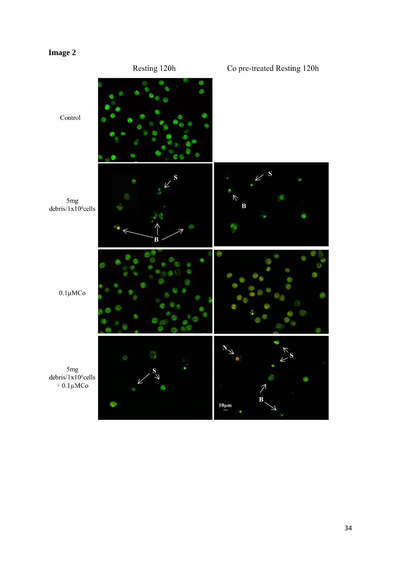
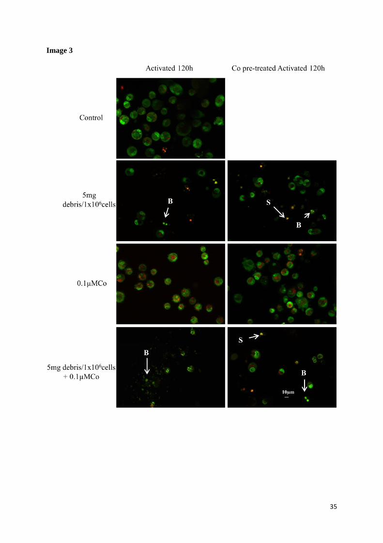
Viability was assessed by AO/PI double staining. PI is impermeable to intact plasma 262
membranes, but it easily penetrates the plasma membrane of dead or dying cells and 263
intercalates with DNA or RNA forming a bright red fluorescent complex. AO is a membrane-264
permeable, monovalent, cationic dye which binds to nucleic acids in the cells to produce a 265
fluorescent green product (Bank, 1988). Images 2 and 3 summarise the 120h results, with 266
there being no significant changes in cell viability for either resting or activated U937 cells 267
after 24h of exposure to the different treatments (data not shown). After 120h treatment, there 268
was a decrease in cell number of both resting and activated cells in the presence of debris but 269
not Co ions. Furthermore, the few cells observed appeared to be mainly apoptotic. 270
271
272
12
3.3 Cell proliferation 273
Effects on cell proliferation were assessed with the BrdU cell proliferation assay after 24h of 274
exposure to the different treatments, at a time when control cells were in the log phase of 275
growth. The results are summarized in Figure 3. 276
277
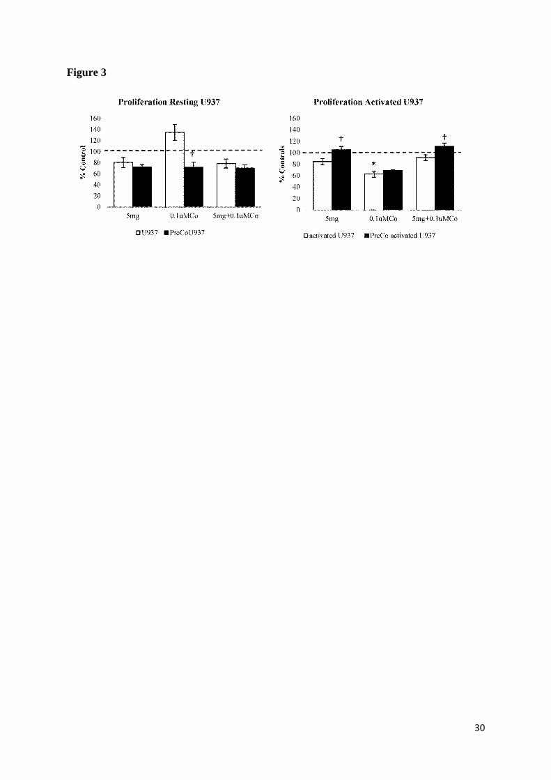
Resting cells in the presence of metal debris had a lower rate of proliferation compared with 278
control cells, although this was not a significant difference. Additionally, the effects on 279
resting U937 and Co pre-treated resting U937 cells were compared in order to establish if the 280
pre-treatment with Co influenced the proliferative cell response to the treatments. Co pre-281
treated resting cells proliferated significantly less than non Co pre-treated resting cells when 282
exposed to 0.1µM Co alone, suggesting a potential role of Co ions in the inhibition of resting 283
U937 cell proliferation. The exposure to 0.1µM Co also caused a significant decrease in the 284
proliferation of activated U937 cells. Moreover, the effects on activated U937 cells and Co 285
pre-treated activated U937 cells were compared in order to establish if the pre-treatment with 286
Co influenced the proliferative cell response to the treatments. Co pre-treated activated cells 287
proliferated significantly more than non Co pre-treated activated cells in the presence of 288
metal wear debris alone. However, the same effect was observed when cells were exposed to 289
the combination of metal wear debris and Co ions, showing a significantly higher 290
proliferation of Co pre-treated activated cells compared to non Co pre-treated activated cells. 291
No difference was observed in the presence of Co ions alone. These findings differ from the 292
results of resting cells, which indicates that the cellular activation state may influence the 293
biological response to metal particles and ions. Although not statistically significant, Co ions 294
alone increased the rate of proliferation of resting cells, and while at different endpoints, 295
analogous to the results found with MTT and NR. 296
297
13
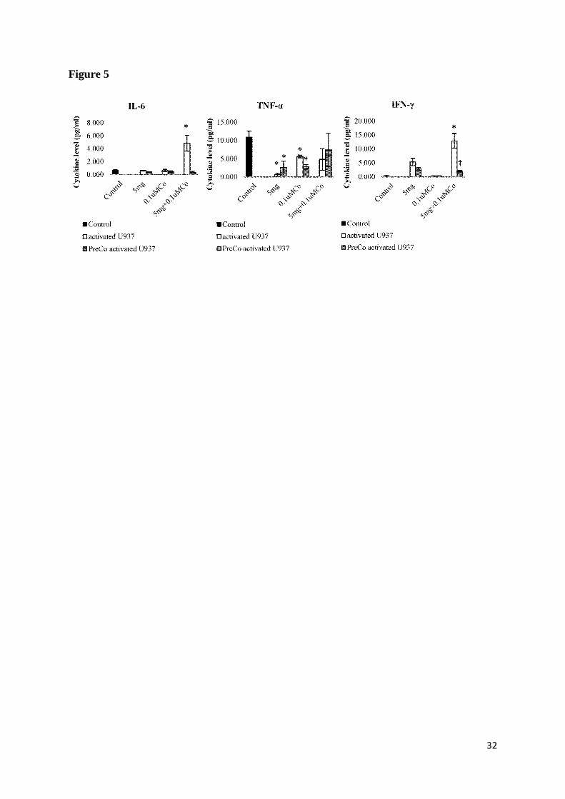
3.4 Cytokine production measured by ELISA 298
Levels of IL-6, TNF-α, and IFN-γ released were determined in the culture medium after 120h 299
of exposure to the different treatments. The results are summarised in Figure 4 and 5. 300
301
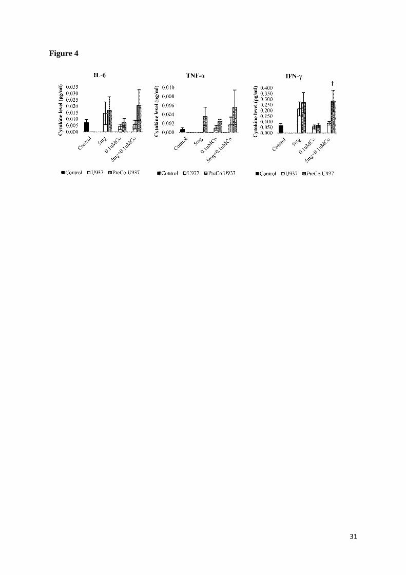
Both untreated resting cells and PMA-activated cells (controls on both graphs) secreted the 302
three cytokines at the 120h culture end point. There was no statistical difference between the 303
levels of cytokines secreted by treated and untreated resting cells. The Co pre-treatment did 304
not cause a significant difference to the levels of cytokine secretion by resting U937 cells. 305
Activated cells exposed to the combination of metal debris and Co ions secreted significantly 306
more IL-6 and IFN-γ than control cells. Contrary to this, treatments caused a decrease in the 307
secretion of TNF-α by activated cells. Co pre-treated activated cells exposed to 0.1µM Co 308
secreted significantly less IFN-γ than non Co pre-treated cells. 309
310
4. Discussion 311
MoM hip implants are composed of CoCr alloys, and as such, during wear, both CoCr 312
nanoparticulate debris and Co and Cr ions are released. In this study the effects of a high 313
wear debris concentration and Co ions on U937 cells were studied. After release from MoM 314
implants, Cr ions tend to bind to the local tissue whereas Co ions tend to remain mobile and 315
enter the circulation (Simonsen et al., 2012). The Cr forms stable complexes which deposit 316
around the implant, whereas Co concentrations in patients’ blood are generally higher, and 317
thus the ions interact with distant tissues and cells to a greater extent. 318
319
The wear debris used in this study was produced from a 39mm ASRTM
prosthesis, and 320
previous work emulating natural gait has shown that approximately 8 mm3 of debris is 321
produced per million cycles on a simulator from this prosthesis (Leslie et al., 2008). The 322
14
density of the CoCr alloy used was 8.32mg/mm3 (Medley et al., 1996), so 8mm
3 of wear 323
would be equivalent to 66.56mg debris. An active person might walk 3.5 million cycles per 324
year, so 232.96mg debris/year would be produced locally in the environment of the 325
prosthesis. The 156.25μg/cm2
metal debris concentration used in this investigation was 326
chosen to mimic metallosis, a situation where metallic debris infiltrates into the periprosthetic 327
tissues, with resulting severe adverse effects. The range of debris and ion concentrations 328
measured locally varies hugely in the literature, for example, in catastrophic failure of a 329
prosthesis amounts of wear debris up to 67mg have been reported (Matziolis et al., 2003). 330
The high metal ion concentrations released from debris in the current experiments simulates 331
this local situation. 332
333
Exposure to 156.25μg debris/cm2
proved to be toxic for both resting and activated U937 cells 334
in the presence and absence of Co ions, which could be in part due to the high concentration 335
of ions released from the particles. However, there was no evidence of toxicity observed after 336
cell exposure to 0.1μM Co ions alone. In fact, the cell numbers of resting cultures increased 337
after exposure to the Co ions alone, and this type of response has been reported previously 338
(Zijlstra et al., 2012), and is thought to represent a protective response to a non-toxic insult. 339
The statistically significant difference in proliferation between Co pre-treated and non-Co 340
pre-treated resting cells exposed to 0.1µM Co ions alone, suggests that longer exposure to Co 341
ions ( >4 days) may result in a toxic response. These results seem to indicate once more that 342
Co ions, particularly at high concentrations, are important in the adverse tissue response to 343
metal wear debris and their effect may differ between cell activation states. 344
345
In the current investigation, a pre-treatment with cobalt ions was carried out to find out if 346
such tissue pre-exposure and ion dissemination in vivo would have an effect on cell responses 347
15
to subsequent exposure to metal wear debris and ions. Such an effect would be important if a 348
patient received a second MoM implant or indeed had a revision procedure to replace a 349
failing implant. Many patients with failing MoM implants have shown high circulating Co 350
ions in their blood (Antoniou et al., 2008; Tkaczyk et al., 2010b; Bisseling et al., 2011; 351
Lavigne et al., 2011; Friesenbichler et al., 2012; Penny et al., 2013), and recently the 352
importance of cobalt release in the inflammatory response to CoCr debris has been 353
demonstrated (Caicedo et al., 2009; Hart et al., 2012). Results from the present study indicate 354
that exposure to wear debris had a pronounced detrimental effect on cell number and 355
metabolic activity of cells pre-treated with Co after an incubation period of 120h. Even 356
though effects observed in resting cells were exacerbated to some extent by the pre-treatment 357
with Co ions, such an effect was not seen after cell activation by PMA. PMA exerts its 358
biologic effects by altering gene expression through the activation of protein kinase C (PKC) 359
and modulating the activity of transcriptional factors such as nuclear factor kappa-light-360
chain-enhancer of activated B cells (NFkB) and activator protein 1 (AP1) (Garcia et al., 361
1999; Daigneault et al., 2010). Thus, it seems unlikely that PKC was directly involved in the 362
pathways leading to the decrease in cell numbers. Despite the changes in proliferation at 24h 363
by BrdU assay, no changes in cell number or metabolic activity were observed at this same 364
endpoint by NR and MTT. Due to the intrinsic characteristics of the assays themselves and 365
complex dynamics of the balance of cell proliferation and cell death, it is unlikely that the 366
changes in cell proliferation (cell division) measured at 24h would be simultaneously 367
apparent as an increase in cell numbers at the same time point. On retrospect, such changes in 368
terms of cell numbers would have been detectable had we measured this at later time points. 369
Cells were treated with the Co ion concentration that has been advised by MHRA as the 370
recommended maximum safe circulating blood level for patients with MoM implants 371
((MDA/2010/069); http://www.mhra.gov.uk/home/groups/dts-372
16
bs/documents/medicaldevicealert/con093791.pdf). It should be noted that cells pre-treated 373
with Co ions at a 120h end point would have been exposed to Co for 9 days in total. In 374
patients, the effects of Co ions and nanoparticles released from metal implants may be 375
additive in terms of adverse effects. Contrary to Cr, Co ions tend to remain mobile and as a 376
result greater Co concentration (compared to Cr) in blood and remote organs have been 377
reported (Afolaranmi et al., 2012) and will be more likely to affect the responses of distant 378
organs to metal nanoparticles than the Cr ions. The importance of Co ions in the 379
inflammatory responses to Co-Cr particles has been recognised, and chronic exposure to 380
circulating levels of ions, plus high local concentrations may act synergistically in vivo to 381
trigger and promote implant loosening (Hallab et al., 2001; Caicedo et al., 2010; Hart et al., 382
2012) . Although circulating systemic Cr levels are generally lower in vivo, it may be 383
interesting in the future to investigate the influence of prior exposure to Cr ions in the CoCr 384
wear debris in vitro exposure experiments. The presence of wear debris in the peri-implant 385
area leads to macrophage phagocytosis of particulate debris and activation, the release of a 386
variety of mediators, such as free radicals and nitric oxide, and a myriad of proinflammatory 387
cytokines and chemokines (Sethi et al., 2003). The uptake of the CoCr particles into the cells 388
was not measured due to technical difficulties in detecting the particles in the cells, but in a 389
preliminary study phagocytosis by the activated cells was demonstrated using 1µm size 390
FITC-labelled dextran beads (results not shown). Differences in the uptake of the CoCr 391
particles could have occurred in resting and/or activated cells after the different treatments, 392
and a contribution towards any differences in response cannot be excluded. Generally, the 393
magnitude of the response to both the particles and the Co ions was greater in the resting cells 394
than in the activated cells, and so the role of phagocytosis in the toxic responses is not clear. 395
It has been reported that local acidification may develop during acute and chronic 396
inflammation (Rajamaki et al., 2013) and high hydrogen ion concentrations down to pH 5.4 397
17
have been found in inflamed tissue (Steen et al., 1995). In turn, such an acidic environment 398
created by actively metabolizing immune cells may enhance the corrosion process of the 399
nanoparticles increasing the amount of metal ions being released (Afolaranmi et al., 2011). 400
401
A number of studies have shown that metal wear particles and high levels of metal ions, 402
particularly Co and Cr, have a cytotoxic effect on a variety of cells such as human osteoblast-403
like cell lines (SaOS-2 and MG-63), and human monocytic-like U937 cells in vitro (Allen et 404
al., 1997; Fleury et al., 2006; Petit et al., 2006). Most of these studies have been focused 405
mainly on the short-term exposure, acute cell response, or have been limited to evaluation of 406
one parameter like cell viability or cytokine levels. Studies carried out with U937 cells have 407
been performed either with resting U937 cells (Ingham et al., 2000; Howling et al., 2003; 408
Tkaczyk et al., 2010a) or activated U937 cells (Lucarelli et al., 2004; Yagil-Kelmer et al., 409
2004; Wang et al., 2011), and have investigated the effects of different kinds of metals, 410
particles, and ions on U937 cells. Papageorgiou et al. (2007) compared the cytotoxic and 411
genotoxic effects of nanoparticles and micron-sized particles of CoCr alloy using human 412
fibroblasts in tissue culture. Their results showed that exposure of human fibroblasts to 413
nanoparticles and micron-sized particles of cobalt chrome alloy, at the same particle mass per 414
cell, cause different types and amounts of cellular damage. In particular, they found 415
nanoparticles to be more cytotoxic and induce more DNA damage than micron-sized 416
particles. This difference in induction of toxicity will contribute to the adverse effects found 417
in vivo from CoCr nanoparticulate debris generated from MoM implants. Dalal et al. (2012) 418
compared the responses of human osteoblasts, fibroblasts, and macrophages exposed to 419
particles of different metal-based particles (i.e., cobalt-chromium (CoCr) alloy, titanium (Ti) 420
alloy, zirconium (Zr) oxide, and Zr alloy). They found that CoCr-alloy particles were by far 421
the most toxic and decreased viability and proliferation of human osteoblasts, fibroblasts, and 422
18
macrophages. Germain et al. (2003) studied the effects cobalt–chromium wear particles at 423
various doses on the viability of U937 cells. CoCr particles at 5µm3 (0.042mg) and 50µm
3 424
(0.42mg) per cell reduced the viability of U937 cells by 42% and 97%, respectively. 425
Papageorgiou et al. (2007) and Germain et al. (2003) used high particle concentrations in 426
comparison to the concentration used in the present study, which indicates that even at lower 427
doses CoCr nanoparticles can exert cytotoxic effects. (Akbar et al., 2011) investigated effects 428
of Cr6+
and Co2+
on primary human lymphocytes in vitro. Their results showed that exposure 429
to 10 and 100μM Cr6+
significantly decreased cell viability and increased apoptosis in both 430
resting and activated lymphocytes. The exposure of resting and activated lymphocytes to 431
100μM Co2+
also resulted in significant decreases in cell viability accompanied by a 432
significant increase in apoptosis and they showed that activated cells were significantly more 433
sensitive to Co2+
toxicity. The concentration of Co ions used in the present study was 1000 434
fold lower (0.1µM) than that used by Akbar et al. (2011), and no significant effect in cell 435
viability and proliferation was observed herein. From the literature there is little doubt that 436
the nanoparticulate debris and metal ions released from MoM implants have toxic effects and 437
the results in the present study point to the potential for interaction between them in vivo. 438
439
Although not statistically significant, there were higher levels of secretion of IL-6, TNF-α 440
and IFN-γ by resting cells after 120h of exposure to 156.25μg/cm2
metal debris and the 441
combination of metal debris and Co ions. In the case of activated cells, there was 442
significantly higher secretion of IL-6 and IFN-γ by cells exposed to the combination of 443
156.25μg/cm2
metal debris and Co ions after 120h. On the other hand, and although not 444
statistically significant, all treatments caused a reduction in the level of secretion of TNF-α. 445
Contrary to what was observed in resting cells, Co pre-treatment did not seem to cause a 446
difference in cytokine secretion by activated cells. It should be noted that the secretion of 447
19
cytokines has not been correlated for cell numbers in these experiments, and changes may 448
reflect cytotoxicity. These results again suggest a larger impact of a chronic exposure and a 449
key role of Co ions. They also suggest that different molecular pathways are affected in 450
activated U937 cells when compared to resting U937 cells. 451
452
Bone remodelling involves tight regulation of three proteins, receptor activator of NF-κB 453
ligand (RANKL), receptor activator of NF-κB (RANK), and osteoprotegerin (OPG). These 454
proteins are key determinants of osteoclastogenesis and regulate bone resorption 455
(Takayanagi, 2005). Pro-inflammatory cytokines such as IL-6, and TNF-α, can upregulate 456
RANKL expression on osteoblasts and accelerate RANKL signalling, and thus directly 457
contribute to bone destruction (Oishi et al., 2012). In biomaterials research, TNF-α, IL-1, and 458
other pro-inflammatory cytokines are also known mediators of the foreign body reaction, an 459
inflammatory response that can cause both severe tissue damage and premature failure of 460
implanted materials (Mountziaris and Mikos, 2008). IFN-γ promotes innate immune 461
responses by activating macrophages. In parallel, IFN-γ exerts regulatory functions to limit 462
tissue damage associated with inflammation like suppressing osteoclastogenesis (Hu and 463
Ivashkiv, 2009). Results from this study suggest that high concentrations of metal wear 464
debris, particularly in the presence of Co ions, promote an immune response with the 465
secretion of pro-inflammatory cytokines, which can contribute to tissue damage and 466
ultimately result in aseptic loosening. 467
468
5. Conclusions 469
The results from this study suggest that a high concentration of metal debris in combination 470
with Co ions not only have a direct effect on cell viability but also influence cell function. 471
Previous exposure to Co ions seems to sensitise U937 cells to the toxic effects of both Co 472
20
ions themselves and to nanoparticles, pointing to the potential for interaction in vivo. The 473
increase in TNF-α secretion by the resting U937 cells could be a factor contributing to the 474
osteolysis process, while the increase in IFN-γ production by the activated cells could be a 475
cellular effort to counteract tissue damage. This also suggests that cellular activation state 476
affects the biological response to wear debris and for this reason caution should be taken 477
when choosing in vitro models to study immune and molecular responses. Moreover, these 478
findings mean that the survival and well-functioning of a second implanted MoM device 479
could be compromised in patients undergoing revision surgery or receiving a second device, 480
due to the interactions between recirculating Co ions and CoCr nanoparticles. 481
482
Acknowledgements 483
This study was supported by funds from University of Strathclyde and by an Overseas 484
Research Studentship to OMP. The authors are grateful to Dr C Hardaker (DePuy 485
International) who prepared the CoCr nanoparticles. 486
487
6. References 488
Afolaranmi, G.A., Akbar, M., Brewer, J., Grant, M.H., 2012. Distribution of metal released 489
from cobalt-chromium alloy orthopaedic wear particles implanted into air pouches in 490 mice. Journal of Biomedical Materials Research Part A 100A, 1529-1538. 491
Afolaranmi, G.A., Al-Mufti, H., Grant, M.H., 2011. Release of soluble metal ions from 492 copper based dental alloys measured by ICPMS. Toxicology 290, 119-119. 493
Akbar, M., Brewer, J.M., Grant, M.H., 2011. Effect of chromium and cobalt ions on primary 494
human lymphocytes in vitro. J. Immunotoxicol. 8, 140-149. 495 Allen, M.J., Myer, B.J., Millett, P.J., Rushton, N., 1997. The effects of particulate cobalt, 496
chromium and cobalt-chromium alloy on human osteoblast-like cells in vitro. Journal 497 of Bone and Joint Surgery-British Volume 79B, 475-482. 498
Andrews, R.E., Shah, K.M., Wilkinson, J.M., Gartland, A., 2011. Effects of cobalt and 499 chromium ions at clinically equivalent concentrations after metal-on-metal hip 500 replacement on human osteoblasts and osteoclasts: Implications for skeletal health. 501 Bone 49, 717-723. 502
Antoniou, J., Zukor, D.J., Mwale, F., Minarik, W., Petit, A., Huk, O.L., 2008. Metal ion 503
levels in the blood of patients after hip resurfacing: A comparison between twenty-504 eight and thirty-six-millimeter-head metal-on-metal prostheses. Journal of Bone and 505
Joint Surgery-American Volume 90A, 142-148. 506
21
Bank, H.L., 1988. Rapid assessment of islet viability with acridine-orange and propidium 507
iodide. In Vitro Cellular & Developmental Biology 24, 266-273. 508 Bisseling, P., Zeilstra, D.J., Hol, A.M., van Susante, J.L.C., 2011. Metal ion levels in patients 509
with a lumbar metal-on-metal total disc replacement. Should we be concerned? 510 Journal of Bone and Joint Surgery-British Volume 93B, 949-954. 511
Brown, C., Lacharme-Lora, L., Mukonoweshuro, B., Sood, A., Newson, R.B., Fisher, J., 512 Case, C.P., Ingham, E., 2013. Consequences of exposure to peri-articular injections of 513 micro- and nano-particulate cobalt-Chromium alloy. Biomaterials 34, 8564-8580. 514
Brown, C., Williams, S., Tipper, J.L., Fisher, J., Ingham, E., 2007. Characterisation of wear 515 particles produced by metal on metal and ceramic on metal hip prostheses under 516
standard and microseparation simulation. Journal of Materials Science-Materials in 517 Medicine 18, 819-827. 518
Caicedo, M.S., Desai, R., McAllister, K., Reddy, A., Jacobs, J.J., Hallab, N.J., 2009. Soluble 519 and particulate Co-Cr-Mo alloy implant metals activate the inflammasome danger 520 signaling pathway in human macrophages: A novel mechanism for implant debris 521
reactivity. Journal of Orthopaedic Research 27, 847-854. 522 Caicedo, M.S., Pennekamp, P.H., McAllister, K., Jacobs, J.J., Hallab, N.J., 2010. Soluble 523
ions more than particulate cobalt-alloy implant debris induce monocyte costimulatory 524 molecule expression and release of proinflammatory cytokines critical to metal-525
induced lymphocyte reactivity. Journal of Biomedical Materials Research Part A 93A, 526 1312-1321. 527
Dadda, F., Borleri, D., Migliori, M., Mosconi, G., Medolago, G., Virotta, G., Colombo, F., 528 Seghizzi, P., 1994. Cardiac-function study in hard metal workers. Science of the Total 529 Environment 150, 179-186. 530
Daigneault, M., Preston, J.A., Marriott, H.M., Whyte, M.K.B., Dockrell, D.H., 2010. The 531 identification of markers of macrophage differentiation in PMA-stimulated THP-1 532
cells and monocyte-derived macrophages. Plos One 5. 533 Dalal, A., Pawar, V., McAllister, K., Weaver, C., Hallab, N.J., 2012. Orthopedic implant 534
cobalt-alloy particles produce greater toxicity and inflammatory cytokines than 535
titanium alloy and zirconium alloy-based particles in vitro, in human osteoblasts, 536
fibroblasts, and macrophages. Journal of Biomedical Materials Research Part A 100A, 537 2147-2158. 538
Doorn, P.F., Campbell, P.A., Worrall, J., Benya, P.D., McKellop, H.A., Amstutz, H.C., 1998. 539
Metal wear particle characterization from metal on metal total hip replacements: 540 Transmission electron microscopy study of periprosthetic tissues and isolated 541
particles. Journal of Biomedical Materials Research 42, 103-111. 542 Fleury, C., Petit, A., Mwale, F., Antoniou, J., Zukor, D.J., Tabrizian, M., Huk, O.L., 2006. 543
Effect of cobalt and chromium ions on human MG-63 osteoblasts in vitro: 544
Morphology, cytotoxicity, and oxidative stress. Biomaterials 27, 3351-3360. 545 Friesenbichler, J., Maurer-Ertl, W., Sadoghi, P., Lovse, T., Windhager, R., Leithner, A., 546
2012. Serum metal ion levels after rotating-hinge knee arthroplasty: comparison 547 between a standard device and a megaprosthesis. International Orthopaedics 36, 539-548
544. 549 Garcia, A., Serrano, A., Abril, E., Jimenez, P., Real, L.M., Canton, J., Garrido, F., Ruiz-550
Cabello, F., 1999. Differential effect on U937 cell differentiation by targeting 551 transcriptional factors implicated in tissue- or stage-specific induced integrin 552 expression. Experimental Hematology 27, 353-364. 553
Germain, M.A., Hatton, A., Williams, S., Matthews, J.B., Stone, M.H., Fisher, J., Ingham, E., 554 2003. Comparison of the cytotoxicity of clinically relevant cobalt-chromium and 555
alumina ceramic wear particles in vitro. Biomaterials 24, 469-479. 556

22
Gilbert, C.J., Cheung, A., Butany, J., Zywiel, M.G., Syed, K., McDonald, M., Wong, F., 557
Overgaard, C., 2013. Hip Pain and Heart Failure: The Missing Link. Canadian Journal 558 of Cardiology 29. 559
Hallab, N., Merritt, K., Jacobs, J.J., 2001. Metal sensitivity in patients with orthopaedic 560 implants. Journal of Bone and Joint Surgery-American Volume 83A, 428-436. 561
Hart, A.J., Quinn, P.D., Lali, F., Sampson, B., Skinner, J.A., Powell, J.J., Nolan, J., Tucker, 562 K., Donell, S., Flanagan, A., Mosselmans, J.F.W., 2012. Cobalt from metal-on-metal 563 hip replacements may be the clinically relevant active agent responsible for 564 periprosthetic tissue reactions. Acta Biomaterialia 8, 3865-3873. 565
Hosman, A.H., van der Mei, H.C., Bulstra, S.K., Busscher, H.J., Neut, D., 2010. Effects of 566
metal-on-metal wear on the host immune system and infection in hip arthroplasty. 567 Acta Orthopaedica 81, 526-534. 568
Howling, G.I., Sakoda, H., Antonarulrajah, A., Marrs, H., Stewart, T.D., Appleyard, S., 569 Rand, B., Fisher, J., Ingham, E., 2003. Biological response to wear debris generated in 570 carbon based composites as potential bearing surfaces for artificial hip joints. Journal 571
of Biomedical Materials Research Part B-Applied Biomaterials 67B, 758-764. 572 Hu, X., Ivashkiv, L.B., 2009. Cross-regulation of Signaling Pathways by Interferon-gamma: 573
Implications for Immune Responses and Autoimmune Diseases. Immunity 31, 539-574 550. 575
Ingham, E., Green, T.R., Stone, M.H., Kowalski, R., Watkins, N., Fisher, J., 2000. Production 576 of TNF-alpha and bone resorbing activity by macrophages in response to different 577
types of bone cement particles. Biomaterials 21, 1005-1013. 578 Keegan, G.M., Learmonth, I.D., Case, C.P., 2007. Orthopaedic metals and their potential 579
toxicity in the arthroplasty patient - Review of current knowledge and future 580
strategies. Journal of Bone and Joint Surgery-British Volume 89B, 567-573. 581 Lavigne, M., Belzile, E.L., Roy, A., Morin, F., Amzica, T., Vendittoli, P.A., 2011. 582
Comparison of whole-blood metal ion levels in four types of metal-on-metal large-583 diameter femoral head total hip arthroplasty: The potential influence of the adapter 584 sleeve. Journal of Bone and Joint Surgery-American Volume 93A, 128-136. 585
Leslie, I., Williams, S., Brown, C., Isaac, G., Jin, Z.M., Ingham, E., Fisher, J., 2008. Effect of 586
bearing size on the long-term wear, wear debris, and ion levels of large diameter 587 metal-on-metal hip replacements - An in vitro study. Journal of Biomedical Materials 588 Research Part B-Applied Biomaterials 87B, 163-172. 589
Lucarelli, M., Gatti, A.M., Savarino, G., Quattroni, P., Martinelli, L., Monari, E., Boraschi, 590 D., 2004. Innate defence functions of macrophages can be biased by nano-sized 591
ceramic and metallic particles. Eur. Cytokine Netw. 15, 339-346. 592 Luo, L., Petit, A., Antoniou, J., Zukor, D.J., Huk, O.L., Liu, R.C.W., Winnik, F.M., Mwale, 593
F., 2005. Effect of cobalt and chromium ions on MMP-1 TIMP-1, and TNF-alpha 594
gene expression in human U937 macrophages: A role for tyrosine kinases. 595 Biomaterials 26, 5587-5593. 596
Lutz, M.B., Kukutsch, N., Ogilvie, A.L.J., Rossner, S., Koch, F., Romani, N., Schuler, G., 597 1999. An advanced culture method for generating large quantities of highly pure 598
dendritic cells from mouse bone marrow. Journal of Immunological Methods 223, 77-599 92. 600
Maezawa, K., Nozawa, M., Matsuda, K., Sugimoto, M., Shitoto, K., Kurosawa, H., 2009. 601 Serum chromium levels before and after revision surgery for loosened metal-on-metal 602 total hip arthroplasty. Journal of Arthroplasty 24, 549-553. 603
Matthews, J.B., Green, T.R., Stone, M.H., Wroblewski, B.M., Fisher, J., Ingham, E., 2001. 604 Comparison of the response of three human monocytic cell lines to challenge with 605
23
polyethylene particles of known size and dose. Journal of Materials Science-Materials 606
in Medicine 12, 249-258. 607 Matziolis, G., Perka, C., Disch, A., 2003. Massive metallosis after revision of a fractured 608
ceramic head onto a metal head. Archives of Orthopaedic and Trauma Surgery 123, 609 48-50. 610
MDA/2010/069, M.a.H.p.R.A., Medical Device Alert. Ref: MDA/2010/069. Issues: 7 611 September 612 2010. http://www.mhra.gov.uk/home/groups/dtsbs/documents/medicaldevicealert/con613 093791.pdf, pp. 614
Medley, J.B., Chan, F.W., Krygier, J.J., Bobyn, J.D., 1996. Comparison of alloys and designs 615
in a hip simulator study of metal on metal implants. Clinical Orthopaedics and 616 Related Research, S148-S159. 617
Mosmann, T., 1983. Rapid colorimetric assay for cellular growth and survival - Application 618 to proliferation and cyto-toxicity assays. Journal of Immunological Methods 65, 55-619 63. 620
Mountziaris, P.M., Mikos, A.G., 2008. Modulation of the inflammatory response for 621 enhanced bone tissue regeneration. Tissue Engineering Part B-Reviews 14, 179-186. 622
Naal, F.D., Pilz, R., Munzinger, U., Hersche, O., Leunig, M., 2011. High revision rate at 5 623 years after hip resurfacing with the durom implant. Clinical Orthopaedics and Related 624
Research 469, 2598-2604. 625 Oishi, Y., Watanabe, Y., Shinoda, S., Naka, M., Ozawa, Y., Matsuyama, T., Morozumi, K., 626
Fuke, Y., 2012. The IL6 gene polymorphism -634C > G and IL17F gene 627 polymorphism 7488T > C influence bone mineral density in young and elderly 628 Japanese women. Gene 504, 75-83. 629
Oldenburg, M., Wegner, R., Baur, X., 2009. Severe cobalt intoxication due to prosthesis wear 630 in repeated total hip arthroplasty. The Journal of arthroplasty 24, 825.e815-820. 631
Papageorgiou, I., Brown, C., Schins, R., Singh, S., Newson, R., Davis, S., Fisher, J., Ingham, 632 E., Case, C.P., 2007. The effect of nano- and micron-sized particles of cobalt-633 chromium alloy on human fibroblasts in vitro. Biomaterials 28, 2946-2958. 634
Penny, J.O., Varmarken, J.E., Ovesen, O., Nielsen, C., Overgaard, S., 2013. Metal ion levels 635
and lymphocyte counts: ASR hip resurfacing prosthesis vs. standard THA 2-year 636 results from a randomized study. Acta Orthopaedica 84, 130-137. 637
Petit, A., Mwale, F., Tkaczyk, C., Antoniou, J., Zukor, D.J., Huk, O.L., 2006. Cobalt and 638
chromium ions induce nitration of proteins in human U937 macrophages in vitro. 639 Journal of Biomedical Materials Research Part A 79A, 599-605. 640
Rajamaki, K., Nordstrom, T., Nurmi, K., Akerman, K.E.O., Kovanen, P.T., Oorni, K., 641 Eklund, K.K., 2013. Extracellular Acidosis Is a Novel Danger Signal Alerting Innate 642 Immunity via the NLRP3 Inflammasome. Journal of Biological Chemistry 288, 643
13410-13419. 644 Repetto, G., del Peso, A., Zurita, J.L., 2008. Neutral red uptake assay for the estimation of 645
cell viability/cytotoxicity. Nature Protocols 3, 1125-1131. 646 Seghizzi, P., Dadda, F., Borleri, D., Barbic, F., Mosconi, G., 1994. Cobalt myocardiopathy - a 647
critical-review of literature. Science of the Total Environment 150, 105-109. 648 Sehatzadeh, S., Kaulback, K., Levin, L., 2012. Metal-on-metal hip resurfacing arthroplasty: 649
An analysis of safety and revision rates. Ontario health technology assessment series 650 12, 1-63. 651
Sethi, R.K., Neavyn, M.J., Rubash, H.E., Shanbhag, A.S., 2003. Macrophage response to 652
cross-linked and conventional UHMWPE. Biomaterials 24, 2561-2573. 653 Simonsen, L.O., Harbak, H., Bennekou, P., 2012. Cobalt metabolism and toxicology-A brief 654
update. The Science of the total environment 432, 210-215. 655
24
Singh, R., Dahotre, N.B., 2007. Corrosion degradation and prevention by surface 656
modification of biometallic materials. Journal of Materials Science-Materials in 657 Medicine 18. 658
Steen, K.H., Steen, A.E., Reeh, P.W., 1995. A dominant role of acid ph in inflammatory 659 excitation and sensitization of nociceptors in rat skin, in-vitro. Journal of 660
Neuroscience 15, 3982-3989. 661 Takayanagi, H., 2005. Mechanistic insight into osteoclast differentiation in 662
osteolmmunology. Journal of Molecular Medicine-Jmm 83, 170-179. 663 Tkaczyk, C., Huk, O.L., Mwale, F., Antoniou, J., Zukor, D.J., Petit, A., Tabrizian, M., 2010a. 664
Effect of chromium and cobalt ions on the expression of antioxidant enzymes in 665
human U937 macrophage-like cells. Journal of Biomedical Materials Research Part A 666 94A, 419-425. 667
Tkaczyk, C., Petit, A., Antoniou, J., Zukor, D.J., Tabrizian, M., Huk, O.L., 2010b. 668 Significance of elevated blood metal ion levels in patients with metal-on-metal 669 prostheses: An evaluation of oxidative stress markers. The open orthopaedics journal 670
4, 221-227. 671 Tower, S.S., 2010. Arthroprosthetic cobaltism: Neurological and cardiac manifestations in 672
two patients with metal-on-metal arthroplasty. A case report. Journal of Bone and 673 Joint Surgery-American Volume 92A, 2847-2851. 674
Wang, J., Xiang, G., Mitchelson, K., Zhou, Y., 2011. Microarray profiling of monocytic 675 differentiation reveals miRNA-mRNA intrinsic correlation. J. Cell. Biochem. 112. 676
Wang, J.Y., Wicklund, B.H., Gustilo, R.B., Tsukayama, D.T., 1996. Titanium, chromium and 677 cobalt ions modulate the release of bone-associated cytokines by human 678 monocytes/macrophages in vitro. Biomaterials 17, 2233-2240. 679
Xia, Z.D., Kwon, Y.M., Mehmood, S., Downing, C., Jurkschat, K., Murray, D.W., 2011. 680 Characterization of metal-wear nanoparticles in pseudotumor following metal-on-681
metal hip resurfacing. Nanomedicine-Nanotechnology Biology and Medicine 7, 674-682 681. 683
Yagil-Kelmer, E., Kazmier, P., Rahaman, M.N., Bal, B.S., Tessman, R.K., Estes, D.M., 2004. 684
Comparison of the response of primary human blood monocytes and the U937 human 685
monocytic cell line to two different sizes of alumina ceramic particles. Journal of 686 Orthopaedic Research 22, 832-838. 687
Zijlstra, W.P., Bulstra, S.K., van Raay, J., van Leeuwen, B.M., Kuijer, R., 2012. Cobalt and 688
chromium ions reduce human osteoblast-like cell activity in vitro, reduce the OPG to 689 RANKL ratio, and induce oxidative stress. Journal of Orthopaedic Research 30, 740-690
747. 691
692
Figure Captions (all figures are 2-column fitting images) 693
Image 1. Scanning Electron Microscopy image of simulator generated wear debris from an 694
ASRTM
hip implant. Image taken at 15kX with a FE-SEM Hitachi SU-6600. 695
Image 2. Fluorescence microscopy images (40X) following PI (Dead cells, red)/AO (Live 696
cells, green) staining of resting U937 and Co pre-treated resting U937 cells exposed to 697
156.25μg debris/cm2 (5mg debris/1x10
6 cells) , 0.1µM Co and 156.25μg debris/cm
2 (5mg 698
25
debris/1x106 cells) + 0.1µM Co for 120h. Images are representative of 5 independent images 699
from each sample at each end point. “B” indicates cell blebbing, “S” indicates cell shrinkage, 700
and “N” indicates necrosis (colour reproduction only on the web). 701
702
Image 3. Fluorescence microscopy images (40X) following PI (Dead cells, red)/AO (Live 703
cells, green) staining of activated U937 and Co pre-treated resting U937 cells exposed to 704
156.25μg debris/cm2 (5mg debris/1x10
6 cells) , 0.1µM Co and 156.25μg debris/cm
2 (5mg 705
debris/1x106 cells) + 0.1µM Co for 120h. Images are representative of 5 independent images 706
from each sample at each end point. “B” indicates cell blebbing, and “S” indicates cell 707
shrinkage (colour reproduction only on the web). 708
709
Figure 1. Neutral Red and MTT assays measured in resting cells at 120h. Results are 710
percentage values (Mean ± SEM, n=9) where 100% corresponds to control values (dash 711
lines). PreCo-debris: cells pre-treated with Co ions and then the Co ions kept in culture 712
medium throughout the experiment. *Significantly different from control values (p<0.05) by 713
one-way ANOVA followed by Dunnett’s multiple comparison test. †Significantly different 714
from non Co pre-treated cell values (p<0.05) by 2 sample t-Test. 715
716
Figure 2. Neutral Red and MTT assays measured in activated cells at 120h. Results are 717
percentage values (Mean ± SEM, n=9) where 100% corresponds to control value (dash lines). 718
PreCo+debris: cells pre-treated with Co ions and then exposed to treatments. PreCo-debris: 719
cells pre-treated with Co ions and then the Co ions kept in culture medium throughout the 720
experiment. *Significantly different from control values (p<0.05) by one-way ANOVA 721
followed by Dunnett’s multiple comparison test. †Significantly different from non Co pre-722
treated cell values (p<0.05) by 2 sample t-Test. 723
26
724
Figure 3. Cell proliferation at 24h measured by BrdU. Results are percentage values 725
(Mean±SEM, n=8) where 100% corresponds to control untreated cells (dash lines). PreCo: 726
cells pre-treated with Co ions. *Significantly different from control values (p<0.05) by one-727
way ANOVA followed by Dunnett’s multiple comparison test. †Significantly different from 728
non Co pre-treated cell values (p<0.05) by 2 sample t-Test. 729
730
Figure 4. Resting U937 cell cytokine secretion measured by ELISA after 120h of 731
treatment. Results are expressed as cytokine concentration values per 1000 cells (± SEM, 732
n=4). Untreated resting U937 cells were used as control. *Significantly different from control 733
values (p<0.05) by one-way ANOVA followed by Dunnett’s multiple comparison test. 734
†Significantly different from non Co pre-treated resting cell values (p<0.05) by 2 sample t-735
Test. 736
737
Figure 5. Activated U937 cell cytokine secretion measured by ELISA after 120h of 738
treatment. Results are expressed as cytokine concentration values per 1000 cells (± SEM, 739
n=5). Untreated activated U937 cells were used as control. *Significantly different from 740
control values (p<0.05) by one-way ANOVA followed by Dunnett’s multiple comparison 741
test. †Significantly different from non Co pre-treated activated cell values (p<0.05) by 2 742
sample t-Test. 743
744
27
Table 1. Release of Co and Cr ions into culture medium from metal wear debris in vitro.
Incubation Cr release
(ng/ml) Co release
(ng/ml) Mo release
(ng/ml)
RPMI control 0.19 +/-0.06 0.09 +/- 0.09 6.32 +/- 0.16
RPMI plus CoCr
wear debris
18.18 +/- 2.64 1259.41 +/- 39.58 124.60 +/- 2.70
5mg wear debris were incubated for 24h at 37 oC in 1ml complete RPMI-1640 medium in the
presence of 10% foetal bovine serum. Metal ion release was measured by ICPMS Results are
expressed as mean +/- SEM (n=3). Release of each ion (ng/ml) from wear debris was
significantly different from the concentration in control RPMI medium by one-way ANOVA
followed by Dunnett’s multiple comparison test (P<0.05). The dissolution rate of the CoCr
wear debris in terms of Co release was 0.000042% over the 24h period.
28
Figure 1
29
Figure 2
30
Figure 3
31
Figure 4
32
Figure 5
33
Image 1
34
Image 2
35
Image 3
36
Highlights 745
Metal debris in combination with Co ions influence cell function 746
Pre-exposure to Co ions seems to sensitise cells to the toxic effects particles 747
Experimental conditions may not allow to discriminate between cytotoxic and cytostatic 748
Cellular activation state affects the biological response to wear debris 749
Interaction between circulating ions and particles may threaten MoM device survival 750
751