Embed Size (px)
Citation preview

1
Department of Biochemistry 2013 (E.T.)
Gluconeogenesis Glycogen metabolism

2
Resorption phase
Postresorption phase, fasting
Saccharides from food
Glycogenolysis (liver)
Gluconeogenesis (liver, kidney)
3,1-5,0 mmol/l
Concentration of glucose in blood
Glucose in blood

3
Hormone Source Effect on the level of glucose
Insulin -cells of pancreas
Glucagon -cells of pancreas
Adrenaline
Cortisol
Adrenal medulla
Adrenal cortex
Main hormones in metabolism of glucose

4
Gluconeogenesis - synthesis of glucose de novo
• Organ: liver (kidney)
• Location: cytoplasma
• Substrates for synthesis: non-saccharide compounds (lactate, pyruvate, glucogenic amino acid, glycerol)
• Reactions: enzymes of glycolysis are used for gluconeogenesis, only 3 irreversible reactions are circumvented by alternate reactions that energetically favor synthesis of glucose
Enzymes are regulated so that either glycolysis or gluconeogenesis predominates, depending on physiologic conditions

5
glucose
Glc-6-P
Fru-6-P
Fru-1,6-bisP
Glyceraldehyde-3-P Dihydroxyaceton -2-P
1,3-bis-P-glycerate
3-P-glycerate
2-P-glycerate
phosphoenolpyruvate
pyruvate
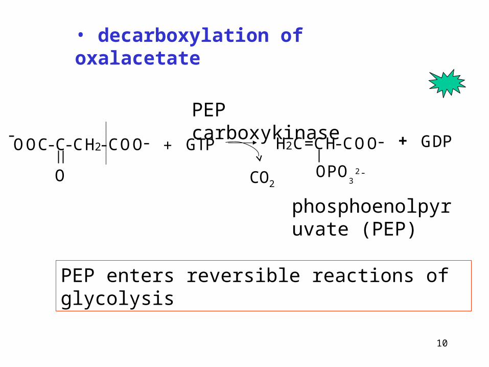
Irreversible reactions of glycolysis
Glycolysis x gluconeogenesis

6
1. Glc + ATP Glc-6-P + ADP
(reverse reaction is catalyzed by different enzyme)
2. Fru-6-P + ATP Fru-1,6-bisP
(reverse reaction is catalyzed by different enzyme)
3. PEP + ADP pyruvate + ATP
(reverse reaction is replaced by „by-pass“)
Irreversible reactions of glycolysis (kinase reactions)

7
Reactions unique to gluconeogenesis
1. Synthesis of phosphoenolpyruvate
Why the reverse reaction cannot proceed?
Go = -61,9 kJ/mol
Cleavage of ATP does not provide energy sufficient for reverse reaction
ATPADP

8
Formation of phosphoenolpyruvate occurs in two steps:
1. Formation of oxalacetate by carboxylation of pyruvate *
enzyme: pyruvate carboxylase
energy: consumption of 1 ATP
location: mitochondria
2. Conversion of oxalacetate to phosphoenolpyruvate
enzyme: phosphoenolpyruvate carboxykinase
energy: consumption of 1 GTP
location: cytoplasma
*note.: carboxylation of pyruvate is also anaplerotic reaction of citric acid cycle

9
1. Conversion of pyruvate to phosphoenolpyruvate (reaction)
•carboxylation pyruvate
Carboxybiotin
CH3
C=O
COOH pyruvate carboxylase
biotin
PyruvateOxaloacetate
10
• decarboxylation of oxalacetate
PEP carboxykinase
phosphoenolpyruvate (PEP)
PEP enters reversible reactions of glycolysis
-OOC-C-CH2-COO-
O
+ GTP H2C=CH-COO-
OPO3
2 -
+ GDP
CO2

1111
• Carboxylation of pyruvate is located in mitochondrial matrix – at the same time it can serve as anaplerotic reaction of citric acid cycle (se lecture citric acid cycle)
• Oxaloacetate cannot be transported across mitochondrial membrane – it must be transported in form of malate or aspartate
• malate ans aspartate are again converted to oxaloacetate in cytoplasma
Compartmentation of reactions at phosphoenolpyruvate formation

1212
pyruvate
pyruvate
oxalacetate
Glucogenic amino acids
malateacetylCoA
citrate
mitochondria
cytoplasma
oxalacetate
malate
alanin
lactate
aspartate
aspartate
C.C.
Kompartmentation of reactions

13
Synthesis phosphoenolpyruvate from pyruvate or lactate requires consumption of 2 ATP
Pairing of carboxylation and decarboxylation drives the reaction that would be otherwise energetically unfavorable.
(see also the synthesis of fatty acids)

1414
glucose
Glc-6-P
Fru-6-P
Fru-1,6-bisP
Glyceraldehyde-3-P Dihydroxyaceton -2-P
1,3-bis-P-glycerate
3-P-glycerate
2-P-glycerate
phosphoenolpyruvate
pyruvate
Irreversible reactions of glycolysis
Glycolysis x gluconeogenesis

15
3-Phosphoglycerate kinase
3-phosphoglycerate 1,3-bisphosphoglycerate
C
CH
CH2 O P
O
O
O
HO
O
-
-
O P O
O
OC
CH
CH2 O P
O
O
O
HO
O
-
-
O-
ADPATP
Further consumption of ATP at gluconeogesis
Reversal proces of substrate phosphorylation in glycolysis
reversible
1616
glucosa
Glc-6-P
Fru-6-P
Fru-1,6-bisP
Glyceraldehyde-3-P Dihydroxyaceton -2-P
1,3-bis-P-glycerát
3-P-glycerate + ATP
2-P-glyceratefosfoenolpyruvát
Pyruvát + ATP
Glycolysis x gluconeogenesis
Substr.fosforylace
Irreversible reactions of glycolysis

17
2. Dephosphorylation of fructose-1,6-bisphosphate
fructose-1,6-bisphosphatase
allosteric inhibition by AMP,activation by ATP
inhibition by fructose-2,6-bisphosphate (its level is decreased by glucagon)
H2O + Pi
hydrolytic cleavage
Like its glycolytic counterpartphosphofructokinase-1, it participates in the regulation of gluconeogenesis.
The second unique reaction on gluconeogesis
O
OH
OH
OH
O OP
O-
O
O- P
O-O
O-
O
OH
OH
OH
O OHP
O-
O
O-

18
3. Dephosphorylation of glucose-6-P
It is present only in liver.
Not present in muscle!!!
glucose-6-phosphatase
+ Pi
H2O
Enzyme is located in lumen of ER
The third unique reaction on gluconeogesis
O
OHOH
OH
OH
OP
O-
O
O-
O
OHOH
OH
OH
OH

19
Energetic requirements for gluconeogenis
reaction ATP/glucose
2 pyruvate → 2 oxalacetate -2
2 oxalacetate → 2 phosphoenolpyruvate -2 (GTP)
2 3-phosphoglycerate → 2 1,3-bisphosphoglycerate -2
-6 ATP/glucose
Source of energy is mainly -oxidation of fatty acids

20
2 pyruvate + 4 ATP + 2 GTP + 2 NADH + 2H+
glucose + 2NAD+ + 4 ADP + 2 GDP + 6 Pi
Sumary equation of gluconeogenesis
-6 ATPConsumption:
Gluconeogenesis is energy demanding process

21
Lactate
formation in tissues, transport by blood to the liver
lactate + NAD+ pyruvate + NADH + H+ (cytoplasma)
(Cori cycle)
Origin of substrates for gluconeogenesis
Pyruvate
E.g. from transamination of alanine, dehydrogenation of lactate

22
Glycerol
• formation in adipocytes at cleavage of triacylglycerols
• transport by blood to the liver
• in liver (cytoplasma):
glycerol + ATP glycerol-3-P + ADP
glycerol-3-P + NAD+ dihydroxyaceton-P + NADH + H+
What is the energy requirement for synthesis of 1 mol of glucose from glycerol?

23
Glucogenic amino acids
They provide pyruvate or intermediates of citric acid cycle, that can be converted to oxalacetate
Acetyl CoA – is not the substrate for gluconeogenesis !!!
It is metabolised to CO2 in citric acid cycle.
Fatty acid cannot be converted to glucose in animals!

24
The most important amino acid for gluconeogenesis is alanin
It is formed mainly in muscle by transamination of pyruvate and is transported by blood to the liver.
Here is again converted to pyruvate by reverse transamination
liver
muscle
glucose
pyruvate
lactate
alanin
lactate
alanin
pyruvate
glucoseamino acids
2-oxo acid
glutamate
2-oxoglutarate

25
Gluconeogenesis from lactate and glycerol requires NAD+
The ratio NADH/NAD+ may by high at some metabolic conditions – gluconeogenesis can not occur
The ratio NADH/NAD+ is increased e.g. at ethanol metabolism (alcohol dehydrogenase).
Therefore intake of alcohol can decrease gluconeogenesis hypoglycemia at alcoholics

26
The main features of gluconeogenesis regulation
Availability of substrates.
Allosteric and hormonal regulation of irreversible reactions.
Allosteric effects are rapid (they affect the reaction immediately)
Hormons can act through
• direct inhibition or activation by a second messenger (rapid effect)
• induction or repression of enzyme synthesis (slow effect – hours - days)

27
Enzyme Activator Inhibitor
Hexokinase glucose-6-phosphate
Phosphofructo kinase 5´AMP, fructose-6-phosphate, fructose-2,6-bisphosphate
Citrate, ATP, glucagon
Pyruvate kinase fructose-1,6-bisphosphate, ATP, alanin
Pyruvate dehydrogenase CoA, NAD+, ADP, pyruvate
acetylCoA, NADH, ATP
Pyruvate carboxylase acetylCoA ADP
Activation and inhibition of enzymes involved in glycolysis and gluconeogenesis

28
Enzyme Inductor Represor
glucokinase insulin glucagon
phosphofructokinase insulin glucagon
Pyruvate kinase insulin glucagon
Pyruvate carboxylase glucokortikoids
glucagon
Adrenalin
insulin
phosphoenolpyruvate carboxykinase
glucocorticoids
glucagon
adrenalin
insulin
glucose-6-phosphatase glucocorticoids
glucagon
adrenalin
insulin
Effects of hormones on enzyme expression

2929
Conversions of pyruvate at different conditions
pyruvate
acetylCoA Lactate, alanine
oxaloacetate
Pyruvate dehydrogenasePyruvate carboxylase
Aktivation:CoA, NAD+, insulin, ADP, pyruvate
Inhibition:acetylCoA, NADH, ATP
Activation: acetylCoA
Inhibition: ADP

30
Gluconeogenesis in kidneys
Substrates: mainly lactate, glycerol and glutamin
Glucose can be released from kidneys – in post-resorptive state or during starvation, at acidosis

31
Glycogen
- synthesis and degradation

32
• synthesis and degradation of glycogen occurs in most types of cells, the largest stores are in liver and skeletal muscle.
• glycogen is a storage form of glucose in cells, that is rapidly released
• Muscle – the mass of glycogen is about 1-2% of muscle mass, glycogen is degraded during intensive muscle work or stress
• Liver: about 5-10 % of liver mass (after the meal)
Glycogen is degraded when glucose level in blood drops
Glycogen storage

3333
Storage of glucose in human (70 kg)
Tisue % tissue mass
Tissue mass (kg)
Mass of glucose (g)
Liver 5,0 1,8 90 (glycogen)
Muscle 0,7 35 245 (glycogen)
Extracelular glucose
0,1 10 10

34
Glycogen is deposited cytoplasma of cells in form of glycogen particles (10-40 nm)
Enzymes od degradation and synthesis are on the surface of particles
Glycogenolysis is not a reversal proces of synthesis.
Location of synthesis and degradation of glycogen
35
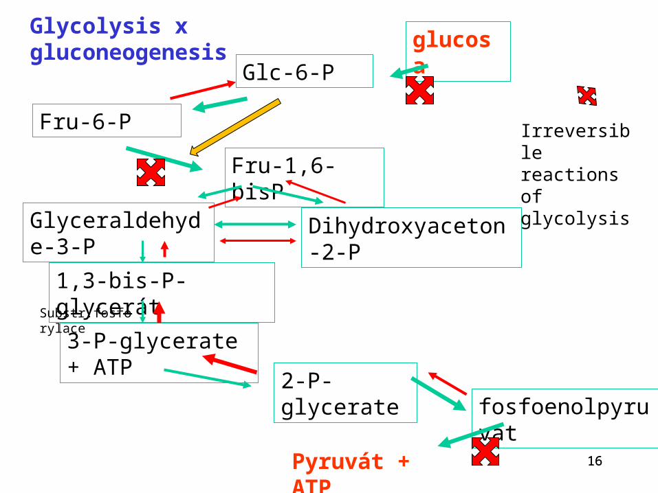
Molecules of glycogen have Mr ~108
The branched structure permits rapid degradation and rapid synthesis, because enzymes can work on several chains simultaneously.
It also increases the solubility in water.

36-1,4-glycosidic bond
Non-reducing end
O
OH
OH
CH2
O
O
OH
OH
CH2OH
OO
O
OH
OH
O
OH
OH
CH2OH
O
CH2OH
OO
CH2OH
O
CH2OH
-1,6-glycosidic bond
Types of bonds in glycogen
Non-reducing end

37
Synthesis of glycogen (glycogenesis)
1. Activation of glucose to UDP-glucose
2. Transfer of glucosyl units from UDP-glucose to the 4´ ends of glycogen chains or primers
3. Formation -1,4 glycosidic bond
4. Branching
It occurs after the meal, activation by insulin
38
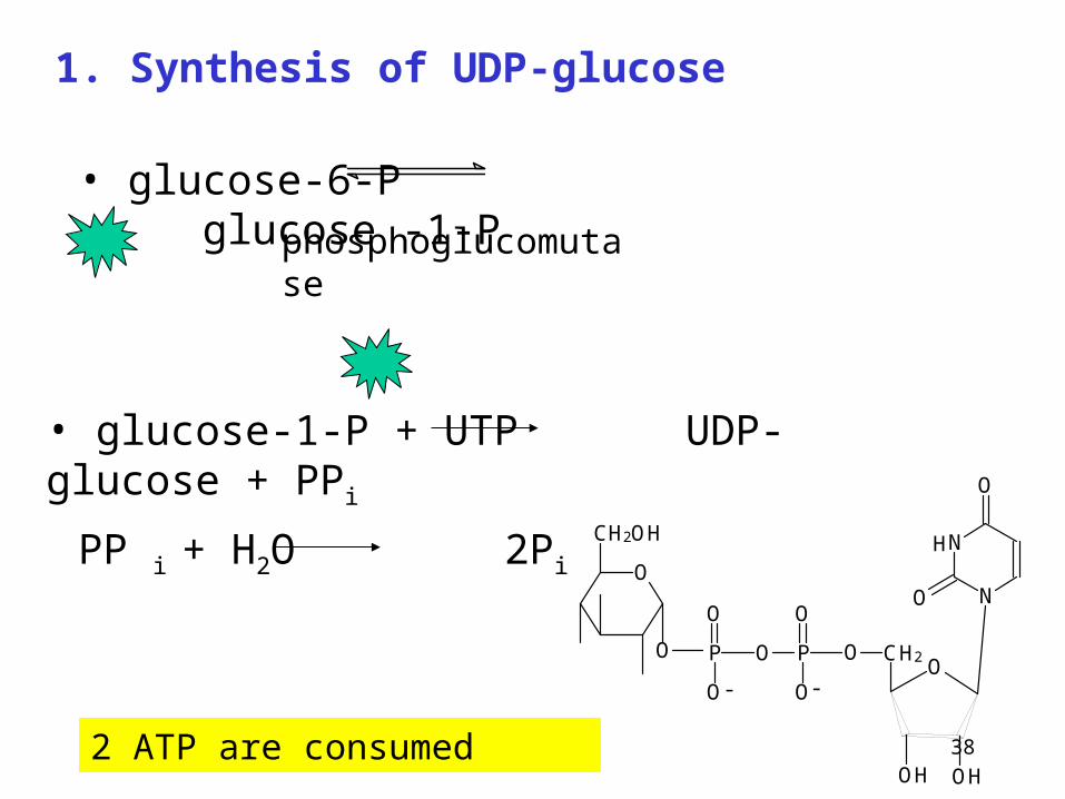
1. Synthesis of UDP-glucose
• glucose-6-P glucose -1-P
phosphoglucomutase
• glucose-1-P + UTP UDP-glucose + PPi
PP i + H2O 2PiN
N
O
O
CH2
OH OH
OO
H
PO
O
O
O
CH2OH
P
O
O
O
--
2 ATP are consumed
39
2. Primer is necessary for synthesis of glycogen
Pre-existing fragment of glycogen
When glycogen stores are totally depleted, specific protein glycogenin serves an acceptor of first glucose residue
Autoglycosylation on serine residues

40
• Iniciation – glucosyl residue is added from UDP-
glucose to the non-reducing terminal of the primer by
glycogen synthase
• Elongation by glycogensynthase - formation of linear
chains with -1,4 glycosidic bond UDP-glucose +
[glucose]n [glucose]n+1 + UDP
3. Formation of -1,4 glycosidic bonds glycogensynthase

41
4. Branching
(branching enzyme)
5-8 glucosyl residues are transferred from non-reducing end to another residue of the chain and attached by 1,6-glycosidic bond
G-G-G-G-G
G-G-G-G-G-G-G-G-G-G-G-G-G -G-G-G-G-G-G-G-G
-1,6 bond
Elongation of both non-reducing ends by glycogensynthase
New branching by branching enzyme
42
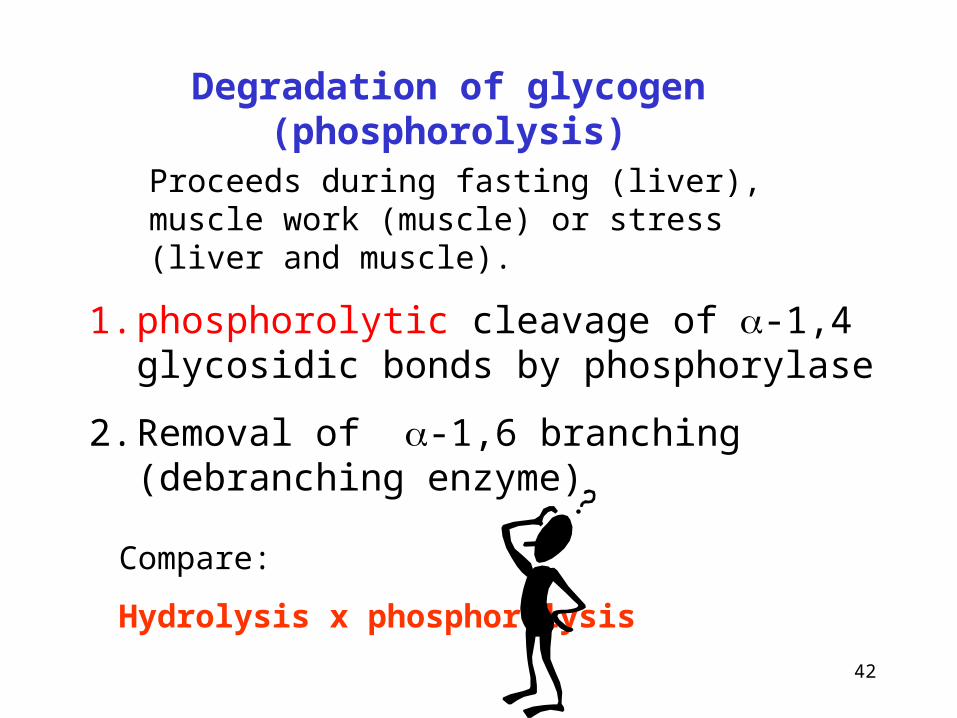
Degradation of glycogen (phosphorolysis)
1. phosphorolytic cleavage of -1,4 glycosidic bonds by phosphorylase
2. Removal of -1,6 branching (debranching enzyme)
Proceeds during fasting (liver), muscle work (muscle) or stress (liver and muscle).
Compare:
Hydrolysis x phosphorolysis

43
1. Phosphorylase - phosphorolytic cleavage of -1,4 glycosidic bonds at the non-reducing ends
O
OH
OH
OH
CH2OH
O
O
OH
OH
CH2OH
O
O
OH
OH
CH2OH
O
O
OH
OH
OH
CH2OH
O P
O
O
O
O
OH
OH
OH
CH2OH
O
O
OH
OH
CH2OH
O-
-
HPO4
2-
glukosa-1-P glykogenn-1
The cleavage continues untill four glucosyl units remain on the chain before a branch point („limit dextrine“)

44
Phosphorylase can split α-1,4-links,its action ends with the production of limit dextrins :
Degradation of glycogen
44
G-G-G-G-G-G-G-G G-G-G-G
G-G--G-G-G-G-G-G-G-G- 8 Pi G-G-G-G-G-G-G-G + 8 G-P
G-G-G-G--G-G-G-G-G- G-G-G-G-G-G-G-
G debranching enzyme
G-G-G-G-G-G-G-G-G-G-G G-G-G-G-G-G-G-G-G-G-G-G + G
G-G-G-G-G-G-G- G-G-G-G-G-G-G
Limit dextrin
transglycosylase

45
2. Debranching enzyme
transferase activity: enzyme transfers unit containing 3 from 4 glucose molecules remaining on the 1,6-branch and adds it to the end of a longer chain by -1,4 glycosidic bond
glucosidase activity: the one glucosyl residue remaining at the end of -1,6 branch is hydrolyzed by the 1,6 –glucosidase activity of debranching enzyme
Free glucose is released ! Not Glc-1-P
46
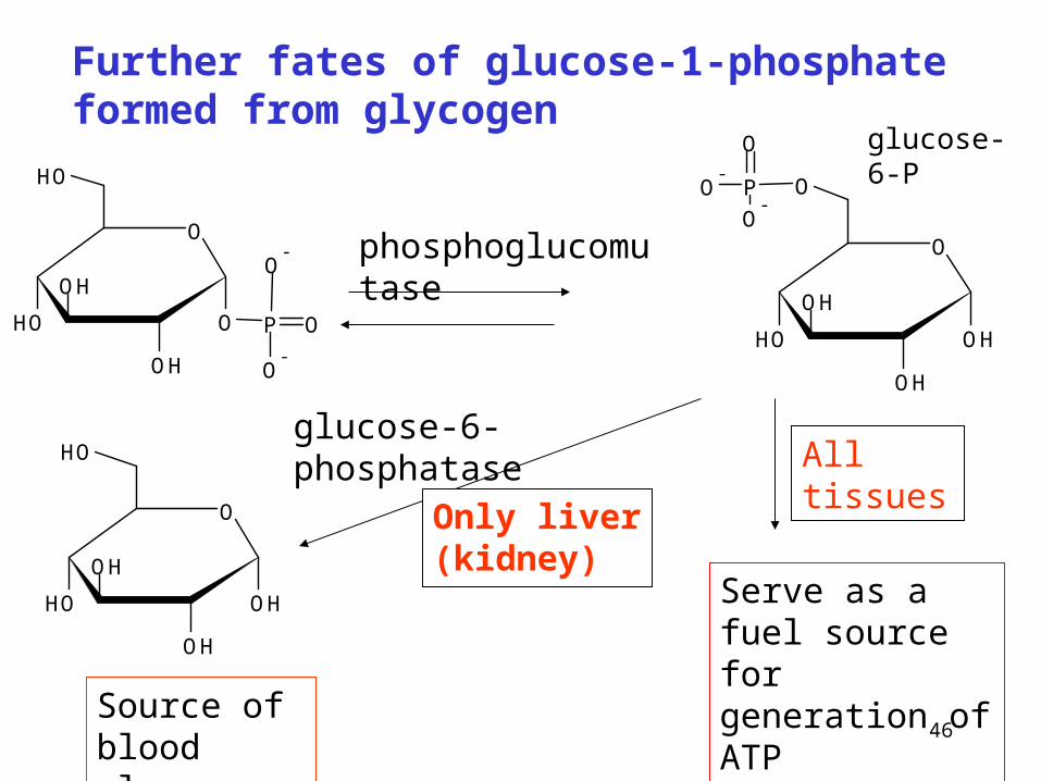
Further fates of glucose-1-phosphate formed from glycogen
phosphoglucomutase
Serve as a fuel source for generation of ATP
Only liver (kidney)
All tissuesglucose-6-phosphatase
Source of blood glucose
glucose-6-P
O
OOH
OH
OH
OH
P
O-
O-
O
O
OHOH
OH
OH
OH
O
OHOH
OH
OH
OP
O-
O
O-

47
glucose-6-P cannot permeate across the cellular membrane, only free glucose can diffuse
Enzyme glucose-6-phosphatase is only in liver and kidneys – it is not present in muscle.
Blood glucose can be maintened only by cleavage of liver glycogen but not by cleavage of muscle glycogen
Cleavage of glycogen in muscle and other cells provides glucose-6-P that can be metabolized only within the given cell (by glycolysis)
Significance of glucose-6-phosphatase
48
Lysosomal degradation of glycogen
Lysosomal acidic glucosidase (pH optimum 4)
Degradation of about 1-3% of cellular glycogen (glycogen particles are surrounded by membranes that then fuse with the lysosomal membrane
-enzyme degrades -1,4-bonds from non-reducing end
- glucose is released
(see also Pompe disease)

49
Regulation glycogen metabolism
Glycogen synthase X glycogen phosphorylase
Hormonal control
Allosteric regulation

50
Hormons affecting synthesis and degradation of glycogen
Hormon synthesis degradation
Insulin
Glucagon
Adrenalin
Hormons action is mediated by their second messengers.

51
Phosphorylation and dephosphorylation plays important role at regulation of glycogen metabolism
• phosphorylation by kinases and ATP
• dephosphorylation by phosphatases

52
H2O
Pi
Protein phosphatase
OH
O-P
ATP
ADP
proteinkinase
Non active enzyme
Active enzyme
H2O
Pi
Protein phosphatase
OH
O-P
ATP
ADP
proteinkinase
Active enzyme
Non active enzyme
Common examples of enzyme activity regulation by phosphorylation and dephosphorylation

53
Activation and inactivation of glycogen synthase
Glycogen synthase a (dephosphorylated - active)
Glycogen synthase b (phosphorylated - inactive)
ATP ADP
H2OPi
glycogensynthase kinase
phosphatase

54
glycogensynthase a
(dephosphorylated, active)
Glycogen synthase b (phosphorylated, non active)
Glucogensynthase phosphatase
(activation by insulin, allosterically by glucose-6-P
Inactivation by ↑ cAMP )
Activation and inactivation of glycogensynthase in liver
ATP
ADP
Glycogene synthase kinase (activation by glucagon /cAMP/ or adrenalin /Ca-calmodulin/
inactivation activation
Pi

55
Activation and inactivation of glycogen phosphorylase
phosphorylase b(non phosphorylated form -low activity)
phosphorylase a (phosphorylated form-active)
ATP ADP
H2OPi
Phosphorylases in liver and muscles are different
phosphorylase kinase
proteinphosphatase

56
Effect of hormons:
Liver:
glucagon (cAMP),
adrenalin (cAMP, Ca2+calmodulin)
Degradation of glycogen
Muscle:
adrenalin (cAMP) at the stress
allosteric regulation
AMP
No effect of glucagon !
Ca2+ during muscle contraction
Glucose, ATP, Glc-6P: allosteric inhibition

57
Glycogen storage diseases - enzyme deffects
Inherited enzyme deficiences. They can be tissue specific, as in various tissues can be various isoenzymes.
Typ Enzyme defect Organ Characteristics
0
I
II
III
IV
V
VI
VII
Glycogen synthase
Glc-6-phosphatase
Lysosome α-glucosidase
Debranching enzyme
Branching enzyme
Muscle phosphorylase
Liver phosphorylase
Phosphofructokinase
Liver
Liver, kidney
All organs
Liver, muscle, heart
Liver
Muscle
Liver
Muscle, ercs
Hypoglycemia
Enlarged liver, kidney. Hypoglykemia. Celly are overloaded by glycogen
Accumulation of glycogen in lyzosomes
Accumulation of branched polysaccharide.
Accumulation of unbranched polysaccharide
High content of glycogen in muscle, exercise induced muscular pain
High content of glycogen in liver, mild hypoglycemia
As in type V
58
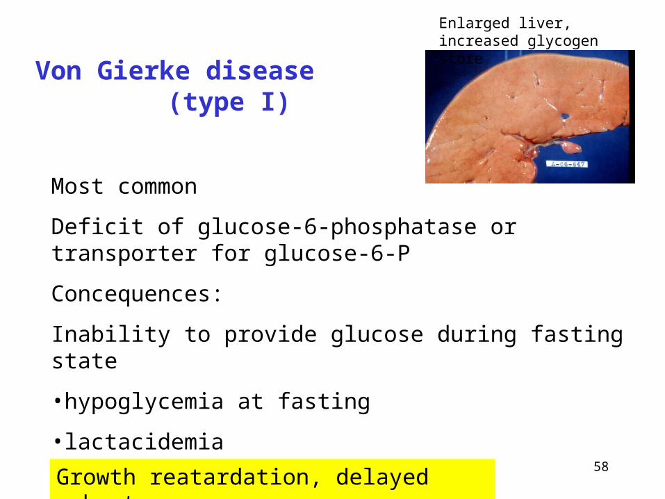
Von Gierke disease (type I)
Most common
Deficit of glucose-6-phosphatase or transporter for glucose-6-P
Concequences:
Inability to provide glucose during fasting state
•hypoglycemia at fasting
•lactacidemia
•(hyperlipidemia, hyperurikemia)
Enlarged liver, increased glycogen store
Growth reatardation, delayed puberty

59
Pompe disease (type II)
Absence of -1,4-glucosidase in lysosomes
Acummulation of glycogen in lysosomes
Loss of lysosomal function
Damage of musclesmuscle weakness
Infantile form: death before age 2 years
Juvenile form: later –onset myopathy with variable cardiac involvment
Adult form: limb-girdle muscular distrophy-like features.

60
McArdle disease (type V)
Absence of muscle phosphorylase
Stores of glycogen are not available for production of energy
Muscle is not able to perform exercise or work