Embed Size (px)
Citation preview
1
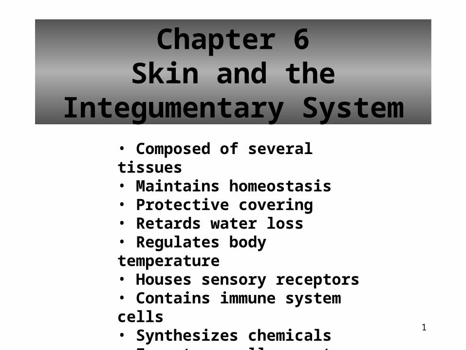
Chapter 6Skin and the Integumentary
System• Composed of several tissues• Maintains homeostasis• Protective covering• Retards water loss• Regulates body temperature• Houses sensory receptors• Contains immune system cells• Synthesizes chemicals• Excretes small amounts of waste
2
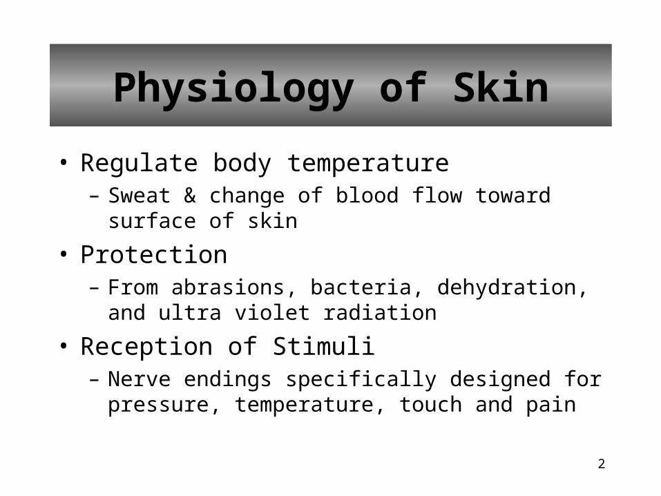
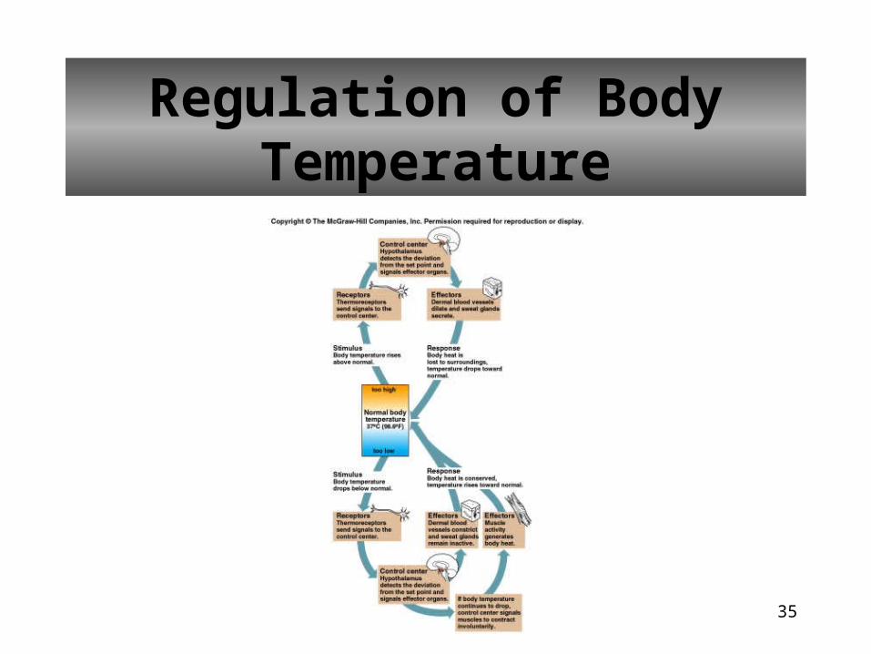
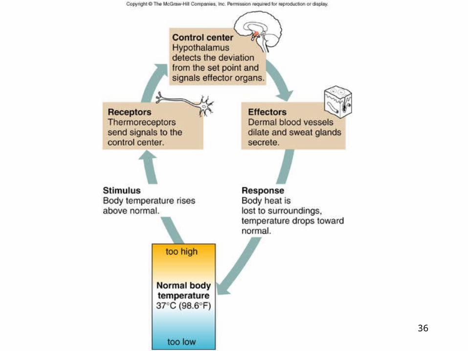
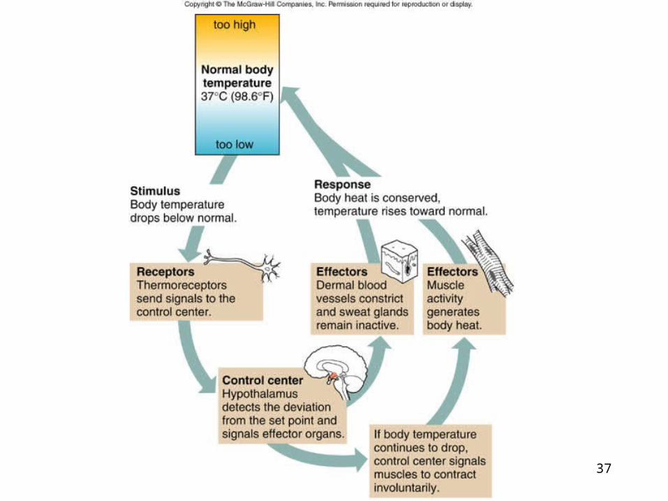
• Regulate body temperature– Sweat & change of blood flow toward surface of skin
• Protection– From abrasions, bacteria, dehydration, and ultra violet
radiation
• Reception of Stimuli– Nerve endings specifically designed for pressure,
temperature, touch and pain
Physiology of Skin
3
• Excretion– sweat helps reduce levels of water, salts, and other
organic compounds
• Synthesis of Vitamin D– Helps manufacture vitamin D
– Helps body absorb calcium and phosphorus from food
• Immunity– Certain cells help in your ability to produce antibodies
Physiology of Skin
4
Skin Cells
• Help produce Vitamin D needed for normal bone and tooth development
• Some cells (keratinocytes) produce substances that simulate development of some WBCs
5
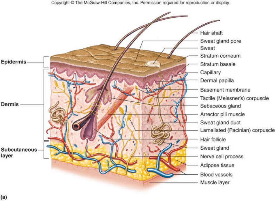
Layers of Skin
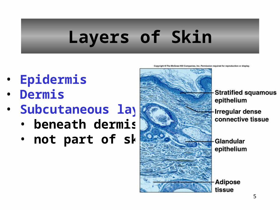
• Epidermis• Dermis• Subcutaneous layer
• beneath dermis • not part of skin
6
Subcutaneous Layer
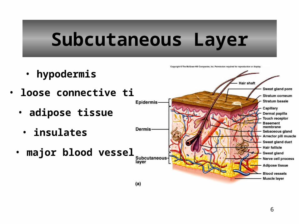
• hypodermis
• loose connective tissue
• adipose tissue
• insulates
• major blood vessels
7
8
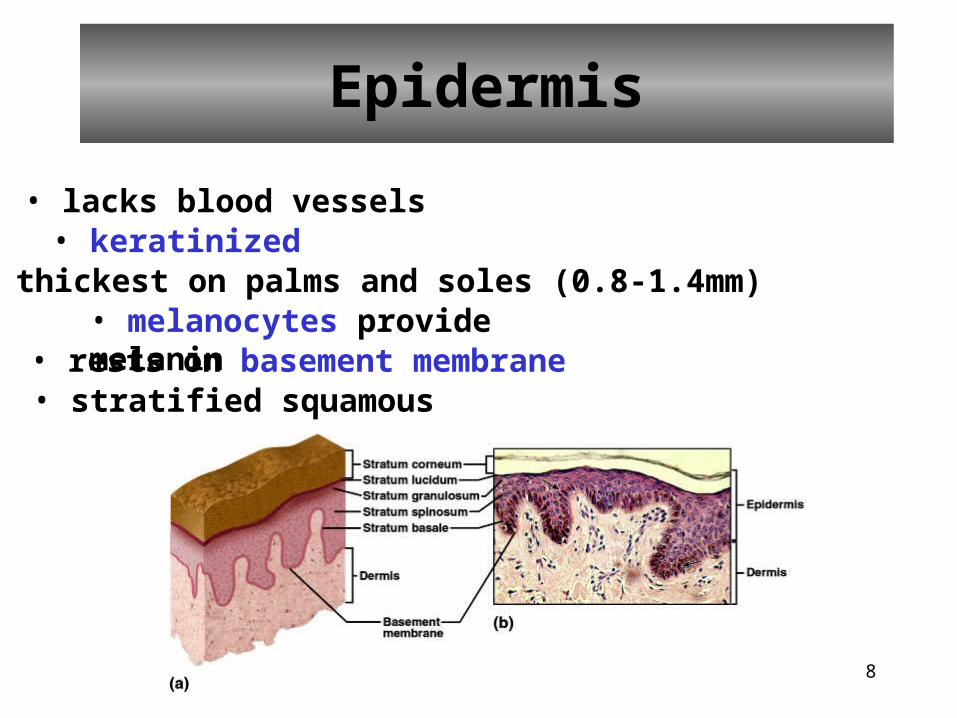
Epidermis
• lacks blood vessels• keratinized• thickest on palms and soles (0.8-1.4mm)• melanocytes provide melanin• rests on basement membrane• stratified squamous
9
10
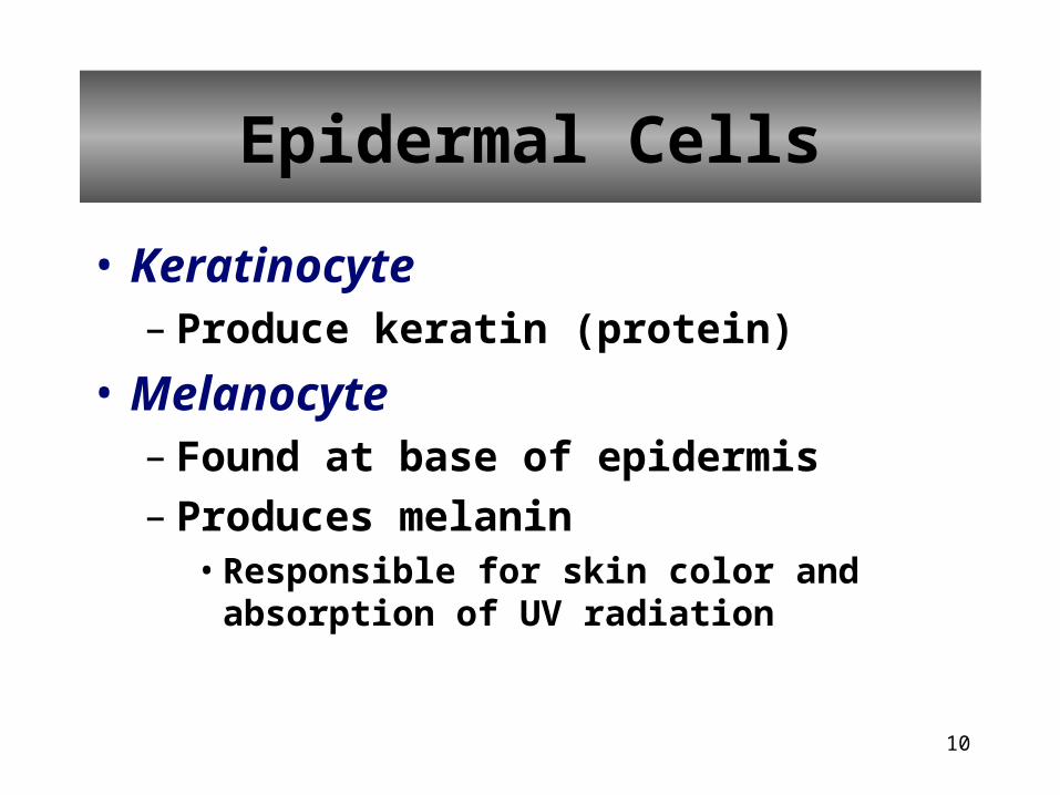
• Keratinocyte– Produce keratin (protein)
• Melanocyte– Found at base of epidermis– Produces melanin
• Responsible for skin color and absorption of UV radiation
Epidermal Cells
11
• Langerhan’s cells– Interact with helper T-cells of the immune
system
• Granstein cells– Resistant to UV radiation– Interact with other T-cells of immune system
Epidermal Cells
12
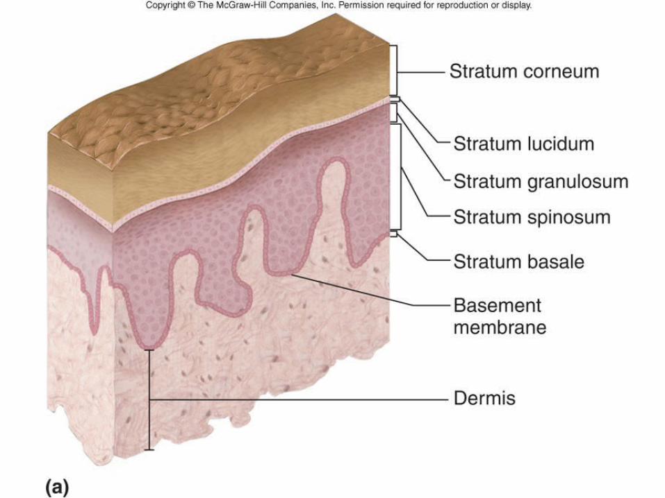
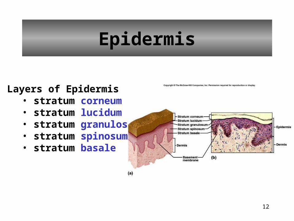
Epidermis
Layers of Epidermis• stratum corneum• stratum lucidum• stratum granulosum• stratum spinosum• stratum basale
13
• Stratum Basale– Deepest layer– Single layer of cells capable of continued cell
division– Also called the stratum germinativum– Cells may migrate to the dermis to become glands
and hair follicles– Areas with no hair contain nerve endings that are
sensitive to touch (tactile disc)
Epidermal Layers
14
• Stratum Spinosum– 8 to 10 rows of close fitting cells– Surface of the cells contain spine-like projections
that help to join cells together
• Stratum Granulosum– 3 to 5 rows flattened cells that contain dark stained
granules of keratohyalin, which is involved in the first steps of keratin formation
– Cells start to die in this layer
Epidermal Layers
15
• Stratum Lucidum– Found only in thick skin of palms, and the
soles of your feet– 3 to 5 rows of clear, flat, dead cells
• Stratum Corneum– 25 to 30 rows of keratinized cells– Continuously replaced and shed
Epidermal Layers

16
17
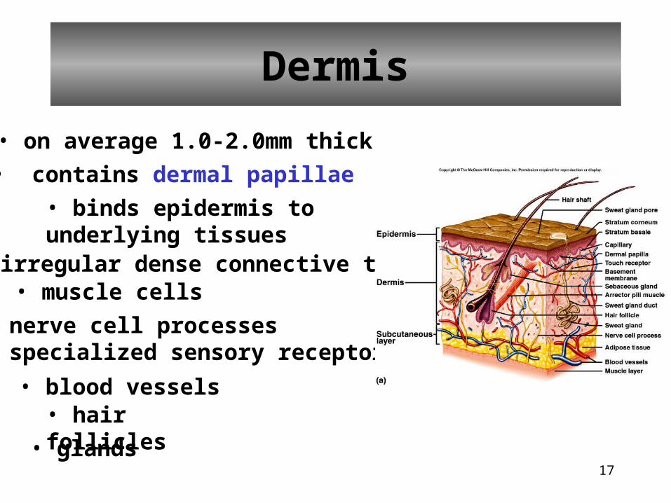
Dermis
• contains dermal papillae
• binds epidermis to underlying tissues• irregular dense connective tissue
• on average 1.0-2.0mm thick
• muscle cells
• nerve cell processes• specialized sensory receptors
• blood vessels• hair follicles
• glands
18
• Papillary Layer– Upper 1/5 of the dermis
– Dermal papillae• Finger-like projections that increase surface area
• Projections that extend into the epidermis and may contain blood vessels or
• Meissner’s corpuscles- endings that are sensitive to touch
• Cause ridges in the overlying epidermis (fingerprints)
Dermal Layers
19
• Reticular layer– Formed by closely packed irregularly arranged
connective tissue– Glands, hair, and nerves fill spaces between– Provides the skin with its strength and elasticity– Attached to underlying organs by the subcutaneous
layer– Pacinian Corpuscles
• Located in the subcutaneous layer• Nerve endings that are sensitive to pressure
Dermal Layers
20
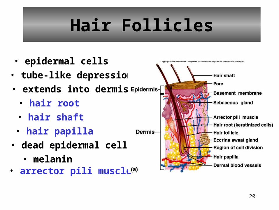
Hair Follicles
• epidermal cells
• tube-like depression
• extends into dermis
• hair root
• hair shaft
• hair papilla
• dead epidermal cells
• melanin• arrector pili muscle
21
• Protects, guards scalp from the sun, eyes from foreign particles
• Hair in the ear and nose protect these structures from foreign particles and insects that might be inhaled or crawl into the ear
Hair
22
• Shaft– Superficial portion that most of which projects above
the skin– Made up of 3 parts
• Medulla– inner portion that contains air spaces
• Cortex– Middle portion that makes the majority of the hair shaft– Contains pigment granules of dark hair & mostly air in light hair
• Cuticle– Outer most layers– Cells heavily keratinized– Arranged like upside-down shingles
Anatomy of the Hair
23
• Root– Portion that penetrates the dermis and even the
subcutaneous layer– Contains the 3 portions like the shaft
• Hair follicle– Surrounds the root– Continuation of the stratum basale and stratum
spinosum layers of the epidermis– Base of each follicle enlarges and looks like an onion
shaped bulb
Anatomy of the Hair
24
• Papillae of the hair– Indentation of the bulb that is filled with
loose connective tissue and many blood vessels
Anatomy of the Hair

25
26
• Sebaceous (oil) glands– Found in association with hair follicles except on the
lips and eyelids– Secrete sebum
• Mixture of fats, cholesterol, proteins, and inorganic salts
– Function• Prevents hair from becoming brittle• Forms a film that prevents excess evaporation of water
from the skin• Keeps the skin soft and flexible• Inhibits the growth of certain bacteria
Associates of the Hair Complex
27
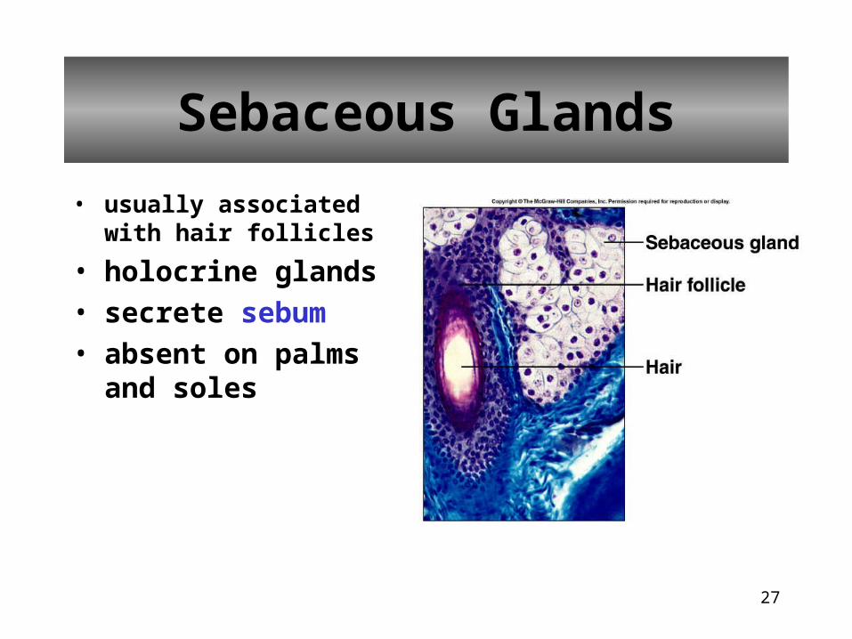
• usually associated with hair follicles
• holocrine glands
• secrete sebum
• absent on palms and soles
Sebaceous Glands
28
• Blackheads– Sebaceous glands enlarge due to
accumulated sebum
– Color of blackhead is due to melanin and oxidized oil, not dirt
Clinical Application: Sebaceous Glands
29
• Sudoriferous Glands– Divided on basis of structure and location– 3 types
• Apocrine– Located in axilla and pubic region– Excretory duct open to hair follicles– Start to function at the onset of puberty– Emit an odor
• Eccrine– More common– Ducts open to the surface of the skin
Associates of the Hair Complex
30
• Both Apocrine and Eccrine glands secrete:– Perspiration
• mixture of water, salts (NaCl), urea, uric acid, amino acids, ammonia, sugar, lactic acid and ascorbic acid
• Function:– Reduces body temperature by evaporation
– Elimination of waste
Associates of the Hair Complex

31
• sudoriferous glands
• widespread in skin
• originates in deeper dermis or hypodermis
Sweat Glands
32
• Mammary glands– Modified sudoriferous glands– Reproductive unit
• Ceruminous glands– Found in external auditory meatus – Produce ear wax that protect your ears from
foreign particles
Associates of the Hair Complex

33
Nails
• protective coverings
• nail plate/nail body
• nail bed
• lunula
34
• Average growth is about 1 mm per week• Protect the end of the digits and aid the
manipulation of small objects• 4 parts of the nail
– Free edge- extends past distal end of the digit– Body- majority of the visible nail
• Lunula- semilunar white part of the body
– Nail Root- hidden part of the nail that lies above the nail matrix
– Nail Matrix- functions to bring the growth of nails• when superficial cells become nail cells, push the whole
nail across the nail bed
Nails
35
Regulation of Body Temperature
36
37
38
Problems in Temperature Regulation
Hyperthermia – abnormally high body temperature
Hypothermia – abnormally low body temperature
39
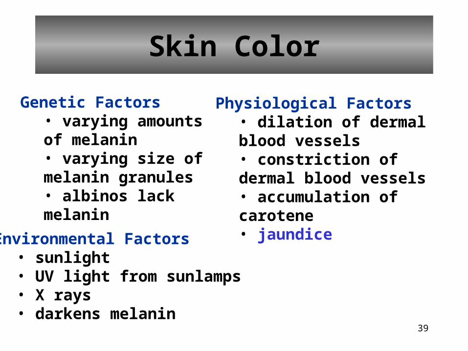
Skin Color
Genetic Factors• varying amounts of melanin• varying size of melanin granules• albinos lack melanin
Environmental Factors• sunlight• UV light from sunlamps• X rays• darkens melanin
Physiological Factors• dilation of dermal blood vessels• constriction of dermal blood vessels• accumulation of carotene• jaundice
40
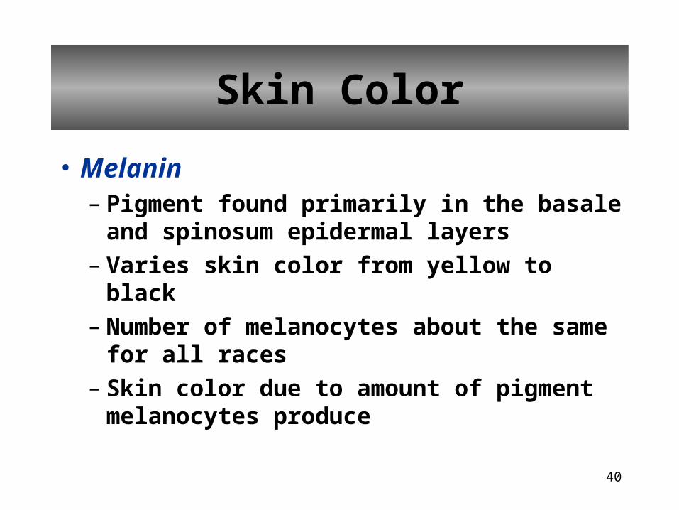
Skin Color
• Melanin– Pigment found primarily in the basale and
spinosum epidermal layers– Varies skin color from yellow to black– Number of melanocytes about the same for
all races– Skin color due to amount of pigment
melanocytes produce
41
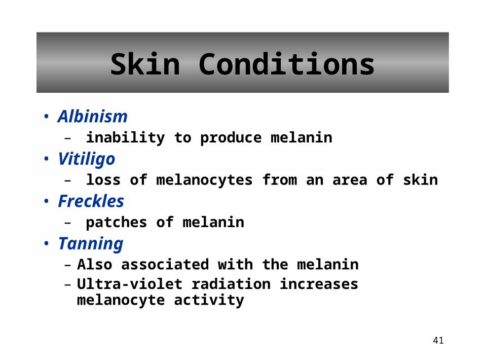
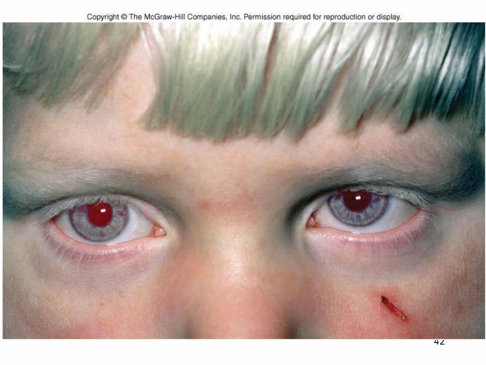
• Albinism– inability to produce melanin
• Vitiligo– loss of melanocytes from an area of skin
• Freckles– patches of melanin
• Tanning– Also associated with the melanin– Ultra-violet radiation increases melanocyte activity
Skin Conditions
42
43
• Carotene– Found in the stratum corneum– People of Asian origin have carotene in fatty
areas of the dermis– Gives a yellowish hue to the skin
• Capillaries– cause the skin to have a pink appearance
Skin Color
44
• Common skin wounds– Abrasions
• portions of the skin has been scraped away
– Laceration• irregular tear of the skin
– Puncture• hole “popped” through the skin
– Incisions• clean out through the skin
– Contusion (bruise)• Tissue below skin damaged• skin is not broken
Epidermal Wound Healing
45
• Superficial Wound Healing1. Basale cells in the area of the wound break
contact with the basement membrane that connects the epidermis to the dermis
2. Basale cells enlarge and migrate across the wound
Epidermal Wound Healing

46
3. Contact inhibition stops the migrating cells and turns cells in a new direction continues until cells are surrounded by similar cells
– Malignant (cancer) cells don’t follow the same rules
• continue to spread and invade other areas
Epidermal Wound Healing
47
4. When the “floor” of the wound is covered• Cells divide to form new strata (layers)
• This thickens the epidermis and fills in the wound from the bottom upward
5. If a scab was formed, it will fall off when the new epidermis is thick enough to protect itself
Epidermal Wound Healing
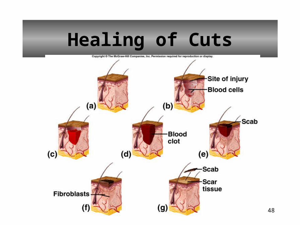
48
Healing of Cuts
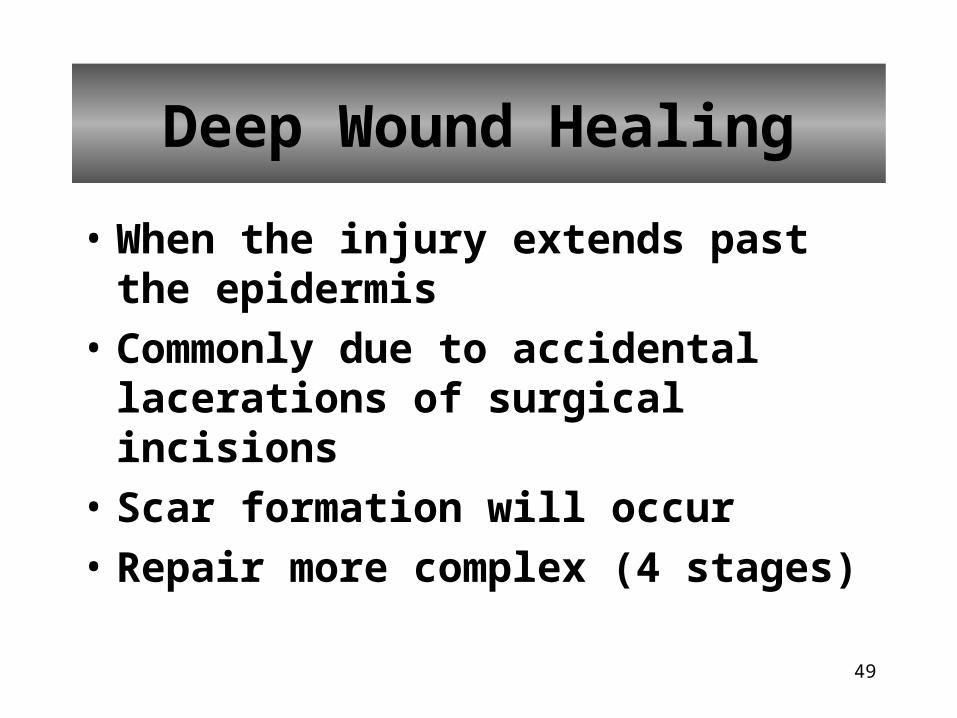
49
• When the injury extends past the epidermis
• Commonly due to accidental lacerations of surgical incisions
• Scar formation will occur
• Repair more complex (4 stages)
Deep Wound Healing
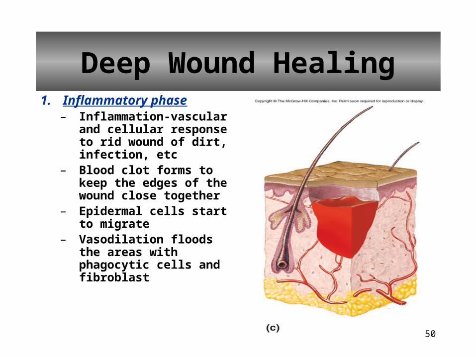
50
1. Inflammatory phase– Inflammation-vascular
and cellular response to rid wound of dirt, infection, etc
– Blood clot forms to keep the edges of the wound close together
– Epidermal cells start to migrate
– Vasodilation floods the areas with phagocytic cells and fibroblast
Deep Wound Healing
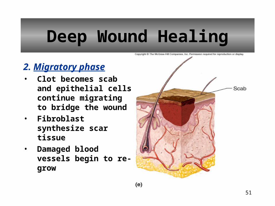
51
2. Migratory phase• Clot becomes scab and
epithelial cells continue migrating to bridge the wound
• Fibroblast synthesize scar tissue
• Damaged blood vessels begin to re-grow
Deep Wound Healing
52
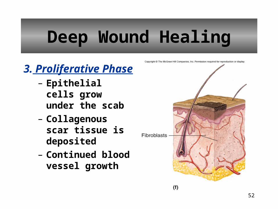
3. Proliferative Phase– Epithelial cells grow
under the scab
– Collagenous scar tissue is deposited
– Continued blood vessel growth
Deep Wound Healing
53
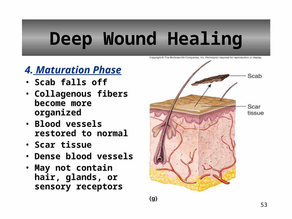
4. Maturation Phase• Scab falls off• Collagenous fibers
become more organized• Blood vessels restored to
normal• Scar tissue• Dense blood vessels• May not contain hair,
glands, or sensory receptors
Deep Wound Healing
54
• First degree burn – superficial partial-
thickness
– Dermal blood vessels dilate causing skin to warm and redden
– Mild edema (swelling)
– Surface layer sometimes shed
– Heals in a few days to 2 weeks
Healing of Burns
QuickTime™ and a decompressor
are needed to see this picture.
55
• Second degree burn – deep partial-thickness
– Damages epidermis and some dermis
– Fluid escapes damaged capillaries causing blisters
– Healing depends on stem cells derived from epidermis but located deep within dermis
Healing of Burns
QuickTime™ and a decompressor
are needed to see this picture.
56
• Third degree burn – full-thickness– Skin becomes dry, leathery– Red, black, white color– autograft
• Thin layer of skin taken from unburned area & transplanted– homograft
• Cadaver skin used to cover burn• Temporary for protection of underlying tissue and prevents infection
– various skin substitutes• Amniotic membrane• Artificial membranes composed of silicone, polyurethane, or nylon• Cultured epithelial cells
Healing of Burns
57
Healing of Burns
QuickTime™ and a decompressor
are needed to see this picture.
58
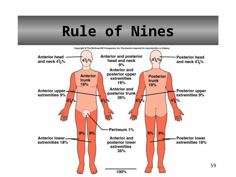
• Divides skin surface into regions, each accounting for 9% (or some multiple of 9%) of the total surface area
• Important for planning to replace body fluids and electrolytes
Rule of Nines
59
Rule of Nines
60
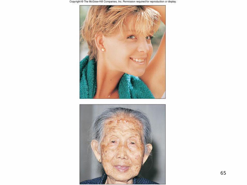
Life Span Changes
• Skin becomes scaly• Age spots appear• Epidermis thins• Dermis becomes reduced• Loss of fat• Wrinkling• Sagging• Sebaceous glands secrete less oil
• Melanin production slows• Hair thins• Number of hair follicles decrease• Nail growth becomes impaired• Sensory receptors decline• Body temperature unable to be controlled• Diminished ability to activate Vitamin D
61
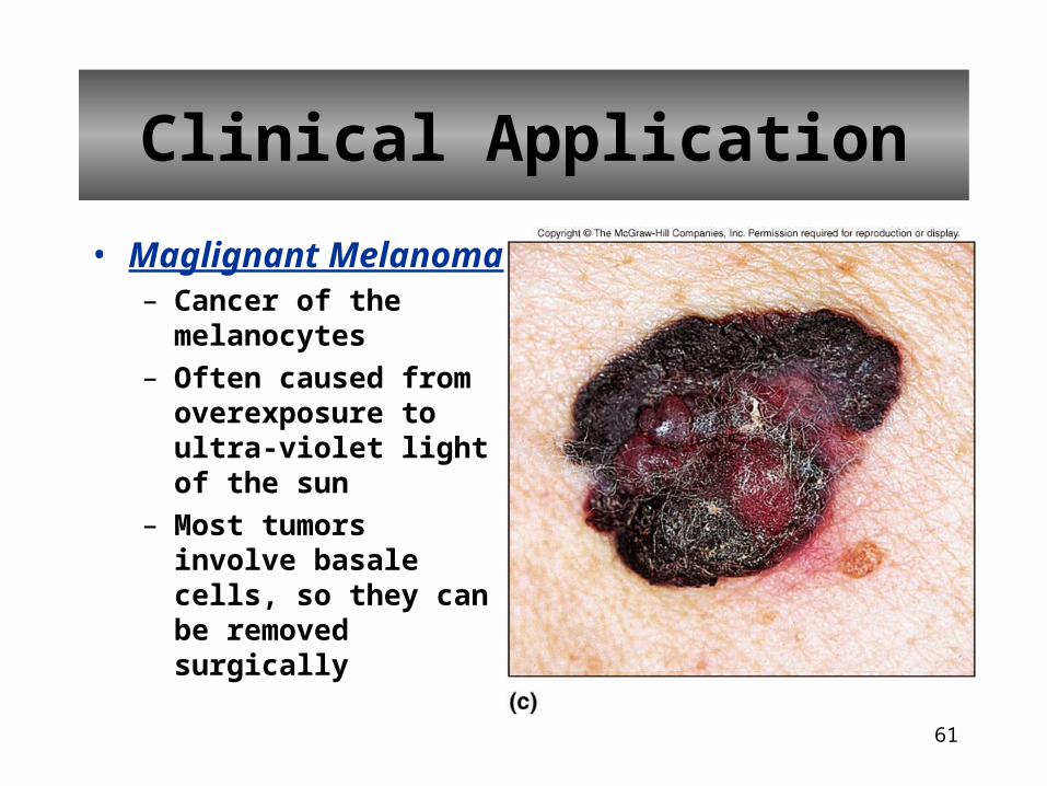
• Maglignant Melanoma– Cancer of the melanocytes
– Often caused from overexposure to ultra-violet light of the sun
– Most tumors involve basale cells, so they can be removed surgically
Clinical Application
62
• Malignant Melanoma– Best treatment is prevention– Examine your skin for moles that develop
irregular appearance– Uneven surfaces or a mixture of colors or
change in size or start to bleed– Many of these may be a sign of developing
melanoma
Clinical Application
63
• Wrinkles– Collagen fibers
• stiffens, break apart and form a shapeless tangle
– Elastic Fibers• some elasticity, thicken
and fray
– Subcutaneous• decreases
– Sebaceous glands• atrophy leads to dry,
cracked skin
Clinical Application
QuickTime™ and a decompressor
are needed to see this picture.
64
• Melanocytes– Decrease of functioning melanocytes lead to gray
hair and atypical skin pigmentation
– Increase of size of some melanocytes can cause liver spots
– Older skin is more susceptible to pathological conditions like cancer and senile pruritis (itching)
Clinical Application
65
66
Clinical Application
Acne Vulgaris
•most common skin disorder•sebum and epithelial cells clog glands•produces whiteheads and blackheads (comedones)•anaerobic bacteria trigger inflammation (pimple)•largely hormonally induced•androgens stimulate sebum production•treatments include antibiotics, topical creams, birth control pills
67
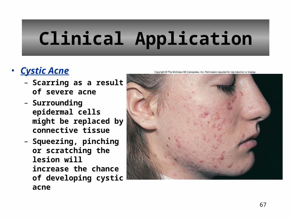
• Cystic Acne– Scarring as a result of
severe acne
– Surrounding epidermal cells might be replaced by connective tissue
– Squeezing, pinching or scratching the lesion will increase the chance of developing cystic acne
Clinical Application
68
• Psoriasis– Symptoms are distinct,
reddish, small round skin elevations covered with scales
– Caused by abnormally high rate of epidermal cell mitosis
– Trauma, infection, stress, seasonal or hormonal changes can irritate
– Treatments include steroid ointments and natural sunlight (ultra-violet light)
Clinical Application
QuickTime™ and a decompressor
are needed to see this picture.
69
• Sunburn– Dead layers of cells
peel off and leave unprotected layers of cells
– Ultra-violet rays of sunlight damage the cells’ DNA and RNA
Clinical Application
QuickTime™ and a decompressor
are needed to see this picture.