Embed Size (px)
Citation preview

1
Cardiovascular System

2
Size of Heart
Average Size of Heart• 14 cm long• 9 cm wide
3
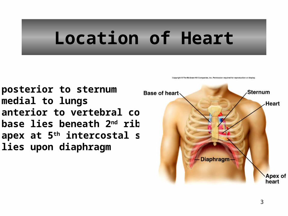
Location of Heart
• posterior to sternum• medial to lungs• anterior to vertebral column• base lies beneath 2nd rib• apex at 5th intercostal space• lies upon diaphragm

4
Coverings of Heart

5
Wall of the Heart

6
Wall of the Heart

7
Heart Chambers
Right Atrium• receives blood from
• inferior vena cava• superior vena cava• coronary sinus
Left Atrium• receives blood from
pulmonary veins
Right Ventricle• receives blood from
right atrium
Left Ventricle• receives blood from
left atrium

8
Heart Valves

9
Coronal Sections of Heart

10
Heart Valves
Tricuspid Valve Pulmonary and Aortic Valve

11
Skeleton of Heart
• fibrous rings to which the heart valves are attached

12
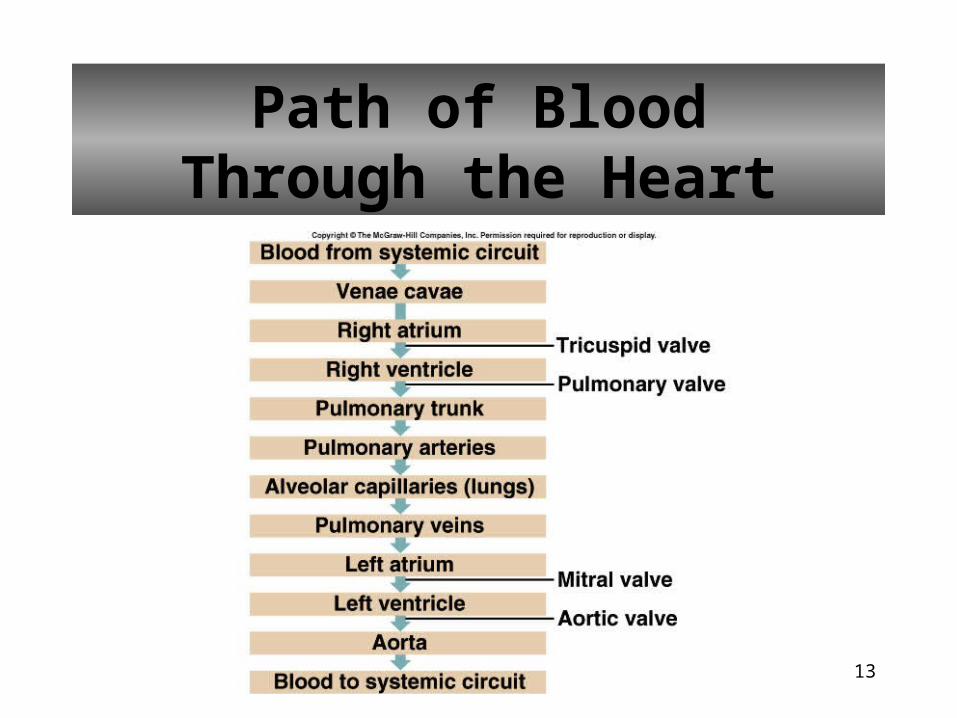
Path of Blood Through the Heart
13
Path of BloodThrough the Heart

14
Blood Supply to Heart

15
Blood Supply to Heart

16
Angiogram of Coronary Arteries

17
Heart Actions
Atrial Systole/Ventricular Diastole Atrial Diastole/Ventricular Systole

18
Cardiac Cycle
Atrial Systole/Ventricular Diastole
• blood flows passively into ventricles
• remaining 30% of blood pushed into ventricles
• A-V valves open/semilunar valves close
• ventricles relaxed
• ventricular pressure increases

19
Cardiac Cycle
Ventricular Systole/Atrial diastole
• A-V valves close
• chordae tendinae prevent cusps of valves from bulging too far into atria
• atria relaxed
• blood flows into atria
• ventricular pressure increases and opens semilunar valves
• blood flows into pulmonary trunk and aorta
20
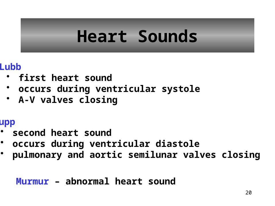
Heart Sounds
Lubb• first heart sound • occurs during ventricular systole• A-V valves closing
Dupp• second heart sound• occurs during ventricular diastole• pulmonary and aortic semilunar valves closing
Murmur – abnormal heart sound

21
Heart Sounds

22
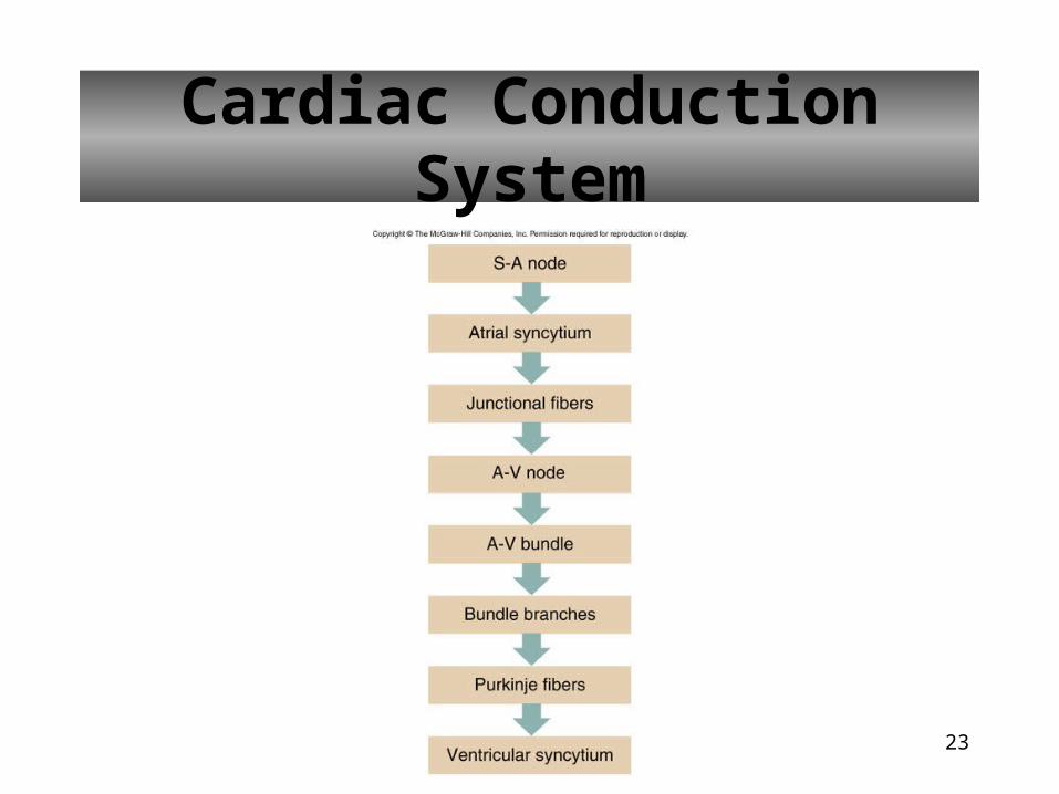
Cardiac Muscle Fibers
Cardiac muscle fibers form a functional syncytium
• group of cells that function as a unit• atrial syncytium• ventricular syncytium
23
Cardiac Conduction System

24
Cardiac Conduction System

25
Muscle Fibers in Ventricular Walls

26
Electrocardiogram
• recording of electrical changes that occur in the myocardium• used to assess heart’s ability to conduct impulses
P wave – atrial depolarizationQRS wave – ventricular depolarizationT wave – ventricular repolarization

27
Electrocardiogram

28
Electrocardiogram
A prolonged QRS complex may result from damage to the A-V bundle fibers

29
Cardiac Cycle

30
Regulation of Cardiac Cycle
Autonomic nerve impulses alter the activities of the S-A and A-V nodes

31
Regulation of Cardiac Cycle
• physical exercise• body temperature• concentration of various ions
• potassium• calcium
• parasympathetic impulses decrease heart action• sympathetic impulses increase heart action• cardiac center regulates autonomic impulses to the heart
Additional Factors that Influence HR

32
Blood Vessels
• arteries• carry blood away from ventricles of heart
• arterioles• receive blood from arteries• carry blood to capillaries
• capillaries• sites of exchange of substances between blood
and body cells• venules
• receive blood from capillaries• veins
• carry blood toward ventricle of heart

33
Arteries and Arterioles
Artery• thick strong wall • endothelial lining• middle layer of smooth
muscle and elastic tissue• outer layer of connective
tissue• carries blood under
relatively high pressure
Arterioles• thinner wall than artery• endothelial lining• some smooth muscle
tissue• small amount of
connective tissue• helps control blood flow
into a capillary
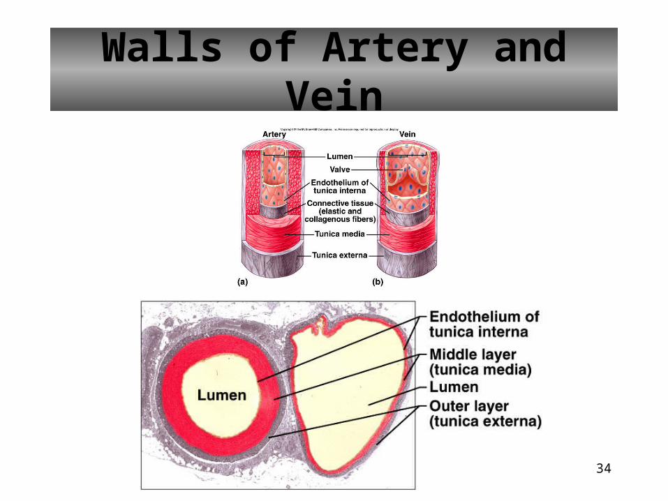
34
Walls of Artery and Vein

35
Arteriole
• smallest arterioles only have a few smooth muscle fibers• capillaries lack muscle fibers

36
Metarteriole
connects arteriole directly to venule

37
Capillaries• smallest diameter blood vessels• extensions of inner lining of arterioles• walls are endothelium only• semipermeable• sinusoids – leaky capillaries

38
Capillary Network
39
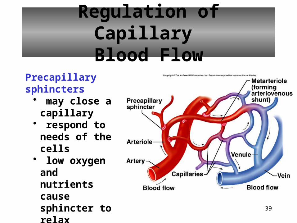
Regulation of Capillary Blood Flow
Precapillary sphincters
• may close a capillary
• respond to needs of the cells
• low oxygen and nutrients cause sphincter to relax

40
Exchange in the Capillaries• water and other substances leave capillaries because of net outward
pressure at the capillaries’ arteriolar ends
• water enters capillaries’ venular ends because of a net inward pressure
• substances move in and out along the length of the capillaries according to their respective concentration gradients

41
Venules and Veins
Venule• thinner wall than arteriole• less smooth muscle and elastic tissue than arteriole
Vein• thinner wall than artery• three layers to wall but middle layer is poorly developed• some have flaplike valves• carries blood under relatively low pressure• serves as blood reservoir

42
Venous Valves

43
Characteristics of Blood Vessels

44
Blood Volumes in Vessels

45
Arterial Blood Pressure
Blood Pressure – force the blood exerts against the inner walls of the blood vessels
Arterial Blood Pressure• rises when ventricles contract• falls when ventricles relax• systolic pressure – maximum pressure• diastolic pressure – minimum pressure

46
Pulse
• alternate expanding and recoiling of the arterial wall that can be felt

47
Factors That InfluenceArterial Blood Pressure

48
Control of Blood Pressure
Controlling cardiac output and peripheral resistance regulates blood pressure

49
Control of Blood Pressure
If blood pressure rises, baroreceptors initiate the cardioinhibitory reflex, which lowers the blood pressure

50
Control of Blood Pressure
Dilating arterioles helps regulate blood pressure

51
Venous Blood Flow
• not a direct result of heart action
• dependent on • skeletal muscle
contraction• breathing• venoconstriction

52
Central Venous Pressure
• pressure in the right atrium
• factors that influence it alter flow of blood into the right atrium
• affects pressure within the peripheral veins
• weakly beating heart increases central venous pressure
• increase in central venous pressure causes blood to back up into peripheral vein

53
Pulmonary Circuit
• consists of vessels that carry blood from the heart to the lungs and back to the heart

54
Blood Flow Through Alveoli
• cells of alveolar wall are tightly joined together• the high osmotic pressure of the interstitial fluid draws
water out of them

55
Systemic Circuit
• composed of vessels that lead from the heart to all body parts (except the lungs) and back to the heart
• includes the aorta and its branches
• includes the system of veins that return blood to the right atrium

56
Major Vessels of Arterial System

57
Major Blood Vessels of the Heart

58
Principal Branches of the Aorta

59
Abdominal Aorta and Its Major Branches

60
Arteries to Neck, Head, and Brain

61
Cerebral Arterial Circle• Circle of Willis• formed by anterior and posterior cerebral arteries, which
join the internal carotid arteries

62
Arteries to Shoulder and Upper Limb

63
Arteries to Thoracic Wall

64
Arteries to Pelvic Region

65
Arteries to the Lower Limb

66
Major Vessels of the Venous System

67
Major Veins of the Brain, Head and Neck

68
Veins from the Upper Limb and Shoulder

69
Veins That Drain the Thoracic Wall

70
Veins That Drain the Abdominal Viscera

71
Veins from the Abdominal Viscera:Hepatic Portal Vein
Hepatic portal vein drains one set of capillaries and leads to another set

72
Veins of the Lower Limb and Pelvis

73
Life-Span Changes• cholesterol deposition in blood vessels
• heart enlargement
• death of cardiac muscle cells
• increase in fibrous connective tissue of the heart
• increase in adipose tissue of the heart
• increase in blood pressure
• decrease in resting heart rate

74
Clinical Application
Arrhythmias
Ventricular fibrillation• rapid, uncoordinated depolarization
of ventricles
Tachycardia• rapid heartbeat
Atrial flutter• rapid rate of atrial
depolarization