Embed Size (px)
Citation preview
1
BIOMARKERS AS BIOLOGICAL INDICATORS TO XENOBIOTIC EXPOSURE
Fernando Gil and Antonio Pla
Department of Legal Medicine and Toxicology. University of Granada.
To whom correspondence and reprint request should be addressed:
Fernando Gil, M.D., Ph.D.
Departamento de Medicina Legal y Toxicología
Facultad de Medicina. Universidad de Granada.
C/ Avda. Madrid 11
18071 Granada (SPAIN)
Phone: 34-58-24 35 46/ 34-58- 24 99 30
Fax: 34-58-24 61 07
e-mail: [email protected]
The presence of a xenobiotic in the environment always represents a risk for
living organisms. However, to talk about impregnation there is a need to detect the toxic
2
in the organism, and the concept of intoxication is related to specific organ alterations
and clinical symptoms. Moreover, the relationship between the toxic levels within the
organism and the toxic response is rather complex and has a difficult forecast since it
depends on several factors, namely toxicokinetic and genetic factors.
One of the methods to quantify the exposure to xenobiotics and its potential
impact on living organisms, including the human being, is the monitoring by the use of
the so-called biomarkers.
1. Definition
The National Academy of Sciences defines a biomarker or biological marker as a
xenobiotically induced alteration in cellular or biochemical components or processes,
structures, or functions that is measurable in a biological system or sample [1].
Silbergeld et al [2] defines biological markers as physiological signals that reflect
exposure, early cellular response or inherent or acquired susceptibilities, which provide
a new strategy for resolving some toxicological problems.
Sensu stricto, we define a biomarker as a biological response to a chemical or a
group of chemical agents [3] but not the presence of the agent or its metabolites within
the body (internal dose). However, there is no doubt that the measurement of a
xenobiotic in a biological system or sample is a bioindicator of exposure, and thus, it
could be considered like a biomarker.
Biological monitoring has advantages over environmental monitoring because it
measures the internal dose of a compound. Interindividual differences in absorption,
bioavailability, excretion, and DNA repair should be taken into account. Moreover,
intraindividual differences, as consequence of particular physiopathological alterations
occurring in a specific period of time, should also be considered. This involves an
individualised biological control to evaluate the exposure to a particular xenobiotic [4].
The organism acts as an integrator of exposure and several physiological factors, which
modulate the uptake of toxic. Thus, we may state that a collective can not be
assimilated as a homogeneous group of individuals exposed to a xenobiotic of physico-
chemical properties under reproducible and standard conditions.
The use of biological markers in the evaluation of disease risk has increased
markedly in the last decade. Biomarkers are observable end points that indicate events
3
in the processes leading to disease. They are particularly useful in the evaluation of
progressive diseases that manifest their symptoms long after exposure to initiating
factors. In such cases, traditional early warning symptoms of developing disease may
be lacking. At the same time, the disease, once clinically apparent, may be essentially
irreversible [5].
The two main research fields in the use of biomarkers in Toxicology are
Enviromental Toxicology and Industrial Toxicology, the latter being one of the most
relevant and important branches of Medical Toxicology.
2. Conditions and limitations of biological monitoring
A rational biological monitoring is only possible when sufficient toxicological
information has been gathered on the mechanism of action and on the toxicokinetic of
xenobiotics including absorption, distribution, metabolism and excretion [6]. Biological
monitoring cannot be applied for assessing exposure to substances that exhibit their
toxic effects at the sites of first contact i.e. primary lung irritants or are poorly absorbed.
For other xenobiotics that are significantly absorbed and exert a systemic toxic
action, a biological monitoring test may provide different information, depending on our
current knowledge of the relationships among external exposure, internal exposure and
the risk of adverse effects. If only the relationships between external exposure and the
internal dose is known, the biological parameter can be used as an index of exposure,
but it provides little information on the health risk. But if a quantitative relationship has
been established between internal dose and adverse effects, and the internal-dose
effects and the internal-dose response relationships are known, biological monitoring
allows for a direct health risk assessment and thus for an effective prevention of the
adverse effects [7].
3. Specificity of biomarkers
The ideal biomarker should have the following characteristics [8]:
a) sample collection and analysis are simple and reliable
b) the biomarker is specific for a particular type of exposure
c) the biomarker only reflects a subclinical and reversible change
d) relevant intervention or other preventive effort can be considered
4
e) use of the biomarker is regarded as ethically acceptable.
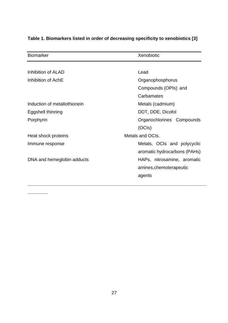
It is clear that if we agree with that last definition only a few biomarkers fit well.
Biomarkers range from those that are highly specific such as an enzyme of the heme
pathway, aminolevulinic acid dehydratase (ALAD), which is inhibited only by lead or the
inhibition of acetyl choline esterase (AChE), which is specific to the organophosphorus
and carbamate pesticides, to those that are non specific namely the effects on the
immune system or DNA that can be caused by a wide variety of chemical agents (Table
1) [3].
An important aspect to be considered is the complementation among biomarkers
that results into a higher degree of specificity. Thus, metalothionein induction may occur
by exposure to a variety of metals. However, if a measurement of one specific metal is
performed in a biological fluid i.e Cadmium in urine, and it is found elevated over normal
values, the evaluation of the induction of metallothionein would enhance the specificity
of the measurement, which in turn would increase even more if a preclinical renal
alteration as renal proteinuria (i.e. ∃2-microglobulin) is detected. This feature is relevant
since currently there is a number of biomarkers of toxic response in different tissues,
organs, and systems that are unspecific. However, the establishment of new
relationships between biomarkers may contribute to increase their specificity.
4. Classification of biomarkers
Biomarkers are generally classified into three groups: exposure, effects, and
susceptibility [2,3,9,10].
4.1 Biomarkers of exposure
They allow measuring the internal dose by chemical analysis of the toxic
compound or metabolite in body fluids or excreta such as blood, urine and exhaled air
(Table 2) [10, 11].
Internal dose may also mean the amount of a chemical stored in one or in
several body compartments or in the whole body. This usually applies to cumulative
toxic chemicals. For example, the concentration of polychlorinated biphenyl (PCB) in
blood is a reflection of the amount accumulated in the main sites of deposition (i.e., fatty

5
tissues). The internal dose reflects the amount of chemical bound to the critical sites of
action [12].
Bernard & Lauwerys classified the biomarkers of exposure into two subgroups,
selective and non-selective, according to their selectivity test, which is based on the
direct measurement of the unchanged chemicals or their metabolites in biological
media. The non-selective tests are used as non-specific indicators of exposure to a
group of chemicals. As an example of non-selective exposure tests, the determination of
diazo-positive metabolites in urine for monitoring exposure to aromatic amines, the
analysis of tioethers in urine, and the determination of the mutagenic activity of urine
can be cited.
When assessing the usefulness of a particular exposure biomarker, one must
consider two aspects of validity: analytical and toxicokinetic. For optimal analytical
quality, standardisation is needed, but the specific requirements vary considerably
between individual toxicants. Major areas of concern include: preparation of the
individual, sampling procedure and sampling handling, and measurement procedure
that encompasses technical factors, such as calibration and quality assurance
procedures. Life events, such as reproduction and senescence, may also affect the
toxicokinetic of a xenobiotic [8].
4.2 Biomarkers of susceptibility
They serve as indicators of a particular sensitivity of individuals to the effect of a
xenobiotic or to the effects of a group of such compounds. Can be genetic markers that
include alterations in chromosomal structure such as restriction fragment length
polymorphism’s (RFLPs), polymorphism of enzyme activities, etc.[13]. After the
exposure of an organism to a xenobiotic it suffers a biotransformation process in two
phases. In the first phase a primary metabolite, usually oxidised and more or less active,
is originated by the specific action of the microsomal P450 cytochrome isoenzymic
family. In the second phase, the primary metabolite is transformed into other secondary
metabolite, which is usually inactive. Some individuals with a low cytochrome P450
activity will be more resistant to the generation of primary active metabolites, whereas
those exhibiting a low activity of enzymes involved in the second phase will show a
lower formation of phase II inactive metabolites, increasing toxicity.
6
Two types of susceptibility biomarkers can be distinguished: polymorphism’s
activating system markers and polymorphisms of detoxicating systems. Polymorphisms
of activating systems are measurements of the activity of cytochrome P-450
isoenzymes. The family of cytochrome P-450 enzymes is involved in the toxicity of
several xenobiotics; associated with the P-450 cytochromes there are a wide range of
enzyme activities referred to as monooxygenase activities. A number of studies have
suggested that the various cytochrome P-450 enzymes differ substantially in their amino
acid sequences and thus they likely are encoded by distinct genes. This has been
confirmed by comparisons of the complete amino acid sequences of over 71 forms of
cytochrome P-450 and of the nucleotide sequences of their corresponding cDNAs and
of several genes [11].
A roman numeral corresponding to its specific class designates each form of
cytochrome P-450. The most important classes that constitute the different forms of
cytochrome P-450 are I through IV. These cytochrome classes comprise several
subclasses that are designated by a combination of a letter (A,B,C,D,..) and an
identifying arabic numeral (i.e. IA1, IIC8,....). The most important are IA1 (represent the
AHH activity), IIC8, IID6 and IIE1. There have been a number of studies trying to
establish a relationship between specific cytochrome P450 activities and some diseases
due to environmental toxic exposure, especially cancer. However, there are no definitive
conclusions [14,15].
Markers of polymorphisms of detoxicating systems are measurements of the
activity of conjugating enzymes such as glutathione-S-transferases, acetyltransferases,
sulfotransferases, glucuronyltransferases and paraoxonase. For instance, predisposition
to cancer has been correlated with genetic polymorphisms of N-acetyltransferases. N-
acetyltransferase is an enzyme involved in the deactivation of aromatic amines. After
acetylation there is enhanced excretion in urine.In a group of arylamine exposed
workers the slow acetylators are at increased risk for bladder cancer versus rapid
acetylators. Another example is glutathione-S-transferase μ, an enzyme involved in the
detoxification of reactive metabolites. Half of the population has no functional allele for
this enzyme and no or low enzyme activity. These persons are at increased risk to
squamous cell carcinoma of the lung [11].
Finally, several organophosphates can be inactivated (hydrolysed) by
7
paraoxonase (PON1). Human paraoxonase exhibits an important polymorphism and in
humans, three genotypes have been detected: individuals homozygous for the low
activity allele, individuals homozygous for the high activity allele, and heterozygous, so
paraoxonase activity can be used as a biomarker of susceptibility to organophosphorus
compounds [16]. The polymorphism is also observed with the oxons of methyl
parathion, chlortion and ethyl 4-nitrophenyl phenylphosphonate (EPN). However, it is
not observed with the oxon of chlorpyrifos.
Several evidences suggest that high levels of serum paraoxonase are protective
against poisoning by organophosphorus pesticides whose active metabolites are
substrates of this enzyme. [17]. Birds, which have very low levels of serum paraoxonase
are very sensitive to parathion, diazinon-oxon, and pirimiphos-oxon compared with
mammals who have higher levels of this enzyme [18]. After injection of partially purified
rabbit paraoxonase into rats an increased resistance to the toxic effects of paraoxon
was observed. Recent studies indicate that administration of paraoxonase might have
therapeutic value in case of organophosphates intoxication [19].
4.3 Response or effect biomarkers
Response or effect biomarkers are indicative of biochemical changes within an
organism as a result of xenobiotic exposure. The ideal biomarkers should be early
detected and be able to show adverse effects before they are irreversible. Those are the
most studied biomarkers and they include modifications in some parameters of blood
composition, alterations of specific enzyme activities, DNA-adducts appearance,
localised mRNA and protein increases, and appearance of specific antibodies
(autoantiboides) against a xenobiotic or a particular cellular fractions [10]. It is noticeable to remark that not always is easy to distinguish between an
exposure and a response biomarker. Perhaps the most typical example is the formation
of an DNA adduct, a exposure biomarker, which formation results from the reaction of a
xenobiotic with the DNA, that in turn constitutes the cellular response. Moreover, it is
evident that a particular response requires a previous exposure to the xenobiotic. Below
we consider some significant examples of response biomarkers.
4.3.1.Respiratory system.
8
Several studies have suggested that low-molecular weight proteins (LMWP)
specific for the lung might serve as peripheral biomarkers of lung toxicity [20]. A lung
biomarker, measurable in serum, bronchoalveolar fluid (BAL) and sputum has recently
been identified. This biomarker is a microprotein initially isolated from urine in 1974
(Urine Protein 1) of patients with renal tubular dysfunction and subsequently identified
as the major secretory product of the lung Clara cells, which are non-ciliated cells
localised predominantly in terminal bronchioles. This protein called Clara cell protein
(CC16) is a homodimer of 15.8 kDa. Clara cells are particularly sensitive to toxic lung
injury and they contain indeed most of the lung cytochrome P-450 activity, which
confers them a high xenobiotic metabolising activity [21,22]. Several lines of evidence
indicate that CC16 is a natural immunoregulator protecting the respiratory tract from
unwanted inflammatory reactions. CC16 has been shown to inhibit the activity of
cytosolic phospholipase A2, a key enzyme in inflammatory processes. Phospholipase
A2 is the rate-limiting enzyme in the production of arachidonic acid, the substrate for the
synthesis of prostaglandin and leukotriene mediators of inflammation [23]. By inhibiting
phospholipase A2, CC16 could also prevent the degradation of lung surfactant
phospholipids [24].
CC16 secreted in the respiratory tract diffuses passively by transudation into
plasma from where it is rapidly eliminated by glomerular filtration before being taken up
and catabolised in proximal tubule cells. Studies reviewed by Bernard suggest that
CC16 in BAL fluid, sputum or serum is a sensitive and relatively specific indicator of
acute or chronic bronchial epithelium injury.
A significant reduction of CC16 in serum is an indicator of Clara cells number and
integrity. After adjustment for age, a linear dose-response relationship was apparent
between smoking history and serum CC16, latter decreasing on average by about 15%
for each 10 pack-year smoking history [25]. Serum CC16 was also found to be
decreased in several occupational groups chronically exposed to silica, dust and
welding fumes, and lung diseases (cancer, asthma and patients with chronic obstructive
pulmonary disease) [26].
The increased concentration of CC16 in serum can also be used to detect an
acute or chronic disruption of the bronchoalveolar/blood barrier integrity. Increased
serum levels of CC16 have been observed in sarcoidosis and adult respiratory distress
9
syndrome (ARDS). This confirm that in pathological conditions the barrier between the
surface of respiratory epithelium and the vascular compartment may be disrupted,
upsetting the diffusional equilibrium between CC16 in serum and in the respiratory tract.
The existence of an enhanced passage of proteins across the blood/brochoalveolar
space barriers, for example in acute exposure in animals by inhalation or systemic
routes with pneumotoxic agents (4-ipomeanol, 3-methylfuran, naphthalene,
trichloroethylene, 1,2-dichloroethylene, etc.), is supported by the significant elevation of
albumin, β2-microglobulin and other plasma proteins in BAL fluids. These findings open
new perspectives in the assessment of lung toxicity by suggesting that readily diffusible
lung-specific proteins may serve as peripheral markers of pneumotoxicity.
4.3.2. Blood system
The most studied biomarkers of effect are those related to the alterations of
heme synthesis. ALAD is an enzyme involved in the heme biosynthetic pathway and the
assay is highly specific for lead exposure and effect. The inhibition of ALAD has been
shown to be a reliable indicator of effect to lead in studies on humans and animals
(specially several species of fish and birds –eagles, starlings, ducks and geese). One of
the most important advantage of this biomarker in Ecotoxicology is that the animal
sacrifice is not required; the effect is slowly reversed, with ALAD values returning to
normal only after about 4 months [3,27-29].
Heme biosynthesis is normally closely regulated, and levels of porphyrins are
ordinarily very low. Some organochlorines (OCs) cause the formation of excess
amounts of hepatic highly carboxylated porphyrins. The two OCs that are most involved
in inducing porphyria in mammals and birds are hexacholorobenzene (HCB) and the
PCBs [3, 30].
Hemoglobin adducts are formed from exposition of several compounds (ethylene
oxide, acrylamide, 3-amino-1,4-dimethyl- 5OH-pyrido-indole, 4-aminobiphenyl, 2,6-
dimethylaniline,etc). Acrylamide is an important neurotoxic agent causing a peripheral
neuropathy to experimental animals as well as to humans and it has been shown to be a
potential carcinogen. The conversion rate of acrylamide to glycidamide (reactive
metabolite epoxide responsible for the neurotoxicity) is significantly correlated with the
hemoglobin adducts of acrylamide. These adducts are useful as biomarkers of
10
acrylamide-induced peripheral neuropathy [31]. Because of the relatively long life span
of the red blood cells (four months in humans), hemoglobin adducts have
advantageously been used for integrating concentrations in the blood of genotoxic
substances.
4.3.3. Nervous system
Despite its obvious importance within toxicology, the area of neurotoxicity seems
to be progressing more slowly than other fields with regard to biological monitoring. The
complexity of the nervous system and its distinctive peculiarities, together with the
problems associated with the determination of the precise targets for neurotoxic action
are certainly responsible for this limited advancement. Neurochemical measurements
for detecting neurotoxicity in humans are limited by the inaccessibility of target tissue.
Thus, a necessary approach for identifying and characterising neurotoxicity is the
search for neurochemical parameters in peripheral tissues easily and ethically obtained
in humans, which could represent a marker for the same parameters in nerve tissue
[32].
Perhaps, the most significant and useful example of specific biomarker of
neurotoxicity is the inhibition of acetyl choline esterase (AChE) caused by
organophosphorus compounds or carbamate pesticides. The enzyme activity is present
in several tissues though their inhibition is generally determined from blood samples
(whole blood or plasma) and brain. This biomarker has been used in human toxicology
and is widely studied in ecotoxicology (birds, mammals and aquatic species). For
example, inhibition of AChE in brain can be taken as proof of mortality in birds, whereas
in other animals, such as fish, there is a bigger variability founding lethal inhibition in a
range of 40-80% [33-35].
The decrease in AChE activity in brain may remain for several weeks after the
toxic exposure, which is adequately correlated with the effect, in contrast to that
occurring in blood with a lower life span. Nevertheless, measuring the blood AchE
activity has the advantage of easy sampling since there is no need of animal sacrifice.
[36].
Butyrylcholinesterase (BuChE), example of an unspecific biomarker of
neurotoxicity, is sometimes studied in parallel with AChE but its physiological role is
11
unknown and its degree of inhibition is not simply related to toxic effect. Other
parameters involved in neurotransmission are the target for a variety of neurotoxicant
xenobiotics. The measurement of these parameters is done in red blood cells,
lymphocytes and fibroblasts [32,37].
Several active bioamines are liberated from the nerve ending by exocytosis, a
process which is triggered by influx of Ca2+ and are inactivated by reuptake and
methylation mediated by catechol-O-methyltransferase (COMT). Because of its
intracellular localisation, monoamine oxidase (MAO) plays a strategic role in inactivating
catecholamines that are free within the nerve terminal and not protected by storage
vesicles. Isoenzymes of MAO have been characterised with differential substrate
specificities; MAO-A preferentially deaminates norepinephrine and serotonin, whereas
MAO-B acts on a broad spectrum of phenylethylamines. MAO-B is a microsomal
enzyme and the aminoacidic sequences of the enzymes from human cerebral cortex
and consequently platelets were shown to be identical and platelet MAO-B activity
appears to reliably reflect enzyme activity in the nervous system.
MAO-B activity is used clinically as a marker of the pharmacological effects of
MAO inhibitors, such as in the treatment of Parkinson’s disease. MAO-B activity in
platelets has been used as a biomarker of effects of styrene and perchloroethylene
occupational exposures [38], which is known to cause dopamine depletion. Changes in
MAO-B could represent an adaptive response to dopamine depletion and alternatively,
styrene or its metabolite(s) might exert a direct inhibitory effect on the enzyme [37, 39].
Another example of neurotoxic biomarker, involved in delayed toxicity , is the
inhibition of neuropathy target esterase (NTE). Several organophosphorus compounds
(Mipafox, Methamidofos,etc), after a single dose, induce delayed neuropathy,
characterised by symmetrical axonal degeneration that implicate the NTE inhibition and
not the acethyl cholinesterase enzyme. Organophosphate-induced delayed neuropathy
(OPIDN) is characterised by a lag period of about 1 to 3 weeks, from the moment of
intoxication to the appearance of clinical symptoms. The first intoxication was described
with TOCP (triorthocresylphosphate) [40-41]. In experimental assays the measurement
of NTE in lymphocytes has been used as biomarker of effect and there is a good
correlation between NTE activity in brain and lymphocytes after 24 h of an acute
exposure to neurotoxic organophosphorus compounds [42].
12
In conclusion, the complexity of the nervous system does not allow a rapid and
easy development of sensitive, specific and reliable biomarkers for neurotoxicity,
although the biomarkers presented in this review appear promising.
4.3.4. Urinary biomarkers
Long-term exposure to certain nephrotoxic compounds (heavy metals –lead,
mercury, cadmium and chromium-, halogenated hydrocarbons –chloroform-, organic
solvents –toluene-, therapeutic agents –aminoglucosides, amphotericine B,
acetaminophen, etc.) may cause progressive degenerative changes in the kidney.
However, because of its large reserve capacity, the clinical signs of renal damage are
not apparent until the injury is extensive and consequently irreversible. The prevention
of renal diseases requires the use of more sensitive tests capable of detecting renal
effects at a stage when they are still reversible or at least not so advanced as to trigger
a progressive renal disease [43-47].
In practice, one usually recommends the determination in urine of at least two
plasma-derived proteins, a high molecular weight protein (HMWP) such us albumin for
the early detection of glomerular-barrier defect and a low molecular weight protein
(LMWP) such as retinol-binding protein for the early screening of proximal damage
(Table 3) [48,49].
Injury to the kidney can be detected by measuring the urinary activity of kidney-
derived enzymes. As index of nephrotoxicity it has been proposed the lysosomal
enzyme β-N-acetyl-D-glucosaminidase (NAG). Advantages of this enzyme include its
stability in urine and its high activity in the kidney. The diagnostic value of NAG can be
further improved by measuring the B isoenzyme (lesional form released with fragments
of cell membranes) [50].
Destruction of renal tissue can also be detected by measuring in urine kidney
components which, when they are quantified by immunochemical methods, are
referred to as renal antigens. These have been proposed as urinary markers of
nephrotoxicity and include: carbonic anhydrase, alanine aminopeptidase and adenosine
deaminase-binding protein for the proximal tubule, fibronectin for the glomerulus and
Tamm-Horsfall glycoprotein for the thick ascending limb of the loop of Henle [50-55].
It is important to realise that this battery of tests, does not permit the detection of
13
effects on all areas or segments of the kidney or nephron. No sensitive biomarker is
available to detect effects on the deep medulla, the papilla or the distal tubule. There is
also no biomarker to detect and follow the progression of active fibrotic processes that
may insidiously and irreversibly reduce the renal function (i.e.interstitial fibrosis).
4.3.5. Immune system
Direct effects of xenobiotics can affect the immune system and can lead to
decreased resistance to infections or tumours, may alter the course of autoimmunity or
induce hypersensitivity reactions. Data of several immunotoxic agents (dioxin,
polychlorinated biphenyls, immunotherapeutic drugs, etc.) are mainly derived from
animal research (mouse and rat), although few biomarkers exist that provide specific
information on the immunotoxicity in man [56,57].
The biomarkers proposed to assess immunotoxicity in man are listed in Table 4
[58] and include full blood count, antibody-mediated immunity (immunoglobulin
concentrations in serum) phenotypic analysis of lymphocytes by flow cytometry, cellular
immunity study, measurement of antibodies and markers of inflammatory response and
finally, examination of non-specific immunity.
Although a variety of factors may modify the immune function including drugs
(non-steroidal anti-inflammatory, vitamin complexes,etc.), biological parameters (sex,
age, pregnancy) and other factors (diet, alcohol consumption, circadian rhythms, stress,
nutritional state, sleep disturbances ,etc).
Within the field of Ecotoxicology, the resistance to infection in ducks exposed to
organochloride pesticides has been studied measuring the cellular activities involved in
the immune response; particularly the in vitro phagocytic capacity from kidney isolated
macrophages in an number of species has been evaluated.
4.3.6. Biomarkers of DNA damage
At the present time many technological approaches permit the detection of
covalent interactions of xenobiotics with proteins and other macromolecules. For
example, several biomolecules (hemoglobin, serum albumin, etc.) have carboxyl, amino
or sulfhydryl reacting groups that can interact with electrophilic compounds. Human
DNA-adduct formation (covalent modification of DNA with chemical carcinogens) has
14
been shown to correlate with the incidence of a carcinogenic process and is a promising
biomarker for elucidating the molecular epidemiology of cancer [59].
There is a sequence of events between the first interaction of a xenobiotic with
DNA and consequent mutation. The first stage is the formation of adducts; the next
stage, may be secondary modifications of DNA such as strand breakage or an increase
in the rate of DNA repair. The third stage is reached when the structural perturbations in
the DNA become fixed and the affected cells often shown altered function. One of the
most widely used assays to measure chromosomal aberrations is the sister chromatid
exchange (SCE). Finally, when the cells divide, damage caused by xenobiotics can lead
to the DNA mutation and consequent alterations in the descent [3, 60].
Some examples of toxics capable to form human DNA adducts are given in Table
5, including polycyclic aromatic hydrocarbons, aromatic amines, heterocyclic amines,
micotoxins and alquilant chemotherapeutic agents [61-62]. The biologic monitoring to
detection of human and animal DNA adducts include 32P post-labelling and recently
immunoassays using adduct-specific antibodies [63-64]. They can be detected in blood
(lymphocytes), urine or tissue homogenates from biopsy (gastric mucosa, liver,etc.)
although the study of DNA-adducts is not feasible in the routine analysis. The damage
of DNA has also been studied in Ecotoxicology on several marine species (fish of fresh
water, snapping turtle, etc.) that can be exposed to benzo [a] pyrene [3, 65-67].
Future investigations will focus on the implementation and design of studies to
assess the association between DNA-adduct formation and cancer risk from toxic
compounds. Whereas this association is strongly supported by animal studies, it
remains to be ascertained whether adducts are also a necessary component of
carcinogenesis in humans. Many studies are now being designed to correlate metabolic
polymorphisms, urinary metabolites, chromosomal aberrations and protein and DNA-
adducts and it is possible in the next future to obtain promising results from the
combined use of these biomarkers in the evaluation of cancer risk.
4.3.7. Biomarkers of gene expression
The development of many tumours related with xenobiotics is associated with the
aberrant expression of genes that encode proteins involved in cellular growth. This
aberrant expression can involve a quantitative difference (overexpression of the protein)
15
and a qualitative difference (expression of a mutant form of the protein). Although these
biomarkers are affected not only by toxic compounds, is very important to establish
potential confounding factors and to assess of sensitivity, specificity and predictive value
of these tests. The Table 6 shows the biomarkers of gene expression, which include
[63,68,69]:
* growth factors
* oncoproteins: - growth factor receptors
- other oncogene proteins
- tumour suppresser gene proteins
These biomarkers have been studied in easily obtainable biological fluids such as
serum, plasma, urine and bronchoalveolar liquid by enzyme-linked immunoadsorbent
assay (ELISA), radioimmunoassay (RIA) or immunoblotting.
It has been reported that in subjects developing cancer, during the first stages of
the disease, show a significant increase in those gene-expression biomarkers related
with the specific cancer.
4.3.8. Biomarkers of oxidative damage
Contaminants such as polycyclic aromatic hydrocarbons (PAHs), halogenated
aromatic hydrocarbons, heavy metals, selenium, pesticides and industrial solvents are
capable of causing oxidative damage, especially by free radicals. In response to
oxidative stress, there may be adaptive responses of the antioxidant systems,
modification of cellular macromolecules and finally tissue damage.
Changes in the antioxidant systems and modified macromolecules can serve as
biomarkers for a variety of xenobiotics. The protective systems included oxidised
glutathione/reduced glutathione, glutathione reductase, catalase, superoxide dismutase
and peroxidase activities, ascorbate and α-tocopherol. Macromolecules that may be
affected by free-radical damage include lipids, proteins and nucleic acids [70-72].
4.3.9. Metallothioneins
Metallothioneins are small proteins with low molecular weight (approximately 61
16
amino acids in most mammals), cysteine-rich and capable of binding metals (Cd, Cu,
Hg, Zn, Co, Bi, Ni and Ag). The cellular role of metallothioneins is complex and partially
unknown; it is believed to protect cells against free heavy metal ion-induced damage.
These proteins are implicated in metabolism regulation of Zn and Cu making these ions
available to the cells as necessary, and may act as sulfhydryl-rich scavengers to
prevent damage from stress-induced free radicals.
Metallothioneins have been proposed as biomarkers for exposure to metal ions
since they are induced by the own metals . Nevertheless, recent studies indicate that
they are also inducible by a variety of non-metal toxic agents (glucocorticoids) and
physiological conditions (nutritional changes and pregnancy). Another limitation concern
to analytical measurements. Often, methods of evaluating are slower and more
expensive than analysis of metals themselves. The recent development of antibody-
based methods and of messenger-RNA assays probably can make metallothionein a
more valid bioindicator [58,73].
4.3.10. Others biomarkers
* Heat stress proteins (HSP) Heat stress proteins are an important group of ATP-dependent proteins, which
facilitate the folding of nascent proteins by preventing their aggregation and then
chaperoning them to sites of membrane translocation. They were previously referred to
as heat shock proteins because of their rapid appearance following heat stress,
although they increased in response to a variety of xenobiotics, including metals and
metalloids (especially, arsenite), heavy metals and oxidising agents [74-77]. Those
biomarkers are of becoming importance in the next future.
Heat shock proteins include four types: hsp 90 (~ 90 kDa), hsp 70 (~ 70 kDa),
hsp 60 (~ 60 kDa) y hsps of low molecular weight (~ 15-30 kDa):
* hsp 90 (stress-90): there are two forms, the Hsp 83 is located in the eucaryotic
cytosol and Grp 94 that is localised in the endoplasmic reticulum of mammals.
* hsp 70 (stress-70): is the most widely studied and is located in the cytosol of
mammals. It belongs to a multigene family with at least one form that is constitutively
expressed (hsc 70) in unstressed cells, and one or more isoforms (hsp 70) that are only
stress inducible. Both (hsp and hsc 70) help to refold denatured proteins that occur in
17
cells following heat shock or exposure to other proteotoxic stresses. Because these
proteins show a very strong response to protein damage, they are ideal candidates for
biomarkers of sub-lethal damage.
* hsp 60 (chaperonins 60): belongs to the family of chaperonins (cpn 60) involved
in protein folding. They are localised in the mitochondria and may be useful as an
organelle-specific biomarker.
* hsps of low molecular weight : they show substantial homology with α−
crystallins of vertebrates, the may be involved in actin binding to stabilise microfilaments
and have a good potential as biomarkers of effect on some targets as the cytoskeleton.
With the development of antibodies to some heat stress proteins, Western
blotting is the best for evaluating heat stress proteins. Nevertheless, the heat stress
protein responses have been studied only in experimental assays on laboratory species,
culture systems or invertebrates.
* Eggshell thinning
Severe eggshell thinning may be lead to breakage of eggs. This fact has been
proposed as biomarker of reproductive damage. Dichloro-diphenyl-ethane (DDE), a
metabolite of DDT, is the most widely studied in many species of birds (pelican,
eagle,etc). The pathogenic mechanism is not yet unknown but could be related with
hormone alterations that interfere in the calcium metabolism, which is essential in the
formation of the eggshell.
Eggshell thickness may be estimated by two methods: one utilises direct
measurement with a micrometer and the other calculates the thickness index as the
weight of the shell (mg) divided by the product of shell length and breadth (mm2) [3].
* Vitamin A, Thyroxine and Thyroxine-TBPA complex.
Normaly, thyroxine is binding in plasma to transthyretin or TBPA (Thyroxine
Binding Prealbumin) and RBP (Retinol binding protein)-vitamin A and form thyroxine-
TBPA-RBP complex. Certain monohydroxymetabolites of 3,3’,4,4’ tetrachlorobiphenyl
(OH-TCBs) formed by the monooxygenase system (Cytochrome P450IA1) compete
strongly with thyroxine for its binding site upon the transthyretin. The consequence of
the presence of these metabolites in blood is the quickly lost of thyroxine from
18
circulation and the appearance of symptoms associated with hypothyroidism. Also the
binding of OH-TCBs to transtyretin can cause a conformational change which leads to a
reduction in the attachment of the RBP to TBPA. RBP and attached retinol (vitamin A) is
then lost from the blood by glomerular filtration (this complex has a low molecular
weight). The consequences of this feature are the appearance of hypovitaminosis A
(dermal and epithelial lesions [78]. These finding have been shown in laboratory studies
with mice, rats and monkeys [79]. The increase of thyroxine and vitamin A in urine
represent a biomarkers of TCBs exposition.
* Biomarkers for anticoagulant rodenticides Warfarin and second generation anticoagulant rodenticides (flocoumafen) act as
inhibitors of the vitamin K cycle, which operates in the liver of vertebrates. These
compounds act by competing with vitamin K or derivatives and the consequence of this
antagonism is a failure of the vitamin K cycle to carboxylate the precursors of clotting
proteins. This leads to an extension of blood clotting time, haemorrhaging and death.
The effects of anticoagulant rodenticides may be detected at two different levels:
monitoring changes in the vitamin K cycle or monitoring increases in precursors of
clotting proteins in blood. The development of ELISA assays in the last years have
allowed the detection of forerunners of clotting proteins in mammals and birds [3].
* Plant biomarkers
Plants have widely been used as biomonitors to analyse the environmental
impact of pollutants (especially gaseous air pollutants). Specific biomarkers have been
identified in sensitive plants. In a few cases, it is known that excess of a specific
compound will give rise to the production of a metabolite, which is different between
tolerant and sensitive plants.
For example, in the presence of an excess of selenium, Se-sensitive plants fail to
differentiate between S and Se and incorporate Se in sulphur amino acids (essentially,
selenomethionine and selenocysteine) leading to the synthesis of enzymes of lower
activity (selenoproteins), which can lead to plant death. On the contrary, Se-tolerant
plants biosynthesise and accumulate non-protein seleno-amino acids (such as
selenocystathioneine and Se-methylselenocysteine) which do not cause metabolic
19
problems for the plant. Thus, the occurrence of selenoproteins in plants are excellent
biomarkers, although their use has not been widely reported.
Another example is the synthesis of fluorocitrate after an exposure to an excess
of fluorine. The plants synthesise fluoroacetyl-CoA and then convert it, via the
tricarboxylic acid cycle to fluorocitrate. This compound is not recognised by aconitase
and as a result the fluorocitrate is accumulated being a very reliable biomarker for
fluorine poisoning.
Phytochelatins are proteins plenty of sulfhydryl groups and synthesised during
exposure to a heavy metal and anions as SeO42-, SeO3
2- y AsO43- . Dose and time
dependent relationships have been established under laboratory conditions for
cadmium, copper and zinc. Nevertheless, for biological monitoring more research is
needed.
Plant biomarkers respond to a wide variety of environmental compounds and
may be useful to indicate a hazard to plant life. For example, the activity of the enzyme
peroxidase has been used to biomarker exposure of plants to air pollution, especially
SO2. Changes of enzyme systems during the development of the plant (seasonal and
climatic processes) are not yet well enough known and plant biomarkers are not as well
advanced as animal end point though a good future in the ecotoxicology field has been
predicted [3].
In conclusion, the markers of biological toxicity represent an important tool in
Toxicology by three main reasons:
1) they permit to estimate the biological effect on target tissue;
2) they are markers of subclinic alterations and sensible indicators of pathology
and thus, may be useful in diagnostic and preventive strategies;
3) the biomarkers consider inter- and intra-individual variability in the response
to xenobiotics.
BIBLIOGRAFÍA
[1] ENTOX/TIWET (The Faculty of the Department of Environmental Toxicology and
The Institute of Wildlife and Environmental Toxicology-Clemson University). Aquatic and
Terrestrial Ecotoxicology. In: Casarett and Doull=s Toxicology. The basic Science of
Poisons. 50 ed. Ed. CD Klaassen. McGraw-Hill, USA, pp. 883-905, 1996.
20
[2] Silbergeld EK, Davis DL. Role of biomarkers in identifying and understanding
environmentally induced disease. Clin Chem 40 (7), 1363-1367, 1994.
[3] Walker CH, Hopkin SP, Sibly RM y Peakall DB. Biomarkers. In: Principles of
Ecotoxicology. Ed. CH Walker, SP Hopkin, RM Sibly y Peakall DB. Taylor and Francis,
London, pp. 175-194, 1996.
[4] Lauwerys RR. Introduction. In: Industrial Chemical Exposure: Guidelines for
Biological Monitoring. 20 ed. Ed. RR Lauwerys, P Hoet. Lewis, USA, pp. 1-13, 1993.
[5] Ward JBJr, Henderson RE. Identification of needs in biomarker research. Environ
Health Perspect 104: 895-900 (1996).
[6] Lauwerys R. Occupational Toxicology. In: Casarett and Doull=s Toxicology. The
basic Science of Poisons.40 ed. Eds. M Amdur, J Doull, C Klaassen. Pergamon, New
York, 1991.
[7] Lauwerys RR. Evaluación de la exposición a los agentes químicos en la industria.
In: Toxicología Industrial e Intoxicaciones Profesionales. Ed. R Lauwerys. Masson,
Barcelona, pp. 73-95, 1994.
[8] Grandjean P, Brown SS, Reavey P, Young DS. Biomarkers of’chemical exposure:
State of the art. Clin Chem 40(7): 1360-1362, 1994.
[9] Derosa CT, Stevens Y-W, Wilson JD, Ademoyero AA, Buchanan SD, Cibulas W, et
al. The agency for toxic substances and disease registry’s role in development and
application of biomarkers in public health practice. Toxicol Ind Health 9(6): 979-995,
1993.
[10] Repetto M. Diagnóstico de la intoxicación. In: Toxicología Fundamental. Ed. M
Repetto. Díaz de Santos, Madrid, pp. 327-333, .
[11] Van Cauteren H, de Kok MCM, Van Schooten FJ. Cancer risk evaluation. In:
Toxicology. Principles and applications. Ed. RJM Niesink, J de Vries, MA Hollinger.
CRC, New York, 1996.
[12] Bernard A, Lauwerys R. Present status and trends in biological monitoring of
exposure to industrial chemicals. J Occup Med 28: 559, 1986.
13] Sanz P. Los polimorfismos genéticos como causa de variabilidad individual de la
toxicidad. En: Toxicología avanzada. M. Repetto (ed.), Díaz de Santos, Madrid, 87-116
(1995).
21
[14]Tukey RH, Johnson EF. Molecular aspects of regulation and structure of the drug-
metabolizing enzymes. En: Principles of drug action. The basis of Pharmacology, WB
Pratt and P Taylor (eds.), 30 ed., Churchill Livingstone, New York, 423-468 (1990).
[15] Paine AJ. Heterogeneity of cytochrome P450 and its toxicological significance. Hum
Exp Toxicol 14: 1-7 (1995).
[16] Geldmacher-von Mallinckrodt M, Diepgen TL. The human serum paraoxonase-
polymorphism and specificity. Toxicol Environ Chem 18: 79-196 (1988).
[17] Smolen A, Eckerson HW, Gan KN, Hailat N, La Du BN. Characteristic of the
genetically determined allozymic forms of human serum paraoxonase/arylesterase.
Drug Metabol Dispos 19: 107-112 (1991).
[18] Brealey CJ, Walker CH and Baldwin BC . A-esterase activities in relation to
differential toxicity of pirimiphos-methyl to birds and mammals. Pestic Sci 11: 546-554
(1980).
[19] Li WF, Furlong CE, Costa LG. Paraoxonase protects against chlorpyrifos toxicity in
mice. Toxicol Lett 76: 219-226 (1995).
[20] Hermans C, Bernard A. Clara cell protein (CC16): characteristics and potential
applications as biomarker of lung toxicity. Biomarkers 1: 3-8 (1996).
[21] Bernard A, Roels H, Lauwerys R, Witters R, Gielens C, Soumillion A, Van Damme J
and De Ley M. Human urinary protein 1: evidence for identity with the Clara cell protein
and occurrence in respiratory tract and urogenital tract secretions. Clin Chim Acta 207:
239-249 (1992).
[22] Boyd MR. Evidence for the Clara cells as a site of cytochrome P-450 dependent
mixed-function oxidase activity in lung. Nature 269: 713-715 (1977).
[23] Mantile C, Miele L, Cordella-Miele E, Singh G, Kaytal S and Mukherjee AB. Human
Clara cell 10 kDa protein is the counterpart of rabbit uteroglobin. J Biol Chem 268:
20343-20351 (1993).
[24] Guy J, Dhanireddy R and Mukherjee AB. Surfactant-producing rabbit pulmonary
alveolar type 2 cells synthetize and secrete an antiinflammatory protein, uteroglobin.
Biochem Biophys Res Com 189: 662-669 (1992).
[25] Bernard A, Roels H, Buchet JP, Lauwerys R. Serum Clara cell protein: an indicator
of bronchial cell dysfunction caused by tobacco smoking. Environ Res 66: 96-104
(1994).
22
[26] Bernard A, Gonzalez-Lorenzo JM, Siles E, Trujillano G, Lauwerys R. Early
decrease of serum Clara cell in silica-exposed workers. Eur Res J 7: 1932-1937 (1994).
[27] Scheuhammer AM. Erythrocyte delta-aminolevulinic acid dehydratase in birds. I.
The effects of lead and other metals in vitro. Toxicol 45: 155 (1987).
[28] Peakall D. Thyroid function, retinols, haem and regulatory enzymes. In: Animal
biomarkers as pollution indicators, Peakall D (ed), Chapman and Hall, London, 108
(1992).
[29] Melancon MJ, Alscher R, Benson W, Kruzynski G, Lee RF, Sikka HC and Spies
RB. Metabolic products as biomarkers. In: Biomarkers: biochemical, physiological and
histological markers of anthropogenic stress, Hugget RJ, Kimerle RA. Mehrle PM Jr,
Bergman HL (eds.), Lewis Publ., Chelsea, MI, 87 (1992).
[30] Fox GA, Kennedy SW, Norstrom RJ and Wigfield DC. Porphyria in herring gulls: a
biochemical response to chemical contamination of Great Lakes food chains. Environ
Toxicol Chem 7: 831 (1988).
[31] Bergmark E, Calleman CJ, Costa LG. Formation of hemoglobin adducts of
acrylamide and its epoxide metabolite glycidamide in the rat. Toxicol Appl Pharmacol
111: 352-363 (1991).
[32] Costa LG and Manzo L. Biochemical markers of neurotoxicity: research strategies
and epidemiological applications. Toxicol Letters 77: 137-144 (1995).
[33] Fairbrother A, Bennett JK. The uselfulness of cholinesterase measurement. J Wildl
Dis 24: 587-590 (1988).
[34] Mineau P. Cholinesterase inhibiting insecticides: their impact on wildlife and the
environment, Elsevier Science, Amsterdam (1991).
[35] Peakall D. Biomarkers od the nervous system. En: Animal Biomarkers as pollution
indicators, Peakall D (ed), Chapman and Hall, London, 19 (1992).
[36] Hill EF. Brain cholinesterase activity of apparently normal wild birds. J Wildl Dis 24:
51 (1988).
[37] Manzo L, Artigas F, Martínez E, Mutti A, Bergamaschi E, Nicotera P, Tonini M,
Candura SM, Ray DE, Costa LG. Biochemical markers of neurotoxicity. A review of
mechanistic studies and applications. Hum Experim Toxicol 15:S20-S35 (1996).
[38] Checkoway H, Echeverria D, Moon JD, Heyer N, Costa LG. Platelet monoamine
oxidase B activity in workers exposed to styrene. Int Arch Occup Environ Health 66:
23
359-362 (1994).
[39] Mutti A, Franchini I. Toxicity of metabolites to dopaminergic systems and the
behavioural and neuroendocrine effects of organic solvents. Br J Ind Med 44: 721-723
(1987).
[40] Lotti M. Organophosphate-induced delayed polyneuropathy in humans:
perspectives for biomonitoring. Trends Pharmacol Sci 8: 175-176 (1987).
[41] Hernández-Jérez AF. Neurotoxicidad retardada inducida por organofosfatos
(OPIDN): alteraciones de la glucolisis durante la fase de desarrollo. Estudio in vitro e in
vivo. Tesis Doctoral. Universidad de Granada. Granada (1989).
[42] Lotti M. Mechanism of toxicity and risk assessment. Toxicol Lett 77: 9-14 (1995).
[43] Bernard A and Lauwerys R. Epidemiological application of early markers of
nephrotoxicity. Toxicol Letters 46: 293-306 (1989).
[44] Bernard A, Hermans C. Biomonitoring of early effects on the kidney or the lung. Sci
Total Environ 199: 205-211 (1997).
[45] Mueller PW, Price RG, Porter GA. Proceedings of the Joint US/EU Workshop:
urinary biomarkers to detect significant effects of environmental and occupational
exposure to nephrotoxins. Ren Fail 19(4): 501-504 (1997).
[46] Mueller PW, Lash LH, Price RG, Stolte H, Gelpi E, Maack T, Berndt WO. Urinary
biomarkers to detect significant effects of environmental and occupational exposure to
nephrotoxins.I. Categories of test for detecting effects of nephrotoxins. Ren Fail 19(4):
505-521 (1997).
[47] Urinary biomarkers to detect significant effects of environmental and occupational
exposure to nephrotoxins.VI. Future research needs.Ren Fail 19(4): 575-594 (1997).
[48] Bernard A, Thielemans N and Lauwerys R. Urinary protein 1 or Clara cell protein: a
new sensitive marker of proximal tubular dysfunction. Kidney Int 46: S34-S37 (1994).
[49] Nortier J, Deschodt-Lanckman M, Simon S, Thielemans N, De Prez E, Depierreux
M, Richard C, Lauwerys R, Bernard A and Vanherweghem JL. Proximal tubular injury in
Chinese herbs nephropathy: monitoring by neutral endopeptidase and microprotein
measurements. Kidney Int 51: 288-293 (1997).
[50] Bernard A, Thielemans N, Roels H and Lauwerys R. Association between NAG-B
and cadmium in urine with no evidence of a threshold. Occup. Environ Med 52: 177-
180.
24
[51] Buchet JP, Lauwerys R, Roels H, Bernard A, Bruaux P, Claeys F, Ducoffre G, De
Plaen P, Staessen J, Amery A, Lijnen P, Thijs L, Rondia D, Sartor F, Saint Remy A,
Nick L. Renal effects of cadmium body burden of the general population. Lancet 336:
699-702 (1990).
[52] Bernard A, Vyskocil A, Roels H, Kriz J, Kodl M and Lauwerys R. Renal effects in
children living in the vicinity of a lead smelter. Environ Res 68: 91-95 (1995).
[53] Roels H, Bernard A, Cardenas A, Buchet JP, Lauwerys R, Hotter G, Raims I, Mutti
A, Franchin I, Bundschuh I, Stolte H, De Broe ME, Nuyts GD, Taylor SA and Price RG.
Markers of early renal changes induced by industrial pollutants. III. Application to
workers exposed to cadmium. Br J Ind Med 50: 37-48 (1993).
[54] Mutti A, Alinovi E, Bergamaschi E, Biagini C, Cavazzini S, Franchini I, Lauwerys R,
Bernard A, Roels H, Gelpi E, Rosello J, Ramis I, Price RG, Taylor SA, De Broe M,
Nutys GD, Stolte H, Fels LM and Herbort C. Nephropathies and exposure to
perchloroethylene in dry-cleaners. Lancet 340: 189-193 (1992).
[55] Hotz P, Lorenzo J, Fuentes E, Cortes G, Lauwerys R and Bernard A. Subclinical
signs of kidney dysfunction following short exposure to silica in the absence of silicosis.
Nephron 70: 438-442 (1995).
[56] Van Loveren H, Steerenberg PA, Vos JG. Early detection of inmunotoxicity: from
animal studies to human biomonitoring. Toxicol Lett 77: 73-80 (1995).
[57] Kimber I. Biomarkers of inmunotoxicity in man. Human Exp Toxicol 14: 148-149
(1995).
[58]. Melancon MJ. Bioindicators used in aquatic and terrestrial monitoring. In:
Handbook of ecotoxicology. Hoffman DJ, Rattner BA, Allen Burton Jr, Cairns J (ed),
Lewis Publ., Boca Raton, Florida, pp. 220-240 (1995).
[59] Meyer MJ, Bechtold WE. Protein adduct biomarkers: state of the art. Environ Health
Perspect 104: 879-882 (1996).
[60] Shugart LR. Biomarkers of DNA Damage. In: Ecotoxicity and Human Health. A
Biological Approach to Environmental Remediation. Ed. FJ de Serres, AD Bloom. Lewis
Publisher, USA, pp. 123-141, 1996.
[61] Poirier MC, Weston A. Human DNA adduct measurements: state of the art. Environ
Health Perspect 104: 883-893 (1996).
[62] Wogan GN. Markers of exposure to carcinogens. Environ Health Perspect 81: 9-17
25
(1989).
[63] Poirier MC. DNA adducts as exposure biomarkers and indicators of cancer risk.
Environ Health Perspect 105: 907-912 (1997).
[64] Chang LW, Hsia SMT, Chan P-C, Hsieh L-L. Macromolecular Adducts: Biomarkers
for toxicity and carcinogénesis. Annu Rev Pharmacol Toxicol 34: 41-67 (1994).
[65] Dunn B, Black J and Maccubbin A. 32P-postlabeling analysis of aromatic DNA
adducts in fish from polluted areas. Cancer Res 47: 6543 (1987).
[66] Shugart LR. An alkaline unwinding assay for the detection of DNA damage in
aquatic organisms. Marine Env Res 24: 321 (1988).
[67] Meyers-Schone L, Shugart LR, Beauchamp JJ and Walton BT. Comparison of two
freshwater turtle species as monitors of radionuclide and chemical contamination: DNA
damage and residue analysis. Environ Toxicol Chem 12: 1487 (1993).
[68] Chang LW, Hsia SMT, Chan P-C, Hsieh L-L. Macromolecular Adducts: Biomarkers
for toxicity and carcinogenesis. Annu Rev Pharmacol Toxicol 34: 41-67 (1994).
[69] Brandt-Rauf PW. Biomarkers of gene expression: growth factors and oncoproteins.
Environ Health Perspect 105: 807-816 (1997).
[70] Hoffman DJ, Heinz GH, Krynitsky AJ. Hepatic glutathione metabolism and lipid
peroxidation in response to excess dietary selenomethionine and selenite in mallard
ducklings. J Toxicol Environ Health 27: 263 (1989).
[71] Di Guilio RT, Wasburn PC, Wenning RJ, Winston GW, Jewell CS. Biochemical
responses in aquatic animals: a review of determinants of oxidative stress. Environ
Toxicol Chem 8: 1103 (1989).
[72] Hogson E, Levi PE. Introduction to Biochemical Toxicology. 2nd ed. Appleton and
Lange, Connecticut, 1994.
[73] Benson WH, Baer KN, Watson CF. Metallothionein as a biomarker of environmental
metal contamination. In: Biomarkers of environmental Contamination, McCarthy JF,
Shugart LR (eds.), Lewis Publ., Boca Ratón, Florida, 255-266 (1990).
[74] Caltabiano MM, Koestler TP, Poste G, Grieg RG. Induction of 32 and 34 kDa stress
proteins by sodium arsenite, heavy metals and thiol-reactive agents. J Biol Chem 261:
13381 (1986).
[75] Sanders BM. Stress proteins: potential as multitiered biomarkers. In: Biomarkers of
environmental contamination, McCarthy JF, Shugart LR (eds), Lewis Publ., Boca Raton,
26
Florida, 165 (1990).
[76] Sanders BM. Stress proteins in aquatic organisms: an environmental perspective.
Crit Rev Toxicol 23: 49 (1993).
[77] Ryan JA, Hightower LE. Stress proteins as molecular biomarkers for environmental
toxicology. EXS 77: 411-424 (1996).
[78] Walker CH. Biochemical biomarkers in ecotoxicology- some recent developments.
Sci Total Environ 171: 189-195 (1995).
[79] Brouwer A. Role of biotransformation in PCB-induced alterations in Vitamin A and
thyroid hormone metabolism in laboratory and field species. Biochem Soc Trans 19:731-
737 (1991).
27
Table 1. Biomarkers listed in order of decreasing specificity to xenobiotics [3]
Biomarker Xenobiotic
Inhibition of ALAD Lead
Inhibition of AchE Organophosphorus
Compounds (OPIs) and
Carbamates
Induction of metallothionein Metals (cadmium)
Eggshell thinning DDT, DDE, Dicofol
Porphyrin Organochlorines Compounds
(OCIs)
Heat shock proteins Metals and OCIs.
Immune response Metals, OCIs and polycyclic
aromatic hydrocarbons (PAHs)
DNA and hemeglobin adducts HAPs, nitrosamine, aromatic
amines,chemoterapeutic
agents
______________________________________________________________________
________
28
Table 2. Examples of selective exposure biomarkers
I. INORGANIC COMPOUNDS Cadmium Cd urine < 2 μg/g creat.
Cd blood < 0.5 μg/100 ml Mercury Hg urine < 5 μg/g creat.
Hg blood < 1 μg/100 ml Lead Pb blood < 30 μg/100 ml
Pb urine < 50 μg/g creat. Pb (after 1g EDTA) urine < 600 μg/24 h
Cinc Zn urine < 0.7 mg/g creat.
Zn serum < 15 μg/L II. ORGANIC COMPOUNDS n-Hexane 2-hexanol urine 0.2 mg/g creat.
2,5-hexanodione urine 2 mg/g creat. Benzene phenol urine < 20 mg/g creat.
benzene blood < 5 μg/100 ml benzene exhaled air < 0,022 ppm
Styrene mandelic acid urine 1g /g creat.
phenylglioxilic acid urine 350 mg/g creat. styrene blood 0.055 mg/100 ml styrene exhaled air 18 ppm
Aniline aniline urine 0.75 mg/g creat.
p- aminophenol urine 10 mg/g creat. metahemeglobin blood < 2 %
Ethylenglicol oxalic acid urine < 50 mg/g creat.
M-n-butylcetone 2,5 hexanodione urine 4 mg/g creat. Acetone acetone urine < 2 mg/g creat.
acetone blood < 0.2 mg/100 ml
29
Table 3. Biomarkers of renal effects [44] Serum * Markers of glomerular filtration Cretainine, β2-microglobulin * Markers of the glomerular basal
membrane (GBM) integrity Laminin and anti-GBM antibodies
Urine
* Plasma-derived proteins
High molecular weight Albumin, transferrin
Low molecular weight β2-microglobulin, retinol binding
protein, α1-microglobulin, Clara Cell
protein, α-amilase
Kidmey-derived components
Enzymes Gluathione-S-transferase, β-N-
Acetylglucosaminidase
Antigens
* Glomerulus Fibronectin, laminin
* Proximal tubule Brush border antigens (alkaline phosphatase)
* Loop of Henle Tamm-Horsfall glycoprotein
Others Glycosaminoglycans, prostanoids
Table 4. Biological markers of immunotoxicity in humans [58]
A) Full blood count (include the lymphocytes count).
30
B) Study of antibody-mediated immunity:
- immunoglobulin concentrations in serum (IgM, IgG, IgA, IgE)
C) Phenotypic analysis of lymphocytes by flow cytometry:
- surface markers (CD3, CD4, CD8, CD20, CD23, etc)
D) Study of cellular immunity:
- delayed-type hypersensitivity on skin
- natural immunity to blood group antigens (anti-A, anti-B)
E) Autoantibodies and markers of inflammatory response:
- C-reactive protein
- Autoantibodies to nuclei, DNA and mitochondria
F) Measure of non-specific immunity:
- Interleukines analysis (ELISA or RT/PCR)
- Natural Killer cell activity (CD56 or CD60)
- Phagocytosis (chemiluminescence)
- Measurement of complement components
Table 5. Xenobiotics capables of human DNA-adducts formation
N- nitrosamines 4-(N-nitrosomethylamino-1-(3-pyridyl)-1-butanone (NNK) N-nitrosodimethylamine (NDMA) Diethylnitrosamine (DEN)
31
Polycyclic Aromatic Hidrocarbons Benzo-a-pyrene (BP)
7,12 Dimethylbenzo[a] antracene (DMBA) Aromatic Amines 2- acetylaminophluorane (2-AAF)
4- aminobiphenyl (4-ABP) 4- iminobiphenyl (4-IBP)
Heterocyclyc Amines 2-amino-3,8-dimethylimidazo-quinoxaline Micotoxins Aflatoxin B1
Ochratoxin A Chemotherapeutic Agents Cisplatin
Mitomycin C Procarbazine Dacarbazine 8-methoxypsoralen
Others Ulltraviolet light
Oxidative damage Malondialdehyde (endogenous)
Table 6. Biomarkers of gene expression [69] I) Growth factors * Platelet-derived (PDGF): breast cancer, various carcinomas, sarcomas, lynphomas, lung fibrosis, pneumoconiosis, atherosclerosis. * Transforming α (TGFα): breast cancer, various carcinomas and pneumoconiosis. * Transforming β (TGFβ): liver cancer, bladder cancer, breast cancer, leukemia, liver
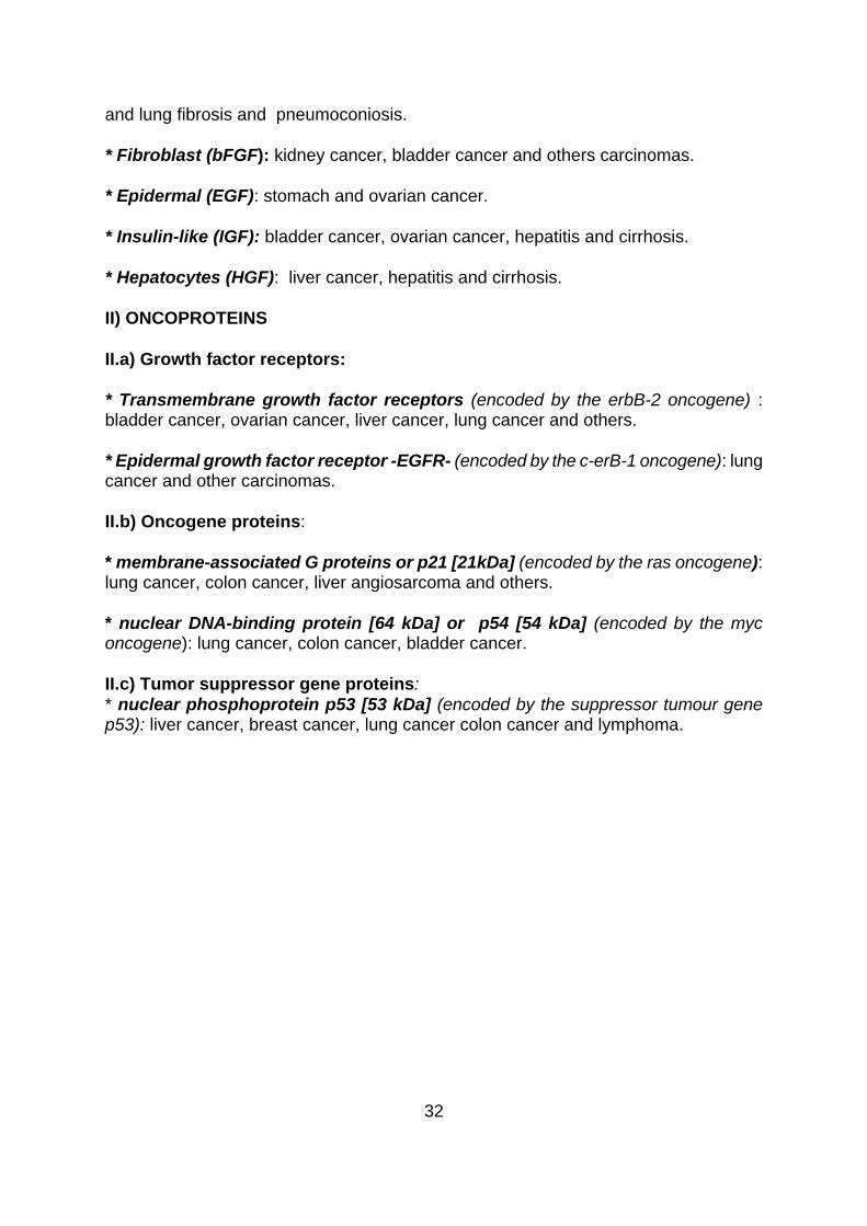
32
and lung fibrosis and pneumoconiosis. * Fibroblast (bFGF): kidney cancer, bladder cancer and others carcinomas. * Epidermal (EGF): stomach and ovarian cancer. * Insulin-like (IGF): bladder cancer, ovarian cancer, hepatitis and cirrhosis. * Hepatocytes (HGF): liver cancer, hepatitis and cirrhosis. II) ONCOPROTEINS II.a) Growth factor receptors: * Transmembrane growth factor receptors (encoded by the erbB-2 oncogene) : bladder cancer, ovarian cancer, liver cancer, lung cancer and others. * Epidermal growth factor receptor -EGFR- (encoded by the c-erB-1 oncogene): lung cancer and other carcinomas. II.b) Oncogene proteins: * membrane-associated G proteins or p21 [21kDa] (encoded by the ras oncogene): lung cancer, colon cancer, liver angiosarcoma and others. * nuclear DNA-binding protein [64 kDa] or p54 [54 kDa] (encoded by the myc oncogene): lung cancer, colon cancer, bladder cancer. II.c) Tumor suppressor gene proteins: * nuclear phosphoprotein p53 [53 kDa] (encoded by the suppressor tumour gene p53): liver cancer, breast cancer, lung cancer colon cancer and lymphoma.