Embed Size (px)
Citation preview

7/8/2016
1
©2014 MFMER | slide-1
Basics and Current Implementations of Ultrasound Imaging of Shear Wave
Speed and Elasticity
Shigao Chen, PhD
Mayo Clinic College of Medicine
August 2016
1©2014 MFMER | slide-2
Disclosures
• Some technologies described here have been licensed. Mayo and Dr. Chen have financial interests in the technologies.
2
©2014 MFMER | slide-3
Outline
• Significance and principle• Generation of shear waves• Detection of shear waves • Shear wave speed calculation• Sources of bias and variation
3©2014 MFMER | slide-4
1. Background and Principle of Elastography
4
©2014 MFMER | slide-5
Why Tissue Stiffness?
THE BOOK OF PROGNOSTICS:“… Such swellings as are soft, free from pain, and yield to the finger, …and are less dangerous than the others.…then, as are painful, hard, and large, indicate danger of speedy death…”
Hippocrates, 400 B.C. 5©2014 MFMER | slide-6
Inherent Contrast
Ultrasound in Medicine & Biology 24(9): 1419-1435, 1998
• Limitations of palpation: subjective; small/deep lesions
6

7/8/2016
2
©2014 MFMER | slide-7
Ultrasound Elastography
US Elastography
Strain elastography
Shear wave elastography
• Qualitative and Indirect• Operator-dependent• Insufficient for diffuse diseases
• Quantitative and direct• Higher repeatability
2sc
Shear modulus Density (1000 kg/m3)
Shear wave speed
Ultrasound Transducer
kPa0
5
10
15
20
25
7©2014 MFMER | slide-8
2. Generation of Shear Waves
8
©2014 MFMER | slide-9
A. External Vibration: Fibroscan
Shear Stiffness: μ = c2ρ
9
Image courtesy of
Dr. Meng Yin.
©2014 MFMER | slide-10
B. Single Push BeamUltrasound Transducer
10
©2014 MFMER | slide-11
11
Amplitude of Shear Waves by a Push Beam
10 15 20 25 30 35 400
2
4
6
8
x (mm)
Dm
ax(
m)
• Increase push amplitude: Mechanical Index• Increase push duration: heating
©2014 MFMER | slide-12
C. Supersonic Shear Imaging
www.supersonicimagine.com; Bercoff et al., 2004 12
7/8/2016
3
©2014 MFMER | slide-13
D. Comb-pushUltrasound Transducer
Comb-push
x (mm)S
low
tim
e (m
s)
5 10 15 20 25 30 35
5
10
15
20
25
Frequency (Hz)
Wav
e N
um
ber
(m
-1)
-500 0 500-500
0
500
2D FFT
Manduca et al., 2003 Deffieux et al., 2011
x (mm)
Slo
w t
ime
(ms)
5 10 15 20 25 30 35
5
10
15
20
25
x (mm)
Slo
w t
ime
(ms)
5 10 15 20 25 30 35
5
10
15
20
25
Directional Filter
x
13©2014 MFMER | slide-14
Comb-push (cont’d)Ultrasound Transducer
∆t
∆d
s
dc
t
2sc
Song et al., IEEE TMI 201214
©2014 MFMER | slide-15
Heterogeneous Medium
x (mm)
z (m
m)
10 20 30
10
20
30
40
50
x (mm)
z (m
m)
10 20 30
10
20
30
40
50
m/s0
1
2
3
4
5
x (mm)
10 20 30
10
20
30
40
50
m/s0
1
2
3
4
5
x (mm)
z (m
m)
10 20 30
10
20
30
40
50
m/s0
1
2
3
4
5
Song et al., IEEE TMI, 201215
©2014 MFMER | slide-16
3. Detection of Shear Waves
16
©2014 MFMER | slide-17
Plane wave imager vs. Line-by-line scanner
Plane wave imager Line-by-line scanner
d
Frame rate = /
d = 4cm -> Frame rate ≈ 20 kHz
Frame rate = ∗ /
d = 4cm, N = 128-> Frame rate ≈ 150 Hz
17©2014 MFMER | slide-18
Plane wave imager
d
Frame rate = /
d = 4cm -> Frame rate ≈ 20 kHz
Line-by-line scanner
Frame rate = ∗ /
d = 4cm, N = 128, PB = 4-> Frame rate ≈ 600 Hz
18
Plane wave imager vs. Line-by-line scanner
7/8/2016
4
©2014 MFMER | slide-19
Challenge in Shear Wave Detection
Plane wave imagers
Research platform
Less than 10% market
Line-by-line scanners
more than 90% of market
Clinical scanner
19©2014 MFMER | slide-20
Time Aligned Sequential Tracking (TAST)
Transducer
1 2 3 4 5
Vectors
20
©2014 MFMER | slide-21
Data Alignment
•••
True data sample
Interpolated data sample
Data tracking trajectory
Aligned data points
Truncated data points
Tim
e
Vector 1
Vector 2
Vector 3
Vector 4
Vector 5
Lateral dimension
TAST
•••
•••
•••
•••
21©2014 MFMER | slide-22
GE LOGIQ E9 (LE9) with CUSELE9
SSI
Diam. (mm) 16.7 10.4 6.5 4.1 2.5
LE9 (kPa) 84.7 74.7 60.1 46.6 33.7
SSI (kPa) 87.3 73.2 61.6 42.5 32.1
Song et al., IEEE UFFC 201422
©2014 MFMER | slide-23
Breast cancer study
Song et al., IEEE UFFC, 2014
Invasive mammary carcinoma Nottingham grade III of III
Normal
LE9 SSI
23©2014 MFMER | slide-24
4. Shear Wave Speed Calculation
24

7/8/2016
5
©2014 MFMER | slide-25
25
A. Time-to-Peak (TTP)
softstiff
C = inverse slope
Image courtesy of Dr. Kathy Nightingale
©2014 MFMER | slide-26
26
B. Random Sample Consensus (RANSAC)
C=inverse slope
=c2
Image courtesy of Dr. Kathy Nightingale
Wang MH et al. Ultrasound Medicine and Biology, 36(5): 802-813, 2010.
©2014 MFMER | slide-27
C. Cross CorrelationUltrasound Transducer
∆t
∆d
s
dc
t
27©2014 MFMER | slide-28
5.Sources of Bias
28
©2014 MFMER | slide-29
A. Tissue Viscosity
29• Frequency of shear wave: push duration, f number…
2sc
22
2211
22
2212
sc
Voigt model
0 200 400 6000
1
2
3
1 = 0.8 kPa, 2 = 0.8 Pa*s
Shear wave speed (m/s)
©2014 MFMER | slide-30
B. Compression
30
Barr et. al. Journal of Ultrasound in Medicine 31:895, 2012
• Probe compression, perfusion pressure
7/8/2016
6
©2014 MFMER | slide-31
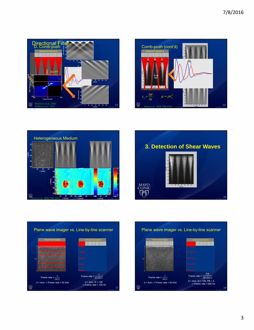
C. Shear Wave Reflections
31Deffieus et. al. IEEE Trans. UFFC, 58:2032, 2011
©2014 MFMER | slide-32
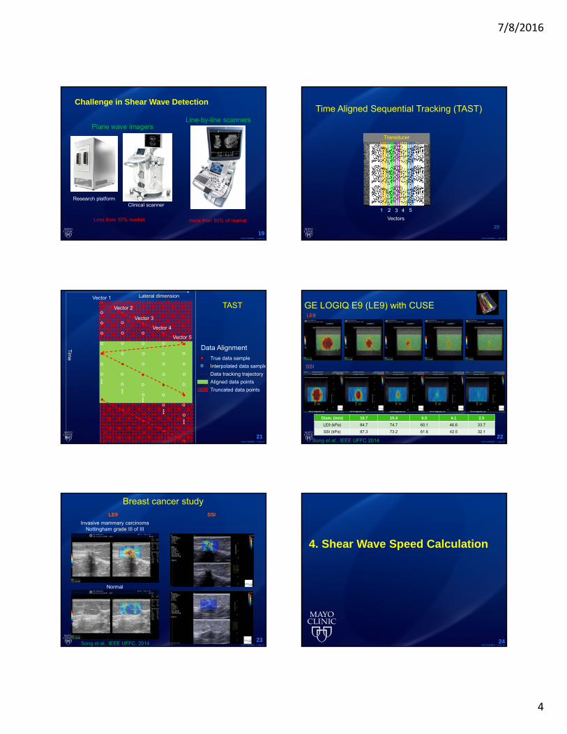
Summary
• Significance and principle• Generation of shear waves:
mechanical, single beam, Supersonic, Comb
• Detection of shear waves: TAST• Shear wave speed calculation:
Time to peak, RANSAC, correlation• Sources of bias and variation:
viscosity, compression, boundary32
©2014 MFMER | slide-33
33