Embed Size (px)
Citation preview
1
Adrenocortical carcinoma: Clinical outcomes and prognosis of 330 patients at a tertiary 1
care center 2
Running Title: Adrenocortical carcinoma outcomes and prognosis 3
Authors: Montserrat Ayala-Ramirez1*, MD; Sina Jasim
2*, MD; Lei Feng
3, MS; Shamim Ejaz
1, 4
MD; Ferhat Deniz1, MD; Naifa Busaidy
1, MD; Steven G. Waguespack
1, MD; Aung Naing
4, MD; 5
Kanishka Sircar5, MD; Christopher G. Wood
6, MD; Lance Pagliaro
7, MD; Camilo Jimenez
1, 6
MD; Rena Vassilopoulou-Sellin1, MD; Mouhammed Amir Habra
1, MD 7
1Department of Endocrine Neoplasia and Hormonal Disorders, The University of Texas MD 8
Anderson Cancer Center (UTMDACC), Houston, TX; 2 Department of Medicine, St. Louis 9
University, St. Louis, MO; 3Department of Biostatistics, UTMDACC;
4 Department of 10
Investigational Cancer Therapy, UTMDACC; 5Department of Pathology, The UTMDACC; 11
6Department of Urology, UTMDACC;
7Department of Genitourinary Medical Oncology, 12
UTMDACC; 8
Department of Investigational Cancer Therapy, UTMDACC. 13
Corresponding Authors: 14
Mouhammed Amir Habra and Rena Vassilopoulou-Sellin, MD 15
Department of Endocrine Neoplasia and Hormonal Disorders, Unit 1461 16
The University of Texas MD Anderson Cancer Center 17
1515 Holcombe Boulevard 18
Houston, Texas 77030 19
E-mail: [email protected], [email protected] 20
Phone: (713) 792-2841; Fax: (713) 794-4065 21
Word Count: 3299 22
Page 1 of 29 Accepted Preprint first posted on 1 October 2013 as Manuscript EJE-13-0519
Copyright © 2013 European Society of Endocrinology.
Accepted Preprint first posted on 1 October 2013 as Manuscript EJE-13-0519
Copyright © 2013 European Society of Endocrinology.
2
Number of Tables: 5 23
Number of Figures: 4 24
Précis for the table of contents: Adrenocortical carcinoma remains a lethal malignancy and 25
recent major breakthroughs in understanding tumor biology did not alter the course of this 26
disease. Factors such as age, hormonal overproduction, stage, and treatment outside major 27
referral centers have repeatedly been shown to contribute to reduce chances of survival. 28
Keywords: Adrenocortical Carcinoma, Survival, Recurrence, secondary malignancies 29
Financial disclosures: None 30
*Both authors have contributed equally to this paper and should be acknowledged as first 31
authors. 32
33
34
35
36
37
ABSTRACT 38
Objective: Adrenocortical carcinoma (ACC) is a rare malignancy with a poor prognosis. Herein, 39
we describe the clinical features and outcomes for a large series of ACC patients. 40
Page 2 of 29
3
Design and Methods: Retrospective review of ACC patients seen at The University of Texas 41
MD Anderson Cancer Center from 1998 through 2011. 42
Results: 330 patients with median age at diagnosis of 48.5 years; 12 (3.6%) patients were under 43
18 years. Hormonally functioning tumors represented 41.8% (n=138) of all cases. Surgical 44
resection for the primary tumor was done in 275 (83.3%) patients [45 at MD Anderson (16.4%)]. 45
For those who had surgical resection, the median local-recurrence-free time was 1.04 years. 46
Factors associated with local recurrence included positive surgical margins (P= 0.007) and 47
advanced disease stage (P=0.026). Median overall survival time for all patients was 3.21 years. 48
Median survival times were 24.1, 6.08, 3.47, and 0.89 years for stages I, II, III, and IV, 49
respectively. In multivariable analysis, older age, functioning tumors, and higher disease stage 50
remained significant prognostic factors associated with poor survival. 51
Conclusion: ACC prognosis remains poor with the use of currently available treatments. Older 52
age, functioning tumors, and incomplete resections are clinical factors associated with poor 53
survival. Surgical expertise is important to achieve complete resections and to improve outcome. 54
55
56
57
58
INTRODUCTION 59
Page 3 of 29
4
Adrenocortical carcinoma (ACC) is a rare and aggressive malignancy with an estimated annual 60
incidence of about 2 cases per million people 1-3
. Most of our knowledge about ACC is derived 61
from case series that reflected national databases and tertiary referral centers experience 4-8
. 62
Since the first case series of malignant suprarenal tumors was described by Otto Ramsay in 63
18999, many milestones have been reached in the treatment and management of ACC, including 64
the discovery of cortisone in the 1940s10
and the introduction of mitotane in the 1960s11
. In the 65
past few decades, better characterization of the molecular alterations that may occur in these 66
tumors (TP53 mutation found in Li-Fraumeni syndrome12
, the APC and CTNNB1 genes in 67
familial adenomatous polyposis coli13, 14
, and the CDKN1C15
and IGF-216, 17
genes associated with 68
Beckwith-Wiedemann syndrome) have led to the proposal of therapeutic interventions and 69
prognostic markers in ACC18-20
. Since the publication of the last MDACC case series5, multiple 70
important developments have occurred, including the introduction of two staging systems (by the 71
International Union Against Cancer in 200421
and by the European Network for the Study of 72
Adrenal Tumors [ENSAT] in 200922, 23
and the completion of the first phase III clinical trial in 73
ACC24
. 74
Despite these promising developments, the estimated 5-year overall survival rate for ACC 75
patients remains poor at 15-44%7, 25-27
. In this study, we summarize important clinical features of 76
a large cohort of ACC patients to assess outcomes and treatment utilization in ACC over the past 77
decade, including factors affecting prognosis. We also compared those findings with earlier 78
reports from the same institution 4, 5, 25
. Finally, we presented our current algorithms that we use 79
to manage patients with ACC. 80
81
Page 4 of 29
5
MATERIALS AND METHODS 82
Patient records and study design 83
With the approval of the institutional review board, we retrospectively reviewed the data for 84
ACC patients from the Tumor Registry Database of the Department of Medical Informatics at 85
The University of Texas MD Anderson Cancer Center from 1998 through 2011. We selected 86
1998 as our previous series was published in 2001 and included patients from 1980-19975. 87
To ensure the accuracy of this retrospective analysis, data were extracted and entered into 88
duplicate datasets by two independent groups of investigators (S.J, S.E, F.D, and M.A.H). Data 89
fields for demographics, clinical outcomes, laboratory tests, imaging, pathologic diagnosis, and 90
treatments were subsequently reviewed, verified, and reconciled into one database using 91
Microsoft SQL Server version 2008 (Microsoft Corporation, Redmond, WA). It is our standard 92
practice to confirm the diagnosis of ACC upon referral to MD Anderson for those patients who 93
had outside surgery or biopsy prior to referral. The reporting of Weiss score and other markers 94
of cell proliferation was not routinely performed and thus were not included in this report. 95
Functional status of the tumor was determined through documentation in the medical records of 96
cortisol, aldosterone, and/or androgen hypersecretion. Overall survival time was calculated from 97
the date of tissue diagnosis to the date of death or to the last follow-up date. Time to local 98
recurrence was calculated from the date of first surgical intervention. Patients who died without 99
local recurrence were censored at the date of death. Patients were censored at the last follow-up 100
if local recurrence or death had not occurred. 101
Resection margins were determined by reviewing pathology, operative reports, and perioperative 102
records and defined as follows: R0, no evidence of tumor; R1, microscopically positive resection 103
margins; R2, macroscopic residual disease; RX, status of resection margins is unknown. 104
Page 5 of 29
6
We used the ENSAT staging classification because of its better prognostic accuracy when 105
compared to the International Union Against Cancer staging classification for adrenocortical 106
carcinoma22, 23
. The ENSAT staging system defines stage I as ACC measuring ≤5 cm in greatest 107
dimension confined to the adrenal gland, stage II as tumor >5 cm without extra-adrenal invasion, 108
stage III by the presence of positive lymph nodes, infiltration of surrounding tissue, or vascular 109
tumor extension, while stage IV includes only patients with distant metastases22
. This staging 110
system has been in use in our institution since 1995 as proposed by Lee et al 28
and was done at 111
the time of operation for the 275 patients who underwent surgery and at the time of diagnosis for 112
the 55 patients who did not have resection of the primary tumor. 113
We also compared patients’ characteristics and outcomes from this cohort with those reported 114
previously from the same institution. As we did not have the original datasets for patients 115
reported before 1998, we defined disease burden as follows: local (stage I and stage II), regional 116
(stage III), and distant (stage IV) to facilitate comparing the results of different series. 117
118
Statistical Analyses 119
Frequencies and percentages were reported for categorical variables. Fisher’s exact test or Chi-120
square test was used to evaluate association between two categorical variables. The Wilcoxon 121
rank-sum test was used to evaluate the difference in the distribution of continuous variables 122
between patient groups. The Kaplan-Meier method was used to analyze time-to-event endpoints, 123
including overall survival and time to local recurrence. The log-rank test was used to evaluate 124
differences in these endpoints between patient groups. Multivariable Cox proportional hazards 125
models were fitted to include important demographic and clinical variables. All tests were two-126
sided. P-values less than 0.05 were considered statistically significant. Statistical software 127
Page 6 of 29
7
packages SAS 9.1.3 (SAS Institute, Cary, NC) and S-Plus 8.0 (TIBCO Software Inc., Palo Alto, 128
CA) were used for all analyses. 129
RESULTS 130
Patients’ characteristics: 330 patients with ACC were included in the current study. Most were 131
Caucasian (n= 281, 85%) and female (n= 212, 64.2%). The median age was 48.5 years (range, 0-132
86 years); 12 patients (3.6%) were under the age of 18 years. ACC was associated with hormonal 133
overproduction in 138 patients (41.8%) as follows: cortisol overproduction 76 patients (55.1%), 134
aldosterone overproduction14 patients (10.1 %), androgens overproduction 21 patients (15.2 %), 135
and overproduction of more than one hormone in 27 patients (19.6 %). Median tumor size was 136
11 cm (range, 1-27 cm) and median tumor weight was 308 grams (range, 4-3500 grams). There 137
were no cases of bilateral ACC in our cohort. Table 1 summarizes the important clinical features 138
of this cohort and Table 2 describes patients’ characteristics and outcomes in this study 139
compared with those reported previously from the same institution. 140
Associated malignancies and hereditary syndromes 141
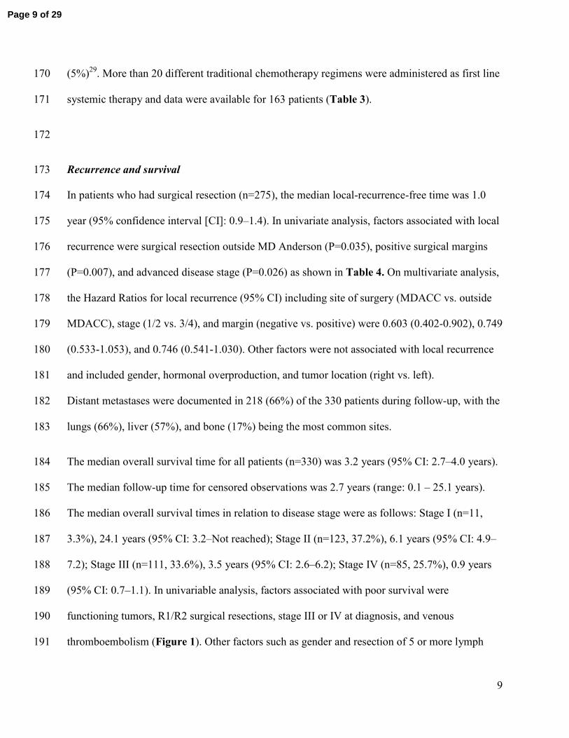
Of the patients without hereditary cancer syndromes, 38 out of the 330 patients (11.5%), had 142
other malignancies before or after their diagnosis with ACC. Breast and prostate cancers were 143
the most common [7 patients with breast cancer (18.4%) and 7 patients with prostate cancer 144
(18.4%)]. Other malignancies were skin cancer (4 patients, 10.5%); non-small cell lung cancer (4 145
patients, 10.5%), endometrial carcinoma (3 patients, 7.9%), papillary thyroid cancer (2 patients, 146
5.2%), renal cell carcinoma (2 patients, 5.2%), melanoma (2 patients, 5.2%), bladder cancer (1 147
patient, 2.6%), colorectal carcinoma (1 patient, 2.6%), cervical cancer (1 patient, 2.6%), ovarian 148
Page 7 of 29
8
carcinoma (1 patient, 2.6%), acute lymphoblastic leukemia (1 patient, 2.6%), and malignant 149
tumors of undetermined etiology (2 patients, 5.2%). 150
Six patients had Li-Fraumeni syndrome (diagnosed clinically or through genetic testing), one had 151
multiple endocrine neoplasia type 1, and another patient had a familial history of ACC. No 152
patients had Beckwith-Wiedemann syndrome or familial adenomatous polyposis. 153
Treatment Utilization: 154
Resection of the primary tumor was performed in 275 (83.3%) patients (n=45 [16.4%] at MD 155
Anderson and n=230 [83.6%] outside MD Anderson). Open resection was performed in 244 156
(88.7%) patients and laparoscopic in 31 (11.2%) patients. Negative resection margins (R0) were 157
achieved in 153 patients (55.6%). Positive margins (R1) were found in 47 patients (17.1%) and 158
(R2) resection margins were found in 28 patients (10.2%). Margin status was unknown (RX) in 159
47 patients (17.1%). 160
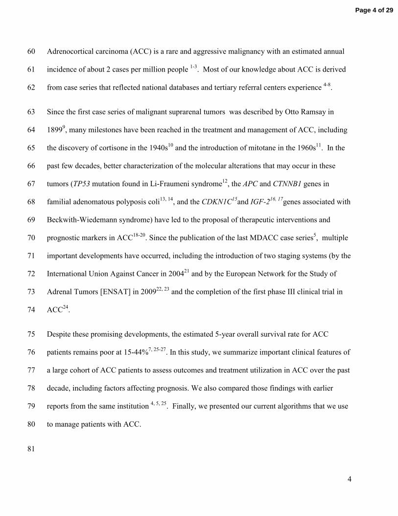
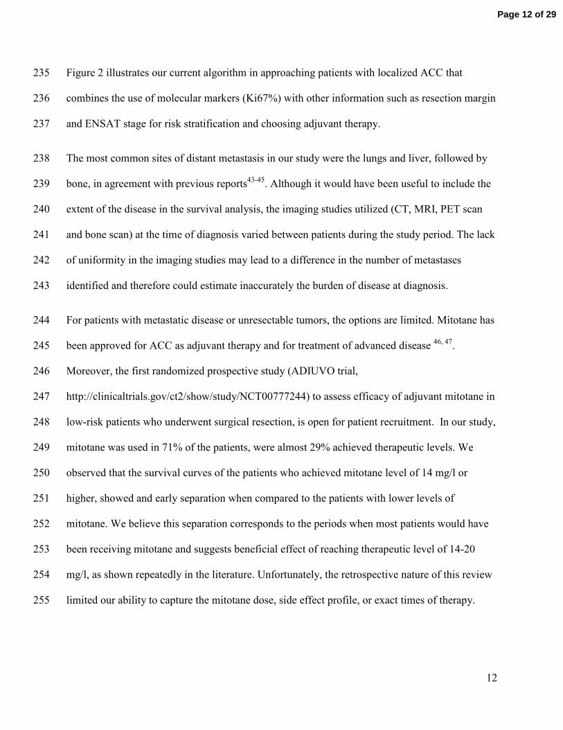
Mitotane was used in 235 (71.2%) of the 330 patients either as monotherapy or in combination 161
with other systemic chemotherapy. Of these, 67 (28.5%) achieved serum mitotane level of 14 162
mg/l or higher. The median OS for those patients with levels of 14 mg/l and higher was 4.1 years 163
(95% CI 2.8 , 7.0 years ) compared with 2.9 years (95% CI 2.2 , 3.8 years) for those who had 164
lower mitotane levels. At 5-years after diagnosis, overall survival was 44% (95% CI 32%-60%)3 165
in patients with mitotane levels 14 mg/l or higher compared with 45% (95% CI 27%-44%) in 166
those with lower mitotane levels. Figure 4 illustrates overall survival curves in both groups. 167
Radiation therapy was used in 58 patients (18%) mostly as a palliative measure in metastatic 168
disease while adjuvant radiation therapy after primary resection was used only in 16 patients 169
Page 8 of 29
9
(5%)29
. More than 20 different traditional chemotherapy regimens were administered as first line 170
systemic therapy and data were available for 163 patients (Table 3). 171
172
Recurrence and survival 173
In patients who had surgical resection (n=275), the median local-recurrence-free time was 1.0 174
year (95% confidence interval [CI]: 0.9–1.4). In univariate analysis, factors associated with local 175
recurrence were surgical resection outside MD Anderson (P=0.035), positive surgical margins 176
(P=0.007), and advanced disease stage (P=0.026) as shown in Table 4. On multivariate analysis, 177
the Hazard Ratios for local recurrence (95% CI) including site of surgery (MDACC vs. outside 178
MDACC), stage (1/2 vs. 3/4), and margin (negative vs. positive) were 0.603 (0.402-0.902), 0.749 179
(0.533-1.053), and 0.746 (0.541-1.030). Other factors were not associated with local recurrence 180
and included gender, hormonal overproduction, and tumor location (right vs. left). 181
Distant metastases were documented in 218 (66%) of the 330 patients during follow-up, with the 182
lungs (66%), liver (57%), and bone (17%) being the most common sites. 183
The median overall survival time for all patients (n=330) was 3.2 years (95% CI: 2.7–4.0 years). 184
The median follow-up time for censored observations was 2.7 years (range: 0.1 – 25.1 years). 185
The median overall survival times in relation to disease stage were as follows: Stage I (n=11, 186
3.3%), 24.1 years (95% CI: 3.2–Not reached); Stage II (n=123, 37.2%), 6.1 years (95% CI: 4.9–187
7.2); Stage III (n=111, 33.6%), 3.5 years (95% CI: 2.6–6.2); Stage IV (n=85, 25.7%), 0.9 years 188
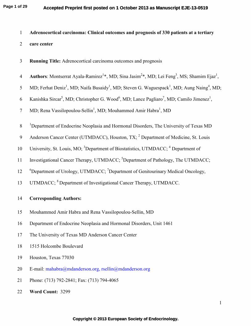
(95% CI: 0.7–1.1). In univariable analysis, factors associated with poor survival were 189
functioning tumors, R1/R2 surgical resections, stage III or IV at diagnosis, and venous 190
thromboembolism (Figure 1). Other factors such as gender and resection of 5 or more lymph 191
Page 9 of 29
10
nodes at time of initial surgery were not associated with overall survival. Older age, functioning 192
tumors, and high disease stage at diagnosis remained significant prognostic factors associated 193
with poor survival in a multivariable Cox proportional hazards model (Table 5). 194
DISCUSSION 195
The current study investigates a large number of patients with ACC treated at a single institution. 196
Compared with previous reports from our institution obtained during different time periods, 197
patients in the current study tended to be diagnosed at an older age, have more localized tumors 198
and more functioning tumors. Our cohort’s 5-year survival rate of 38% is similar to those 199
reported by others6, 30-32
although we noticed a slightly lower 5-year survival rate than those 200
reported from the same institution in the past. As we did not review the source documents from 201
the older series, direct comparisons could not be done to assess if these differences are truly 202
statistically significant. In addition, the variability in methodology may explain some of the 203
differences in retrospectively collected data. The variation is likely small for certain outcomes 204
(such as age, gender, date of diagnosis, death, and treatments received) but may be significant for 205
other variables (such as performance status and certain operative complications)33-35
. In this 206
large cohort, patients were older than patients reported in previous reports from our institution 207
and had higher percentage of localized disease and functioning tumors at the time of diagnosis. It 208
remains unclear if these differences are due to increased detection of tumors at an earlier stage or 209
a change in referral pattern. The presence of functioning tumors could lead to distinct clinical 210
manifestations associated with excessive hormonal production and prompts further imaging 211
studies. The theoretical benefit of earlier detection of functioning tumors is likely negated by the 212
increased morbidity associated hormonally active tumors compared to non functioning tumors. 213
Page 10 of 29
11
Surgery remains the treatment of choice for ACC 32, 36, 37
, as it is the only therapeutic approach 214
that can be curative for localized disease. In our study, complete resections of primary tumors 215
were associated with both decreased disease recurrence and better overall survival36
, in 216
agreement with findings reported in previous studies7, 26, 31
. Median time to recurrence was about 217
1 year. Factors associated with local recurrence, other than incomplete resection, were resection 218
performed outside of MD Anderson and advanced disease stage at diagnosis. In addition, we 219
have recently described higher recurrence rates (especially peritoneal carcinomatosis) with 220
laparoscopic resection when compared to open resection38
. The improved survival in patients 221
operated on at MD Anderson is in line of literature from Europe that suggested improved 222
outcome in ACC patients who received their care in referral centers known for their expertise in 223
ACC39
. Adequate pre-operative imaging is crucial to plan initial surgical treatment as well as 224
subsequent adjuvant therapy and should include imaging of the chest, abdomen, and pelvis. In 225
the past few years, there has been an increasing use of markers of cell proliferation (such as 226
Ki67%) as prognostic markers to help with treatment decisions especially after primary tumor 227
resection40-42
. In our study, almost 83% of the patients had their initial surgery outside our 228
institution and this referral pattern resulted in lack of consistency in reporting proliferation 229
markers (including Ki67%) and Weiss score. The absence of this information is a shortcoming of 230
our study and similar large cohort studies published in the past decade 5 7, 8, 22
. This limitation 231
reinforces the need to have standardized pathological template that would contain key 232
pathological features (e.g. Weiss score, Ki-67%, resection margins) to facilitate a uniform 233
interpretation and generalization of pathological data in ACC. 234
Page 11 of 29
12
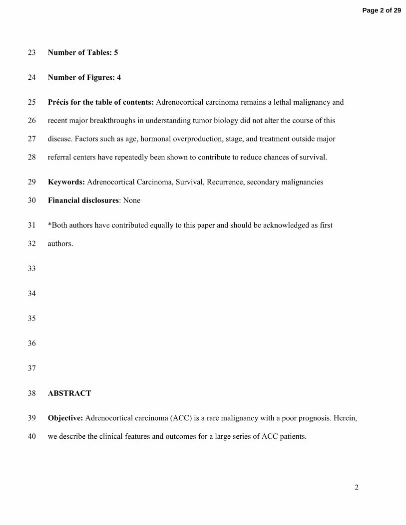
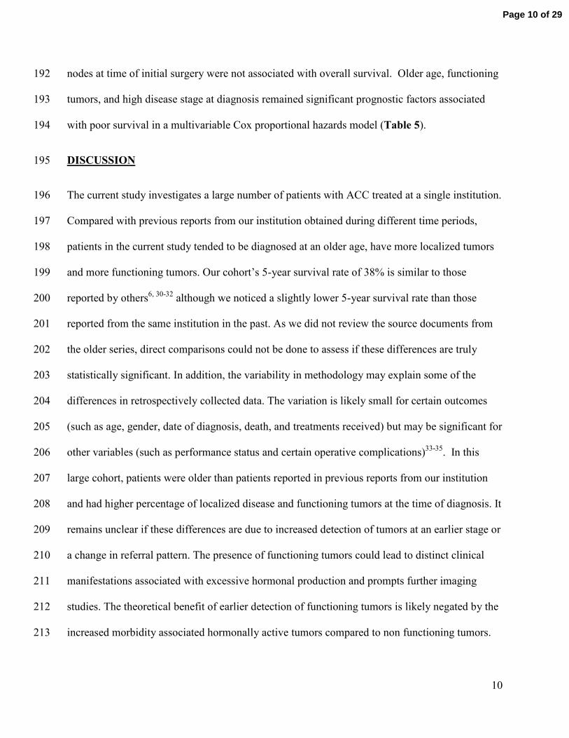
Figure 2 illustrates our current algorithm in approaching patients with localized ACC that 235
combines the use of molecular markers (Ki67%) with other information such as resection margin 236
and ENSAT stage for risk stratification and choosing adjuvant therapy. 237
The most common sites of distant metastasis in our study were the lungs and liver, followed by 238
bone, in agreement with previous reports43-45
. Although it would have been useful to include the 239
extent of the disease in the survival analysis, the imaging studies utilized (CT, MRI, PET scan 240
and bone scan) at the time of diagnosis varied between patients during the study period. The lack 241
of uniformity in the imaging studies may lead to a difference in the number of metastases 242
identified and therefore could estimate inaccurately the burden of disease at diagnosis. 243
For patients with metastatic disease or unresectable tumors, the options are limited. Mitotane has 244
been approved for ACC as adjuvant therapy and for treatment of advanced disease 46, 47
. 245
Moreover, the first randomized prospective study (ADIUVO trial, 246
http://clinicaltrials.gov/ct2/show/study/NCT00777244) to assess efficacy of adjuvant mitotane in 247
low-risk patients who underwent surgical resection, is open for patient recruitment. In our study, 248
mitotane was used in 71% of the patients, were almost 29% achieved therapeutic levels. We 249
observed that the survival curves of the patients who achieved mitotane level of 14 mg/l or 250
higher, showed and early separation when compared to the patients with lower levels of 251
mitotane. We believe this separation corresponds to the periods when most patients would have 252
been receiving mitotane and suggests beneficial effect of reaching therapeutic level of 14-20 253
mg/l, as shown repeatedly in the literature. Unfortunately, the retrospective nature of this review 254
limited our ability to capture the mitotane dose, side effect profile, or exact times of therapy. 255
Page 12 of 29
13
Currently used systemic therapies often combine mitotane with systemic agents. In the only 256
completed phase III trial of ACC, the combination of etoposide, doxorubicin, and cisplatin with 257
mitotane was superior to the combination of streptozocin with mitotane in terms of progression-258
free survival (5 months versus 2.1 months); however, the two groups did not differ significantly 259
in overall survival24
. In our cohort, cisplatin/etoposide and etoposide/doxorubicin/cisplatin 260
regimens were the most common. In a previous study from our group, neither regimen conferred 261
a significant advantage 48
. Therefore, there is an urgent need for more efficacious treatment for 262
this lethal disease. In fact, we have recently published our experience of dual inhibition of the 263
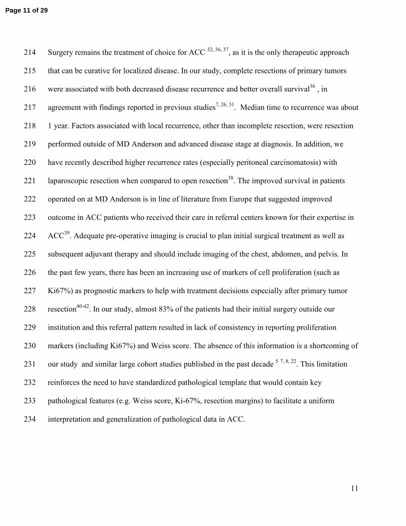
IGF1 receptor and mTOR pathway, were stable disease was achieved for more than 6 months in 264
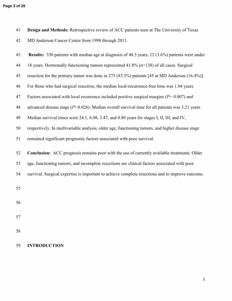
42% of the patients 49
. Figure 3 illustrates our current management plan for patients with 265
advanced/metastatic ACC. 266
The use of adjuvant radiotherapy in the context of ACC remains unclear. While some authors 267
have proposed a decreased in local recurrences after adjuvant radiotherapy48, 50
; in a recent study 268
published by our group that included the 16 patients who received adjuvant radiotherapy, we 269
were unable to demonstrate improved survival, recurrence rate or time to recurrence.29
Further 270
prospective, multicenter study is needed to better determine the impact of radiotherapy on 271
recurrence and survival. 272
ACC in children is extremely rare, with an estimated incidence of 19 new cases per year in the 273
United States51
. Carriers of TP53 mutations and some genetic syndromes are conditions that have 274
been associated with ACC in children. In fact, it is estimated that about 50-80% of children with 275
ACC carry a germline TP53 mutation52, 53
, making Li-Fraumeni syndrome the most common 276
inherited condition in young patients with ACC. In our cohort, 12 patients (3.6%) were younger 277
Page 13 of 29
14
than 18 years old at the age of diagnosis, and 50% of the pediatric patients (6 of 12) had Li-278
Fraumeni syndrome based upon clinical grounds or genetic testing. 279
Similar to older reports25, 54
, approximately one out of ten patients with non-hereditary ACC 280
patients had other malignancies, with breast and prostate cancer being the most frequent ones. 281
Despite this apparent risk for other malignancies, we only recommend age- appropriate cancer 282
screening that is adjusted per personal and family history in the few patients who have long-term 283
survival. 284
This study was limited by the inherent shortcomings of retrospective reviews and potential 285
referral bias. Referral bias is likely more pronounced in rare diseases that require special 286
expertise compared to more common illnesses that require well set standards of care and 287
commonly available treatments55
. The main factors to cause this bias include the tendency to 288
refer patients if they have unusual presentation, after failing prior treatments, and/or if they have 289
advanced disease that requires special expertise. In ACC, referral bias is expected to result in 290
worse outcome of the whole group if most included subjects were referred after failing prior 291
treatments. 292
Also, it is unknown how many patients are treated outside major centers and never referred to be 293
included in such study design. Some of these patients may have been cured and did not require 294
further treatment or they may have accepted their diagnosis as being terminal and chose to stay 295
in their local communities56
. In our series, most of the patients had their initial surgical resection 296
outside MDACC and then referred and it was often difficult to ascertain the cause of referral in 297
all cases or the temporal relationship between recurrence and referral. 298
Page 14 of 29
15
Nevertheless, the current study’s has multiple strengths including the summary of clinical 299
experience with a large cohort of ACC patients treated at a single institution over the past decade 300
and description our treatment approach in this rare disease. 301
The unchanged mortality observed over the decades in our cohort and that has been also 302
described by a recent population study done in the Netherlands8 underscores the urgency to find 303
better treatments for ACC. 304
Conclusions 305
Despite better understanding of molecular pathways involved in ACC and the availability of new 306
classes of anti-cancer therapy, the prognosis of ACC remains poor. Older age at diagnosis, 307
functioning tumors, and incomplete resections are clinical factors associated with worse survival. 308
Surgical expertise is important to achieve complete resections and to improve outcome. There is 309
an urgent need for more efficacious systemic treatments than what is currently used, as distant 310
recurrence and ultimate death is very common despite the best efforts at locoregional control of 311
disease. 312
313
314
315
316
317
318
319
320
321
Page 15 of 29
16
Funding Sources: This paper is supported in part by the National Institutes of Health through 322
The University of Texas MD Anderson Cancer Center Support Grant, CA016672, and The 323
Beverlin Fund for Adrenal Cancer Research 324
CONFLICT OF INTEREST DISCLOSURES 325
The authors made no disclosures 326
327
328
329
References 330
331
1. Gerhardt PR, Handy VH & Ferber B. Trends in cancer incidence, mortality, and probability in the 332 state of New York. N Y State J Med 1957 57 1387-1390. 333
2. Griswold MH & Cutler SJ. The Connecticut cancer register. Seventeen years of experience. 1956. 334 Conn Med 2006 70 323-328. 335
3. Soreide JA, Brabrand K & Thoresen SO. Adrenal cortical carcinoma in Norway, 1970-1984. World 336 J Surg 1992 16 663-667; discussion 668. 337
4. Nader S, Hickey RC, Sellin RV & Samaan NA. Adrenal cortical carcinoma. A study of 77 cases. 338 Cancer 1983 52 707-711. 339
5. Vassilopoulou-Sellin R & Schultz PN. Adrenocortical carcinoma. Clinical outcome at the end of 340 the 20th century. Cancer 2001 92 1113-1121. 341
6. Abiven G, Coste J, Groussin L, Anract P, Tissier F, Legmann P, Dousset B, Bertagna X & Bertherat 342 J. Clinical and biological features in the prognosis of adrenocortical cancer: poor outcome of 343 cortisol-secreting tumors in a series of 202 consecutive patients. J Clin Endocrinol Metab 2006 344 91 2650-2655. 345
7. Icard P, Goudet P, Charpenay C, Andreassian B, Carnaille B, Chapuis Y, Cougard P, Henry JF & 346 Proye C. Adrenocortical carcinomas: surgical trends and results of a 253-patient series from the 347 French Association of Endocrine Surgeons study group. World J Surg 2001 25 891-897. 348
8. Kerkhofs TM, Verhoeven RH, Van der Zwan JM, Dieleman J, Kerstens MN, Links TP, Van de Poll-349 Franse LV & Haak HR. Adrenocortical carcinoma: A population-based study on incidence and 350 survival in the Netherlands since 1993. Eur J Cancer 2013. 351
9. Ramsay O. Malignant tumors of the suprarrenal glands. Johns Hopkins Hosp Bull 1899 94-96 20-352 29. 353
10. Hench PS, Slocumb CH & et al. The effects of the adrenal cortical hormone 17-hydroxy-11-354 dehydrocorticosterone (Compound E) on the acute phase of rheumatic fever; preliminary 355 report. Proc Staff Meet Mayo Clin 1949 24 277-297. 356
Page 16 of 29
17
11. Hutter AM, Jr. & Kayhoe DE. Adrenal cortical carcinoma. Results of treatment with o,p'DDD in 357 138 patients. Am J Med 1966 41 581-592. 358
12. Varley JM, McGown G, Thorncroft M, Cochrane S, Morrison P, Woll P, Kelsey AM, Mitchell EL, 359 Boyle J, Birch JM & Evans DG. A previously undescribed mutation within the tetramerisation 360 domain of TP53 in a family with Li-Fraumeni syndrome. Oncogene 1996 12 2437-2442. 361
13. Groden J, Thliveris A, Samowitz W, Carlson M, Gelbert L, Albertsen H, Joslyn G, Stevens J, Spirio 362 L, Robertson M & et al. Identification and characterization of the familial adenomatous polyposis 363 coli gene. Cell 1991 66 589-600. 364
14. Tissier F, Cavard C, Groussin L, Perlemoine K, Fumey G, Hagnere AM, Rene-Corail F, Jullian E, 365 Gicquel C, Bertagna X, Vacher-Lavenu MC, Perret C & Bertherat J. Mutations of beta-catenin in 366 adrenocortical tumors: activation of the Wnt signaling pathway is a frequent event in both 367 benign and malignant adrenocortical tumors. Cancer Res 2005 65 7622-7627. 368
15. Hatada I, Ohashi H, Fukushima Y, Kaneko Y, Inoue M, Komoto Y, Okada A, Ohishi S, Nabetani A, 369 Morisaki H, Nakayama M, Niikawa N & Mukai T. An imprinted gene p57KIP2 is mutated in 370 Beckwith-Wiedemann syndrome. Nat Genet 1996 14 171-173. 371
16. Gicquel C, Raffin-Sanson ML, Gaston V, Bertagna X, Plouin PF, Schlumberger M, Louvel A, Luton 372 JP & Le Bouc Y. Structural and functional abnormalities at 11p15 are associated with the 373 malignant phenotype in sporadic adrenocortical tumors: study on a series of 82 tumors. J Clin 374 Endocrinol Metab 1997 82 2559-2565. 375
17. Weksberg R, Shen DR, Fei YL, Song QL & Squire J. Disruption of insulin-like growth factor 2 376 imprinting in Beckwith-Wiedemann syndrome. Nat Genet 1993 5 143-150. 377
18. Heaton JH, Wood MA, Kim AC, Lima LO, Barlaskar FM, Almeida MQ, Fragoso MC, Kuick R, Lerario 378 AM, Simon DP, Soares IC, Starnes E, Thomas DG, Latronico AC, Giordano TJ & Hammer GD. 379 Progression to adrenocortical tumorigenesis in mice and humans through insulin-like growth 380 factor 2 and beta-catenin. Am J Pathol 2012 181 1017-1033. 381
19. Soon PS, Gill AJ, Benn DE, Clarkson A, Robinson BG, McDonald KL & Sidhu SB. Microarray gene 382 expression and immunohistochemistry analyses of adrenocortical tumors identify IGF2 and Ki-67 383 as useful in differentiating carcinomas from adenomas. Endocr Relat Cancer 2009 16 573-583. 384
20. Ozata DM, Caramuta S, Velazquez-Fernandez D, Akcakaya P, Xie H, Hoog A, Zedenius J, Backdahl 385 M, Larsson C & Lui WO. The role of microRNA deregulation in the pathogenesis of adrenocortical 386 carcinoma. Endocr Relat Cancer 2011 18 643-655. 387
21. DeLellis RA LR, Heitz PU, Eng C. Pathology and genetics of tumours of endocrine 388
organs. World Health Organization Classification of Tumours. IARC Press, 2004. 389 22. Fassnacht M, Johanssen S, Quinkler M, Bucsky P, Willenberg HS, Beuschlein F, Terzolo M, 390
Mueller HH, Hahner S, Allolio B, German Adrenocortical Carcinoma Registry G & European 391 Network for the Study of Adrenal T. Limited prognostic value of the 2004 International Union 392 Against Cancer staging classification for adrenocortical carcinoma: proposal for a Revised TNM 393 Classification. Cancer 2009 115 243-250. 394
23. Lughezzani G, Sun M, Perrotte P, Jeldres C, Alasker A, Isbarn H, Budaus L, Shariat SF, Guazzoni G, 395 Montorsi F & Karakiewicz PI. The European Network for the Study of Adrenal Tumors staging 396 system is prognostically superior to the international union against cancer-staging system: a 397 North American validation. Eur J Cancer 2010 46 713-719. 398
24. Fassnacht M, Terzolo M, Allolio B, Baudin E, Haak H, Berruti A, Welin S, Schade-Brittinger C, 399 Lacroix A, Jarzab B, Sorbye H, Torpy DJ, Stepan V, Schteingart DE, Arlt W, Kroiss M, Leboulleux S, 400 Sperone P, Sundin A, Hermsen I, Hahner S, Willenberg HS, Tabarin A, Quinkler M, de la 401 Fouchardiere C, Schlumberger M, Mantero F, Weismann D, Beuschlein F, Gelderblom H, Wilmink 402 H, Sender M, Edgerly M, Kenn W, Fojo T, Muller HH, Skogseid B & Group F-AS. Combination 403 chemotherapy in advanced adrenocortical carcinoma. N Engl J Med 2012 366 2189-2197. 404
Page 17 of 29
18
25. Venkatesh S, Hickey RC, Sellin RV, Fernandez JF & Samaan NA. Adrenal cortical carcinoma. 405 Cancer 1989 64 765-769. 406
26. Pommier RF & Brennan MF. An eleven-year experience with adrenocortical carcinoma. Surgery 407 1992 112 963-970; discussion 970-961. 408
27. Schulick RD & Brennan MF. Long-term survival after complete resection and repeat resection in 409 patients with adrenocortical carcinoma. Ann Surg Oncol 1999 6 719-726. 410
28. Lee JE, Berger DH, el-Naggar AK, Hickey RC, Vassilopoulou-Sellin R, Gagel RF, Burgess MA & 411 Evans DB. Surgical management, DNA content, and patient survival in adrenal cortical 412 carcinoma. Surgery 1995 118 1090-1098. 413
29. Habra MA, Ejaz S, Feng L, Das P, Deniz F, Grubbs EG, Phan A, Waguespack SG, Ayala-Ramirez M, 414 Jimenez C, Perrier ND, Lee JE & Vassilopoulou-Sellin R. A retrospective cohort analysis of the 415 efficacy of adjuvant radiotherapy after primary surgical resection in patients with adrenocortical 416 carcinoma. J Clin Endocrinol Metab 2013 98 192-197. 417
30. Bilimoria KY, Shen WT, Elaraj D, Bentrem DJ, Winchester DJ, Kebebew E & Sturgeon C. 418 Adrenocortical carcinoma in the United States: treatment utilization and prognostic factors. 419 Cancer 2008 113 3130-3136. 420
31. Paton BL, Novitsky YW, Zerey M, Harrell AG, Norton HJ, Asbun H, Kercher KW & Heniford BT. 421 Outcomes of adrenal cortical carcinoma in the United States. Surgery 2006 140 914-920; 422 discussion 919-920. 423
32. Kendrick ML, Lloyd R, Erickson L, Farley DR, Grant CS, Thompson GB, Rowland C, Young WF, Jr. & 424 van Heerden JA. Adrenocortical carcinoma: surgical progress or status quo? Arch Surg 2001 136 425 543-549. 426
33. Shiloach M, Frencher SK, Jr., Steeger JE, Rowell KS, Bartzokis K, Tomeh MG, Richards KE, Ko CY & 427 Hall BL. Toward robust information: data quality and inter-rater reliability in the American 428 College of Surgeons National Surgical Quality Improvement Program. J Am Coll Surg 2010 210 6-429 16. 430
34. Hutter MM, Rowell KS, Devaney LA, Sokal SM, Warshaw AL, Abbott WM & Hodin RA. 431 Identification of surgical complications and deaths: an assessment of the traditional surgical 432 morbidity and mortality conference compared with the American College of Surgeons-National 433 Surgical Quality Improvement Program. J Am Coll Surg 2006 203 618-624. 434
35. Beard CM, Yunginger JW, Reed CE, O'Connell EJ & Silverstein MD. Interobserver variability in 435 medical record review: an epidemiological study of asthma. J Clin Epidemiol 1992 45 1013-1020. 436
36. Grubbs EG, Callender GG, Xing Y, Perrier ND, Evans DB, Phan AT & Lee JE. Recurrence of adrenal 437 cortical carcinoma following resection: surgery alone can achieve results equal to surgery plus 438 mitotane. Ann Surg Oncol 2010 17 263-270. 439
37. Schteingart DE, Doherty GM, Gauger PG, Giordano TJ, Hammer GD, Korobkin M & Worden FP. 440 Management of patients with adrenal cancer: recommendations of an international consensus 441 conference. Endocr Relat Cancer 2005 12 667-680. 442
38. Cooper AB, Habra MA, Grubbs EG, Bednarski BK, Ying AK, Perrier ND, Lee JE & Aloia TA. Does 443 laparoscopic adrenalectomy jeopardize oncologic outcomes for patients with adrenocortical 444 carcinoma? Surg Endosc 2013. 445
39. Hermsen IG, Kerkhofs TM, den Butter G, Kievit J, van Eijck CH, Nieveen van Dijkum EJ, Haak HR & 446 Dutch Adrenal N. Surgery in adrenocortical carcinoma: Importance of national cooperation and 447 centralized surgery. Surgery 2012 152 50-56. 448
40. Terzolo M, Boccuzzi A, Bovio S, Cappia S, De Giuli P, Ali A, Paccotti P, Porpiglia F, Fontana D & 449 Angeli A. Immunohistochemical assessment of Ki-67 in the differential diagnosis of 450 adrenocortical tumors. Urology 2001 57 176-182. 451
Page 18 of 29
19
41. Stojadinovic A, Ghossein RA, Hoos A, Nissan A, Marshall D, Dudas M, Cordon-Cardo C, Jaques DP 452 & Brennan MF. Adrenocortical carcinoma: clinical, morphologic, and molecular characterization. 453 J Clin Oncol 2002 20 941-950. 454
42. Morimoto R, Satoh F, Murakami O, Suzuki T, Abe T, Tanemoto M, Abe M, Uruno A, Ishidoya S, 455 Arai Y, Takahashi K, Sasano H & Ito S. Immunohistochemistry of a proliferation marker 456 Ki67/MIB1 in adrenocortical carcinomas: Ki67/MIB1 labeling index is a predictor for recurrence 457 of adrenocortical carcinomas. Endocr J 2008 55 49-55. 458
43. Datrice NM, Langan RC, Ripley RT, Kemp CD, Steinberg SM, Wood BJ, Libutti SK, Fojo T, Schrump 459 DS & Avital I. Operative management for recurrent and metastatic adrenocortical carcinoma. J 460 Surg Oncol 2012 105 709-713. 461
44. Erdogan I, Deutschbein T, Jurowich C, Kroiss M, Ronchi C, Quinkler M, Waldmann J, Willenberg 462 HS, Beuschlein F, Fottner C, Klose S, Heidemeier A, Brix D, Fenske W, Hahner S, Reibetanz J, 463 Allolio B, Fassnacht M & on behalf of the German Adrenocortical Carcinoma Study G. The Role of 464 Surgery in the Management of Recurrent Adrenocortical Carcinoma. J Clin Endocrinol Metab 465 2012. 466
45. Bellantone R, Ferrante A, Boscherini M, Lombardi CP, Crucitti P, Crucitti F, Favia G, Borrelli D, 467 Boffi L, Capussotti L, Carbone G, Casaccia M, Cavallaro A, Del Gaudio A, Dettori G, Di Giovanni V, 468 Mazziotti A, Marrano D, Masenti E, Miccoli P, Mosca F, Mussa A, Petronio R, Piat G, Marazano L 469 & et al. Role of reoperation in recurrence of adrenal cortical carcinoma: results from 188 cases 470 collected in the Italian National Registry for Adrenal Cortical Carcinoma. Surgery 1997 122 1212-471 1218. 472
46. Haak HR, Hermans J, van de Velde CJ, Lentjes EG, Goslings BM, Fleuren GJ & Krans HM. Optimal 473 treatment of adrenocortical carcinoma with mitotane: results in a consecutive series of 96 474 patients. Br J Cancer 1994 69 947-951. 475
47. Terzolo M, Angeli A, Fassnacht M, Daffara F, Tauchmanova L, Conton PA, Rossetto R, Buci L, 476 Sperone P, Grossrubatscher E, Reimondo G, Bollito E, Papotti M, Saeger W, Hahner S, Koschker 477 AC, Arvat E, Ambrosi B, Loli P, Lombardi G, Mannelli M, Bruzzi P, Mantero F, Allolio B, Dogliotti L 478 & Berruti A. Adjuvant mitotane treatment for adrenocortical carcinoma. N Engl J Med 2007 356 479 2372-2380. 480
48. Fareau GG, Lopez A, Stava C & Vassilopoulou-Sellin R. Systemic chemotherapy for adrenocortical 481 carcinoma: comparative responses to conventional first-line therapies. Anticancer Drugs 2008 482 19 637-644. 483
49. Naing A, Lorusso P, Fu S, Hong D, Chen HX, Doyle LA, Phan AT, Habra MA & Kurzrock R. Insulin 484 growth factor receptor (IGF-1R) antibody cixutumumab combined with the mTOR inhibitor 485 temsirolimus in patients with metastatic adrenocortical carcinoma. Br J Cancer 2013. 486
50. Polat B, Fassnacht M, Pfreundner L, Guckenberger M, Bratengeier K, Johanssen S, Kenn W, 487 Hahner S, Allolio B & Flentje M. Radiotherapy in adrenocortical carcinoma. Cancer 2009 115 488 2816-2823. 489
51. Lack EE, Mulvihill JJ, Travis WD & Kozakewich HP. Adrenal cortical neoplasms in the pediatric 490 and adolescent age group. Clinicopathologic study of 30 cases with emphasis on epidemiological 491 and prognostic factors. Pathol Annu 1992 27 Pt 1 1-53. 492
52. Varley JM, McGown G, Thorncroft M, James LA, Margison GP, Forster G, Evans DG, Harris M, 493 Kelsey AM & Birch JM. Are there low-penetrance TP53 Alleles? evidence from childhood 494 adrenocortical tumors. Am J Hum Genet 1999 65 995-1006. 495
53. Varley JM. Germline TP53 mutations and Li-Fraumeni syndrome. Hum Mutat 2003 21 313-320. 496 54. Didolkar MS, Bescher RA, Elias EG & Moore RH. Natural history of adrenal cortical carcinoma: a 497
clinicopathologic study of 42 patients. Cancer 1981 47 2153-2161. 498
Page 19 of 29
20
55. Layde PM, Broste SK, Desbiens N, Follen M, Lynn J, Reding D & Vidaillet H. Generalizability of 499 clinical studies conducted at tertiary care medical centers: a population-based analysis. J Clin 500 Epidemiol 1996 49 835-841. 501
56. Sutherland LR & Verhoef MJ. Patients who seek a second opinion: are they different from the 502 typical referral? J Clin Gastroenterol 1989 11 308-313. 503
504
Figure legends 505
Figure 1. Factors associated with the overall survival of patients with adrenocortical 506
carcinoma. R0, no evidence of tumor; R1, microscopically positive resection margins; R2, 507
macroscopic residual disease ENSAT, European Network for the Study of Adrenal Tumors; 508
VTE, Venous Thromboembolism 509
Figure 2. A Suggested algorithm for the management of localized adrenocortical 510
carcinoma. R0, no evidence of tumor; R1, microscopic evidence of tumor; R2, macroscopic 511
residual disease; RX, margins unknown. We recommend mitotane for 2-3 years. 512
Figure 3. An Algorithm for the management of locally advanced/metastatic adrenocortical 513
carcinoma 514
Figure 4. Overall survival (OS) in patients who achieve mitotane levels of 14 mg/l or higher 515
(YES, red line) compared with the patients with lower levels (NO, green line). 516
517
Page 20 of 29
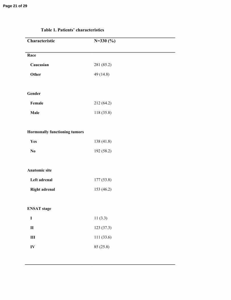
Table 1. Patients’ characteristics
Characteristic N=330 (%)
Race
Caucasian
Other
281 (85.2)
49 (14.8)
Gender
Female
Male
212 (64.2)
118 (35.8)
Hormonally functioning tumors
Yes
No
138 (41.8)
192 (58.2)
Anatomic site
Left adrenal
Right adrenal
177 (53.8)
153 (46.2)
ENSAT stage
I
II
III
IV
11 (3.3)
123 (37.3)
111 (33.6)
85 (25.8)
Page 21 of 29
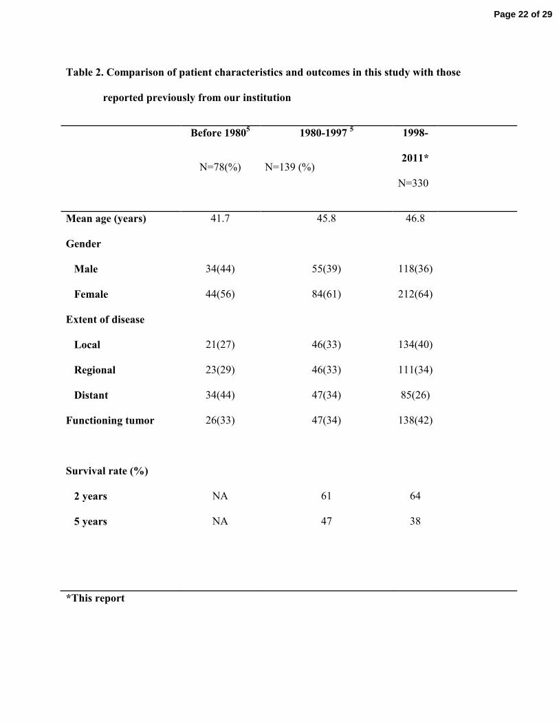
Table 2. Comparison of patient characteristics and outcomes in this study with those
reported previously from our institution
Before 19805
N=78(%)
1980-1997 5
N=139 (%)
1998-
2011*
N=330
Mean age (years) 41.7 45.8 46.8
Gender
Male 34(44) 55(39) 118(36)
Female 44(56) 84(61) 212(64)
Extent of disease
Local 21(27) 46(33) 134(40)
Regional 23(29) 46(33) 111(34)
Distant 34(44) 47(34) 85(26)
Functioning tumor 26(33) 47(34) 138(42)
Survival rate (%)
2 years
5 years
NA
NA
61
47
64
38
*This report
Page 22 of 29
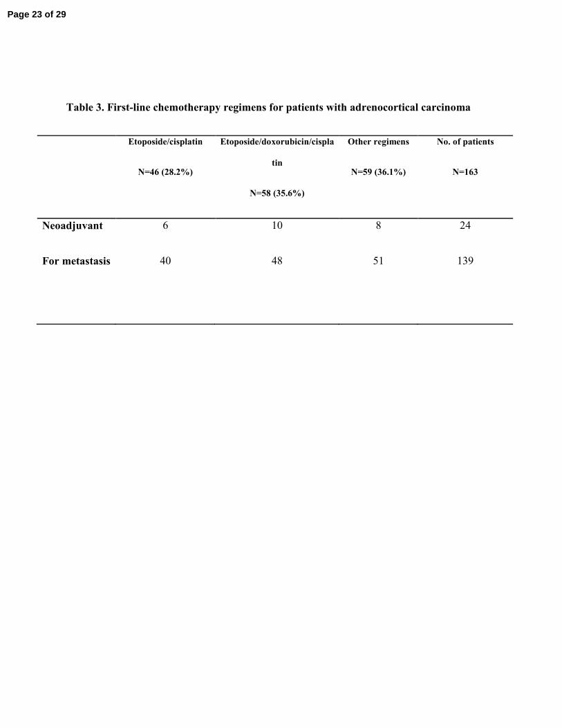
Table 3. First-line chemotherapy regimens for patients with adrenocortical carcinoma
Etoposide/cisplatin
N=46 (28.2%)
Etoposide/doxorubicin/cispla
tin
N=58 (35.6%)
Other regimens
N=59 (36.1%)
No. of patients
N=163
Neoadjuvant 6 10 8 24
For metastasis 40 48 51 139
Page 23 of 29
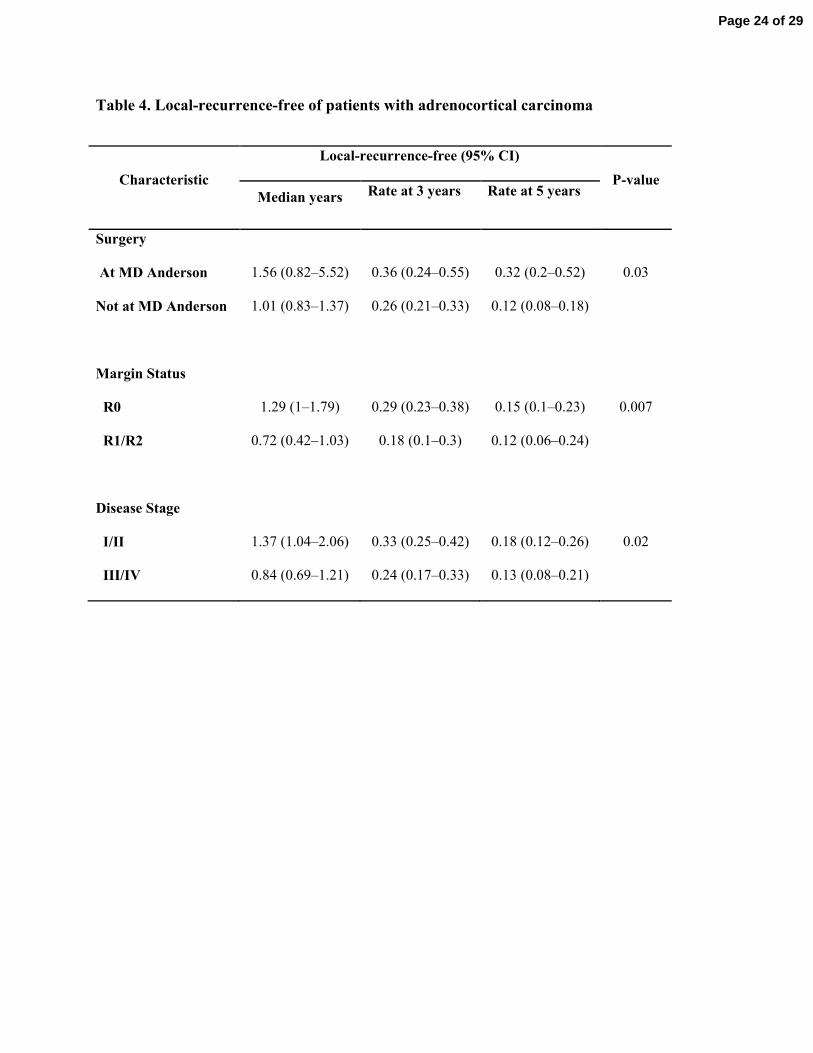
Table 4. Local-recurrence-free of patients with adrenocortical carcinoma
Characteristic
Local-recurrence-free (95% CI)
P-value
Median years Rate at 3 years Rate at 5 years
Surgery
At MD Anderson
Not at MD Anderson
1.56 (0.82–5.52)
1.01 (0.83–1.37)
0.36 (0.24–0.55)
0.26 (0.21–0.33)
0.32 (0.2–0.52)
0.12 (0.08–0.18)
0.03
Margin Status
R0
R1/R2
1.29 (1–1.79)
0.72 (0.42–1.03)
0.29 (0.23–0.38)
0.18 (0.1–0.3)
0.15 (0.1–0.23)
0.12 (0.06–0.24)
0.007
Disease Stage
I/II
III/IV
1.37 (1.04–2.06)
0.84 (0.69–1.21)
0.33 (0.25–0.42)
0.24 (0.17–0.33)
0.18 (0.12–0.26)
0.13 (0.08–0.21)
0.02
Page 24 of 29
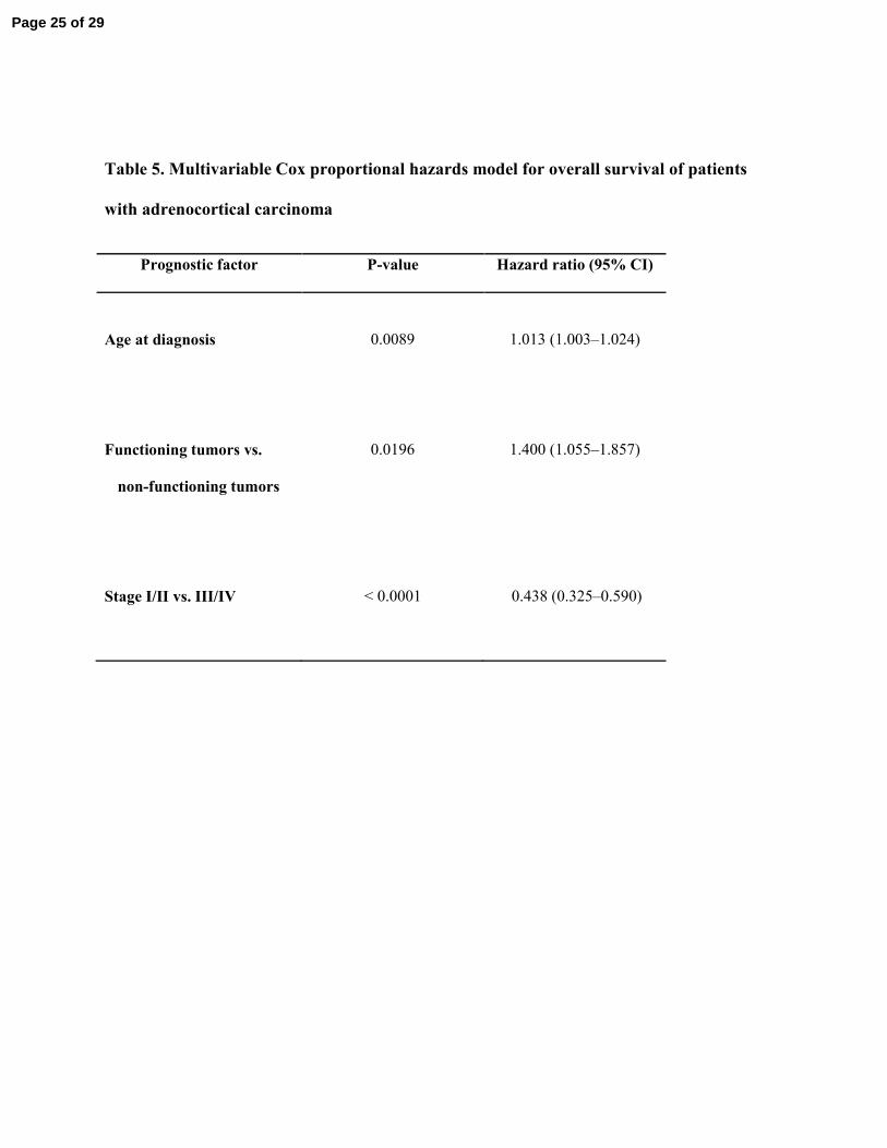
Table 5. Multivariable Cox proportional hazards model for overall survival of patients
with adrenocortical carcinoma
Prognostic factor P-value Hazard ratio (95% CI)
Age at diagnosis
0.0089
1.013 (1.003–1.024)
Functioning tumors vs.
non-functioning tumors
0.0196
1.400 (1.055–1.857)
Stage I/II vs. III/IV
< 0.0001
0.438 (0.325–0.590)
Page 25 of 29
Overall survival (OS) in patients who achieve mitotane levels of 14 mg/l or higher (YES, red line) compared with the patients with lower levels (NO, green line).
18x14mm (300 x 300 DPI)
Page 26 of 29
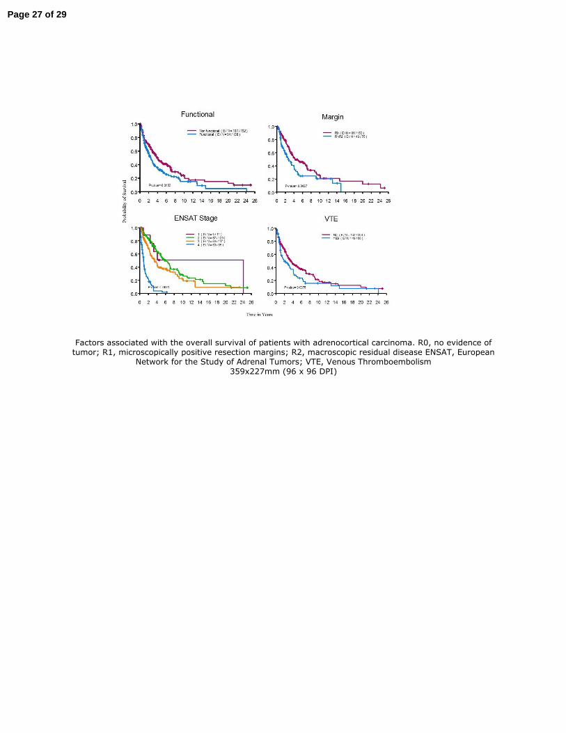
Factors associated with the overall survival of patients with adrenocortical carcinoma. R0, no evidence of tumor; R1, microscopically positive resection margins; R2, macroscopic residual disease ENSAT, European
Network for the Study of Adrenal Tumors; VTE, Venous Thromboembolism
359x227mm (96 x 96 DPI)
Page 27 of 29
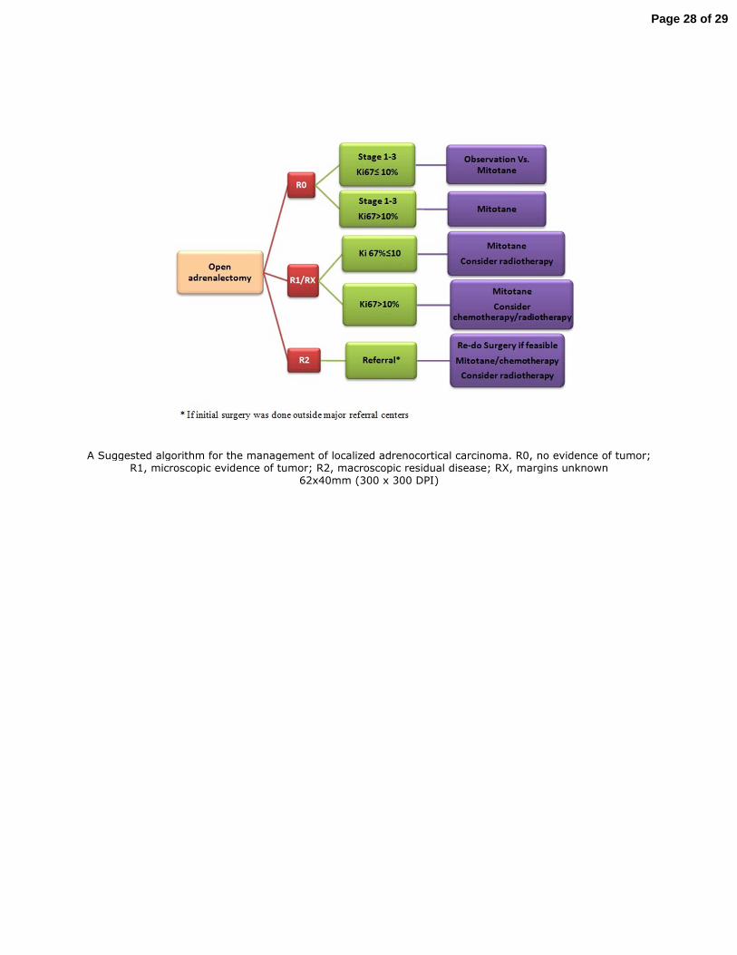
A Suggested algorithm for the management of localized adrenocortical carcinoma. R0, no evidence of tumor; R1, microscopic evidence of tumor; R2, macroscopic residual disease; RX, margins unknown
62x40mm (300 x 300 DPI)
Page 28 of 29
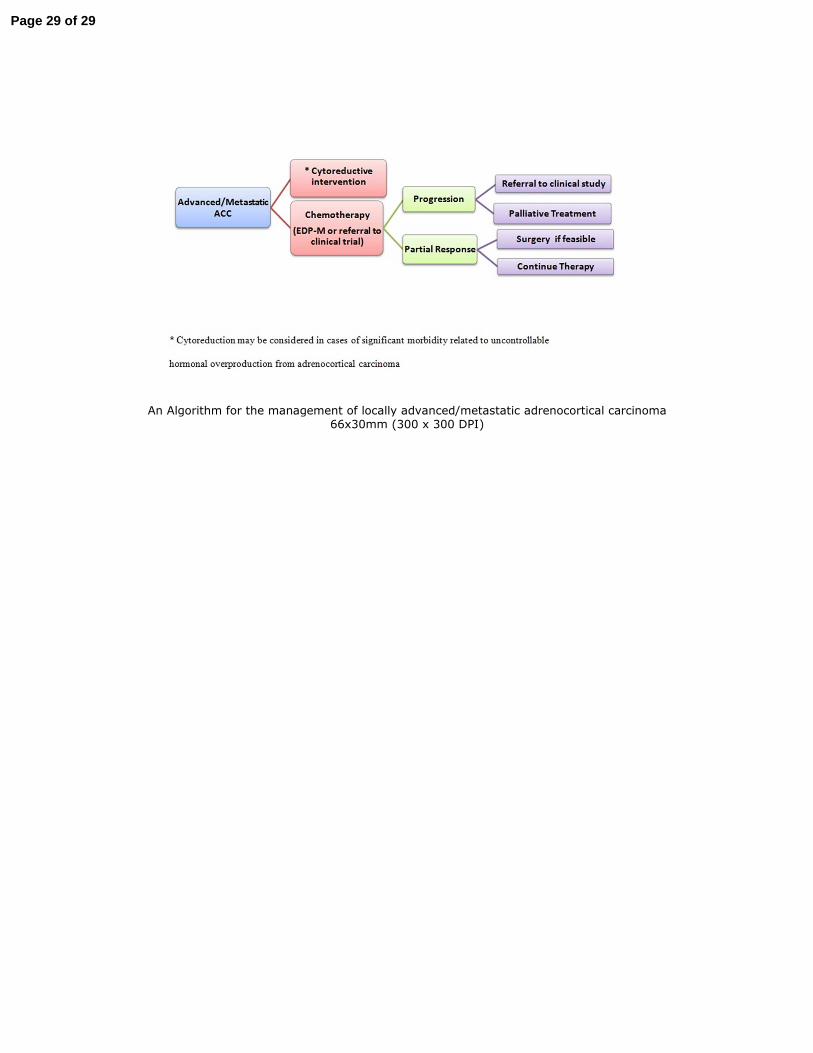
An Algorithm for the management of locally advanced/metastatic adrenocortical carcinoma 66x30mm (300 x 300 DPI)
Page 29 of 29


![Adrenocortical carcinoma in a Ghanaian girl: Report …Adrenocortical carcinoma is a rare tumour in the paediatric population and it is thought to have a poor prognosis [1,2]. It arises](https://img.pdfslide.us/doc/110x75/5f0c24a17e708231d433f37f/adrenocortical-carcinoma-in-a-ghanaian-girl-report-adrenocortical-carcinoma-is.jpg)