Embed Size (px)
Citation preview

1
5The Integumentary System
2
Skin (Integument)
Consists of three major regions
Epidermis – outermost superficial region
Dermis – middle region
Hypodermis (superficial fascia) – deepest region
3
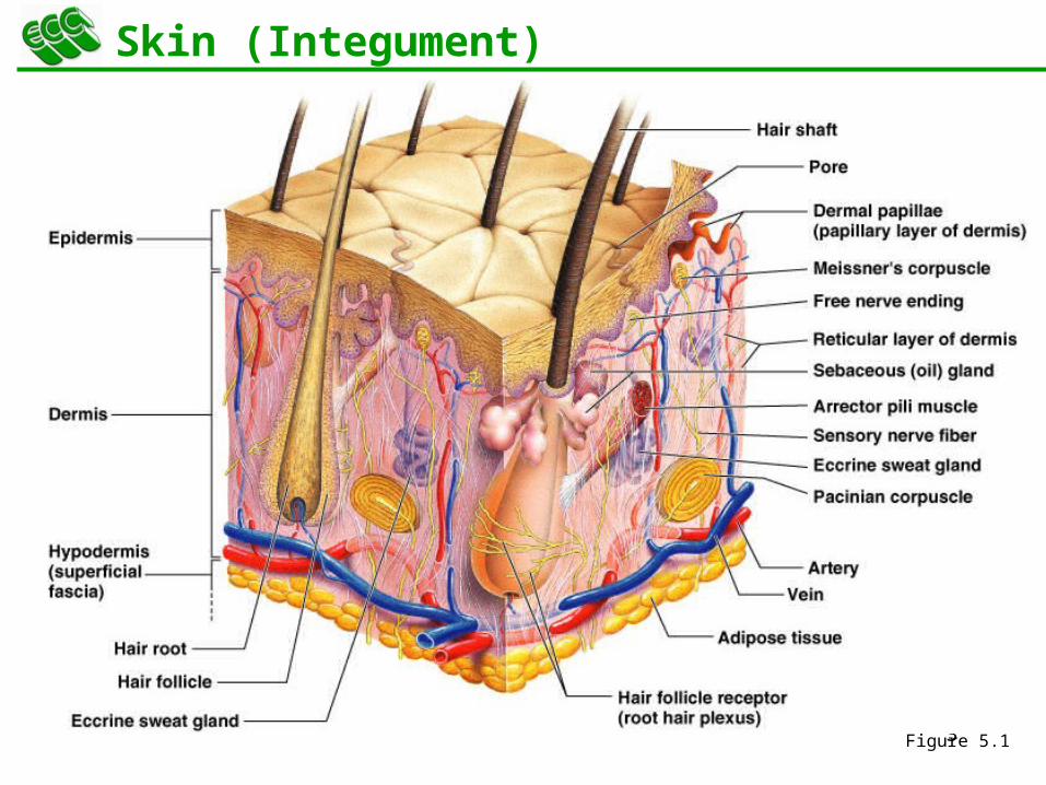
Skin (Integument)
Figure 5.1
4
Epidermis
Composed of keratinized stratified squamous epithelium, consisting of four distinct cell types and four or five layers
Cell types include keratinocytes, melanocytes, Merkel cells, and Langerhans’ cells
Outer portion of the skin is exposed to the external environment and functions in protection from bacteria
5
Cells of the Epidermis
Keratinocytes – produce the fibrous protein keratin
Melanocytes – produce the brown pigment melanin
Langerhans’ cells – epidermal macrophages that help activate the immune system
Merkel cells – function as touch receptors in association with sensory nerve endings
These are the main reasons for protection
6
Deepest epidermal layer firmly attached to the dermis
Consists of a single row of the youngest keratinocytes
Cells undergo rapid division, hence its alternate name, stratum germinativum
Layers of the Epidermis: Stratum Basale (Basal Layer)
7
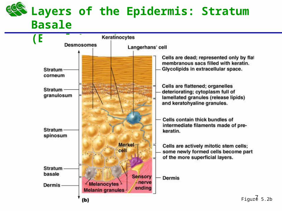
Layers of the Epidermis: Stratum Basale (Basal Layer)
Figure 5.2b
8
Cells contain a weblike system of intermediate filaments attached to desmosomes
Melanin granules and Langerhans’ cells are abundant in this layer
Layers of the Epidermis: Stratum Spinosum (Prickly Layer)
9
Thin; three to five cell layers in which drastic changes in keratinocyte appearance occurs
Keratohyaline and lamellated granules accumulate in the cells of this layer
Layers of the Epidermis: Stratum Granulosum (Granular Layer)
10
Thin, transparent band superficial to the stratum granulosum
Consists of a few rows of flat, dead keratinocytes
Present only in thick skin
Layers of the Epidermis: Stratum Lucidum (Clear Layer)
11
Outermost layer of keratinized cells
Accounts for three quarters of the epidermal thickness
Functions include:
Waterproofing
Protection from abrasion and penetration
Rendering the body relatively insensitive to biological, chemical, and physical assaults
Layers of the Epidermis: Stratum Corneum (Horny Layer)
12
Dermis
Second major skin region containing strong, flexible connective tissue
Responsible for the dermal ridges that produce whorled ridges on the epidermal surfaces
Cell types include fibroblasts, macrophages, and occasionally mast cells and white blood cells
Composed of two layers – papillary and reticular
13
Layers of the Dermis: Papillary Layer
Papillary layer
Areolar connective tissue with collagen and elastic fibers
Its superior surface contains peglike projections called dermal papillae
Dermal papillae contain capillary loops, Meissner’s corpuscles, and free nerve endings
14
Layers of the Dermis: Reticular Layer
Reticular layer
Accounts for approximately 80% of the thickness of the skin
Collagen fibers in this layer add strength and resiliency to the skin
Elastin fibers provide stretch-recoil properties
15
Hypodermis
Subcutaneous layer deep to the skin
Composed of adipose and areolar connective tissue
Responsible for shock absorption
16
Skin Color
Three pigments contribute to skin color
Melanin – yellow to reddish-brown to black pigment, responsible for dark skin colors
Freckles and pigmented moles – result from local accumulations of melanin
Carotene – yellow to orange pigment, most obvious in the palms and soles of the feet
Hemoglobin – reddish pigment responsible for the pinkish hue of the skin
17
Sweat Glands
Different types prevent overheating of the body; secrete cerumen and milk
Eccrine sweat glands – found in palms, soles of the feet, and forehead
Sudoriferous glands serve as thermoregulators.
Apocrine sweat glands – found in axillary and anogenital areas
Ceruminous glands – modified apocrine glands in external ear canal that secrete cerumen (wax)
Mammary glands – specialized sweat glands that secrete milk
18
Sebaceous Glands
Simple alveolar glands found all over the body
Soften skin when stimulated by hormones
Secrete an oily secretion called sebum
Associated with acne
19
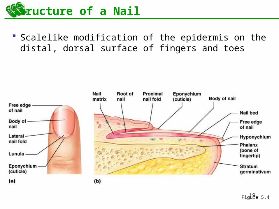
Structure of a Nail
Scalelike modification of the epidermis on the distal, dorsal surface of fingers and toes
Figure 5.4
20
Hair
Filamentous strands of dead keratinized cells produced by hair follicles
Contains hard keratin which is tougher and more durable than soft keratin of the skin
Made up of the shaft projecting from the skin, and the root embedded in the skin
Consists of a core called the medulla, a cortex, and an outermost cuticle
Pigmented by melanocytes at the base of the hair
21
Hair Function and Distribution
Functions of hair include:
Helping to maintain warmth
Alerting the body to presence of insects on the skin
Guarding the scalp against physical trauma, heat loss, and sunlight
Hair is distributed over the entire skin surface except
Palms, soles, and lips
Nipples and portions of the external genitalia
22
Hair Follicle
Root sheath extending from the epidermal surface into the dermis
Deep end is expanded forming a hair bulb
A knot of sensory nerve endings (a root hair plexus) wraps around each hair bulb
Bending a hair stimulates these endings, hence our hairs act as sensitive touch receptors
Arrector pili muscles pulls the hair follicle into an upright position
23
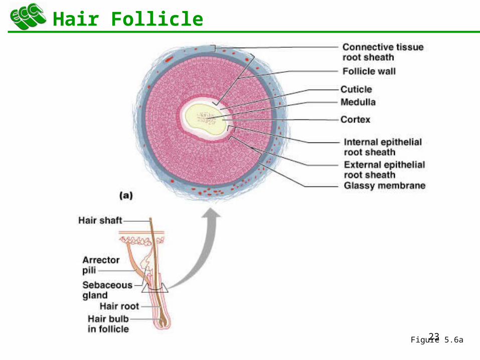
Hair Follicle
Figure 5.6a
24
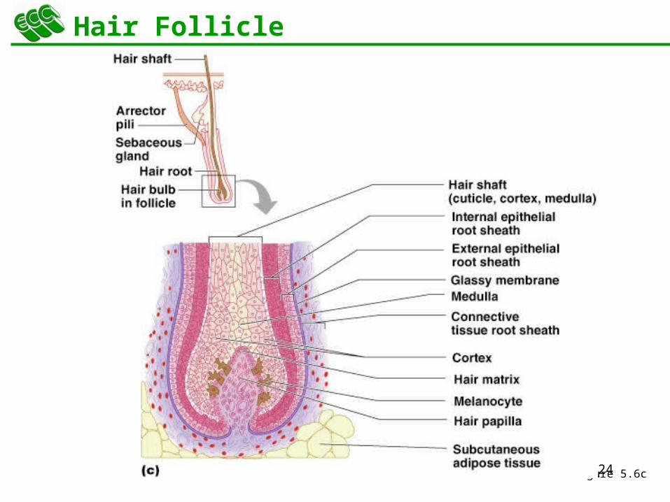
Hair Follicle
Figure 5.6c
25
Types of Hair
Vellus – pale, fine body hair found in children and the adult female
Terminal – coarse, long hair of eyebrows, scalp, axillary, and pubic regions
26
Hair Thinning and Baldness
Alopecia – hair thinning in both sexes
True, or frank, baldness
Genetically determined and sex-influenced condition
Male pattern baldness – caused by follicular response to DHT
27
Functions of the Integumentary System
Protection – chemical, physical, and mechanical barrier
Body temperature regulation is accomplished by:
Dilation (cooling) and constriction (warming) of dermal vessels
Increasing sweat gland secretions to cool the body
Cutaneous sensation – exoreceptors sense touch and pain
28
Functions of the Integumentary System
Metabolic functions – synthesis of vitamin D in dermal blood vessels
Blood reservoir – skin blood vessels store up to 5% of the body’s blood volume
Excretion – limited amounts of nitrogenous wastes are eliminated from the body in sweat
29
Skin Cancer
Most skin tumors are benign and do not metastasize
A crucial risk factor for nonmelanoma skin cancers is the disabling of the p53 gene
Newly developed skin lotions can fix damaged DNA
30
Skin Cancer
The three major types of skin cancer are:
Basal cell carcinoma
Squamous cell carcinoma
Melanoma
31
Basal Cell Carcinoma
Least malignant and most common skin cancer
Stratum basale cells proliferate and invade the dermis and hypodermis
Slow growing and do not often metastasize
Can be cured by surgical excision in 99% of the cases
32
Squamous Cell Carcinoma
Arises from keratinocytes of stratum spinosum
Arise most often on scalp, ears, and lower lip
Grows rapidly and metastasizes if not removed
Prognosis is good if treated by radiation therapy or removed surgically
33
Melanoma
Cancer of melanocytes is the most dangerous type of skin cancer because it is:
Highly metastatic
Resistant to chemotherapy
34
Melanoma
Melanomas have the following characteristics (ABCD rule)
A: Asymmetry; the two sides of the pigmented area do not match
B: Border is irregular and exhibits indentations
C: Color (pigmented area) is black, brown, tan, and sometimes red or blue
D: Diameter is larger than 6 mm (size of a pencil eraser)
35
Melanoma
Treated by wide surgical excision accompanied by immunotherapy
Chance of survival is poor if the lesion is over 4 mm thick
36
Burns
First-degree – only the epidermis is damaged
Symptoms include localized redness, swelling, and pain
Second-degree – epidermis and upper regions of dermis are damaged
Symptoms mimic first degree burns, but blisters also appear
Third-degree – entire thickness of the skin is damaged
Burned area appears gray-white, cherry red, or black; there is no initial edema or pain (since nerve endings are destroyed)
In danger of dehydrating
37
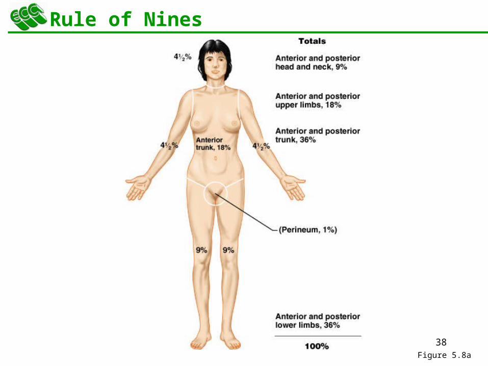
Rule of Nines
Estimates the severity of burns
Burns considered critical if:
Over 25% of the body has second-degree burns
Over 10% of the body has third-degree burns
There are third-degree burns on face, hands, or feet
38
Rule of Nines
Figure 5.8a
39
Developmental Aspects of the Integument: Fetal
Epidermis develops from ectoderm
Dermis and hypodermis develop from mesoderm
Lanugo – downy coat of delicate hairs covering the fetus
Vernix caseosa – substance produced by sebaceous glands that protects the skin of the fetus in the amnion
40
Skin and hair become oilier and acne may appear
Skin shows the effects of cumulative environmental assaults around age 30
Scaling and dermatitis become more common
Developmental Aspects of the Integument: Adolescent to Adult
41
Epidermal replacement of cells slows and skin becomes thinner
Skin becomes dry and itchy
Subcutaneous fat layer diminishes, leading to intolerance of cold
Decreased elasticity and loss of subcutaneous tissue leads to wrinkles
Decreased numbers of melanocytes and Langerhans’ cells increase the risk of skin cancer
Developmental Aspects of the Integument: Old Age