Embed Size (px)
Citation preview

1
2nd & 3rd Week Devp
Dr. Lubna Nazli
2
Objectives
• Chorion and amnion• Gastrulation • Prochordal plate• Primitive streak and formation of notochord.• Neurulation & neural crest cells.• Divisions of mesoderm• Foldind of embryo• Allantois• Derivatives of all germ layers
3
Bilaminar germ disc
• The inner cell mass differentiates at first into 2 layers.
• Some of the cells differentiate into flattened cells in the lower part called endodermal layer. This is the first layer to be formed.
• The remaining cells become columnar and form the second layer, the ectoderm.
• Space between ectoderm and trophoblast --- amniotic cavity.
4
• The endodermal cells then spread and line the inside of blastocyst--- Heuser’s membrane.
• Thus a cavity lined by endodermal cells form called primary yolk sac.

5
Extraembryonic mesoderm
• The trophoblast gives origin to a mass of cells which lie between trophoblast and endodermal cells. This is called extraembryonic mesoderm.
• Extraembryonic coelom or chorionic cavity.
• Extraembryonic mesoderm is split into 2 layers: somatopleuric and sphlanchnopleuric mesoderm.

6

7
Formation of chorion & amnion
• Chorion is formed by : parietal extra-emb mesoderm on the inside and trophoblast on outside
• Amnion is amniogenic cells forming the amniotic cavity.

8
Sec yolk sac
• Prim yolk sac becomes smaller – sec yolk sac
• Embryo becomes disc like, bilaminar.
• No head or tail end.

9
Prochordal plate
• At one end cubical cells become columnar – prochordal plate.
• This determines the central axis and also the head and tail ends.
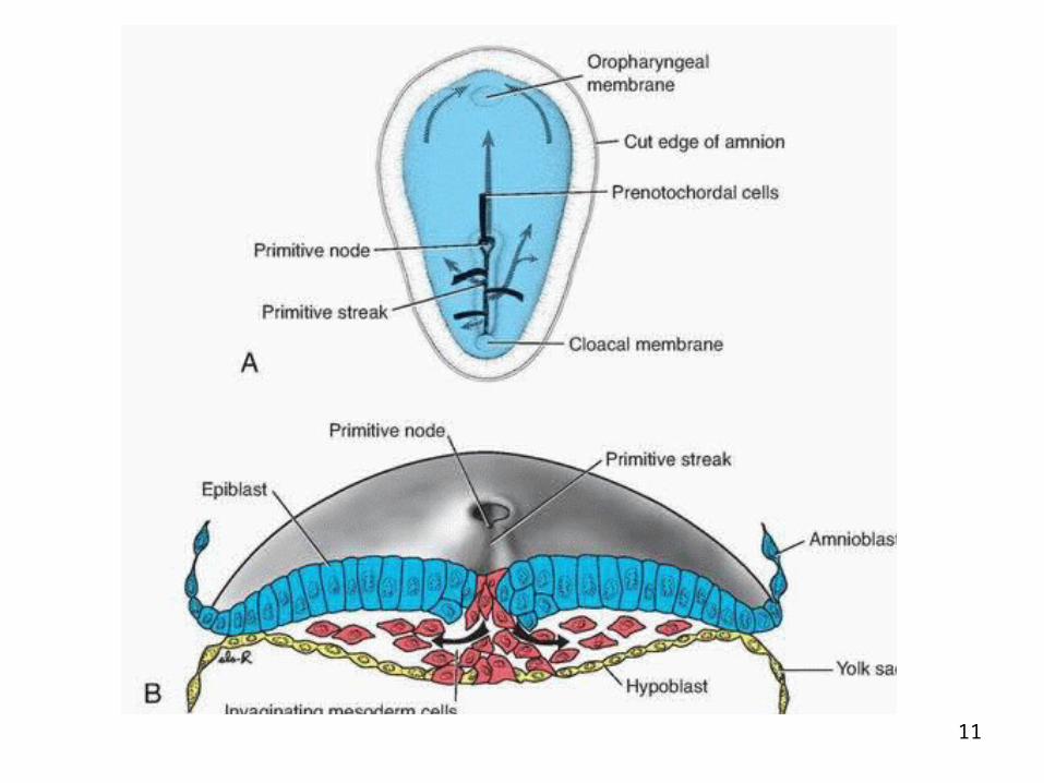
• Soon after this the ectodermal cells at the tail end begin to proliferate, form elevation that bulges into amniotic cavity called the primitive streak.

10
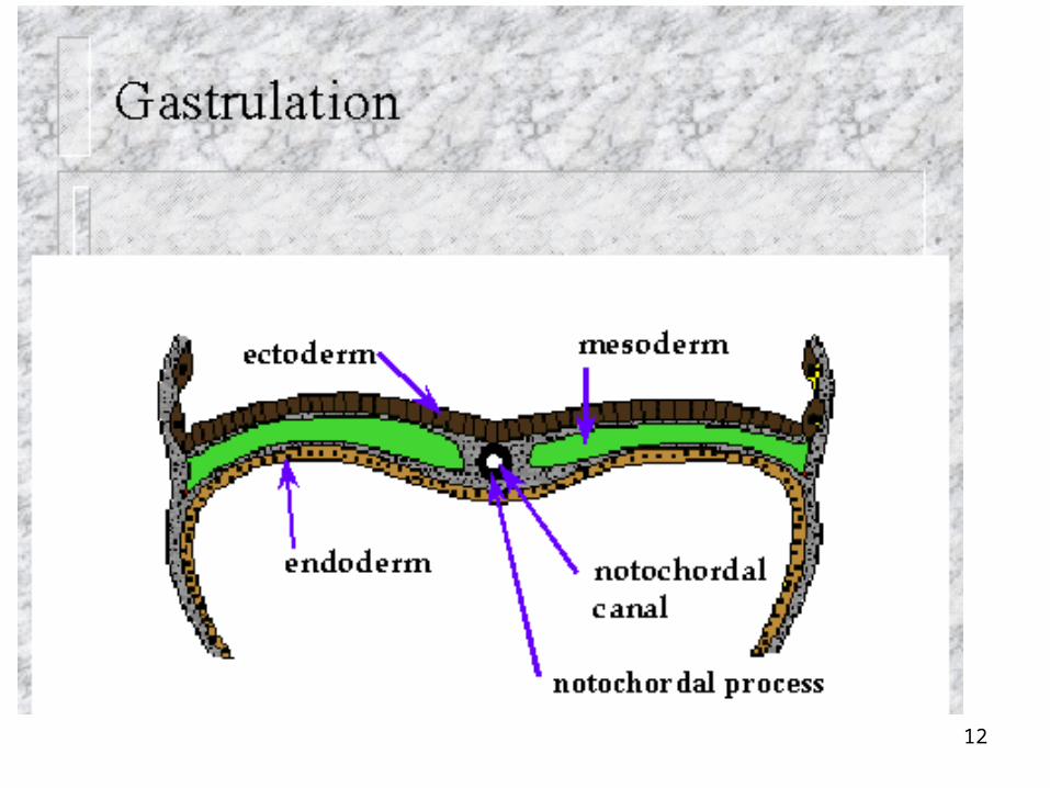
• Primitive streak cells proliferate and pass between the ecto & endoderm to form a third layer called intraembyonic mesoderm.
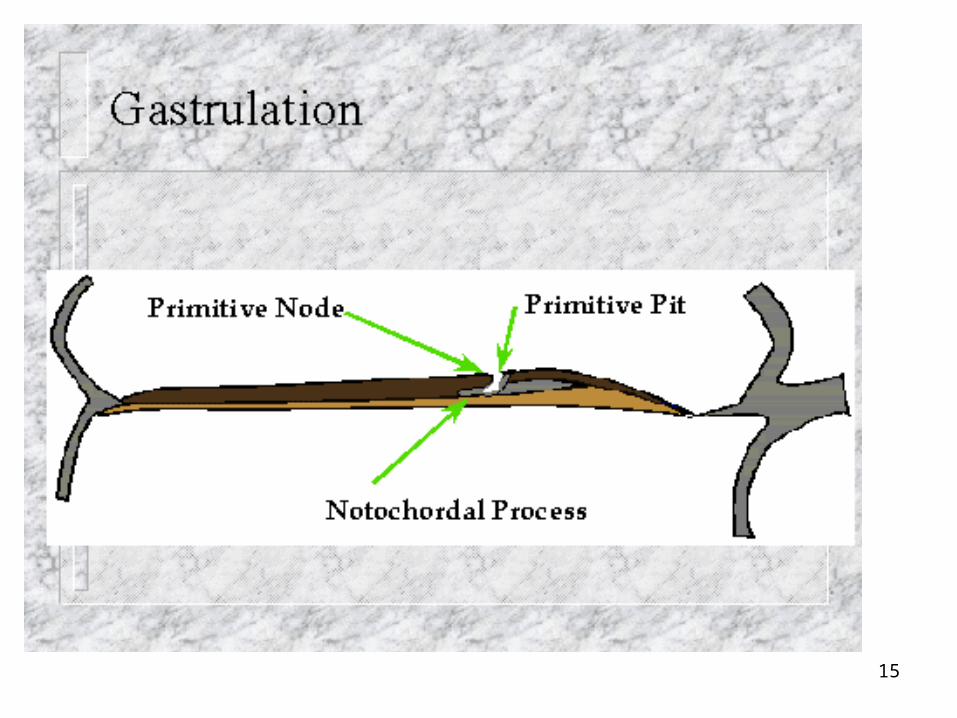
• This process is called gastrulation.
• Intraemby meso spreads throughout except prochordal plate. This forms future bucco pharyngeal membrane.
11
12
13
• The embryonic disc is suspended from trophoblast by connecting stalk.
• As disc enlarges the stalk becomes small & is confined to tail end.
• The mesoderm passes backwards in the stalk but leaves an area caudally which forms the cloacal membrane.
14
Formation of Notochord
• Cranial end of primitive streak becomes thickened – primitive node or Hensen’s node.
• Depression appears in the center of it – blastopore.
• Cells in primitive node multiply & pass cranially between ecto & endo reaching upto prochordal plate.
• They form solid cord called notochord.
15

16
17
• As embryo enlarges notochord elongates and is later occupied by vertebral column.
• It does not give rise to vertebral column but persists as nucleus pulposus in the disc.

18
Neurulation
• Neural tube is formed from the ectoderm overlying the notochord, extends from prochordal plate to primitive knot.
• It forms the future brain and spinal cord.
19
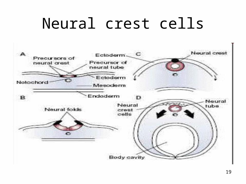
Neural crest cells
20
Neural Crest cells
• As the edges of the ectoderm layer curl upwards to form the neural tube, another cell type forms at the tip of the curl.
• These cells differentiate into neural crest cells.
• Neural crest cells eventually give rise to the dorsal root ganglia, Schwann cells, the autonomic nervous system, meninges, sensory ganglia, bones of the face, teeth, lens of the eyes, melanocytes, adrenal medulla, and many glands.
• (The neural crest cells are sometimes referred to as the fourth germ layer.)
21
Subdivisions of mesoderm
• It is divided into 3 parts.
• On either side of the notochord it becomes thick and is called paraxial mesoderm.
• More laterally is lateral plate mesoderm.
• In between is the intermediate mesoderm.

22
23
• Paraxial meso gets segmented to form somites.
• Lateral plate forms cavities which coalesce to form intraembryonic coelom. This splits it into 2 layers : somato and splanchno
• The coelom forms pericardial, pleural and peritoneal cavities.

24
Folding of embryo
• With increasing size the disc becomes folded on itself at both ends.
• These are the head and tail folds.
• This part of yolk sac becomes enclosed within the embryo. This is the primitive gut from which most of GIT is formed.
• Midgut communicates with the definitive yolk sac by vitellointestinal duct.

25
• Lateral folds form on the sides. Thus the embryo is enclosed on all sides by ectoderm except at vitellointestinal duct.
• Amniotic cavity expands & surrounds the embryo & the embryo now floats in amniotic fluid.
• With the formation of head fold the pericardial cavity comes on the ventral side of the foregut

26
Allantoic diverticulum
• Before the formation of tail fold a small endodermal diverticulum called Allantoic diverticulum arises from the yolk sac.
• This grows into the mesoderm of connecting stalk. This forms the future urinary bladder.
27
The ectoderm derivatives
• Brain & spinal cord
• Lens, cornea &retina
• Skin epidermis
• Pituitary gland
• Nails, hairs, sweat glands & sebaceous glands
• Mammary glands

28
The mesoderm derivatives
• Muscles…skeletal/cardiac / smooth• Skin...dermis• Vertebrae/sternum/all bones &cartilages• Blood• Testis/ovary/epidydimis/vas
deferens/uterus/fallopian tubes• Kidneys/ureter/muscles of urinary bladder• Spleen
29
The Endoderm derivatives
• Epithelium of tongue/digestive system
• Liver/gallbladder/pancreas
• Epithelium of urinary bladder
• Trachea/lungs
• Thyroid/parathyroid/thymus
• Tonsils
• Middle ear cavity/auditory tube