Embed Size (px)
Citation preview
1

2
CHAPTER 5
EXTRACELLULAR MATRIX AND ORGANELLES

Extracellular Matrix
Extracellular matrix (ECM) is a meshwork composed of macro molecules secreted by cells:
Collagen (Over 30%)
Non-collagen glycoprotein
Aminoglycan and proteoglycan
Elastin

4
Components of ECM

5
The ECM of epithelial tissue
6
Collagen:
The ratio of collagen is over 30% of total protein quantity in human body. Collagen forms the meshwork in matrix. Collagen is synthesized and transferred out by fibroblasts, chondrocytes, osteoblasts, and some of epithelial cells.
So far, more than 20 types of collagen have been established at least. The type I, II, III, and are the fibro collagens that have striation on Ⅴ Ⅺthem. The collagen I from embryo or new born baby is easy to be extracted because the conjugation between molecules is inefficient. With aging, the conjugation is increased and collagen I will become hard that cause the hard skin, vascular tissue, that means “Old”.
Vitamin C is an important helper factor to the synthesis of collagen I. The inefficiency of vitamin C will cause blood vessels easy to be broken and bleeding that we call as “Scurvy”.

7
Collagen fiber
Fibroblast
The collagen fibers surrounding fibroblast

8The types of collagen

9
The structure of collagen
L: a model fig; R: an image taken by electron microscope

10
Fibronectin (FN):
FN is a type of large glycoprotein. FN can link cell on to ECM.
FN can exist in blood plasma or body fluid ( 0 . 3mg/ml ) as soluble type (Plasma FN) and in ECM or cell surface as insoluble type (Cellular FN).
Plasma FN exists in a molecule formed a dimer by two subunits linked at C terminal as a “V” shape. Cell FN is a polymer.
Some short fragments of the FN chain are the smallest structural units identified by FN receptors on cell surface. For example, the RGD (Arg-Gly-Asp) sequence existing in the cell binding domain located in the middle of chain is the site that can be recognized by some integrin receptors located on cell surface.

11
Model structure of FN

12FN links cell on to ECM

13
Laminin (LN):
LN is a large type of glycoprotein too. Matrix is formed by LN and collagen IV. LN is the earliest expressed component of ECM during the embryonic development. LN molecule is composed of one heavy chain and two light chains as a cross shape. At least, there are 8 cell binding sites in LN molecule. For example, the 5 mers peptide, IKVAV sequence, is located in the site near to the ball domain of long arm that can bind to neuron cells and enhance the neuron growth. 7 types of LN molecules and 8 types of subunit (α1,α2,α3,β1,β2, β3,γ1,γ2) have been identified so far. But, it is different from FN that each subunit is encoded by different structural gene. About 50 sugar chains are linked to the N terminals of LN protein, so, LN is the most complicated glycoprotein that we known so far. Excepting LN and collagen IV, entactin, perlecan, decorin and other proteins exist in ECM.

14Model structure of LN
N terminals(Short arms)
C terminals(Long arm)
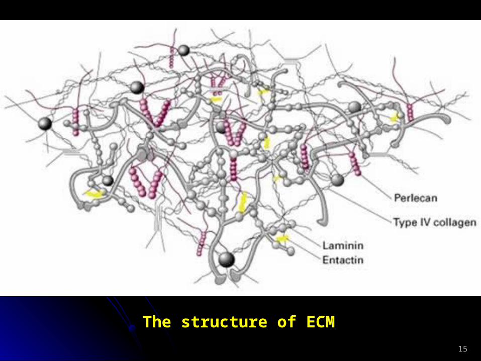
15
The structure of ECM

16
Glycosaminoglycan (GAG): GAG is a big family of polysaccharides formed by repeated disaccharide units.
The features of GAG and distribution
GAG Disaccharide unit Sulfate Distribution radicalsHyaluronic acid Glucuronic acid 0 Connective tissue, skin N-acetylglucose Cartilage, vitreous body Synovial fluidChondroitin sulfate Glucuronic acid 0.2-2.3 Cartilage, bone, skin N-acetylgalactose Cornea, arteryDermatan sulfate Glucuronic acid 1.0-2.0 Skin, blood vessel N-acetylglucose Heart, cardiac valvesHeparitin sulfate Glucuronic acid 0.2-3.0 Lungs, artery, cell surface N-acetylglucoseHeparin Glucuronic acid 2.0-3.0 Lungs, liver, skin, N-acetylglucose Mastocyte
Keratin sulfate Galactose 0.9-1.8 Cartilage, cornea, N-acetylglucose Intervertebral discs

17
L: Proteoglycan; M: Polymers of proteoglycan; R: Glycosaminoglycan

18
Proteoglycan: Proteoglycan is the compound of GAG (except hyaluronic acid) and core protein (Shown as the fig above). The polymer of proteoglycan is composed of single proteoglycan and hyaluronic acid. The molecule weight of polymer of proteoglycan may be over 108KD, and its volume may be bigger than bacteria. Chondroitin sulfate and keratin sulfate are the main members of GAG from the aggrecan that forms cartilage. The inefficiency of the quantity of the both GAGs can cause the inhibition of bones development and short limbs and trunk. We call these persons “Dwarf”.
Elastin: Elastin meshwork makes tissue soft and elastic. The extension of elastin is stronger 5 times than rubber band at least. In the tissues of old people, the synthesis of elastin is decreased and the degeneration increased, that is why the tissues of old people became hard, rough.

19
Conjugation
Model structure of elastin

20
The functions of ECM:1. Plays important role to the survival, growth, and death of cells. Eukaryotic cells
must attach to ECM for their growth excepting blood cells, that we call as anchorage dependence. For example, epithelial cells will turn to apoptosis if they are separated from ECM. Different ECM will give cell different effects. The proliferation of fibroblast will be quick up on a fibronectin matrix, and slow down on a laminin matrix, but the response of epithelial cells to the matrix is just opposite.
2. Controlls the shape of cell. Cells will be spherical if they grow with ECM separately. A cell can present a different shape if it grow on a different ECM. The mechanism of this regulation is mediated by the receptors on ECM that regulates the cytoskeleton.
3 . Regulates the differentiation by the interaction between cell and special component of ECM. For example, myoblast (sarcoblast) can keep its original shape on fibronectin, but it will stop its proliferation to differentiate and fuse to myotube.
4 . Mediates cell migration. ECM can regulate the speed and direction of the migration. Laminin can enhance the migration of tumor cells, and the migration of other cells is dependent on ECM too. This dependence is very important during the embryo development and wounds healing.
So, ECM mediate almost every event in cell and life story.

21
ECM
Cell survival
Cell differentiation
Cell proliferation
Cell migration
ApoptosisCell death
Cell shape
Cell growth
ECM plays role in almost each event of cell

22
Organelles Many complicated function areas are packaged by inner membrane inside eukaryotic cells that we call as organelle, or endomembrane system. Organelles include nucleus, endoplasmic reticulum, Golgi body (Golgi complex), lysosome, mitochondrion, chloroplast, and others. Every organelle is associated with some special protein for protein synthesis, modification, transportation or storage.
Protein sorting: The proteins and lipids synthesized inside cells must be transported into specific organelles firstly, then transported out cells like the follows:
23
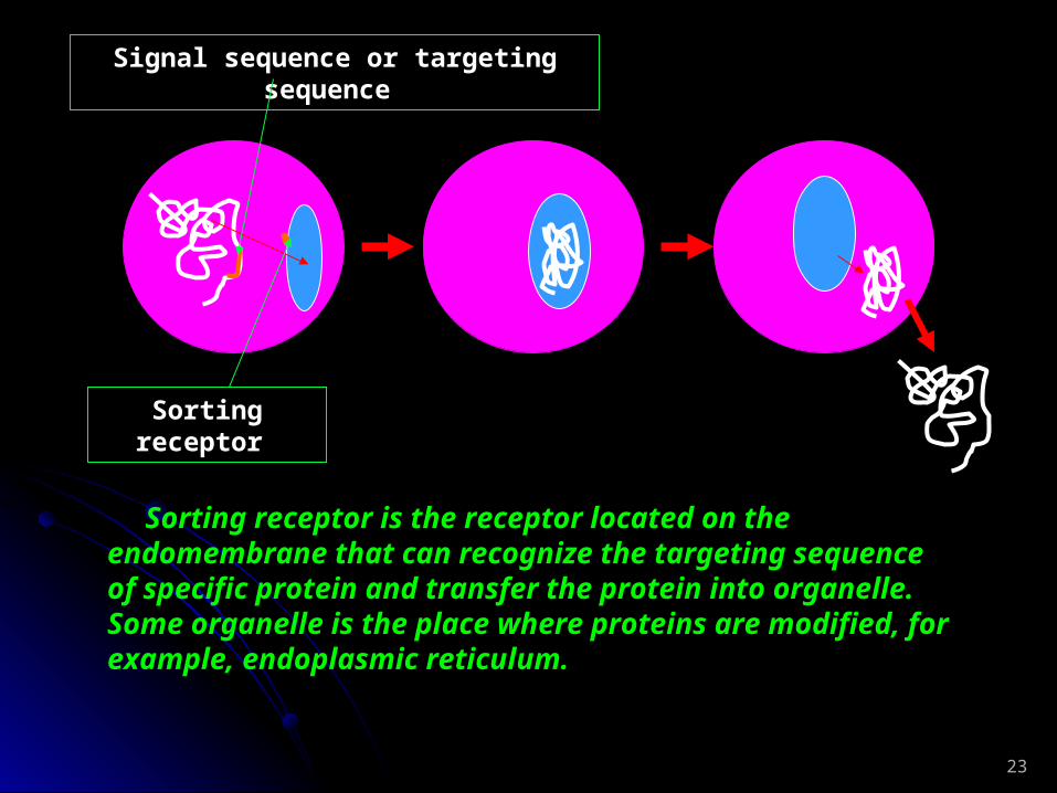
Signal sequence or targeting sequence
Sorting receptor
Sorting receptor is the receptor located on the endomembrane that can recognize the targeting sequence of specific protein and transfer the protein into organelle. Some organelle is the place where proteins are modified, for example, endoplasmic reticulum.
24
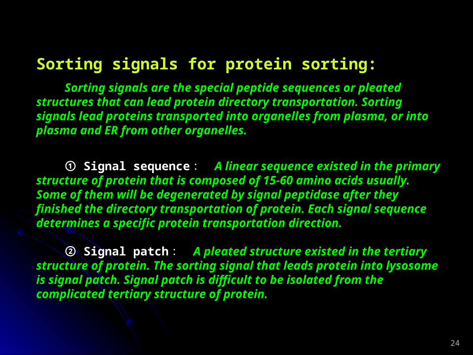
Sorting signals for protein sorting: Sorting signals are the special peptide sequences or pleated structures that can lead protein directory transportation. Sorting signals lead proteins transported into organelles from plasma, or into plasma and ER from other organelles.
① Signal sequence : A linear sequence existed in the primary structure of protein that is composed of 15-60 amino acids usually. Some of them will be degenerated by signal peptidase after they finished the directory transportation of protein. Each signal sequence determines a specific protein transportation direction.
② Signal patch : A pleated structure existed in the tertiary structure of protein. The sorting signal that leads protein into lysosome is signal patch. Signal patch is difficult to be isolated from the complicated tertiary structure of protein.

25
Signal sequence and signal patch
26
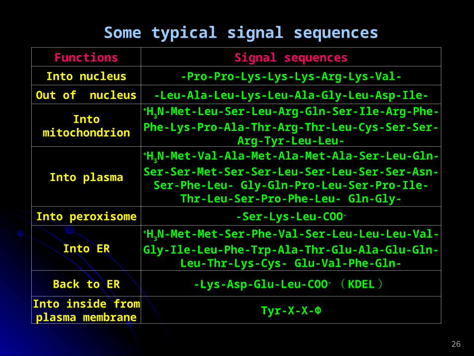
Functions Signal sequencesInto nucleus -Pro-Pro-Lys-Lys-Lys-Arg-Lys-Val-
Out of nucleus -Leu-Ala-Leu-Lys-Leu-Ala-Gly-Leu-Asp-Ile-
Into mitochondrion
+H3N-Met-Leu-Ser-Leu-Arg-Gln-Ser-Ile-Arg-Phe-Phe-Lys-
Pro-Ala-Thr-Arg-Thr-Leu-Cys-Ser-Ser-Arg-Tyr-Leu-Leu-
Into plasma
+H3N-Met-Val-Ala-Met-Ala-Met-Ala-Ser-Leu-Gln-Ser-Ser-
Met-Ser-Ser-Leu-Ser-Leu-Ser-Ser-Asn-Ser-Phe-Leu- Gly-Gln-Pro-Leu-Ser-Pro-Ile-Thr-Leu-Ser-Pro-Phe-Leu- Gln-Gly-
Into peroxisome -Ser-Lys-Leu-COO-
Into ER
+H3N-Met-Met-Ser-Phe-Val-Ser-Leu-Leu-Leu-Val-Gly-Ile-
Leu-Phe-Trp-Ala-Thr-Glu-Ala-Glu-Gln-Leu-Thr-Lys-Cys- Glu-Val-Phe-Gln-
Back to ER -Lys-Asp-Glu-Leu-COO- ( KDEL)Into inside from
plasma membrane
Tyr-X-X-Φ
Some typical signal sequences

27
The ways by that proteins are sorted and transported:
1. Gated transportation: For example, nucleopore can transport macromolecules selectively and
automatically. 2. Transmembrane transportation: For example, passing through the translocator on the mitochondrion, the
synthesized proteins located in plasma can be transported into mitochondrion under the leading of signal sequence with a linear molecule.
3. Vesicular transportation: Proteins are selectively packaged as vesicles and directively transported
into target organelles. For examples, transportation from ER to Golgi body, formation of lysosome from Golgi body, adsorption of nutrition and hormone by cells.
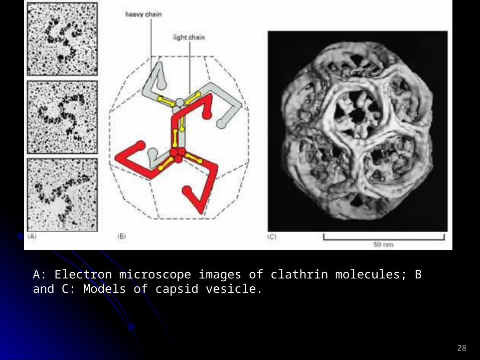
There are three capsid proteins can form vesicles to vesicular transportation: clathrin, COPI and COPII.
28
A: Electron microscope images of clathrin molecules; B and C: Models of capsid vesicle.

29
Electron microscope images of clathrin capsid vesicles

30
Forming of clathrin capsid vesicle

31
The direction of vesicles is dependent on the marker protein and receptor of target. The key proteins for that are SNAREs (soluble NSF attachment protein receptor) and Rabs (targeting GTPase).
The fusion between HIV and CD4 is almost same to SNAREs way:

32
Adsorption of low density lipoproteins (LDL) by cells:
LDL is very important to cause severe cardiovascular diseases. Cholesterols can form LDL (a vesicle actually) with phospholipids and proteins. LDL are released into blood from liver cells. The molecule weight of LDL vesicle is 3X10⁶Da in 20~30nm diameter. There are about 1500 cholesterol molecules packaged inside the vesicle. So, the LDL outer layer is single layer of lipid and a very huge protein (apolipoprotein B-100) that can bind to the receptor of target cell.
Usually, cells will synthesize the LDL receptors when they need cholesterol. But, if too much cholesterol is accumulated in cell, cell will stop the synthesis of receptor. As the result, LDL level in blood will go up. For some persons, a mutation of LDL receptor gene causes their blood LDL level going up. LDL is much easier to attach on to the inner wall of artery than high density lipoprotein (HDL), especially in brain and heart to form the atherosclerosis that is number one killer to human being now in the world.

33A model of LDL

34
Endocytosis (vesicular transportation) of LDL

35
Endoplasmic reticulum:
Rough endoplasmic reticulum (RER)
Endoplasmic reticulum (ER)
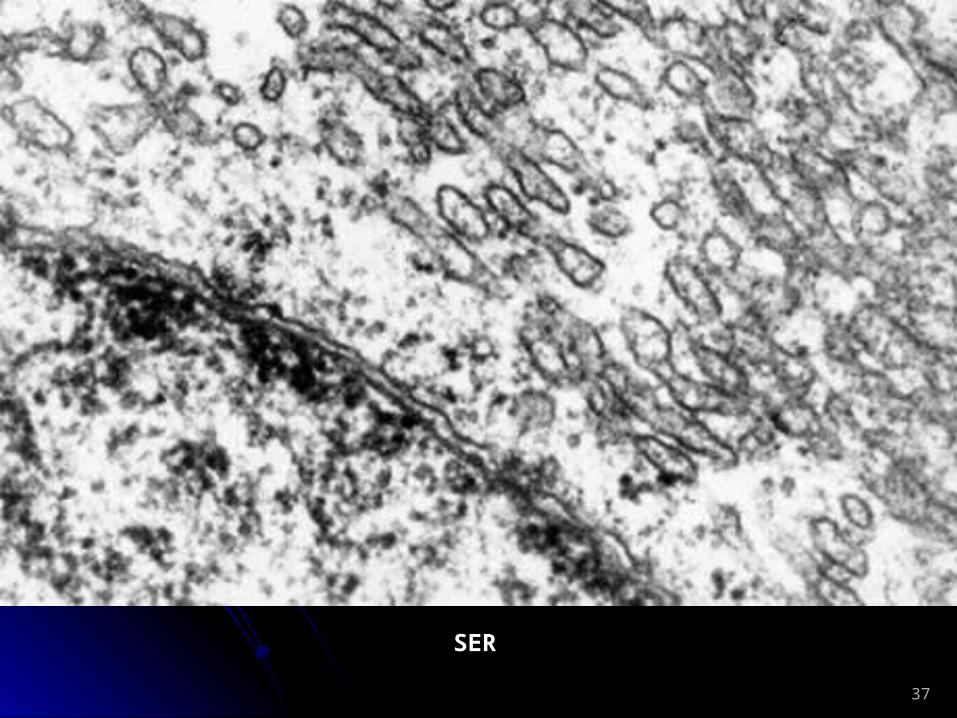
Smooth endoplasmic reticulum (SER)
There are ribosome particles attached on RER, and no any on SER, that is why we see RER under microscope with rough membrane and gave it the name, rough endoplasmic reticulum.
The function of ER is to synthesize proteins and lipids.

36RER
37
SER

38
Functions of ER:
1. Synthesis of proteins:
Usually, proteins synthesis is originally started in plasma and finished on ribosome. But, some proteins synthesis will be finished in RER after synthesis starting. For examples, secreted proteins, such as, antibodies, hormones; ① ②transmembrane proteins; Divided enzymes, such as hydrolases in ③lysosome; Modified proteins, such as glycoprotein. ④
There are 5 factors associated with the transferred synthesis at least: ① Signal peptide: A polypeptides that leads new synthesized peptide chain
to ER, and it located at the N terminal of new synthesized peptide chain. Signal peptide is composed of 16~30 amino acids, and 6-15 of them are the positive charged and non-polarized amino acids. Signal peptide is named as start transfer sequence also.
② Signal recognition particle (SRP): SRP is composed of 6 different polypeptides, and binds to a 7S RNA. Its molecule weight is 325KD. SRP can bind to signal sequence to cause the protein synthesis paused.
③ SRP receptor: A integral protein can bind to SRP specifically. ④ Stop transfer sequence: A special peptide sequence with a high affinity to
ER membrane. It can stop the peptide chain goes into ER and change the chain as a transmembrane protein.
⑤ Translocator.

39
ProteinsProteins Sequences of signal Sequences of signal peptidespeptides
PreproalbuminPreproalbumin Met-Lys-Trp-Val-Thr-Met-Lys-Trp-Val-Thr-Phe-Leu-Leu-Leu-Leu-Phe-Leu-Leu-Leu-Leu-Phe-Ile-Ser- Gly-Ser-Ala-Phe-SerPhe-Ile-Ser- Gly-Ser-Ala-Phe-Ser↓Arg↓Arg......
Pre-IgG light Pre-IgG light chainchain
Met-Asp-Met-Arg-Ala-Pro-Ala-Gln-Met-Asp-Met-Arg-Ala-Pro-Ala-Gln-Ile-Phe-Gly-Ile-Phe-Gly-Phe-Leu- Leu-Leu-Leu-PhePhe-Leu- Leu-Leu-Leu-Phe-Pro-Gly- Thr-Arg--Pro-Gly- Thr-Arg-Cys↓AspCys↓Asp......
PrelysozymePrelysozyme Met-Arg-Ser-Met-Arg-Ser-Leu-Leu-Ile-Leu-Val-Leu-Cys-Leu-Leu-Ile-Leu-Val-Leu-Cys-Phe-Leu- Phe-Leu- Pro-Leu-Ala-Ala-Leu-Gly↓LysPro-Leu-Ala-Ala-Leu-Gly↓Lys......
Some sequences of signal sequences
2. Protein modification:
The modifications include glycosylation (saccharification), hydroxylation, acylation, and bisulfide bond formation. The glycosylation is the most important here.
Glycosylation can: have proteins resistant to the digestion by enzymes of ①digestion system; give proteins markers, signals, or cluster of determining ②(CD); have some proteins pleated correctly. ③
O-linked glycosylation: Link galactose or N-acetylgalactosamine to the OH of Ser, Thr and Hyp. The reaction is carried out on Golgi body. N-linked glycosylation: Link N-acetylglucosamine to NH2 of Asn. The reaction is carried out on ER.

40
N-linked glycosylation

41
3. Fold, assemble, and transportation of new synthesized peptide chain. 4. Synthesis of membrane lipids: Most of membrane lipids are synthesized in ER. Synthesized membrane lipids will be transported to Golgi body and lysosome with vesicular transportation way. 5. Detoxication: The P450 of SER is a monooxygenase (mixed function oxidase or hydroxylase) that is distributed on SER and other organelles. P450 can metabolize the fat soluble (liposoluble) toxic substance into water soluble substance to be ejected out of body. But, sometime, P450 can activate the cancer inducer during the detoxication. 6. Synthesis of steroid hormones: The enzymes in the SER, mitochondrion, Golgi body of the endocrine cells of genital glands and adrenal glands take duty to synthesize steroid hormones. 7. Regulation to the level of blood sugar.8. Construction of some special structures: For example, the sarcoplasmic reticulum specialized by the SER in muscle cells can store Ca+ as the signal substance for the muscle cell excitation.

42
Golgi body:

43
The Golgi bodies in cultured epithelial cells
(Golgi body: red; Nucleus: green)
44
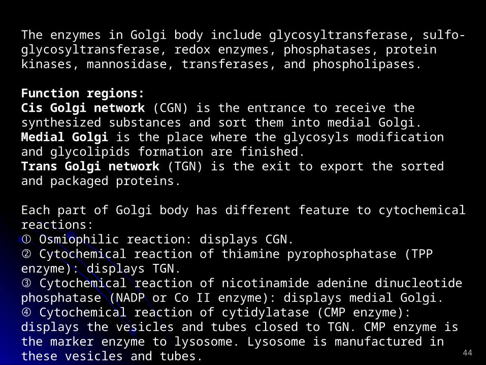
The enzymes in Golgi body include glycosyltransferase, sulfo-glycosyltransferase, redox enzymes, phosphatases, protein kinases, mannosidase, transferases, and phospholipases.
Function regions: Cis Golgi network (CGN) is the entrance to receive the synthesized substances and sort them into medial Golgi. Medial Golgi is the place where the glycosyls modification and glycolipids formation are finished. Trans Golgi network (TGN) is the exit to export the sorted and packaged proteins.
Each part of Golgi body has different feature to cytochemical reactions: ① Osmiophilic reaction: displays CGN. ② Cytochemical reaction of thiamine pyrophosphatase (TPP enzyme): displays
TGN. ③ Cytochemical reaction of nicotinamide adenine dinucleotide phosphatase (NADP
or Co II enzyme): displays medial Golgi. ④ Cytochemical reaction of cytidylatase (CMP enzyme): displays the vesicles and
tubes closed to TGN. CMP enzyme is the marker enzyme to lysosome. Lysosome is manufactured in these vesicles and tubes.

45
The regions of Golgi
body
Osmiophilic reaction
TPP enzyme
NADP or Co II enzyme
CMP enzyme

46
Function of Golgi body:1. Glycosylation of protein: N-linked glycosylation is started on ER and finished on Golgi body. Proteins will be serially modified when they pass through the Golgi body from Cis region to Trans region. O-linked glycosylation is started and finished in Golgi body. Glycosylation can mark protein, change the structure of polypeptide, and stabilize protein molecules. Proteoglycan is formed in Golgi body also. 2. Exportation, secretion and transportation of manufactured and modified proteins: Based on the signal peptide and patch, Golgi body can sort synthesized proteins on SER. Protein molecules CGN Medial region (modified there) form vesicles in TGN vesicles are fused with plasma of TGN vesicles are released from TGN. 3. Transformation of membrane: New synthesized membrane in ER can be transferred into Golgi body to be remanufactured and modified, then, transferred into plasma membrane with a transporting vesicle that can be fused to plasma membrane, that recruits plasma membrane. 4. Hydrolyze proteins as active molecules: The N or C terminals can be cut off or hydrolyze protein molecule as polypeptides. For examples, insulin and neuropeptides. 5. Play role to form lysosome. 6. Play role to form the cell wall of plant. 7. Synthesize the cellulose and pectine in cell wall of plant.

47
Lysosome:
Primary lysosome
Lysosome Secondary lysosome
Residual body
Primary lysosome:
Primary lysosome is formed and secreted by Golgi body. It obtains over 60 hydrolases without activity. When the lysosome was broken or substance else entered the lysosome, the enzymes can be activated immediately. The hydrolases include protease, nuclease, lipase, phosphatase, sulfatase, phospholipases, and others. All of them are the acidic hydrolases with a favorite pH (5.0) to their activity.
A proton pump is located on lysosome membrane to input H+ into cell for the low pH maintenance.
Lysosome membrane is highly glycosylated to protect the membrane protein of themselves from digested.
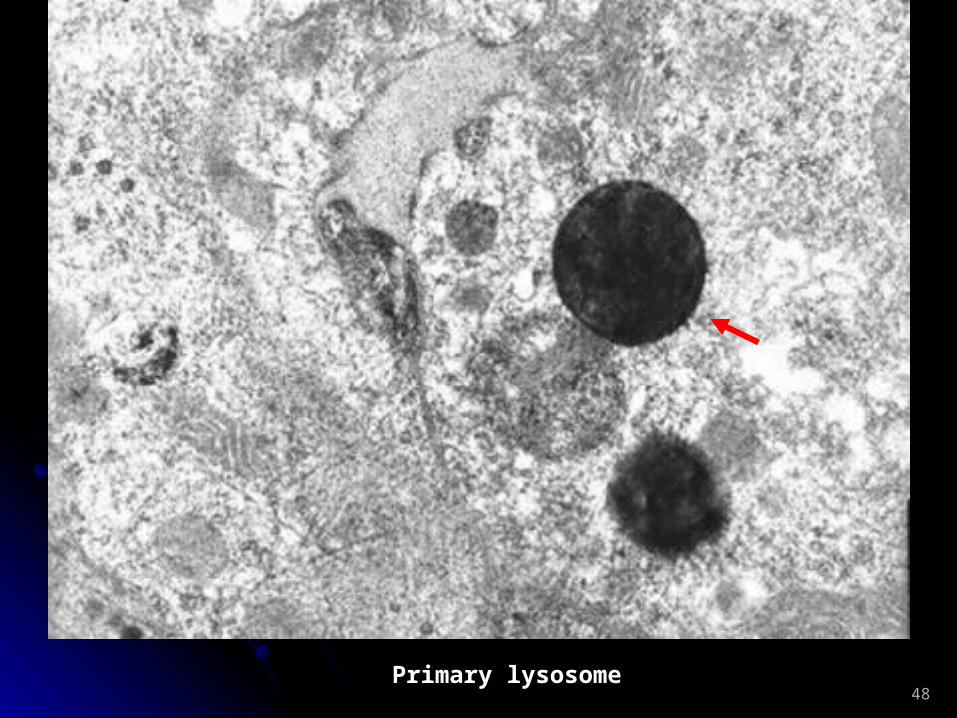
48Primary lysosome

49
Secondary lysosome: Secondary lysosome is the lysosome that is digesting the substances from outside of itself (phagolysosome) or inside of itself (autophagolysosome).
50
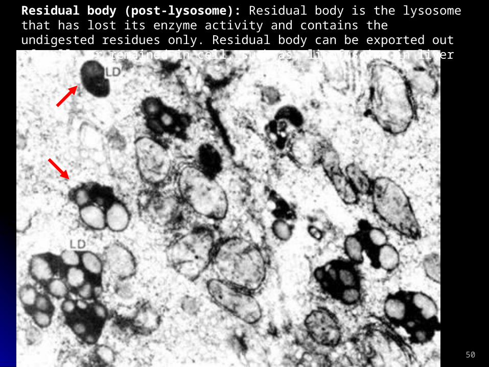
Residual body (post-lysosome): Residual body is the lysosome that has lost its enzyme activity and contains the undigested residues only. Residual body can be exported out of cell, or remained in cell, such as, lipofuscins in liver cell.

51
Function of lysosome:
Lysosome is a digestion organelle in cell. It is associated with cell autolysis, defense, and utilization to some substances.
Digestion in cell: The macromolecules entered by phagocytosis can be digested by lysosome to be utilized by cell. For example, the LDL can be digested for the sterol utilization. Lysosome is unique digestion organ in unicellular organism. Apoptosis: The cell that is going to apoptosis will form an apoptotic body that can be swallowed by macrophage and form a phagosome (secondary lysosome). The apoptotic cell will be eliminated away from tissue by this way. Autolysis: To clear away the wasted bio macromolecules and old organelles. Defense: Macrophage kills pathogens using its lysosome. Regulation to endocrine: Thyroglobulin can be digested in lysosome as thyroxin. Formation of acrosome of sperm: Acrosome helps sperm to enter ovum.

52
Lysosome and diseases: The damage, function inhibition, non-digestion, gene mutation or genetic deficiency of lysosome can cause many diseases for human body, for examples, silicosis (pneumosilicosis), pulmonary tuberculosis, rheumatoid arthritis (The lysosome is easy to break and release out the enzymes that can cause the inflammation), and storage diseases (caused by the mutation of the enzymes in lysosome). The storage diseases include Tay-Sachs disease ( 台 - 萨氏综合征 ), Pompe disease (II 型糖原累积症 ), Gaucher disease ( 脑苷脂沉积症 ), Inclusion-cell disease ( 细胞内含物症 ), and others. All patient children will die within two years.
Peroxisome (microbody): 0.2~1.5μm in diameter. Over 40 oxidases have be found in peroxisome. Oxidases can oxidize substrates and release out hydrogen peroxide (H2O2): RH2+O2 → R+H2O2
Peroxisome take the β-oxidation of the fatty acids in animals. If you use some medicine of fat cleaner to rat, you will detect a 10 folds higher concentration of oxidases in rat liver cells than normal. Peroxisome can take a detoxifying function that oxidizes the toxic substances with H2O2. For example, about a quarter of the alcohol you drank in will be oxidized as acetaldehyde in your liver cells. Plant peroxisome can: join photorespiration; take ① ② β-oxidation of fat. The signal to lead oxidases from plasma enter peroxisome is -Ser-Lys-Leu-COO- 。 The syndrome of Zellweger ( 脑肝肾综合征 ) is caused by abnormal peroxisome (blank peroxisome). The infant will die within 3-6 months.

53
Peroxisomes in human liver cells

54
The peroxisome in
the cell of Nicotiana
tabacum (The central square is the crystal
formed by uricoxidase)