Embed Size (px)
Citation preview
1
2
4
5
6
7
8
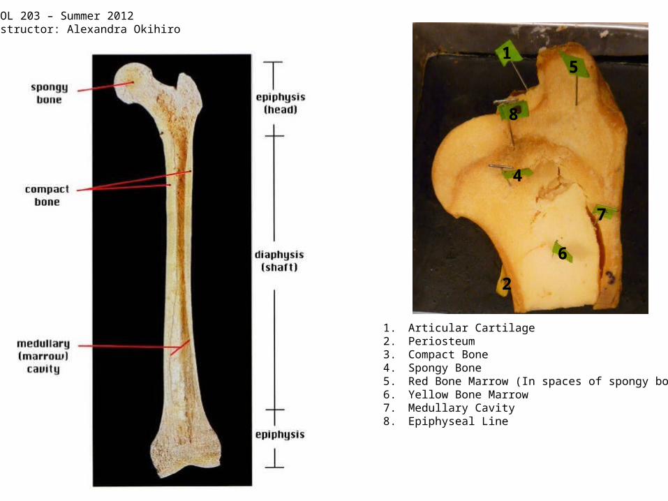
1. Articular Cartilage2. Periosteum3. Compact Bone4. Spongy Bone5. Red Bone Marrow (In spaces of spongy bone)6. Yellow Bone Marrow7. Medullary Cavity8. Epiphyseal Line
BIOL 203 – Summer 2012Instructor: Alexandra Okihiro
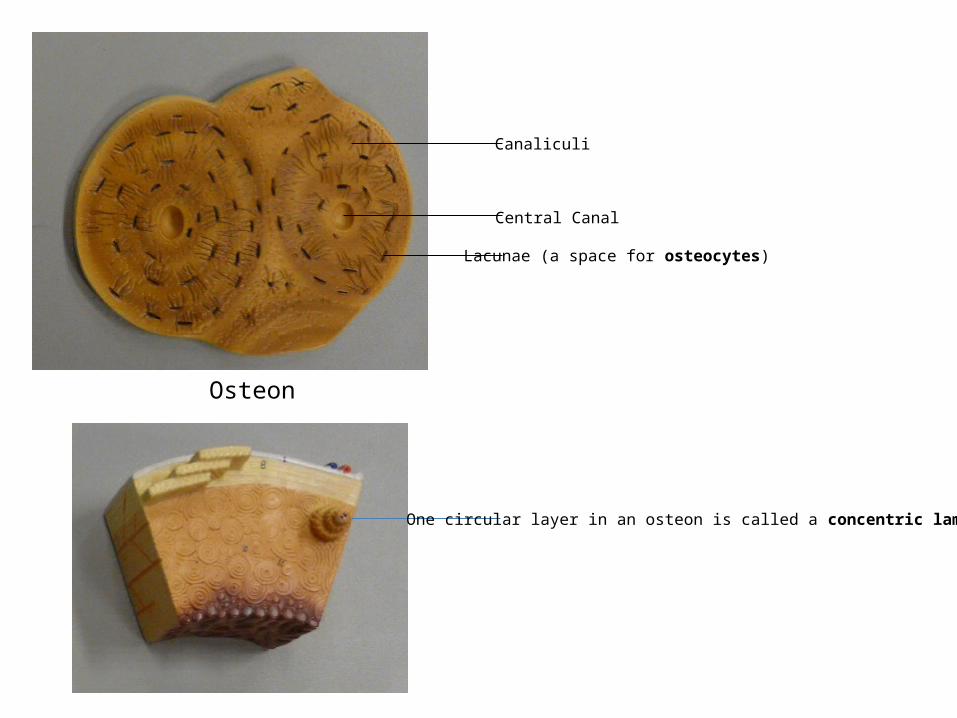
Osteon
Central Canal
Canaliculi
Lacunae (a space for osteocytes)
One circular layer in an osteon is called a concentric lamellae
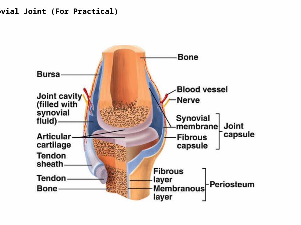
Synovial Joint (For Practical)
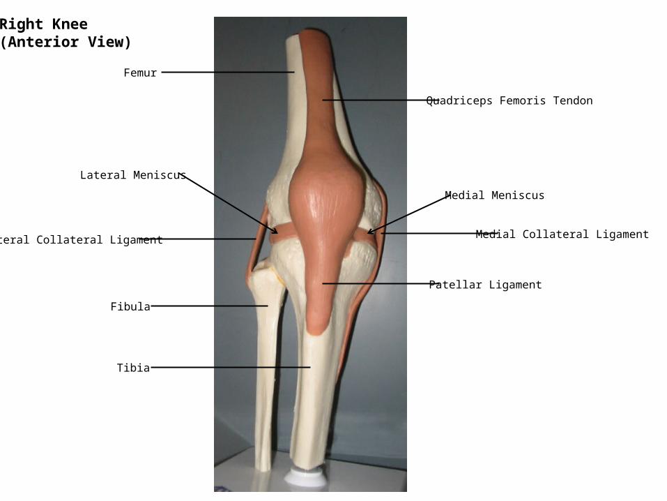
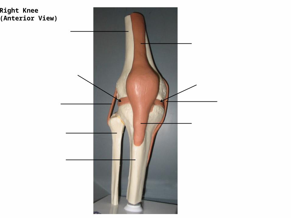
Femur
Tibia
Fibula
Quadriceps Femoris Tendon
Patellar Ligament
Lateral Collateral Ligament Medial Collateral Ligament
Medial Meniscus
Lateral Meniscus
Right Knee(Anterior View)
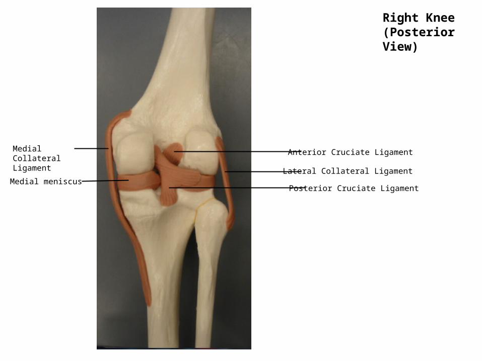
Right Knee(Posterior View)
Anterior Cruciate Ligament
Posterior Cruciate Ligament
Medial Collateral Ligament
Lateral Collateral LigamentMedial meniscus
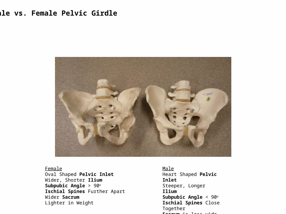
Male vs. Female Pelvic Girdle
FemaleOval Shaped Pelvic InletWider, Shorter IliumSubpubic Angle > 90o
Ischial Spines Further ApartWider SacrumLighter in Weight
MaleHeart Shaped Pelvic InletSteeper, Longer IliumSubpubic Angle < 90o
Ischial Spines Close Together Sacrum is less wideHeavier in Weight
Right Knee(Anterior View)
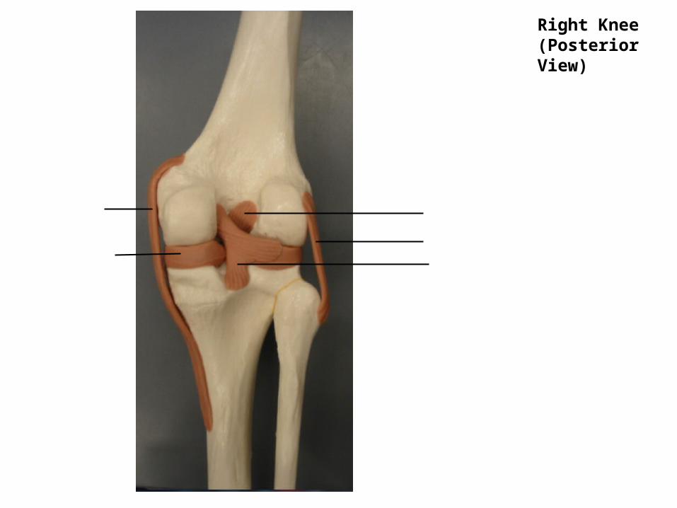
Right Knee(Posterior View)