Embed Size (px)
Citation preview
miR-22 targets BMP-7/6
1
MicroRNA-22 Is A Master Regulator of Bone Morphogenetic Protein-7/6 Homeostasis In The Kidney*
Jianyin Long1*, Shawn S. Badal1,2*, Yin Wang1, Benny H.J. Chang3, Antony Rodriguez4, and Farhad R. Danesh 1,2,5
1From Nephrology Section, The University of Texas MD Anderson Cancer Center, Houston, TX 77030; 2Interdepartmental Graduate Program in Translational Biology and Molecular Medicine, 3Department of
Molecular and Cellular Biology, 4Department of Surgery, and 5Department of Pharmacology, Baylor College of Medicine, Houston, TX 77030
* Both authors contributed equally to the manuscript
*Running title: miR-22 targets BMP-7/6 To whom correspondence should be addressed: Farhad R. Danesh, Section of Nephrology, The University of Texas MD Anderson Cancer Center, 1515 Holcombe Boulevard, Houston, TX 77030, USA, Tel: (713) 745-0858; Fax: (713) 745-0854; E-mail: [email protected] Key words: microRNA, gene regulation, bone morphogenetic proteins (BMPs), kidney, fibrosis. Background: BMPs have emerged as key regulators of kidney fibrosis. Results: Deletion of miR-22 attenuated renal fibrosis via direct targeting of BMP-7/6. BMP-7/6 in turn transcriptionally regulates miR-22 expression. Conclusion: miR-22 and BMP-7/6 form a negative feedback circuit. Significance: The present study is the first to report the effect of miR-22 on BMP signaling and kidney fibrosis. ABSTRACT
Accumulating evidence suggests that microRNAs (miRNA) contribute to a myriad of kidney diseases. However, the regulatory role of miRNAs on the key molecules implicated in kidney fibrosis remains poorly understood. Bone morphogenetic protein-7 (BMP-7) and its related BMP-6 have recently emerged as key regulators of kidney fibrosis. Using the established unilateral ureteral obstruction (UUO) model of kidney fibrosis as our experimental model, we examined the regulatory role of miRNAs on BMP-7/6 signaling. By analyzing the potential miRNAs that target BMP-7/6 in silica, we identified miR-22 as a potent miRNA targeting BMP-7/6. We found that expression levels of BMP-7/6 were significantly elevated in the kidneys of
the miR-22 null mouse. Importantly, mice with targeted deletion of miR-22 exhibited attenuated renal fibrosis in the UUO model. Consistent with these in vivo observations, primary renal fibroblast isolated from miR-22-deficient UUO mice demonstrated a significant increase in BMP-7/6 expression and their downstream targets. This phenotype could be rescued when cells were transfected with miR-22 mimics. Interestingly, we found that miR-22 and BMP-7/6 are in a regulatory feedback circuit, whereby not only miR-22 inhibits BMP-7/6, but miR-22 by itself is induced by BMP-7/6. Finally, we identified two BMP-responsive elements in the proximal region of miR-22 promoter. These findings identify miR-22 as a critical miRNA that contributes to renal fibrosis on the basis of its pivotal role on BMP signaling cascade.
Kidney fibrosis represents a failed wound healing process following a chronic and sustained injury, and regardless of the type of injury and etiology, is the common final outcome of many progressive chronic kidney diseases leading to the destruction and collapse of renal parenchyma and progressive loss of kidney function (1-3).
Activated fibroblasts, also known as
http://www.jbc.org/cgi/doi/10.1074/jbc.M113.498634The latest version is at JBC Papers in Press. Published on October 25, 2013 as Manuscript M113.498634
Copyright 2013 by The American Society for Biochemistry and Molecular Biology, Inc.
by guest on July 5, 2018http://w
ww
.jbc.org/D
ownloaded from

miR-22 targets BMP-7/6
2
myofibroblasts, are commonly regarded as the main effector cells responsible for the excess of extracellular matrix (ECM) production, a key pathological feature of renal fibrosis. Although the origin of the activated fibroblasts pool in the kidney remains controversial, a growing body of evidence indicates that fibrogenic cues in the kidney evoke multiple intracellular signaling pathways that ultimately lead to phenotypic changes in the fibroblasts, resulting in their activation with characteristic expression of several smooth muscle cell markers (1-3). Among the many fibrogenic factors that regulate renal fibrotic processes, transforming growth factor-β (TGF-β) and connective tissue growth factor (CTGF) are regarded as key fibrogenic cytokines (1-4), whereas bone morphogenic protein-7 (BMP-7) and BMP-6, have been known as natural antagonists of TGF-β signaling and anti-fibrogenic factors that have been shown to prevent renal fibrosis in several experimental models (5-11).
BMPs are important signaling molecules that were first identified by their ability to induce bone and cartilage, and subsequently were shown to be pleiotropic cytokines controlling a wide variety of biological processes (12,13). Signaling by BMPs has been long implicated in kidney homeostasis and kidney-injury repair (14). In the BMP canonical signaling pathway, the constitutively active Type-II receptors, upon binding to BMP ligands, phosphorylate and thus activate their Type-I partners, which in turn phosphorylate their intracellular effectors, the receptor-regulated Smad proteins 1, 5 and 8 (Smad1/5/8) (12,13,15). Phosphorylated Smads form complexes with Smad4 and translocate to the nucleus where they regulate expression of their target genes, by binding to BMP-responsive elements (BRE) on the promoter region of their target genes (14-17). Although much is known on the BMP canonical pathway, the precise mechanism of its regulation remains inadequately described.
MicroRNAs (miRNAs) comprise a broad
class of small non-coding RNAs that negatively regulate gene expression by base-pairing to partial or perfect complementary sites in the 3’-untranslated regions (UTR) of specific target mRNAs (18,19). Recent studies from our group (20,21) and others (22-26) suggest that miRNAs are involved in the pathogenesis and progression of a variety of kidney diseases, and hold great therapeutic potential. However, limited information is available on the exact role of miRNAs on renal fibrosis in vivo.
Herein, using the established unilateral ureteral obstruction (UUO) mouse model of renal fibrosis as our experimental model, we found that miR-22 deletion attenuates kidney fibrosis in vivo by targeting BMP-7/6 and BMP receptor type I receptor (BMPR1B). Our findings also suggest that there is a regulatory feedback circuit between miR-22 and BMP-7/6 signaling, whereby miR-22 targets BMP-7/6, but it is by itself induced by this pathway. Furthermore, we identified two BREs in the miR-22 promoter region, which are responsible for the transcriptional regulation of BMP-7/6 on miR-22 expression.
EXPERIMENTAL PROCEDURES
Tissue culture—BALB/c mouse primary kidney fibroblasts were purchased from Cell Biologicals (Chicago, IL). Primary kidney fibroblasts were isolated from wild type or miR-22-deficient kidney cortex as previously described (27). TGF-β1 was purchased from R&D Systems (Minneapolis, MN). HEK 293T cells and HeLa cells were purchased from ATCC and cultured as previously described (28).
miRNA extraction, real-time RT-PCR, in situ hybridization and Northern blot— miRNAs were extracted using Total RNA Purification Kit (Norgen Biotek, Thorold, ON) (15). The TaqMan MicroRNA Reverse Transcription Kit and its compatible TaqMan MicroRNA Assays for U6 snRNA and miR-22-3p (Applied Biosystems) were used to quantify the expression level of mir-22 on a CFX96 Real-Time PCR Detection
by guest on July 5, 2018http://w
ww
.jbc.org/D
ownloaded from
miR-22 targets BMP-7/6
3
System (Bio-Rad). Individual samples were run in duplicate, and each experiment was repeated at least 3 times. Relative gene expression was calculated using the 2-ρρCT
method (29). miR-22 chromogenic in situ hybridization assays were carried out as previously described (30), in collaboration with Bioneer A/S (Hørsholm, Denmark) using specific miR-22-3p probe (ACAGTTCTTCAACTGGCAGCTT) or scramble probe (TGTAACACGTCTATACGCC CA). Northern blots were carried out using γ-32P-ATP (PerkinElmer Life Sciences) end- labeled miRNA locked nucleic acid probes for miR-22 and control U6 snRNA (Exiqon) (31).
In silica targeted gene analysis of miR-22—Three separate algorithms (miRanda, TargetScan, and PicTar) were used to assess potential miRNAs that target BMP-7 and BMP-6. These programs were also used to analyze binding sites in target mRNAs for miR-22. The RNA Hybrid program (32) was used to predict the secondary structure and free energy of the RNA/miRNA duplex.
Plasmids, mutagenesis and miRNAs transfection—The 3’-UTR of the mouse Bmp6 gene (NM_001718), Bmp7 gene (NM_001719) and Bmpr1b gene (NM_001203) were amplified from mouse primary kidney fibroblasts genomic DNA by PCR using HotMaster Taq DNA Polymerase (5PRIME), with the following primers: GTCTCGAGTTGAAGCTGGTGTG TGTGT (Bmp6 forward), GAGAATTCACCAC CGAGAGTCAACACA (Bmp6 reverse); GTCT CGAGCTCTTCCTGAGACCCTGACC (Bmp7 forward), GAGAATTCGTCAGAATAAACCA AGAAACGG (Bmp7 reverse); GTCTCGAGT GCCTGGGACCACAGCTTGG (Bmpr1b forward), GAGAATTCACTGTGCCGTGCAC ACTTCT (Bmpr1b reverse). The PCR products were cloned between XhoI and EcoRI site of luciferase reporter vector 3.1-luc, kindly provided by Dr. Ralph Nicholas (Dartmouth Medical School, Hanover, NH) (33). Putative miR-22 binding site GGCAGCU (nt 153-159) in
mouse Bmp7 was mutated into GAUGAUU by oligonucleotide- directed PCR.
Human miR-22 gene proximal promoter (-1197 to +49) (34) was cloned from HEK 293T genomic DNA by PCR using HotMaster Taq DNA Polymerase, with the following primers: GTAGGTACCGAAGGAAA AGGCATGGATTTCAGTCC (forward), GCAG CTAGCAGGGGGAGCAAATCACTGCGTCC (reverse). The 1246bp PCR product was cloned between KpnI and NheI site of promoter-less luciferase reporter vector pGL4.10 [luc2] (Promega, Madison, WI). Putative mouse miR-22 promoter was retrieved from genebank and aligned to human miR-22 promoter using Clustal Omega. Binding sites for transcription factors were analyzed using rVista 2.0 (35). Potential BREs in this promoter region were mutated through oligonucleotide-directed mutagenesis by regular PCR using the following primers: GCTCGTGCGTCACGAATTCGCCA GCTGATCGG (for BRE-1), CAATCAGAGCC AGAGAATCCGGAGAGGCGGGA (for BRE-2) and CATTGCAAACGCAGAATTCGTGGCTT TCGCCT (for BRE-3), where the mutated BRE sequence is underlined. All constructs were verified by sequencing.
Pre-miRNA precursor and anti-miR miRNAs molecules were purchased from Life Technologies (Grand Island, NY). miRNA mimics and miRNA inhibitors were transfected into cultured cells using Lipofectamine 2000 (Life Technologies, Grand Island, NY) at a final concentration of 40nM (20,21).
For experiments testing the effect of BMP-7/6 signaling on miR-22 expression, HEK 293T cells or Hela cells were serum starved overnight, pretreated with vehicle (DMSO) or 10µM dorsomorphin (Sigma, St. Louis, MO) for 30min before treatment with 100ng/ml BMP-6 or 100ng/ml BMP-7 (R&D Systems) for indicated time (36).
Luciferase reporter assays—For experiments using 3.1-luc luciferase reporter
by guest on July 5, 2018http://w
ww
.jbc.org/D
ownloaded from
miR-22 targets BMP-7/6
4
constructs in vitro, 1.5x10⁵ HeLa cells were plated in 12-well plates. 1.5µg of 3.1-luc empty vector or 3.1- luc-Bmp7, -Bmp6 or -Bmpr1b constructs, 50 ng of pSV-β-gal control vector (Promega) and 30nM of miR-22 mimics were transfected using Lipofectamine 2000 (Life Technologies, Grand Island, NY). Following 48 hrs of transfections, luciferase activity was measured using Steady-Glo Luciferase Assay System (Promega, Madison, WI) on a FLUOstar Omega luminometer (BMG Labtech, Cary, NC) as previously reported (20,21), using β-gal as internal control.
For experiments using pGL4.10 luciferase reporter constructs in vitro, 1.5x10⁵ HEK 293T cells were plated in 12-well plates. 3.3µg of pGL4.10 empty vector or miR-22 promoter constructs (wild type or BRE mutants) and 50 ng of pSV-β-gal control vector (Promega) were transfected into each well using Calcium Phosphate Transfection Kit (Life Technologies, Grand Island, NY) following manufacturer’s instructions. 24hrs post transfection, cells were serum starved overnight and treated with 100ng/ml BMP-6 (R&D Systems, Minneapolis, MN) for 24hrs. Luciferase activity was measured using Steady-Glo Luciferase Assay System on a FLUOstar Omega luminometer, using β-gal as internal control (20,21).
Biotin-miR-22 pulldown— Capture of miR-22 target mRNAs by streptavidin beads pulldown using biotinylated synthetic miR-22 was carried out as previously described (37,38). Briefly, primary renal fibroblasts isolated from kidney cortex of miR-22-/- mice were transfected with 50nM biotin labeled non-targeting mimic or biotin-miR-22 mimic (custom synthesized from Sigma). Cells were harvested 24hrs later in biotin pulldown lysis buffer (20 mM Tris-HCl, pH 8.0, 100mM KCl, 1% NP-40, 1/100 Complete Protease Inhibitor (Sigma) and RNase OUT (50U/ml, Life Technologies). 100µl of M-280 Streptavidin coated Dynabeads at a concentration of 10ng/ml (Life Technologies) were washed 2 times in lysis buffer and added to 1 ml of cell
extract, incubated overnight at 4°C. Pulldowns were washed 5 times in lysis buffer. RNAs from the beads (pull-down RNA) or from 10% of the cell extract (input RNA) were extracted for downstream applications. The level of mRNA in the biotin-labeled control miRNA or miR-22 was quantified by RT-qPCR. For RT-qPCR, mRNA levels were normalized to a housekeeping gene (Gapdh or Ubiquitin C). The enrichment ratio of the control-normalized pull-down RNA to the control-normalized input levels was then calculated (37).
Quantitative Real-time-PCR—SYBR Green-based qPCR on a CFX96 Real-Time PCR Detection System (Bio-Rad) were used to analyze the relative expression levels of the following genes with the different primer sets: collagen I: TGCCGCGACCTCAAGATGTG (forward) and CACAAGGGTGCTGTAGGTG A (reverse); collagen III: GCGGAATTCCTGGA CCAAAAGGTGATGCTG (forward) and CGG ATCCGAGGACCACGTTCCCCATTATG (reverse); fibronectin: 5’-GCGGTTGTCTGACG CTGGCT-3’ (forward) and 5’-TGGGTTCAGC AGCCCCAGGT-3’ (reverse); αSMA: ACTGG GACGACATGGAAAAG (forward) and CAT CTCCAGAGTCCAGCACA (reverse); Bmp7: TGTGGCAGAAAACAGCAGCA (forward); TC AGGTGCAATGATCCAGTCC (reverse); Bmp6: CCAACCACGCCATTGTACAGA (forward); G GAATCCAAGGCAGAACCATG (reverse); Id1: ACCCTGAACGGCGAGATCA (forward) and TCGTCGGCTGGAACACATG (reverse); pri-mir-22: GGAACCTGTGCCTCCCAC (forward) and TTCCCACTGCCACACAGAC (reverse); Gapdh: 5’-CCTTCATTGACCTCAA CTAC-3’ (forward) and 5’-GGAAGGCCATGC CAGTGAGC -3’ (reverse).
Animal studies—All animal studies were conducted according to the “Principles of Laboratory Animal Care” (NIH publication No. 85023, revised 1985) and the guidelines of the IACUC of Baylor College of Medicine. For experiments using UUO model of kidney injury,
by guest on July 5, 2018http://w
ww
.jbc.org/D
ownloaded from
miR-22 targets BMP-7/6
5
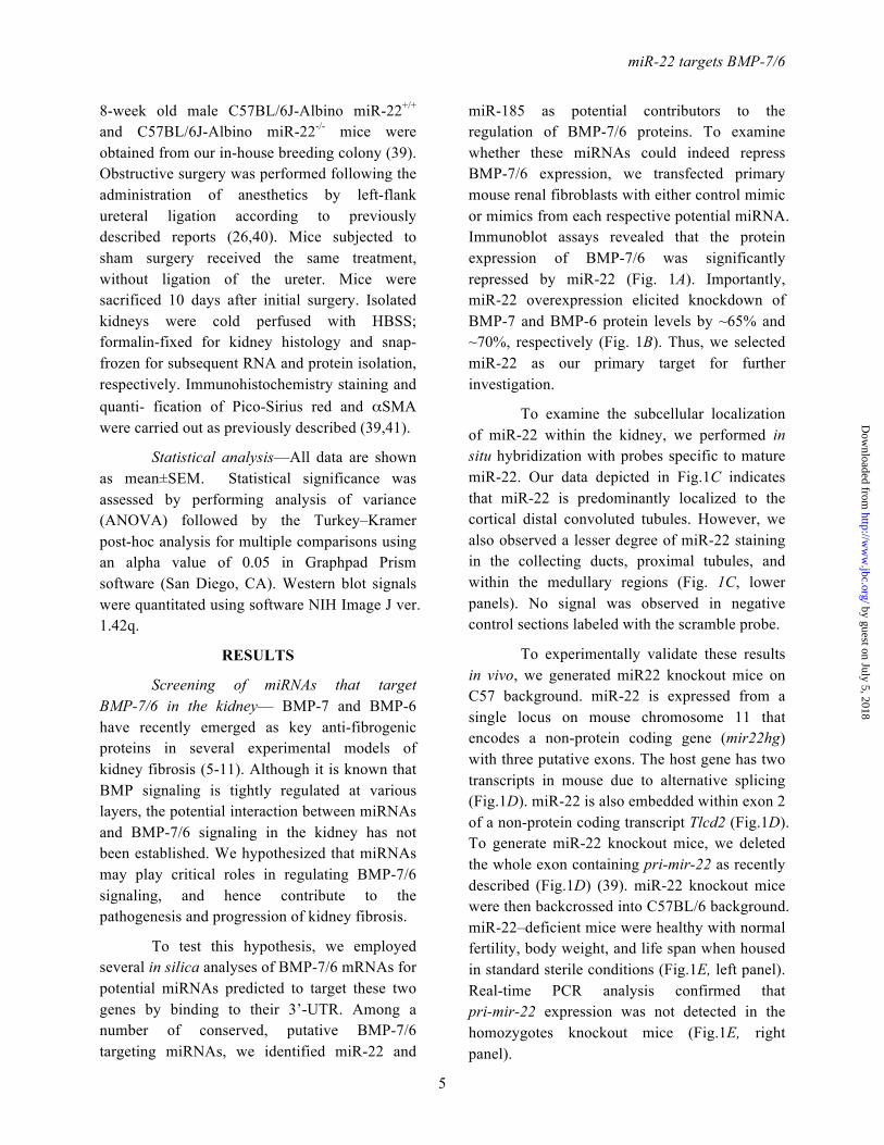
8-week old male C57BL/6J-Albino miR-22+/+ and C57BL/6J-Albino miR-22-/- mice were obtained from our in-house breeding colony (39). Obstructive surgery was performed following the administration of anesthetics by left-flank ureteral ligation according to previously described reports (26,40). Mice subjected to sham surgery received the same treatment, without ligation of the ureter. Mice were sacrificed 10 days after initial surgery. Isolated kidneys were cold perfused with HBSS; formalin-fixed for kidney histology and snap- frozen for subsequent RNA and protein isolation, respectively. Immunohistochemistry staining and quanti- fication of Pico-Sirius red and αSMA were carried out as previously described (39,41).
Statistical analysis—All data are shown as mean±SEM. Statistical significance was assessed by performing analysis of variance (ANOVA) followed by the Turkey–Kramer post-hoc analysis for multiple comparisons using an alpha value of 0.05 in Graphpad Prism software (San Diego, CA). Western blot signals were quantitated using software NIH Image J ver. 1.42q.
RESULTS
Screening of miRNAs that target BMP-7/6 in the kidney— BMP-7 and BMP-6 have recently emerged as key anti-fibrogenic proteins in several experimental models of kidney fibrosis (5-11). Although it is known that BMP signaling is tightly regulated at various layers, the potential interaction between miRNAs and BMP-7/6 signaling in the kidney has not been established. We hypothesized that miRNAs may play critical roles in regulating BMP-7/6 signaling, and hence contribute to the pathogenesis and progression of kidney fibrosis.
To test this hypothesis, we employed several in silica analyses of BMP-7/6 mRNAs for potential miRNAs predicted to target these two genes by binding to their 3’-UTR. Among a number of conserved, putative BMP-7/6 targeting miRNAs, we identified miR-22 and
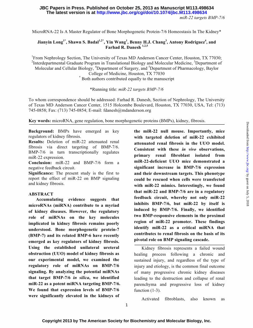
miR-185 as potential contributors to the regulation of BMP-7/6 proteins. To examine whether these miRNAs could indeed repress BMP-7/6 expression, we transfected primary mouse renal fibroblasts with either control mimic or mimics from each respective potential miRNA. Immunoblot assays revealed that the protein expression of BMP-7/6 was significantly repressed by miR-22 (Fig. 1A). Importantly, miR-22 overexpression elicited knockdown of BMP-7 and BMP-6 protein levels by ~65% and ~70%, respectively (Fig. 1B). Thus, we selected miR-22 as our primary target for further investigation.
To examine the subcellular localization of miR-22 within the kidney, we performed in situ hybridization with probes specific to mature miR-22. Our data depicted in Fig.1C indicates that miR-22 is predominantly localized to the cortical distal convoluted tubules. However, we also observed a lesser degree of miR-22 staining in the collecting ducts, proximal tubules, and within the medullary regions (Fig. 1C, lower panels). No signal was observed in negative control sections labeled with the scramble probe.
To experimentally validate these results in vivo, we generated miR22 knockout mice on C57 background. miR-22 is expressed from a single locus on mouse chromosome 11 that encodes a non-protein coding gene (mir22hg) with three putative exons. The host gene has two transcripts in mouse due to alternative splicing (Fig.1D). miR-22 is also embedded within exon 2 of a non-protein coding transcript Tlcd2 (Fig.1D). To generate miR-22 knockout mice, we deleted the whole exon containing pri-mir-22 as recently described (Fig.1D) (39). miR-22 knockout mice were then backcrossed into C57BL/6 background. miR-22–deficient mice were healthy with normal fertility, body weight, and life span when housed in standard sterile conditions (Fig.1E, left panel). Real-time PCR analysis confirmed that pri-mir-22 expression was not detected in the homozygotes knockout mice (Fig.1E, right panel).
by guest on July 5, 2018http://w
ww
.jbc.org/D
ownloaded from

miR-22 targets BMP-7/6
6
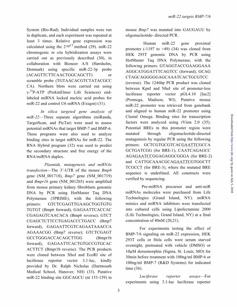
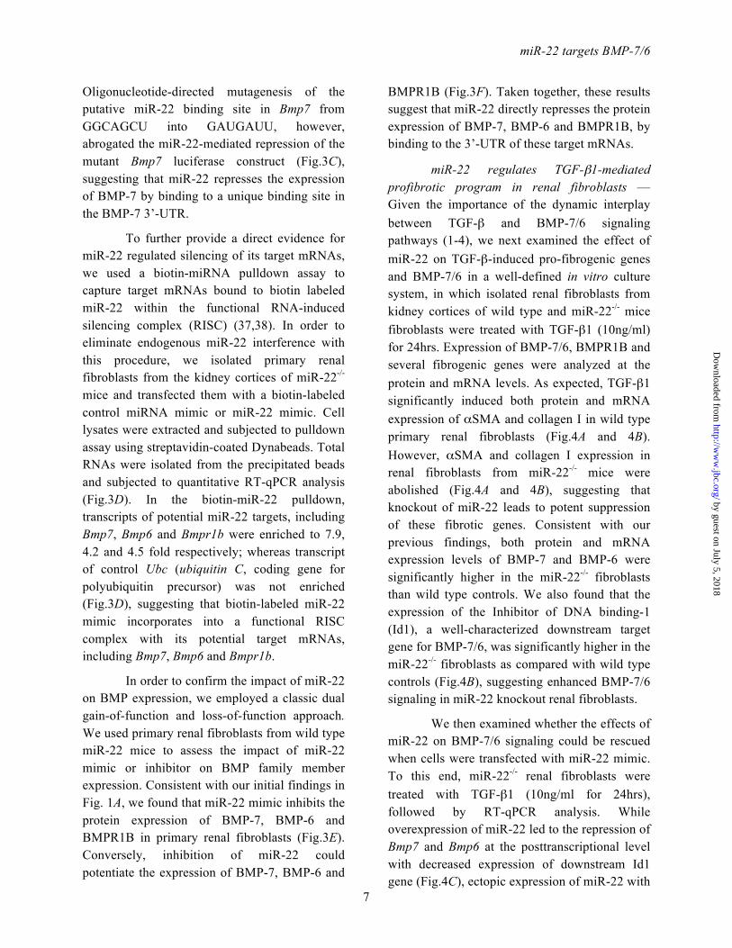
Targeted deletion of miR-22 ameliorates features of renal fibrosis in the unilateral ureteral obstruction (UUO) model of kidney fibrosis— To test the role of miR-22 on renal fibrosis in vivo, we used the well-characterized UUO mouse model of kidney injury, which results in progressive interstitial fibrosis (42). Wild type and miR-22-/- mice were subjected to obstructive or sham injury for 10 days. Following this period, the degree of renal fibrosis was evaluated by Pico-Sirius red staining of kidney sections. As expected, the UUO injury in wild type mice led to a significant increase in type I collagen deposition as measured by relative intensity of Pico-Sirius red positive tissue (Fig.2A). Targeted deletion of miR-22, however, impeded the observed abundance of type 1 collagen in the UUO model (Fig. 2A). We also quantified the expression of αSMA, a key marker of activated myofibroblasts. Immuno- histochemistry staining revealed that UUO led to significantly elevated expression of αSMA in wild type mice (Fig.2B), whereas similar to attenuated type 1 collagen expression, the expression of αSMA was significantly reduced in the miR-22-/- mice compared to wild type (Fig.2B).
In considering the ramifications of the effect of miR-22 on other key fibrotic genes, total RNAs were extracted from kidney cortices of mice after UUO injury from each group, and subjected to quantitative real time PCR. As expected, UUO injury induced the expression of several fibrotic marker genes in wild type mice, (Figs. 2C-2F). In contrast, mRNA expression of each of these genes, including collagen I, collagen III, fibronectin and αSMA, was significantly suppressed in miR-22 deficient mice following UUO (Fig.2C-2F).
To establish the regulatory role of miR-22 on BMP-7/6 protein expression in vivo, we extracted kidney cortex proteins from wild type and miR-22 knockout mice following UUO injury and performed several immunoblot assays. As shown in Fig. 2G, the protein levels of
BMP-7 and BMP-6 were ~3.5 fold and 2.5 fold higher, respectively in miR-22-/- mice compared with wild type controls in UUO conditions. BMPR1B, the type I receptor for BMP-7/6 signaling, was similarly upregulated by ~2.0 fold in miR-22-/- UUO mice compared to UUO control mice (Fig. 2G). To assess whether these genetic changes had downstream functional consequences, we probed our samples for phospho-Smad1/5, the critical downstream mediator of BMP signaling. While total Smad5 levels were comparable among wild type and miR-22-/- mice following UUO injury, deletion of miR-22 led to significantly higher phosphorylation of Smad1/5, indicating enhanced expression of BMP downstream targets in miR-22 deficient mice (Fig. 2G). Thus, our findings suggest that targeted deletion of miR-22 enhances BMP-7/6 signaling in vivo, which may contribute to the amelioration of renal fibrosis.
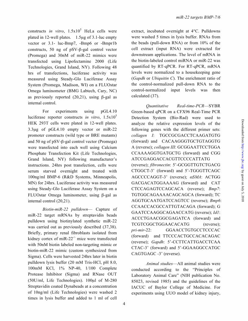
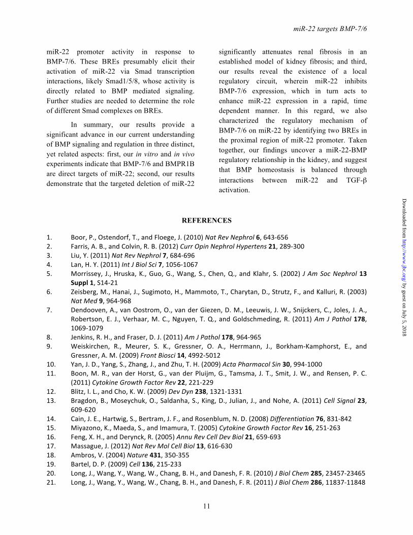
miR-22 directly targets key components of BMP signaling—(18,19). Several algorithms suggest the existence of miR-22 binding sites in the 3’-UTR of mouse Bmp7, Bmp6 and Bmpr1b mRNAs (Fig.3A). Thus, to examine whether BMP-7/6 mRNAs are bona fide targets of miR-22, we evaluated the 3’-UTR sequence of BMP-7/6 mRNAs for the presence of a miR-22 binding site. As shown in Fig. 3B, complementary binding of miR-22 seed sequence to these binding sites in the 3’-UTR of target mRNAs are expected to generate relatively stable secondary structure between miR-22 and Bmp7, Bmp6 or Bmpr1b (ΔG<-20 kcal/mol) (Fig. 3B). To confirm that miR-22 indeed binds directly to 3’-UTR to repress the expression of these target genes, we amplified the 3’-UTR of Bmp7, Bmp6 or Bmpr1b from mouse genomic DNA by PCR, and subcloned them into the pcDNA3.1-luc empty vector. Luciferase reporter assays indicated that when these 3’-UTR reporter constructs were introduced into Hela cells, their transcriptions were markedly repressed by co-transfection of miR-22 mimics but not with the control mimic (Fig.3C).
by guest on July 5, 2018http://w
ww
.jbc.org/D
ownloaded from
miR-22 targets BMP-7/6
7
Oligonucleotide-directed mutagenesis of the putative miR-22 binding site in Bmp7 from GGCAGCU into GAUGAUU, however, abrogated the miR-22-mediated repression of the mutant Bmp7 luciferase construct (Fig.3C), suggesting that miR-22 represses the expression of BMP-7 by binding to a unique binding site in the BMP-7 3’-UTR.
To further provide a direct evidence for miR-22 regulated silencing of its target mRNAs, we used a biotin-miRNA pulldown assay to capture target mRNAs bound to biotin labeled miR-22 within the functional RNA-induced silencing complex (RISC) (37,38). In order to eliminate endogenous miR-22 interference with this procedure, we isolated primary renal fibroblasts from the kidney cortices of miR-22-/- mice and transfected them with a biotin-labeled control miRNA mimic or miR-22 mimic. Cell lysates were extracted and subjected to pulldown assay using streptavidin-coated Dynabeads. Total RNAs were isolated from the precipitated beads and subjected to quantitative RT-qPCR analysis (Fig.3D). In the biotin-miR-22 pulldown, transcripts of potential miR-22 targets, including Bmp7, Bmp6 and Bmpr1b were enriched to 7.9, 4.2 and 4.5 fold respectively; whereas transcript of control Ubc (ubiquitin C, coding gene for polyubiquitin precursor) was not enriched (Fig.3D), suggesting that biotin-labeled miR-22 mimic incorporates into a functional RISC complex with its potential target mRNAs, including Bmp7, Bmp6 and Bmpr1b.
In order to confirm the impact of miR-22 on BMP expression, we employed a classic dual gain-of-function and loss-of-function approach. We used primary renal fibroblasts from wild type miR-22 mice to assess the impact of miR-22 mimic or inhibitor on BMP family member expression. Consistent with our initial findings in Fig. 1A, we found that miR-22 mimic inhibits the protein expression of BMP-7, BMP-6 and BMPR1B in primary renal fibroblasts (Fig.3E). Conversely, inhibition of miR-22 could potentiate the expression of BMP-7, BMP-6 and
BMPR1B (Fig.3F). Taken together, these results suggest that miR-22 directly represses the protein expression of BMP-7, BMP-6 and BMPR1B, by binding to the 3’-UTR of these target mRNAs.
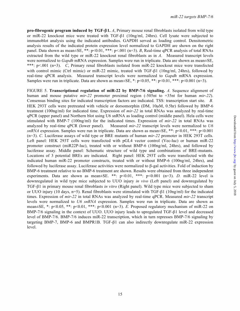
miR-22 regulates TGF-β1-mediated profibrotic program in renal fibroblasts — Given the importance of the dynamic interplay between TGF-β and BMP-7/6 signaling pathways (1-4), we next examined the effect of miR-22 on TGF-β-induced pro-fibrogenic genes and BMP-7/6 in a well-defined in vitro culture system, in which isolated renal fibroblasts from kidney cortices of wild type and miR-22-/- mice fibroblasts were treated with TGF-β1 (10ng/ml) for 24hrs. Expression of BMP-7/6, BMPR1B and several fibrogenic genes were analyzed at the protein and mRNA levels. As expected, TGF-β1 significantly induced both protein and mRNA expression of αSMA and collagen I in wild type primary renal fibroblasts (Fig.4A and 4B). However, αSMA and collagen I expression in renal fibroblasts from miR-22-/- mice were abolished (Fig.4A and 4B), suggesting that knockout of miR-22 leads to potent suppression of these fibrotic genes. Consistent with our previous findings, both protein and mRNA expression levels of BMP-7 and BMP-6 were significantly higher in the miR-22-/- fibroblasts than wild type controls. We also found that the expression of the Inhibitor of DNA binding-1 (Id1), a well-characterized downstream target gene for BMP-7/6, was significantly higher in the miR-22-/- fibroblasts as compared with wild type controls (Fig.4B), suggesting enhanced BMP-7/6 signaling in miR-22 knockout renal fibroblasts.
We then examined whether the effects of miR-22 on BMP-7/6 signaling could be rescued when cells were transfected with miR-22 mimic. To this end, miR-22-/- renal fibroblasts were treated with TGF-β1 (10ng/ml for 24hrs), followed by RT-qPCR analysis. While overexpression of miR-22 led to the repression of Bmp7 and Bmp6 at the posttranscriptional level with decreased expression of downstream Id1 gene (Fig.4C), ectopic expression of miR-22 with
by guest on July 5, 2018http://w
ww
.jbc.org/D
ownloaded from

miR-22 targets BMP-7/6
8
miR-22 mimic in miR-22-/- renal fibroblasts partially restored the expression of αSMA and collagen I, at the basal as well as following induction with TGF-β1- (Fig.4C). Taken together, these findings suggest that miR-22 plays a critical role in the dynamic interactions between BMP-7/6 and TGF-β signaling pathways.
Transcriptional regulation of miR-22 by BMP-7/6 signaling— The regulation of miR-22 expression is largely unknown, although there are reports indicating that miR-22 expression can be induced by several nuclear receptors, including vitamin D3 (43,44), testosterone (45), and retinoic acids (46).
To study the transcriptional regulation of miR-22, we retrieved mouse miR-22 promoter sequence from genomic sequence of mir22hg gene, based on homologous alignment to recently published human miR-22 promoter sequence (34). As shown in Fig.5A, the proximal region (-500bp upstream of transcription starting site) of mouse and human miR-22 promoter is highly homologous (80% identical), suggesting a possible conserved regulation of miR-22 expression. Further careful examination of the miR-22 proximal promoter region for transcription factor binding sites revealed several binding sites for MyoD, CREB and NF-κB (Fig. 5A). Importantly, there were three consensus BMP-responsive elements (BREs) in this region, including GACGCC (BRE-1 and BRE-3) and GGCGCC (BRE-2) (Fig. 5A). This was quite surprising since BREs, which are believed to serve as the binding sites for phosphorylated Smad1/5 transcription complex (12,15-17), are commonly found in BMP target genes (e.g., Id1 GACGCC, Id2 GGCGCC, and Id3 GGCGCC) (47-51).
The existence of several BREs in miR-22 promoter prompted us to examine whether miR-22 and BMP-7/6 are in a regulatory negative feedback loop, in which not only miR-22 inhibits BMP-7/6 gene, but miR-22 by itself is regulated by BMP-7/6. To test this possibility, cultured
human embryonic fibroblasts HEK 293T cells were treated with 100ng/ml BMP-6 in different time points. RT-qPCR analysis of mir-22 gene revealed that BMP-6 treatment indeed leads to increased levels of miR-22 in a time-dependent manner, reaching a plateau around 2hrs (Fig. 5B, upper panel), indicating that mir-22 is an early responsive gene of BMP-6. To further test whether BMP signaling directly enhances miR-22 expression, we examined the effect of BMP inhibition on the endogenous levels of miR-22 RNA. To this end, HEK 293T cells were pretreated with dorsomorphin, a selective small molecule inhibitor of BMP type 1 receptors (36), prior to BMP-6 treatment. Pretreatment of dorsomorphin abrogated the expected response of miR-22 to BMP-6 (Fig. 5B, upper panel), suggesting that induction of miR-22 by BMP-6 requires functional type I BMP receptors. Total RNAs from these cells were also subjected to Northern blot using specific miR-22 probe. Consistent with our previous findings in RT-qPCR (Fig. 5B, upper panel), we observed that miR-22 RNA level was also rapidly induced by BMP-6 within 6hrs as detected by Northern blot analysis (Fig. 5B, middle panel). In a similar study using BMP-7, we observed an almost identical pattern, showing early response of miR-22 to BMP-7 (Fig. 5B, lower panel). Thus, our data suggest that miR-22 is an immediate early gene induced by BMP-7/6 signaling.
We then generated luciferase reporter construct with the human miR-22 proximal promoter and examined its response to BMP-7/6 signaling. Cells were transfected with with empty vector pGL4.1 and wild type miR-22 promoter construct, treated with BMP-6 (100ng/ml for 16hrs) prior to luciferase assay. Under these conditions, wild type human miR-22 promoter construct showed an average of ~500 fold higher luciferase activity compared to empty vector (Fig. 5C). Importantly, BMP-6 treatment further increased miR-22 promoter activity ~ 2.6 fold (Fig. 5C, left panel), indicating that miR-22
by guest on July 5, 2018http://w
ww
.jbc.org/D
ownloaded from

miR-22 targets BMP-7/6
9
responds to BMP-6 stimulus at the transcriptional level.
To delineate the BRE element(s) responsible for BMP induction in miR-22 promoter, we generated a series of miR-22 promoter mutant constructs, where BRE-1, BRE-2, BRE-3 were each separately or in- combination mutated (Fig.5C, middle panel). Subsequent luciferase assays indicated that mutation of BRE-1 and BRE-3, but not BRE-2, significantly reduced the response of miR-22 to BMP-6 (Fig. 5C, right panel). Double mutant BRE-1/BRE-2 had similar BMP-6 response to BRE-1, while triple mutant BRE-1/BRE-2 /BRE-3 was completely unresponsive to BMP-6 treatment (Fig. 5C, right panel). Taken together, these findings indicate that BRE-1 and BRE-3, but not BRE-2, are responsible for BMP-6 induction in miR-22, and support the notion that miR-22 forms a regulatory loop with BMP-7/6 signaling.
In light of the observed transcriptional regulation of miR-22 by BMP-7/6, we decided to verify whether miR-22 underwent a similar pattern of downregulation in vivo under UUO conditions. To this end, we examined miR-22 expression by qPCR in kidney cortices from wild type mice that had undergone UUO, and found that miR-22 was indeed downregulated by ~2 fold when compared to sham controls (Fig. 5D, left panel). This result suggests sustained BMP-7/6 is critical for miR-22 regulation within the kidney.
In addition to BMP signaling, we were also interested in exploring whether known fibrogenic factors, i.e., TGF-β1, could likewise downregulate miR-22 expression. To test this, mouse renal fibroblasts were stimulated with TGF-β1 (10ng/ml) over several time points. We found significant downregulation of miR-22 by qPCR (Fig. 5D, right panel). Taken together, these results suggest that both TGF-β1 and BMP-7/6 may contribute to the expression levels of miR-22 in the kidney.
DISCUSSION
Emerging evidence suggests that BMP-7/6, members of the TGF-β superfamily of cysteine knot cytokines, play pivotal roles in kidney homeostasis mainly by antagonizing TGF-β-induced profibrogenic signaling that contribute to the accumulation of the extracellular matrix (5-11).
In this study, we uncover a critical role of miR-22 on BMP signaling and renal fibrosis by providing strong evidence that miR-22 directly targets BMP-7, BMP-6 and BMPR1B. In support of our conclusions, we found that targeted deletion of miR-22 significantly attenuated renal fibrosis in the well-characterized UUO model of kidney fibrosis. Knockdown of miR-22 both genetically or pharmacologically, resulted in elevated BMP-7/6 protein levels and enhanced BMP-7/6 downstream targets. Importantly, using miR-22-deficient primary renal fibroblasts, we demonstrated that miR-22 mimic could partially rescue the profibrotic effect of TGF-β1. Finally, we made the novel observation that BMP-7/6 per se could induce miR-22 at the transcriptional level through two consensus BREs in the proximal promoter region of miR-22 gene. Our findings suggest that miR-22 is an immediate early target gene of BMP-7/6, requiring an intact BMP-7/6 signaling, since pretreatment with dorsomorphin, a selective small-molecule inhibitor of BMP type 1 receptor, abolished the induction of miR-22 expression by BMP-6. Taken together, our data suggest that miR-22 serves as an important regulator of BMP homeostasis and kidney fibrosis in response to extracellular signals.
Recent studies from our group (20,21) and others (22-26) have demonstrated that microRNAs play important roles in the development and progression of a variety of kidney diseases leading to kidney fibrosis as their final common pathway. Among miRNAs modulating kidney fibrosis, miR-21 has been recently reported by several groups to positively
by guest on July 5, 2018http://w
ww
.jbc.org/D
ownloaded from

miR-22 targets BMP-7/6
10
regulate renal fibrosis in UUO and ischemia reperfusion injury (22,26,52-55). Similarly, miR-200 has also been implicated in renal fibrosis (56,57). The present study is the first to report the effect of miR-22 on BMP homeostasis and kidney fibrosis. However, we recognize that the effect of miR-22 on BMP-7/6 could be kidney and tissue-specific. For instance, we have recently reported an association between miR-22 and cardiomyocyte hypertrophy and cardiac fibrosis (39), where in contrast to the current findings, deletion of miR-22 was associated with enhanced cardiac fibrosis (39,58). Our interpretation of these functional differences on the effect of miR-22 on tissue fibrosis is that the effect of miRNAs on their target mRNAs have been convincingly shown to be tissue and temporal-dependent (59,60). Thus, miR-22 could preferentially target BMP-7/6 in the kidney, where BMP-7/6 are highly expressed, whereas other miR-22 targets [e.g., purine-rich element binding protein B (PURB) (39) or HDAC4 (58) could be preferentially targeted by miR-22 in the heart. Finally, it is a canonical feature of miRNAs to exert their effects on target genes in a pleiotropic manner. Therefore, several other important targets, in addition to BMP-7/6 and BMPR1B, may potentially mediate the effect of miR-22 on renal fibrosis.
Our findings also suggest that miR-22 forms a negative feedback circuit with BMP-7/6 signaling (Fig.5E). This finding is of considerable interest since accumulating evidence suggests that not only miRNAs can regulate their target genes, but also these target genes may in turn interact with and regulate these specific miRNAs positively or negatively, either at the transcriptional or post-transcriptional level (61,62). In our current study, we established such a relationship between miR-22 and BMP-7/6, whereby we identified BMP-7/6 and BMPR1B as direct targets of miR-22. On the other hand, we provided strong evidence that BMP-7/6 are also positive regulators of miR-22 levels mainly via their direct transcriptional regulation on miR-22
expression. These findings further extend the observations of others and validate the regulatory effects of miRNAs as fine-tuning elements (63), critical in the homeostasis of genes in response to a particular stimulus.
Much attention has recently been paid to canonical BMP signaling, which is known to be largely mediated through activation of Smad1/5/8, while TGF-β signaling is mainly mediated by activation of Smad2/3. Both signaling pathways share Smad4 as a common assembly point into a heteromeric signaling complex which translocates into the nucleus to regulate target gene expression in coordination with several other transcription factors (12-17). Cross-talk between TGF-β and BMP signaling is a well known feature of the TGF-β superfamily. BMP-7/6 is a potent antagonistic factor of TGF-β signaling (6) and repression of BMP-7/6 expression leads to increased activation of TGF-β signaling, a key pathogenic factor within renal fibrosis (Fig. 4B). Within our model system, knockout of miR-22 led to increased BMP-7/6 mRNA and protein levels, while concomitantly caused blunting of the TGF-β1 induced profibrogenic genes (Fig.4A-B). These findings suggest that miR-22 plays a critical role in the cross-talk between TGF-β and BMP-7/6 signaling pathways.
Interestingly, miR-22 itself is reduced under wild-type UUO conditions, most likely as a consequence of reduced expression of its key regulators BMP-7/6, and an indirect effect via upregulation of its negative regulator, TGF-β1 (Fig. 5E). The renoprotective effect of complete knockout of miR-22 highlights the importance of sustained miR-22 expression to modulate levels of BMP-7/6 and BMPR1B under normal and pathogenic conditions.
A surprising finding of this study was that we identified two conserved BREs within the proximal promoter region of the miR-22 host gene. A reporter gene expression assay indicated that these elements are necessary to enhance
by guest on July 5, 2018http://w
ww
.jbc.org/D
ownloaded from
miR-22 targets BMP-7/6
11
miR-22 promoter activity in response to BMP-7/6. These BREs presumably elicit their activation of miR-22 via Smad transcription interactions, likely Smad1/5/8, whose activity is directly related to BMP mediated signaling. Further studies are needed to determine the role of different Smad complexes on BREs.
In summary, our results provide a significant advance in our current understanding of BMP signaling and regulation in three distinct, yet related aspects: first, our in vitro and in vivo experiments indicate that BMP-7/6 and BMPR1B are direct targets of miR-22; second, our results demonstrate that the targeted deletion of miR-22
significantly attenuates renal fibrosis in an established model of kidney fibrosis; and third, our results reveal the existence of a local regulatory circuit, wherein miR-22 inhibits BMP-7/6 expression, which in turn acts to enhance miR-22 expression in a rapid, time dependent manner. In this regard, we also characterized the regulatory mechanism of BMP-7/6 on miR-22 by identifying two BREs in the proximal region of miR-22 promoter. Taken together, our findings uncover a miR-22-BMP regulatory relationship in the kidney, and suggest that BMP homeostasis is balanced through interactions between miR-22 and TGF-β activation.
REFERENCES
1. Boor, P., Ostendorf, T., and Floege, J. (2010) Nat Rev Nephrol 6, 643-‐656 2. Farris, A. B., and Colvin, R. B. (2012) Curr Opin Nephrol Hypertens 21, 289-‐300 3. Liu, Y. (2011) Nat Rev Nephrol 7, 684-‐696 4. Lan, H. Y. (2011) Int J Biol Sci 7, 1056-‐1067 5. Morrissey, J., Hruska, K., Guo, G., Wang, S., Chen, Q., and Klahr, S. (2002) J Am Soc Nephrol 13
Suppl 1, S14-‐21 6. Zeisberg, M., Hanai, J., Sugimoto, H., Mammoto, T., Charytan, D., Strutz, F., and Kalluri, R. (2003)
Nat Med 9, 964-‐968 7. Dendooven, A., van Oostrom, O., van der Giezen, D. M., Leeuwis, J. W., Snijckers, C., Joles, J. A.,
Robertson, E. J., Verhaar, M. C., Nguyen, T. Q., and Goldschmeding, R. (2011) Am J Pathol 178, 1069-‐1079
8. Jenkins, R. H., and Fraser, D. J. (2011) Am J Pathol 178, 964-‐965 9. Weiskirchen, R., Meurer, S. K., Gressner, O. A., Herrmann, J., Borkham-‐Kamphorst, E., and
Gressner, A. M. (2009) Front Biosci 14, 4992-‐5012 10. Yan, J. D., Yang, S., Zhang, J., and Zhu, T. H. (2009) Acta Pharmacol Sin 30, 994-‐1000 11. Boon, M. R., van der Horst, G., van der Pluijm, G., Tamsma, J. T., Smit, J. W., and Rensen, P. C.
(2011) Cytokine Growth Factor Rev 22, 221-‐229 12. Blitz, I. L., and Cho, K. W. (2009) Dev Dyn 238, 1321-‐1331 13. Bragdon, B., Moseychuk, O., Saldanha, S., King, D., Julian, J., and Nohe, A. (2011) Cell Signal 23,
609-‐620 14. Cain, J. E., Hartwig, S., Bertram, J. F., and Rosenblum, N. D. (2008) Differentiation 76, 831-‐842 15. Miyazono, K., Maeda, S., and Imamura, T. (2005) Cytokine Growth Factor Rev 16, 251-‐263 16. Feng, X. H., and Derynck, R. (2005) Annu Rev Cell Dev Biol 21, 659-‐693 17. Massague, J. (2012) Nat Rev Mol Cell Biol 13, 616-‐630 18. Ambros, V. (2004) Nature 431, 350-‐355 19. Bartel, D. P. (2009) Cell 136, 215-‐233 20. Long, J., Wang, Y., Wang, W., Chang, B. H., and Danesh, F. R. (2010) J Biol Chem 285, 23457-‐23465 21. Long, J., Wang, Y., Wang, W., Chang, B. H., and Danesh, F. R. (2011) J Biol Chem 286, 11837-‐11848
by guest on July 5, 2018http://w
ww
.jbc.org/D
ownloaded from

miR-22 targets BMP-7/6
12
22. Dey, N., Das, F., Mariappan, M. M., Mandal, C. C., Ghosh-‐Choudhury, N., Kasinath, B. S., and Choudhury, G. G. (2011) J Biol Chem 286, 25586-‐25603
23. Wang, Q., Wang, Y., Minto, A. W., Wang, J., Shi, Q., Li, X., and Quigg, R. J. (2008) FASEB J 22, 4126-‐4135
24. Kato, M., Zhang, J., Wang, M., Lanting, L., Yuan, H., Rossi, J. J., and Natarajan, R. (2007) Proc Natl Acad Sci U S A 104, 3432-‐3437
25. Wei, Q., Bhatt, K., He, H. Z., Mi, Q. S., Haase, V. H., and Dong, Z. (2010) J Am Soc Nephrol 21, 756-‐761
26. Chau, B. N., Xin, C., Hartner, J., Ren, S., Castano, A. P., Linn, G., Li, J., Tran, P. T., Kaimal, V., Huang, X., Chang, A. N., Li, S., Kalra, A., Grafals, M., Portilla, D., MacKenna, D. A., Orkin, S. H., and Duffield, J. S. (2012) Sci Transl Med 4, 121ra118
27. Grimwood, L., and Masterson, R. (2009) Propagation and culture of renal fibroblasts. in Methods in Molecular Biology: Kidney Research (Hewitson, T. D., and Becker, G. J. eds.), Humans Press, Totowa, NJ. pp 25-‐37
28. Long, J., Matsuura, I., He, D., Wang, G., Shuai, K., and Liu, F. (2003) Proc Natl Acad Sci U S A 100, 9791-‐9796
29. Livak, K. J., and Schmittgen, T. D. (2001) Methods 25, 402-‐408 30. Jorgensen, S., Baker, A., Moller, S., and Nielsen, B. S. (2010) Methods 52, 375-‐381 31. Varallyay, E., Burgyan, J., and Havelda, Z. (2008) Nat Protoc 3, 190-‐196 32. Rehmsmeier, M., Steffen, P., Hochsmann, M., and Giegerich, R. (2004) RNA 10, 1507-‐1517 33. Du, M., Roy, K. M., Zhong, L., Shen, Z., Meyers, H. E., and Nichols, R. C. (2006) FEBS J 273, 732-‐745 34. Bar, N., and Dikstein, R. (2010) PLoS ONE 5, e10859 35. Loots, G. G., and Ovcharenko, I. (2004) Nucleic Acids Res 32, W217-‐221 36. Yu, P. B., Hong, C. C., Sachidanandan, C., Babitt, J. L., Deng, D. Y., Hoyng, S. A., Lin, H. Y., Bloch, K.
D., and Peterson, R. T. (2008) Nat Chem Biol 4, 33-‐41 37. Lal, A., Thomas, M. P., Altschuler, G., Navarro, F., O'Day, E., Li, X. L., Concepcion, C., Han, Y. C.,
Thiery, J., Rajani, D. K., Deutsch, A., Hofmann, O., Ventura, A., Hide, W., and Lieberman, J. (2011) PLoS Genet 7, e1002363
38. Orom, U. A., and Lund, A. H. (2007) Methods 43, 162-‐165 39. Gurha, P., Abreu-‐Goodger, C., Wang, T., Ramirez, M. O., Drumond, A. L., van Dongen, S., Chen, Y.,
Bartonicek, N., Enright, A. J., Lee, B., Kelm, R. J., Jr., Reddy, A. K., Taffet, G. E., Bradley, A., Wehrens, X. H., Entman, M. L., and Rodriguez, A. (2012) Circulation 125, 2751-‐2761
40. Satoh, M., Kashihara, N., Yamasaki, Y., Maruyama, K., Okamoto, K., Maeshima, Y., Sugiyama, H., Sugaya, T., Murakami, K., and Makino, H. (2001) J Am Soc Nephrol 12, 317-‐325
41. Wang, W., Wang, Y., Long, J., Wang, J., Haudek, S. B., Overbeek, P., Chang, B. H., Schumacker, P. T., and Danesh, F. R. (2012) Cell Metab 15, 186-‐200
42. Klahr, S., and Morrissey, J. (2002) Am J Physiol Renal Physiol 283, F861-‐875 43. Alvarez-‐Diaz, S., Valle, N., Ferrer-‐Mayorga, G., Lombardia, L., Herrera, M., Dominguez, O., Segura,
M. F., Bonilla, F., Hernando, E., and Munoz, A. (2012) Hum Mol Genet 21, 2157-‐2165 44. Wang, W. L., Chatterjee, N., Chittur, S. V., Welsh, J., and Tenniswood, M. P. (2011) Mol Cancer 10,
58 45. Delic, D., Grosser, C., Dkhil, M., Al-‐Quraishy, S., and Wunderlich, F. (2010) Steroids 75, 998-‐1004 46. Jian, P., Li, Z. W., Fang, T. Y., Jian, W., Zhuan, Z., Mei, L. X., Yan, W. S., and Jian, N. (2011) J
Hematol Oncol 4, 20 47. Lopez-‐Rovira, T., Chalaux, E., Massague, J., Rosa, J. L., and Ventura, F. (2002) J Biol Chem 277,
3176-‐3185 48. Nakahiro, T., Kurooka, H., Mori, K., Sano, K., and Yokota, Y. (2010) Biochem Biophys Res Commun
399, 416-‐421 49. Korchynskyi, O., and ten Dijke, P. (2002) J Biol Chem 277, 4883-‐4891
by guest on July 5, 2018http://w
ww
.jbc.org/D
ownloaded from

miR-22 targets BMP-7/6
13
50. Katagiri, T., Imada, M., Yanai, T., Suda, T., Takahashi, N., and Kamijo, R. (2002) Genes Cells 7, 949-‐960
51. Shepherd, T. G., Theriault, B. L., and Nachtigal, M. W. (2008) Gene 414, 95-‐105 52. Ben-‐Dov, I. Z., Muthukumar, T., Morozov, P., Mueller, F. B., Tuschl, T., and Suthanthiran, M. (2012)
Transplantation 94, 1086-‐1094 53. Glowacki, F., Savary, G., Gnemmi, V., Buob, D., Van der Hauwaert, C., Lo-‐Guidice, J. M., Bouye, S.,
Hazzan, M., Pottier, N., Perrais, M., Aubert, S., and Cauffiez, C. (2013) PLoS ONE 8, e58014 54. Zarjou, A., Yang, S., Abraham, E., Agarwal, A., and Liu, G. (2011) Am J Physiol Renal Physiol 301,
F793-‐801 55. Zhong, X., Chung, A. C., Chen, H. Y., Meng, X. M., and Lan, H. Y. (2011) J Am Soc Nephrol 22,
1668-‐1681 56. Oba, S., Kumano, S., Suzuki, E., Nishimatsu, H., Takahashi, M., Takamori, H., Kasuya, M., Ogawa, Y.,
Sato, K., Kimura, K., Homma, Y., Hirata, Y., and Fujita, T. (2010) PLoS ONE 5, e13614 57. Xiong, M., Jiang, L., Zhou, Y., Qiu, W., Fang, L., Tan, R., Wen, P., and Yang, J. (2012) Am J Physiol
Renal Physiol 302, F369-‐379 58. Huang, Z. P., Chen, J., Seok, H. Y., Zhang, Z., Kataoka, M., Hu, X., and Wang, D. Z. (2013) Circ Res
112, 1234-‐1243 59. Sayed, D., and Abdellatif, M. (2011) Physiol Rev 91, 827-‐887 60. van Rooij, E. (2011) Circ Res 108, 219-‐234 61. Mendell, J. T., and Olson, E. N. (2012) Cell 148, 1172-‐1187 62. Davis, B. N., Hilyard, A. C., Lagna, G., and Hata, A. (2008) Nature 454, 56-‐61 63. Iliopoulos, D., Malizos, K. N., Oikonomou, P., and Tsezou, A. (2008) PLoS ONE 3, e3740
FOOTNOTES We are grateful to Dr. Ralph C. Nicholas at Dartmouth Medical School for luciferase reporter
construct 3.1-luc. We thank the Sequencing Core at Baylor College of Medicine. The work was supported by grants from NIDDK RO1DK091310 and RO1DK078900 (to F.R.D).
ABBREVIATIONS The abbreviations used are: miRNA, microRNA; TGF-β1, transforming growth factor-β1; BMP,
bone morphogenetic protein; BMPR1B, bone morphogenetic protein receptor, type 1B; αSMA, smooth muscle α-actin; UUO, unilateral ureteral obstruction; BRE, BMP-responsive element; UTR, untranslated region; RT-qPCR, Real-time quantitative polymerase chain reaction.
by guest on July 5, 2018http://w
ww
.jbc.org/D
ownloaded from

miR-22 targets BMP-7/6
14
Figure Legends
FIGURE 1. BMP-7/6 are direct targets of miR-22 in the kidney. A, Cultured primary renal fibroblasts were transfected with the indicated miRNA mimics predicted to target BMP-7 in silica. Cell lysate were analyzed for the expression of BMP-6 and BMP-7 by immunoblotting using β-actin as loading control. B, Densitometric analysis of BMP-7 and BMP-6 expression in cells transfected with control mimic (Ctrl mimic) or miR-22 mimics shown in A (n=3). C, Expression pattern of miR-22 in a wild type C57 mouse kidney cortex as detected by in situ hybridization (ISH) with the indicated probes. Blue is the ISH signal and red is nuclear counterstaining. Scale bars denote 200µm. D, Genomic structure of the mouse mir-22 host gene. The miR-22 host gene is depicted along with several alternative splice variants. E. Representative image of wild type and miR-22-/- mice (Left panel). Real-time PCR of kidney cortex RNA to show that pri-mir-22 expression could not be detected in miR-22-/- mice compared to miR-22+/+ controls. Gapdh served as the reference gene (Right panel).
FIGURE 2. Targeted deletion of miR-22 significantly improved renal fibrosis in the UUO model of kidney injury. A, Representative Sirius red staining pictures of kidney tissues from wild type or miR-22 knockout mice subjected to sham or UUO injury (10 days, n=5). Quantitative results of % collagen-positive area are shown as mean±SE, ***: p<0.001 (n=5). Scale bars denote 500µm. B, Representative light micrographs of kidney tissues from wild type or miR-22 knockout mice subjected to sham or UUO injury (10 days, n=5) stained for αSMA protein, Quantitative results of % αSMA area are shown as mean±SE, *: p<0.05 (n=5). Scale bars denote 500µm. C-F, Real-time qPCR analysis depicts expression of several pro-fibrotic genes from kidney cortices of wild type or miR-22 knockout mice subjected to sham or UUO injury (10 days, n=5). Measured transcript levels were normalized to Gapdh mRNA expression. Samples were run in triplicate (n=5). Data are shown as mean±SE. **: p<0.01, ***: p<.001(n=5). G, Immunoblot analysis of kidney cortices of miR-22 knockout mice subjected to UUO injury compared to injured wild-type controls, GAPDH served as a loading control. Densitometric analysis results of the indicated protein expression level normalized to GAPDH are shown, data shown as mean±SE, **: p<0.01, ***: p<0.001 (n=4). FIGURE 3. miR-22 targets several components of BMP-7/6 signaling, including BMPR1B. A Representative table of algorithms highlighting the potential number of miR-22 binding sites within the 3’-UTR of indicated genes in BMP signaling. B. Sequence alignment of miR-22 with the mouse Bmp7, Bmp6 and Bmpr1b 3’-UTR’s based on TargetScan algorithm (Left Panel). miR-22 can potentially form strong secondary structures with the target sequence of 3’-UTR’s of Bmp7, Bmp6 and Bmpr1b (Right panel, predicted by the RNA Hybrid program). C, HeLa cells were co-transfected with empty vector (Vector) and three independent plasmids expressing either 3.1-luc-Bmp7, -Bmp6 or –Bmpr1b wild type 3’-UTR (WT-UTR) or a 3.1-luc-Bmp7 mutant 3’ UTR (BMP-7 mut) and the indicated control mimic (Ctrl mimic) or miR-22 mimic. Luciferase activities were normalized to β-gal activities. Results were obtained from three independent experiments. Data are shown as mean±SE. **: p<0.01 (n=3). D. miR-22 deficient renal fibroblasts were transfected with either biotin-control mimic (Biotin-Ctrl mimic) or biotin-miR-22 mimic (Biotin-miR-22 mimic) and subjected to streptavidin pulldown, followed by qPCR detection of enrichment of indicated miR-22 target genes (see Materials and Methods for details). Results were obtained from three independent experiments. Data are shown as mean±SE. ***: p<0.001 (n=4). E. Wild type renal fibroblasts were transfected with control mimic (Ctrl mimic) or miR-22 mimic followed by immunoblot analysis of expression level of the indicated miR-22 targets. Densitometric analysis results of the indicated protein expression level normalized to GAPDH are shown, data shown as mean±SE, **: p<0.01 (n=3). F. Wild type renal fibroblasts were transfected with control inhibitor (Anti-Ctrl) or miR-22 inhibitor (Anti-miR-22) followed by immunoblot analysis of expression level of the indicated miR-22 targets. Densitometric analysis results of the indicated protein expression level normalized to GAPDH are shown, data shown as mean±SE, **: p<0.01 (n=3). FIGURE 4. Knockout of miR-22 attenuates, and rescue of miR-22 partially restores, the
by guest on July 5, 2018http://w
ww
.jbc.org/D
ownloaded from
miR-22 targets BMP-7/6
15
pro-fibrogenic program induced by TGF-β1. A, Primary mouse renal fibroblasts isolated from wild type or miR-22 knockout mice were treated with TGF-β1 (10ng/ml, 24hrs). Cell lysate were subjected to immunoblot analysis using the indicated antibodies. GAPDH served as loading control. Densitometric analysis results of the indicated protein expression level normalized to GAPDH are shown on the right panel. Data shown as mean±SE, **: p<0.01, ***: p<.001 (n=3). B, Real-time qPCR analysis of total RNAs extracted from the wild type or miR-22 knockout renal fibroblasts as in A. Measured transcript levels were normalized to Gapdh mRNA expression. Samples were run in triplicate. Data are shown as mean±SE, ***: p<.001 (n=3). C, Primary renal fibroblasts isolated from miR-22 knockout mice were transfected with control mimic (Ctrl mimic) or miR-22 mimic, treated with TGF-β1 (10ng/ml, 24hrs), followed by real-time qPCR analysis. Measured transcript levels were normalized to Gapdh mRNA expression. Samples were run in triplicate. Data are shown as mean±SE, *: p<0.05, **: p<0.01, ***: p<0.001 (n=3). FIGURE 5. Transcriptional regulation of miR-22 by BMP-7/6 signaling. A. Sequence alignment of human and mouse putative mir-22 promoter proximal region (-505nt to +35nt for human mir-22). Consensus binding sites for indicated transcription factors are indicated. TSS: transcription start site. B. HEK 293T cells were pretreated with vehicle or dorsomorphin (DM, 10µM, 0.5hr) followed by BMP-6 treatment (100ng/ml) for indicated time. Expression of mir-22 in total RNAs was analyzed by real-time qPCR (upper panel) and Northern blot using U6 snRNA as loading control (middle panel). Hela cells were stimulated with BMP-7 (100ng/ml) for the indicated times. Expression of mir-22 in total RNAs was analyzed by real-time qPCR (lower panel). Measured mir-22 transcript levels were normalized to U6 snRNA expression. Samples were run in triplicate. Data are shown as mean±SE, **: p<0.01, ***: p<0.001 (n=3). C. Luciferase assays of wild type or BRE mutants of human mir-22 promoter in HEK 293T cells. Left panel: HEK 293T cells were transfected with pGL4 vector control (Vec-luc) or human miR-22 promoter construct (miR22P-luc), treated with or without BMP-6 (100ng/ml, 24hrs), and followed by luciferase assay. Middle panel: Schematic structure of wild type and combinations of BRE-mutants. Locations of 3 potential BREs are indicated. Right panel: HEK 293T cells were transfected with the indicated human miR-22 promoter constructs, treated with or without BMP-6 (100ng/ml, 24hrs), and followed by luciferase assay. Luciferase activities were normalized to β-gal activities. Fold of induction by BMP-6 treatment relative to no BMP-6 treatment are shown. Results were obtained from three independent experiments. Data are shown as mean±SE. **: p<0.01, ***: p<0.001 (n=3). D. miR-22 level is downregulated in wild type mice subjected to UUO injury in vivo (Left panel) and downregulated by TGF-β1 in primary mouse renal fibroblasts in vitro (Right panel). Wild type mice were subjected to sham or UUO injury (10 days, n=5). Renal fibroblasts were stimulated with TGF-β1 (10ng/ml) for the indicated times. Expression of mir-22 in total RNAs was analyzed by real-time qPCR. Measured mir-22 transcript levels were normalized to U6 snRNA expression. Samples were run in triplicate. Data are shown as mean±SE, *: p<0.05, **: p<0.01, ***: p<0.001 (n=3). E. Proposed regulatory mechanism of miR-22 on BMP-7/6 signaling in the context of UUO. UUO injury leads to upregulated TGF-β1 level and decreased level of BMP-7/6. BMP-7/6 induces miR-22 transcription, which in turn represses BMP-7/6 signaling by targeting BMP-7, BMP-6 and BMPR1B. TGF-β1 can also indirectly downregulate miR-22 expression level.
by guest on July 5, 2018http://w
ww
.jbc.org/D
ownloaded from

Figure 1
miR-22Scramble
In Situ Hybridization
Mir22hg-001
Tlcd2-004
Mir22hg-002
miR-22
A B
CD
BMP-7
BMP-60.0
0.5
1.0
1.5 Ctrl mimicmiR-22 mimic
*** *** Rel
ativ
e Ex
pres
sion
(Fol
d of
Cha
nge)
BMP-7
BMP-6
β-actin
Neg Ctrl
MockmiR-22
miR-185
miR-138-1
miR-129-5p
miR-340
miR-762
miR-329
miR-592
miRNA mimics
E
miR-22+/+
miR-22-/-
pri-mir-22
Gapdh
miR-22+/+
miR-22-/-
Cotrex
Medulla
50μm 50μm
200μm 200μm
by guest on July 5, 2018http://w
ww
.jbc.org/D
ownloaded from

UUO
Sham
Sirius Red
E Collagen IIICollagen I
αSMA
αSMA
miR-22+/+
UUO
Sham
Figure 2
miR-22-/-miR-22+/+ miR-22-/-
+/+
miR-22-/-
miR-22
0
2
4
6
8
10 ShamUUO
***
***
Rel
ativ
e Ex
pres
sion
(Fol
d of
Cha
nge)
+/+
miR-22-/-
miR-22
0.0
0.5
1.0
1.5
2.0ShamUUO**
**
Rela
tive
Exp
ress
ion
(Fol
d of
Cha
nge)
+/+
miR-22-/-
miR-22
0.0
0.5
1.0
1.5
2.0
2.5ShamUUO
***
***
Rel
ativ
e Ex
pres
sion
(Fol
d of
Cha
nge)
+/+
miR-22-/-
miR-22
0.0
0.5
1.0
1.5
2.0ShamUUO
***
***
Rela
tive
Exp
ress
ion
(Fol
d of
Cha
nge)
+/+
miR-22-/-
miR-22
0
2
4
6ShamUUO
***
***
Colla
gen
Area
(%)
+/+
miR-22-/-
miR-22
0
10
20
30ShamUUO
*
*
ααSM
A Ar
ea (%
)
EFibronectin
BA
C D E F
BMP-7
BMP-6
pSmad1/5
GAPDH
miR-22+/+ UUO miR-22-/- UUO
BMPR1B
Smad5BMP-7
BMP-6
BMPR1B
pSmad1/50
2
4
6 miR-22+/+ UUOmiR-22-/- UUO
******
*****
Rela
tive
Exp
ress
ion
(Fol
d of
Cha
nge)
EG
500 mm
by guest on July 5, 2018http://w
ww
.jbc.org/D
ownloaded from

Figure 3
Biotin miR-22 pulldowns
3' ugUCAAG--AA--GUUGA-CCGUCGAa 5' mmu-miR-22 :|||| ||: ||||||| 5' caGGUUCCAUUCCCAGAAGGGCAGCUc 3' 465-491 Bmp6 3' ugUCAAGAAGUUGACCGUCGAa 5' mmu-miR-22 :|| : : ||||||| 5' ugGGUG---UGGUAGGCAGCUc 3' 652-670 Bmpr1b
3' ugUCAAGAAGU-----UGACCGUCGAa 5' mmu-miR-22 :|||| || :| ||||||| 5' agGGUUC--CAGN11UGCAGGCAGCUg 3' 127-160 Bmp7 5'
mfe: -24.0 kcal/mol
A GGUUCCAU U C
CC A G
A AGGGCAGCU C
AA
GC
UG
CCAG
UUGAAG
AA
CUGU
miR-22/Bmp65'
mfe: -22.3 kcal/mol
G GGUUCCAGA
AACCUG A G
CGUGC A
GGCAGCU G
AA
GC
UG
CCA
GU
UGAAG
AA
CUGU miR-22/Bmp7
5'
mfe: -20.8 kcal/mol
GGGUGU
GGUAGGCAGCU
CA A G C U G C C
A GU U GAA G
AA
CU GU
miR-22/Bmpr1b
3’-UTR luciferase assays
miR-mimic transfections
Ctrl m
imic
MockmiR-22
BMP-7
BMP-6
GAPDH
BMPR1B
BMP-7
BMP-6
GAPDH
BMPR1B
Anti-Ctrl
MockAnti-
miR-22
D
A
C
EF
BMP-7
BMP-6
BMPR1B0.0
0.5
1.0
1.5 Ctrl mimicmiR-22 mimic
****
**
Rela
tive
Expr
essio
n
(Fol
d of
Cha
nge)
miR-inhibitor transfections
B
BMP-7
BMP-6
BMPR1B0
1
2
3
4 Anti-CtrlAnti-miR-22
**
**
**
Rela
tive
Expr
essio
n
(Fol
d of
Cha
nge)
Vector
Bmp7Bmp6
Bmpr1b
Bmp7 mut
0
50
100
150Ctrl mimicmiR-22 mimic
******
WT-UTR
Rela
tive
Lucif
eras
e Un
its
Control
Bmp7Bmp6
Bmpr1b0
2
4
6
8
10Biotin-Ctrl mimicBiotin-miR-22 mimic
***
******
Enric
hmen
t Ra
tio(N
orm
aliz
ed to
Gap
dh)
Mouse DIANAmT miRanda miRDB miRWalk PICTAR4 PICTAR5 PITA RNA22 TargetScan SUM
Bmp7 1 1 0 1 0 1 0 0 1 5
Bmp6 1 1 0 1 0 1 0 0 1 5
Bmpr1b 1 1 0 1 0 1 0 0 1 5
Selection criteria: Minimum seed length- 5mer, longest pontential binding site within 3’ UTR, p-value cutoff of <0.05
by guest on July 5, 2018http://w
ww
.jbc.org/D
ownloaded from
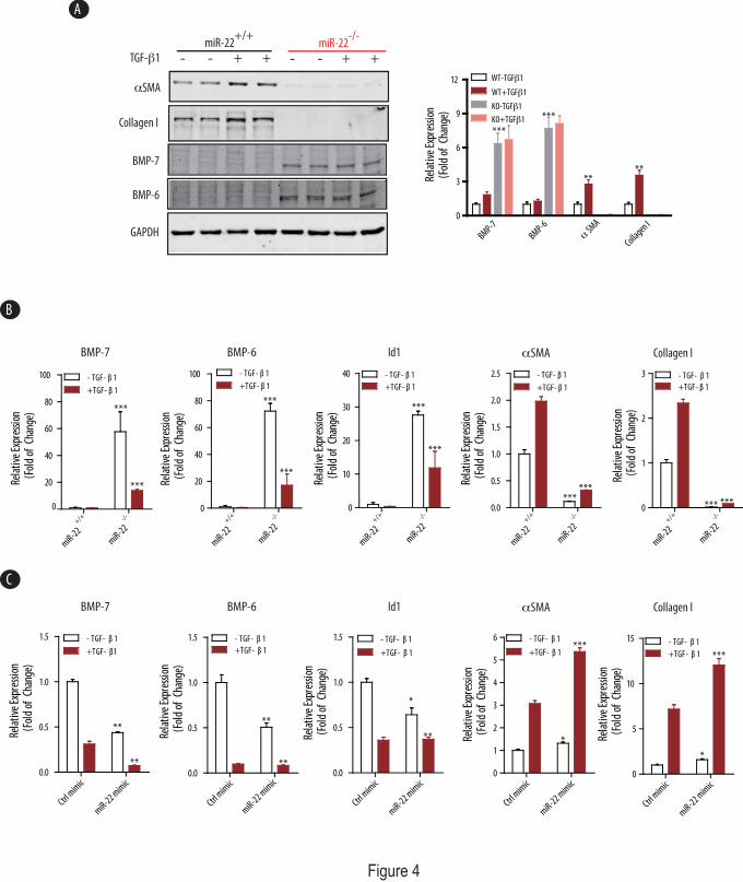
Figure 4
BMP-7
BMP-6
Collagen I
GAPDH
TGF-β1
αSMA
- - + +miR-22
+/+
BMP-7 BMP-6 Id1 αSMA Collagen I
BMP-7 BMP-6 Id1 αSMA Collagen I
+/+
miR-22
-/-
miR-22
0
20
40
60
80
100 - TGF- β 1
+TGF- β 1
***
***Rela
tive
Expr
essio
n(F
old
of C
hang
e)
+/+
miR-22
-/-
miR-22
0
20
40
60
80
100 - TGF- β 1
+TGF- β 1
***
***
Rela
tive
Expr
essio
n(F
old
of C
hang
e)
+/+
miR-22-/-
miR-22
0
10
20
30
40 - TGF- β 1
+TGF- β 1
***
***
Rela
tive
Expr
essio
n(F
old
of C
hang
e)
- - + +miR-22
-/-
Ctrl m
imic
miR-22 mim
ic0
1
2
3
4
5
6 - TGF- β 1
+TGF- β 1***
*Rela
tive
Expr
essio
n(F
old
of C
hang
e)
Ctrl m
imic
miR-22 mim
ic0.0
0.5
1.0
1.5 - TGF- β 1
+TGF- β1
**
**
Rela
tive
Expr
essio
n(F
old
of C
hang
e)
Ctrl m
imic
miR-22 mim
ic0.0
0.5
1.0
1.5 - TGF- β 1
+TGF- β 1
**
**
Rela
tive
Expr
essio
n(F
old
of C
hang
e)
Ctrl m
imic
miR-22 mim
ic0.0
0.5
1.0
1.5 - TGF- β 1
+TGF- β 1
*
**Rela
tive
Expr
essio
n(F
old
of C
hang
e)
Ctrl m
imic
miR-22 mim
ic0
5
10
15 - TGF- β 1
+TGF- β 1 ***
*
Rela
tive
Expr
essio
n(F
old
of C
hang
e)
+/+
miR-22
-/-
miR-22
0.0
0.5
1.0
1.5
2.0
2.5 - TGF- β 1
+TGF- β 1
******
Rela
tive
Expr
essio
n(F
old
of C
hang
e)
+/+
miR-22
-/-
miR-22
0
1
2
3 - TGF- β 1
+TGF- β 1
*** ***
Rela
tive
Expr
essio
n(F
old
of C
hang
e)
C
B
A
BMP-7
BMP-6 SMA
αColla
gen I0
3
6
9
12 WT-TGFβ1
**
***
**
***
Rela
tive
Expr
essio
n(F
old
of C
hang
e)
WT+TGFβ1
KO-TGFβ1
KO+TGFβ1
by guest on July 5, 2018http://w
ww
.jbc.org/D
ownloaded from

Figure 5
hmiR22-P -505 CTCCAACAAGGAGGGGCTGTCGCCCTTTAAGGACTTCCCCGGGCTGGATTCCGAGCTCAGmmiR22-P -502 TTCCAAGAAGGAGGGGCTGTAGCCCTTTAAGGACTTCCCCGCGACCGAGTCAGAGATCAG ***** *************.******************** *. ** **.***.****
hmiR22-P -445 TTTAAAAATGCCAACTCACAGAGCGCGCTGGGTTCTGGGAACGACGGTGTCAGGCTGAGAmmiR22-P -442 TTTAAAAATGCCAACTCACAGAGCGCGCTGCATTCTGGGAAGCTGAGTGTCACCGTAAGA ****************************** .********* : .****** *.***
hmiR22-P -385 ACTACACGGACCAGCATGCATTGCAAACGCAGACGCCGTGGCTTTCGCCTGCTCTTTAGGmmiR22-P -382 ACTTCATTGACCGGAATGCACTGCAAAAATACACGCCTATACTTCCTTCTGCTCTTTA-A ***:** ****.*.***** ******.. * ***** : .*** * ********** .
hmiR22-P -325 ACTCTCGTTTGACGTAGCGCTTTTCTAAGCGGGTCGCTGACGCGGAGGGCTGGGCCACACmmiR22-P -323 ACTGTAGTTTGACGTAAAGCTGGTCTAAGCAAGTCGCCTAGGCCGAGGGT-TAGCCACAC *** *.**********..*** *******..***** * ** ***** .*******
hmiR22-P -265 CTGTCCGTCTATTGGCCGGCTCCTCAGTGGGTGGGCGTGGCTTCGTGCAATTCCGCCCGGmmiR22-P -264 CTTTTCAGCCATTGGCCAGTTGGTTAGTTGGTAGGCGTGGCTTAGAGAAGCTCCTCCAGG ** * *. * *******.* * * *** ***.**********.*:*.*. *** **.**
hmiR22-P -205 CAAAAGGGTGGCCTCCTTGCCAATCAGAGCCAGGGCGCCCGGAGAGGCGGGACCAGGGCGmmiR22-P -204 CAAGGGGGTGGCCTCCTTGCCAATCAGAGCCCAGACGCCTGAATGGGCGGGAGTAAGCAG ***..**************************..*.**** *.* .******* *.* .*
hmiR22-P -145 AGGCCCCGTCGGCCCCCGGGTGGGCGGGGCCACGTTGCCCAGCAGTGGGCGGTGATTGGCmmiR22-P -144 AGGTGC-TGGCGCCCCCGAGTGGGTGTGGTCACGTTGCCCAGCAATGGGCGGTGATTGGC *** * *******.***** * ** **************.***************
hmiR22-P -085 CCCGGGCCGTGCATTCGCAGCTCGTGCGTCACGACGCCGCCAGCTGATCGGAGCCTGGAGmmiR22-P -085 CCTGGGTGGTTCATTCGCAGCTCGTGCGTCACGACGCCGCCAGCTGATCGGAGACTGGAG ** *** ** ******************************************.******
hmiR22-P -025 CCGGTGTGTGCTGGGTGCCGAGAAGAGACAGCGCCGCCGGCCGTGGGGAGCGGACGCAGTmmiR22-P -025 CCGGTGTGTGCTGGGCGCTGGGAAGAGACAGAGCGGTCGGCCGTGCGGACAGGTCGCAGT *************** ** *.**********.** * ******** *** .**:******
MyoD
USF
CREB
STAT
NF-kB
BRE-1
BRE-2
TSS
BRE-3
miR-22
0 0.2 0.5 1 2 4 60
1
2
3
4 BMP-6
DM+BMP-6
**
*** *** ***
Rela
tive
Expr
essio
n(F
old
of C
hang
e)
BMP-6 DM+BMP-6
miR-22
U6 snRNA
0 0.2 0.5 1 2 4 6Hrs 0.2 0.5 1 2 4 6
C
B
E
A
0 0.2 0.5 1 2 4 240
1
2
3
*
**
**
Rela
tive
Expr
essio
n(F
old
of C
hang
e)
BMP-7 (Hrs)
Vec-luc
miR22P-luc
0
510
200
600
1000
1400
1800 - BMP-6+BMP-6
***
**
Rela
tive
Lucif
eras
e Un
its
BMP-6 (Hrs)
D
Fold of Induction
(BMP-6)
BRE-
1BR
E-2
BRE-
3
luc-1197 +49
X
X
luc-1197 +49
luc-1197 +49
luc-1197 +49
X
luc-1197 +49
X
luc-1197 +49
X X X
X
1 2 3 4
BRE123
BRE12
BRE3
BRE2
BRE1
WT
**
****
**
UUO
BMP-7/6TGF-β1
miR-22
--+
0.0
0.5
1.0
1.5
***
Rel
ativ
e m
iR-2
2 E
xpre
ssio
n(F
old
of C
hang
e)
TGF-β1(Hrs)0.0
0.5
1.0
1.5 ShamUUO
***
Rela
tive
miR
-22
Expr
esso
ion
(Fol
d of
Cha
nge)
0 0.5 1 3 6
by guest on July 5, 2018http://w
ww
.jbc.org/D
ownloaded from
Farhad R. DaneshJianyin Long, Shawn S. Badal, Yin Wang, Benny H. J. Chang, Antony Rodriguez and
In The Kidney.MicroRNA-22 Is A Master Regulator of Bone Morphogenetic Protein-7/6 Homeostasis
published online October 25, 2013J. Biol. Chem.
10.1074/jbc.M113.498634Access the most updated version of this article at doi:
Alerts:
When a correction for this article is posted•
When this article is cited•
to choose from all of JBC's e-mail alertsClick here
by guest on July 5, 2018http://w
ww
.jbc.org/D
ownloaded from