Embed Size (px)
Citation preview
Discussion
Improvements in recalcitrant wounds where all other therapies have failed are very encouraging and further studies of the use of chitosan dressings in this group of patients will be helpful. We plan to try the products on neonates suffering wounds from prenatal activity and birth trauma to see if the healing process is accelerated.
Use Of KytoCel® Dressings In The Management Of Chronic Wounds In Children With Severe Epidermolysis Bullosa
Asp
en 5
50(L
) 10.
14
Introduction
The term epidermolysis bullosa (EB) represents a number of genetically determined skin disorders, each having a wide range of severity. The condition is characterised by an extreme fragility of the skin and mucous membranes and a susceptibility of these to blister or break down in response to minimal everyday friction and trauma. The affected protein and a specific gene mutation determine each type of EB 1,2,3.
In its mildest form, EB causes painful blistering to the hands and feet, limiting function and mobility. Other types of EB can lead to death in early infancy or progressive disability resulting from contractural scarring of the skin and mucous membranes.
Whilst work is progressing towards stem cell and other therapies at the present time management focuses on nutritional supplementation and pain control together with skin and wound care. Infection and critical colonisation of wounds predisposes to formation of chronic wounds which impact on quality of life4.
This paper describes the use of gelling fibre dressings containing chitosan for infants and children with severe forms of EB.
Conclusion
Early results have indicated in this small pilot study that KytoCel® dressings are helpful in treating recalcitrant wounds in patients with severe forms of EB. We recommend that further studies should be undertaken extending to adult EB patients to see if the same benefits can improve patients’ outcomes.
References: 1. Denyer J, (2012)Pillay E. Best Practice Guidelines for Skin and Wound Care in Epidermolysis Bullosa. Wounds International. 2012 2. Fine JD, Eady RAJ, Bauer JA, Bauer JW, Bruckner-Tuderman L Hegarty A, et al.(2008) The classification of inherited epidermolysis bullosa (EB): report of the third international consensus meeting on diagnosis and classification of EB. J Am Acad Dermatol 2008;58-931-50 3. Fine JD, Johnson LB, Weiner M, Li KP, Suchindran C. (2009) Inherited epidermolysis bullosa (EB) and the risk of life threatening skin-derived cancers; experience of the national EB Registry, 1986-2006. J AM Acad Dermatol 2009;60:203-11 4. Stephen-Haynes, Gibson E, Greenwood M (2014)Chitosan: a natural solution for wound healing Journal of Community Nursing 2014, Vol 28, No 1 Pp48-53 5. Lee DW, Lim H, Chong HN, Shim WS(2009) Advances in chitosan material and its hybrid derivatives: a review. Open Biomaterial J 1: 10–20 6. Li Q, Dunn ET, Grand maison EW, Goosen MFA (1992) Applications and properties of chitosan. J Bioact Compat Polym 7:370–97 7. Khor E, Lim LY (2003) Implantable applications of chitin and chitosan. Biomaterials 24(13): 2339–49 8. Foda NH, El-laithy HM, Tadros MI (2007)Implantable biodegradable sponges: effect of interpolymer complex formationof chitosan with gelatin on the release behavior of tramadol hydrochloride. Drug Dev Ind Pharm 33(1): 7–17
Lead Author - Jacqueline Denyer - Senior Clinical Nurse Specialist, Epidermolysis Bullosa.Great Ormond Street Hospital, Great Ormond Street, London, WC1N 3JHTel: 0207 829 7808 Email: [email protected]
Results
Due to the rarity of severe EB only a total of 10 children were selected for the study varying from EB Simplex to severe dystrophic disease. Improved wound healing was demonstrated in all cases and bleeding reduced. The dressing was easy to apply and comfortable during wear time. Where complete gelling had not occurred, irrigation with saline facilitated atraumatic removal.
Second Author - Elaine Gibson - Tissue Viability Nurse Specialist,East Kent University Hospitals NHS TrustClinical Manager, Aspen Medical Europe LtdTel: 07584 390803Email: [email protected]
Method
A Total of 10 Children with all types of severe EB were selected for the study. Criteria included those with chronic wounds which were critically colonised or where presence of a biofilm was suspected.
In severe forms of EB, chronic wounds develop and healing is compromised by nutritional deficiencies, continual trauma, colonisation and infection and the underlying gene defect. Those with junctional and dystrophic forms of EB have proved to be especially prone to wounds which are particularly difficult to manage and frequently do not respond to conventional therapies.
The natural gelling fibre dressing used for this study (KytoCel®, Aspen Medical) is a highly absorbent dressing composed of natural, biodegradable acylated chitosan. These fibres bond with wound exudate to form a clear gel that locks-in fluid absorbs, pathogens and is conformable to the wound bed 4,5. Chitosan is a naturally-occurring starch (polymer) derived from the shells of crustaceans. It works by creating a positive charge. The absorbent properties of KytoCel® enable it to bind and lock away commonly encountered wound pathogens such as Escherichia coli, Staphylococcus aureus, Candida Albicans and methicillin-resistant organisms. The positive charge of chitosan fibres facilitates haemostasis by binding to negatively charged red blood cells, resulting in faster coagulation5,6,7. This ability to reduce bio-burden, absorb exudate and encourage coagulation is an advantage in children with EB where skin breakdown, bleeding and resulting infection remains a challenge.
Factors considered were the requirement of the product to: Aid healing, be atraumatic, reduce bleeding, be confortable during wear time, reduce bioburden.
This poster was first presented at Wounds UK Conference Harrogate 2014
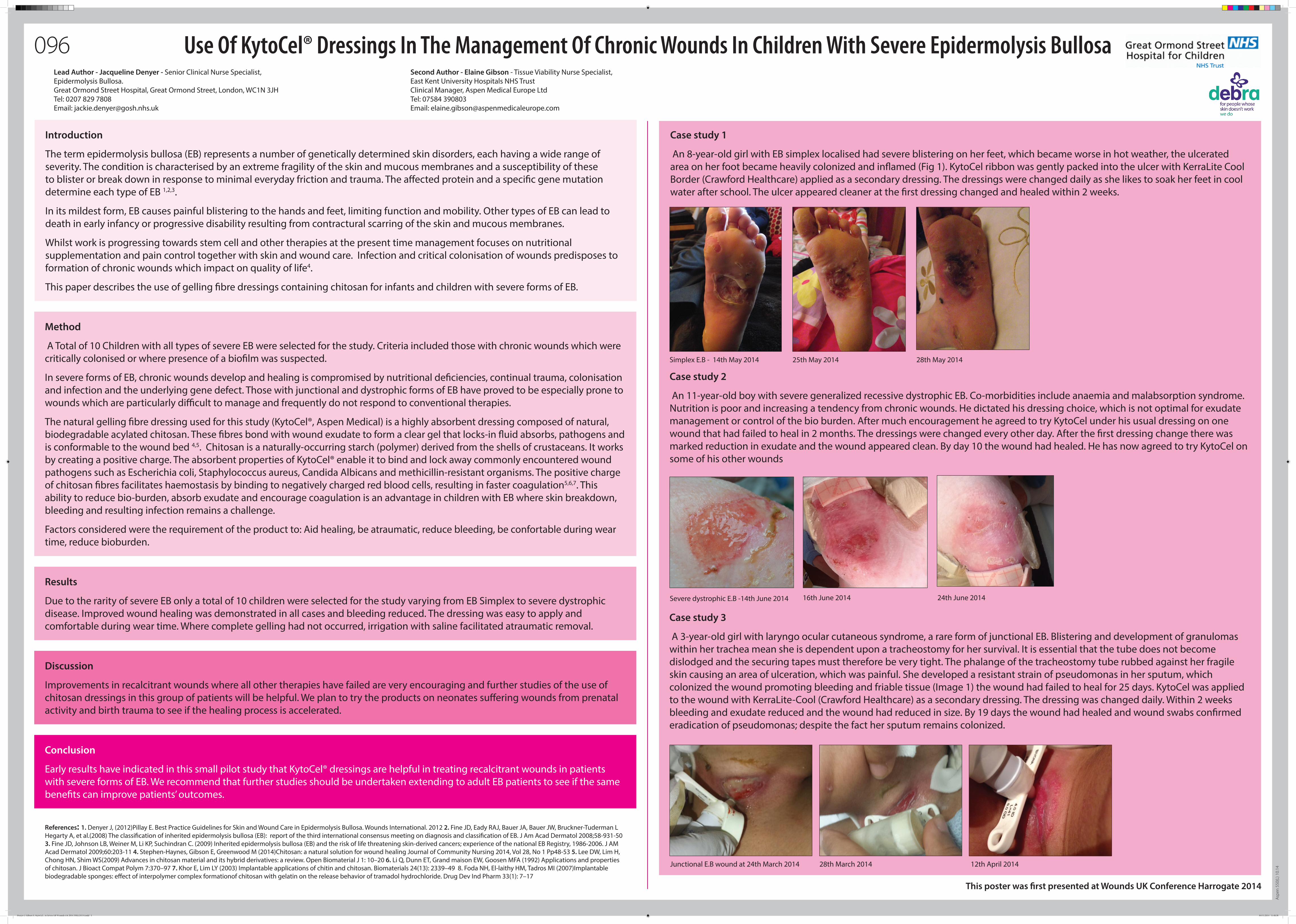
Simplex E.B - 14th May 2014 25th May 2014 28th May 2014
Junctional E.B wound at 24th March 2014 28th March 2014
16th June 2014Severe dystrophic E.B -14th June 2014 24th June 2014
096
Case study 1
An 8-year-old girl with EB simplex localised had severe blistering on her feet, which became worse in hot weather, the ulcerated area on her foot became heavily colonized and inflamed (Fig 1). KytoCel ribbon was gently packed into the ulcer with KerraLite Cool Border (Crawford Healthcare) applied as a secondary dressing. The dressings were changed daily as she likes to soak her feet in cool water after school. The ulcer appeared cleaner at the first dressing changed and healed within 2 weeks.
Case study 2
An 11-year-old boy with severe generalized recessive dystrophic EB. Co-morbidities include anaemia and malabsorption syndrome. Nutrition is poor and increasing a tendency from chronic wounds. He dictated his dressing choice, which is not optimal for exudate management or control of the bio burden. After much encouragement he agreed to try KytoCel under his usual dressing on one wound that had failed to heal in 2 months. The dressings were changed every other day. After the first dressing change there was marked reduction in exudate and the wound appeared clean. By day 10 the wound had healed. He has now agreed to try KytoCel on some of his other wounds
Case study 3
A 3-year-old girl with laryngo ocular cutaneous syndrome, a rare form of junctional EB. Blistering and development of granulomas within her trachea mean she is dependent upon a tracheostomy for her survival. It is essential that the tube does not become dislodged and the securing tapes must therefore be very tight. The phalange of the tracheostomy tube rubbed against her fragile skin causing an area of ulceration, which was painful. She developed a resistant strain of pseudomonas in her sputum, which colonized the wound promoting bleeding and friable tissue (Image 1) the wound had failed to heal for 25 days. KytoCel was applied to the wound with KerraLite-Cool (Crawford Healthcare) as a secondary dressing. The dressing was changed daily. Within 2 weeks bleeding and exudate reduced and the wound had reduced in size. By 19 days the wound had healed and wound swabs confirmed eradication of pseudomonas; despite the fact her sputum remains colonized.
12th April 2014
Denyer J. Gibson E. KytoCel... in Severe EB Wounds UK 2014 550(L)10.14.indd 1 04/11/2014 11:48:38

![Adderley UJ, Holt IGScampusvirtual.farmacoterapia-sanidadmadrid.org/CURSOS/logic/Consejeria... · [Intervention Review] Topical agents and dressings for fungating wounds Una J Adderley](https://img.pdfslide.us/doc/110x75/5e8aa5d7a399d038d37bfad2/adderley-uj-holt-intervention-review-topical-agents-and-dressings-for-fungating.jpg)