Embed Size (px)
Citation preview

1
Placenta and Fetal Membranes
Amnion - Epiblast / Extraembryonic Mesoderm
Yolk Sac - Hypoblast / Extraembryonic Mesoderm
Allantois - Embryonic Hindgut
Chorion - Trophoblasts / Extraembryonic Mesoderm
Placenta - Chorion / Maternal Decidua
Amnion
Amnionic membrane is two cell layers1) epiblast derived extraembryonic ectodermal layer2) thin non-vascular extraembryonic mesoderm
As the amnion enlarges it encompasses the embryo on the ventral side, merging around the umbilical cord.
Amnion forms the epithelial layer of the umbilical cord
With embryo growth the amnion obliterates the chorioniccavity
Amnionic sac is fluid filled called amnionic fluid: the embryo is bathed in the fluid
Extraembryonic Tissues
8 days
9 days
14 days
Amnion

2
Amnion
Amnionic Fluid
Up to week 20 - fluid is similar to fetal serum (keratinization)
After 20 weeks – Contribution from urine, maternal serum filtered thru endothelium of nearby vessels, filtration from fetal vessels in cord
Near birth - can contain fetal feces called meconium
Near birth – amnionic fluid (500-1000 ml) exchanges every 3 hrs 1) across the amnion – exchange with maternal fluids. 2) fetal swallowing (20 ml/hour) – to gut – adsorption by fetus – out the umbilical cord to placenta.
Hydraminos – Excess fluid (>2000 ml), esophageal atresia
Oligohydramnios – Insufficient fluid (<500 ml), renal agenesis
Amnion Function
Mechanical protection: hydrostatic pressure
Allows free movement - which aids in neuromuscular development
Antibacterial
Allow for fetal growth
Protection from adhesions
Amnion Band Syndrome (ABS) Placenta and Fetal Membranes
Amnion - Epiblast / Extraembryonic Mesoderm
Yolk Sac - Hypoblast / Extraembryonic Mesoderm
Allantois - Embryonic Hindgut
Chorion - Trophoblasts / Extraembryonic Mesoderm
Placenta - Chorion / Maternal Decidua

3
Yolk SacHypoblast - the primary yolk sac or Heuser's membrane.
Day 12 - Second wave of cell migration - forms definitive yolk sac
Composed of extrembryonic endoderm
Early nutrition (2-3 weeks) for the embryo - later shrinking -nonfunctional – Meckels diverticulum (outpocketing of small intestine)
Connects to midgut via the yolk sac stalk
Derivatives:Early blood cells forms from blood islandsPrimordial germ cellsThe early gut, epithelium of the respiratory and digestive tracts
Placenta and Fetal Membranes
Amnion - Epiblast / Extraembryonic Mesoderm
Yolk Sac - Hypoblast / Extraembryonic Mesoderm
Allantois - Embryonic Hindgut
Chorion - Trophoblasts / Extraembryonic Mesoderm
Placenta - Chorion / Maternal Decidua
Allantois
Endodermal origin – caudal outpocketing of the yolk sac
Invades the connecting stalk (extraembryonic mesoderm) that suspends the embryo in the chorionic cavity
Involved in early hematopoiesis (up to 2 months)
The allantois blood vessels - artery and vein - becomes the umbilical vessels
Remnants of Allantois becomes the urachus ligament that connects the belly button to the bladder
Placenta and Fetal Membranes
Amnion - Epiblast / Extraembryonic Mesoderm
Yolk Sac - Hypoblast / Extraembryonic Mesoderm
Allantois - Embryonic Hindgut
Chorion - Trophoblasts / Extraembryonic Mesoderm
Placenta - Chorion / Maternal Decidua
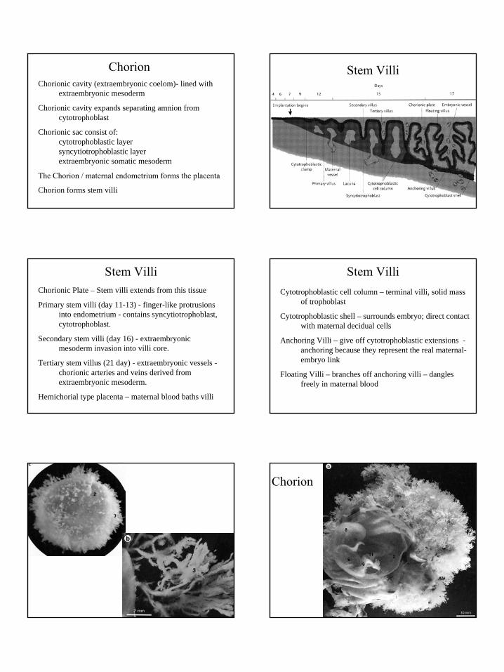
Chorion
4
ChorionChorionic cavity (extraembryonic coelom)- lined with
extraembryonic mesoderm
Chorionic cavity expands separating amnion from cytotrophoblast
Chorionic sac consist of:cytotrophoblastic layersyncytiotrophoblastic layerextraembryonic somatic mesoderm
The Chorion / maternal endometrium forms the placenta
Chorion forms stem villi
Stem Villi
Stem VilliChorionic Plate – Stem villi extends from this tissue
Primary stem villi (day 11-13) - finger-like protrusions into endometrium - contains syncytiotrophoblast,cytotrophoblast.
Secondary stem villi (day 16) - extraembryonicmesoderm invasion into villi core.
Tertiary stem villus (21 day) - extraembryonic vessels -chorionic arteries and veins derived fromextraembryonic mesoderm.
Hemichorial type placenta – maternal blood baths villi
Stem VilliCytotrophoblastic cell column – terminal villi, solid mass
of trophoblast
Cytotrophoblastic shell – surrounds embryo; direct contact with maternal decidual cells
Anchoring Villi – give off cytotrophoblastic extensions -anchoring because they represent the real maternal-embryo link
Floating Villi – branches off anchoring villi – dangles freely in maternal blood
Chorion

5
Placenta and Fetal Membranes
Amnion - Epiblast / Extraembryonic Mesoderm
Yolk Sac - Hypoblast / Extraembryonic Mesoderm
Allantois - Embryonic Hindgut
Chorion - Trophoblasts / Extraembryonic Mesoderm
Placenta - Chorion / Maternal Decidua
DeciduaDecidual Reaction – stromal cells – accumulate glycogen
and lipid, called Decidual Cells
Decidua basalis - forms maternal component of the placenta; associates with the chorion frondosom
Decidua capsularis - superfical layer overlying the entireembryoblast - this layer eventually degenerates; associates with the chorion laeve
Decidua parietalis - all remaining parts of the endometrium- not associated with the embryo
Deciduas Making the PlacentaBy 8 weeks - chorionic stem villi over the entire surface of
the chorionic sacThose villi associated with the decidua basalis increase in
size and more villi form. Enlargement includes further branching of the anchoring
villus - chorion frondosum. The villi continue to enlarge during most of gestation. The villi project into a blood filled intervillous space
resulting from the erosion of the decidua basalis.Endometrial vessels - spiral arteries and endometrial veinsVilli associated with the decidua capsularis degenerate -
this region is called the chorion laeve
DeciduasPlacenta
The erosion of the decidua basalis is incomplete - unerodedregions called decidual septa.
The decidual septa define regions of the placenta called cotyledon.

6
Placental Blood Flow Placental Anatomy
Umbilical Cord
One umbilical vein, two umbilical arteries
Wharton’s jelly – mucoid connective tissue surrounding vessels
Allantois
Yolk Stalk (vitelline duct) and vitelline vessels (early)
Intestinal loop – umbilical hernia (late)
Placental CirculationFetal – Contained within vessels
Umbilical Arteries – chorionic plate – branches to stem villi – capillaries in terminal villi – return via umbilical vein
Maternal – Free-flowing lakeSpiral arteries open into intervillous space and bath the villi150 ml of maternal bloodExchanged - 3-4 times/minuteReduced blood pressure in intervillous spaceOxygenated blood to the chorionic plate, return baths the villi
Placental Anatomy

7
Placental barrier decreases with gestation
Placental Barrier – syncytiotrophoblast + basal lamina, basal lamin+ fetal capillary endothelium
Syncytiotrophoblasts – many microvilli, no major histocompatibility antigens
O2
H2O
Fe
salts
carbohydrates, amino acids, lipids
vitamins, hormones, antibodies
drugs, alcohol
viruses (rubella, varicella-zoster, HIV)
CO2
H2O
salts
urea, uric acid
creatinine
bilirubin, hormones,
RBC antigens
Placenta as an Endocrine Organ
Human Chorionic Gonadotropin – Corpus Luteum (declines after 8 weeks)
Progesterone – High levels by the end of first trimester
Estrogen – Synthesis involves enzymatic activity of fetal adrenal gland and liver
Chorionic Somatomammotropin – Human Placental Lactogen –similar to GH (growth, lactation, lipid and carbohydrate metabolism)
Placental Growth Hormone – similar to GH – Replaces materrnal GH by 15 wks – enhances blood glucose levels
Chorionic Thyrotropin, Chorionic Corticotropin
Multiple Pregnancies
Monochorionic/Dichorionic Monoamnionic/Diamnionic
Hydatiform Mole

8
Erythroblastosis fetalis
Fetus / newborn - hemolytic disease (anemia)
Rh factor is a RBC surface antigen
Rh- mother with Rh+ 1st baby – Maternal antibodies are induced after birth
At risk is second Rh+ baby
Maternal Rh antibodies cross placenta
Hemolysis of fetal Rh+ RBC