Embed Size (px)
Citation preview
103
CASE REPORT
Adipofascial anterolateral thigh free fl ap for hemifacial atrophyIl lembo anterolaterale adipofasciale nell’emiatrofi a facciale
T. AGOSTINI, V. AGOSTINI
Department of Plastic and Reconstructive Surgery, Faculty of Medicine and Surgery, University of Florence, Florence,
Italy
SUMMARY
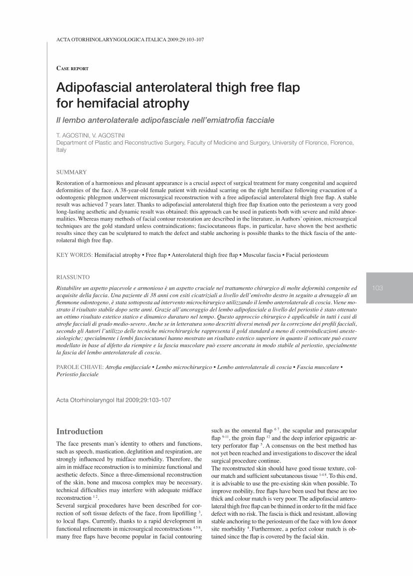
Restoration of a harmonious and pleasant appearance is a crucial aspect of surgical treatment for many congenital and acquired deformities of the face. A 38-year-old female patient with residual scarring on the right hemiface following evacuation of a odontogenic phlegmon underwent microsurgical reconstruction with a free adipofascial anterolateral thigh free fl ap. A stable result was achieved 7 years later. Thanks to adipofascial anterolateral thigh free fl ap fi xation onto the periosteum a very good long-lasting aesthetic and dynamic result was obtained; this approach can be used in patients both with severe and mild abnor-malities. Whereas many methods of facial contour restoration are described in the literature, in Authors’ opinion, microsurgical techniques are the gold standard unless contraindications; fasciocutaneous fl aps, in particular, have shown the best aesthetic results since they can be sculptured to match the defect and stable anchoring is possible thanks to the thick fascia of the ante-rolateral thigh free fl ap.
KEY WORDS: Hemifacial atrophy • Free fl ap • Anterolateral thigh free fl ap • Muscular fascia • Facial periosteum
RIASSUNTO
Ristabilire un aspetto piacevole e armonioso è un aspetto cruciale nel trattamento chirurgico di molte deformità congenite ed acquisite della faccia. Una paziente di 38 anni con esiti cicatriziali a livello dell’emivolto destro in seguito a drenaggio di un fl emmone odontogeno, è stata sottoposta ad intervento microchirurgico utilizzando il lembo anterolaterale di coscia. Viene mo-strato il risultato stabile dopo sette anni. Grazie all’ancoraggio del lembo adipofasciale a livello del periostio è stato ottenuto un ottimo risultato estetico statico e dinamico duraturo nel tempo. Questo approccio chirurgico è applicabile in tutti i casi di atrofi e facciali di grado medio-severo. Anche se in letteratura sono descritti diversi metodi per la correzione dei profi li facciali, secondo gli Autori l’utilizzo delle tecniche microchirurgiche rappresenta il gold standard a meno di controindicazioni aneste-siologiche; specialmente i lembi fasciocutanei hanno mostrato un risultato estetico superiore in quanto il sottocute può essere modellato in base al difetto da riempire e la fascia muscolare può essere ancorata in modo stabile al periostio, specialmente la fascia del lembo anterolaterale di coscia.
PAROLE CHIAVE: Atrofi a emifacciale • Lembo microchirurgico • Lembo anterolaterale di coscia • Fascia muscolare • Periostio facciale
Acta Otorhinolaryngol Ital 2009;29:103-107
IntroductionThe face presents man’s identity to others and functions, such as speech, mastication, deglutition and respiration, are strongly infl uenced by midface morbidity. Therefore, the aim in midface reconstruction is to minimize functional and aesthetic defects. Since a three-dimensional reconstruction of the skin, bone and mucosa complex may be necessary, technical diffi culties may interfere with adequate midface reconstruction 1 2.Several surgical procedures have been described for cor-rection of soft tissue defects of the face, from lipofi lling 3, to local fl aps. Currently, thanks to a rapid development in functional refi nements in microsurgical reconstructions 4 5 8, many free fl aps have become popular in facial contouring
such as the omental fl ap 6 7, the scapular and parascapular fl ap 9-11, the groin fl ap 12 and the deep inferior epigastric ar-tery perforator fl ap 5. A consensus on the best method has not yet been reached and investigations to discover the ideal surgical procedure continue.The reconstructed skin should have good tissue texture, col-our match and suffi cient subcutaneous tissue 1-4 8. To this end, it is advisable to use the pre-existing skin when possible. To improve mobility, free fl aps have been used but these are too thick and colour match is very poor. The adipofascial antero-lateral thigh free fl ap can be thinned in order to fi t the mid face defect with no risk. The fascia is thick and resistant, allowing stable anchoring to the periosteum of the face with low donor site morbidity 4. Furthermore, a perfect colour match is ob-tained since the fl ap is covered by the facial skin.
ACTA OTORHINOLARYNGOLOGICA ITALICA 2009;29:103-107
T. Agostini et al.
104
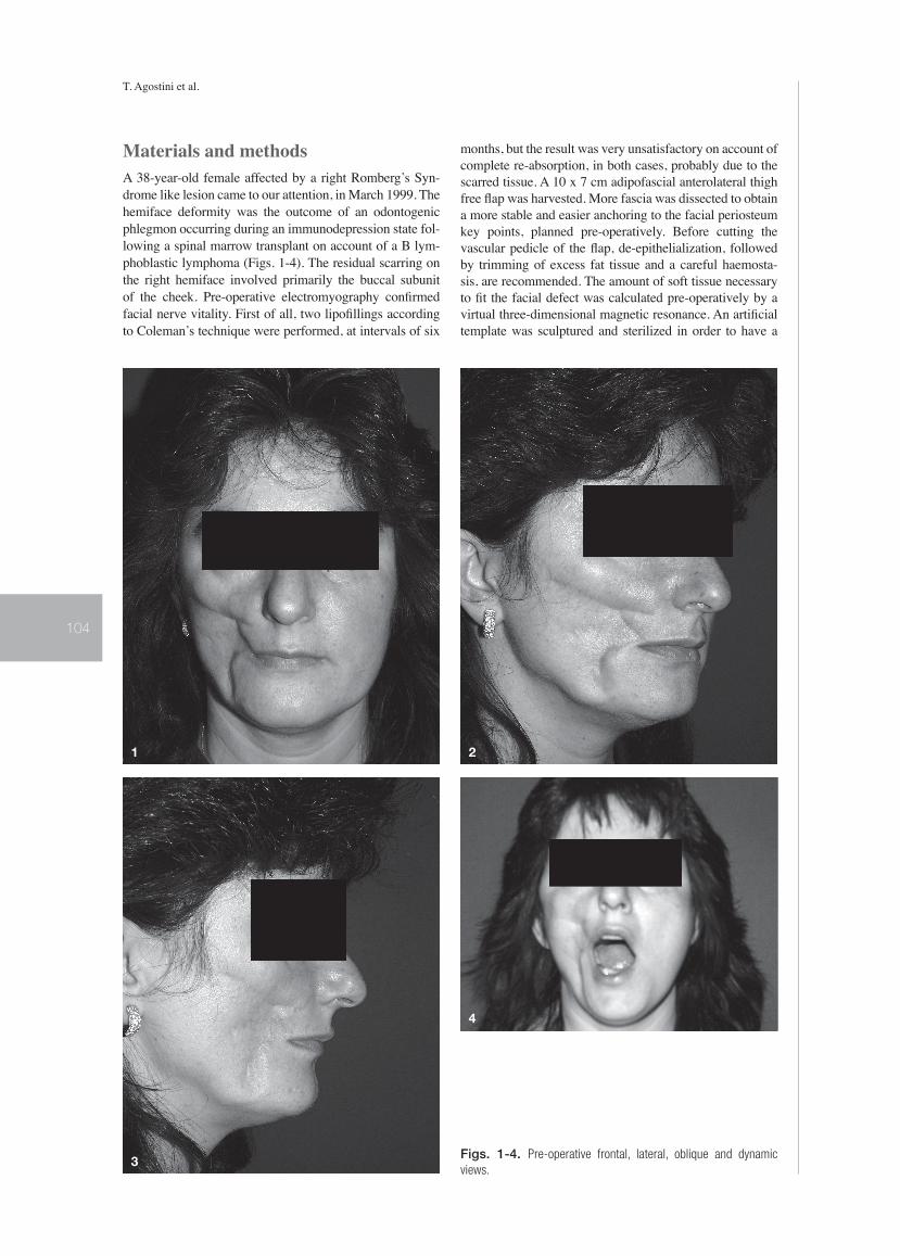
Materials and methodsA 38-year-old female affected by a right Romberg’s Syn-drome like lesion came to our attention, in March 1999. The hemiface deformity was the outcome of an odontogenic phlegmon occurring during an immunodepression state fol-lowing a spinal marrow transplant on account of a B lym-phoblastic lymphoma (Figs. 1-4). The residual scarring on the right hemiface involved primarily the buccal subunit of the cheek. Pre-operative electromyography confi rmed facial nerve vitality. First of all, two lipofi llings according to Coleman’s technique were performed, at intervals of six
months, but the result was very unsatisfactory on account of complete re-absorption, in both cases, probably due to the scarred tissue. A 10 x 7 cm adipofascial anterolateral thigh free fl ap was harvested. More fascia was dissected to obtain a more stable and easier anchoring to the facial periosteum key points, planned pre-operatively. Before cutting the vascular pedicle of the fl ap, de-epithelialization, followed by trimming of excess fat tissue and a careful haemosta-sis, are recommended. The amount of soft tissue necessary to fi t the facial defect was calculated pre-operatively by a virtual three-dimensional magnetic resonance. An artifi cial template was sculptured and sterilized in order to have a
Figs. 1-4. Pre-operative frontal, lateral, oblique and dynamic
views.
1 2
3
4
Anterolateral thigh free fl ap for hemifacial atrophy
105
precise bench mark. The residual scarring from drainage of the phlegmon, on the outer cheek was chosen to fi t the fl ap into the defect.The vascular anastomosis were harvested with facial artery and vein. The muscular fascia was sutured using non-ab-sorbable material to the periosteum of the malar, orbitozy-gomatic, pyriform and mandibular periosteum to contrast
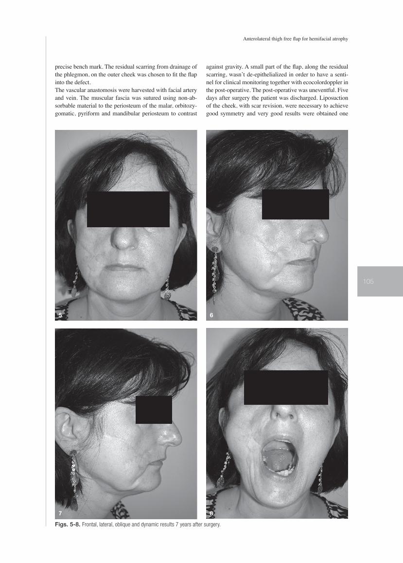
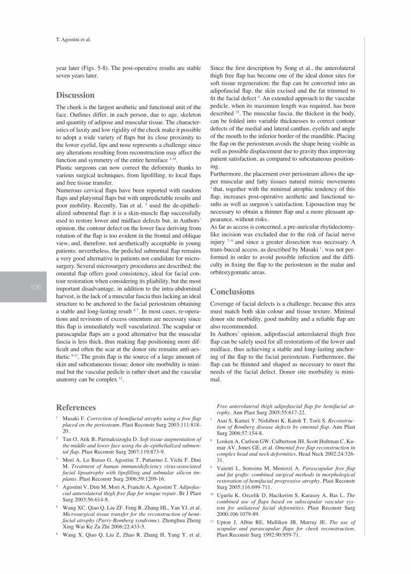
against gravity. A small part of the fl ap, along the residual scarring, wasn’t de-epithelialized in order to have a senti-nel for clinical monitoring together with ecocolordoppler in the post-operative. The post-operative was uneventful. Five days after surgery the patient was discharged. Liposuction of the cheek, with scar revision, were necessary to achieve good symmetry and very good results were obtained one
5 6
7 8
Figs. 5-8. Frontal, lateral, oblique and dynamic results 7 years after surgery.
T. Agostini et al.
106
year later (Figs. 5-8). The post-operative results are stable seven years later.
DiscussionThe cheek is the largest aesthetic and functional unit of the face. Outlines differ, in each person, due to age, skeleton and quantity of adipose and muscular tissue. The character-istics of laxity and low rigidity of the cheek make it possible to adopt a wide variety of fl aps but its close proximity to the lower eyelid, lips and nose represents a challenge since any alterations resulting from reconstruction may affect the function and symmetry of the entire hemiface 1-14.Plastic surgeons can now correct the deformity thanks to various surgical techniques, from lipofi lling, to local fl aps and free tissue transfer.Numerous cervical fl aps have been reported with random fl aps and platysmal fl aps but with unpredictable results and poor mobility. Recently, Tan et al. 2 used the de-epitheli-alized submental fl ap: it is a skin-muscle fl ap successfully used to restore lower and midface defects but, in Authors’ opinion, the contour defect on the lower face deriving from rotation of the fl ap is too evident in the frontal and oblique view, and, therefore, not aesthetically acceptable in young patients; nevertheless, the pedicled submental fl ap remains a very good alternative in patients not candidate for micro-surgery. Several microsurgery procedures are described: the omental fl ap offers good consistency, ideal for facial con-tour restoration when considering its pliability, but the most important disadvantage, in addition to the intra-abdominal harvest, is the lack of a muscular fascia thus lacking an ideal structure to be anchored to the facial periosteum obtaining a stable and long-lasting result 6 7. In most cases, re-opera-tions and revisions of excess omentum are necessary since this fl ap is immediately well vascularized. The scapular or parascapular fl aps are a good alternative but the muscular fascia is less thick, thus making fl ap positioning more dif-fi cult and often the scar at the donor site remains anti-aes-thetic 9-11. The groin fl ap is the source of a large amount of skin and subcutaneous tissue; donor site morbidity is mini-mal but the vascular pedicle is rather short and the vascular anatomy can be complex 12.
Since the fi rst description by Song et al., the anterolateral thigh free fl ap has become one of the ideal donor sites for soft tissue regeneration; the fl ap can be converted into an adipofascial fl ap, the skin excised and the fat trimmed to fi t the facial defect 4. An extended approach to the vascular pedicle, when its maximum length was required, has been described 15. The muscular fascia, the thickest in the body, can be folded into variable thicknesses to correct contour defects of the medial and lateral canthus, eyelids and angle of the mouth to the inferior border of the mandible. Placing the fl ap on the periosteum avoids the shape being visible as well as possible displacement due to gravity thus improving patient satisfaction, as compared to subcutaneous position-ing.Furthermore, the placement over periosteum allows the up-per muscular and fatty tissues natural mimic movements 1 that, together with the minimal atrophic tendency of this fl ap, increases post-operative aesthetic and functional re-sults as well as surgeon’s satisfaction. Liposuction may be necessary to obtain a thinner fl ap and a more pleasant ap-pearance, without risks.As far as access is concerned, a pre-auricular rhytidectomy-like incision was excluded due to the risk of facial nerve injury 7-11 and since a greater dissection was necessary. A trans-buccal access, as described by Masaki 1, was not per-formed in order to avoid possible infection and the diffi -culty in fi xing the fl ap to the periosteum in the malar and orbitozygomatic areas.
ConclusionsCoverage of facial defects is a challenge, because this area must match both skin colour and tissue texture. Minimal donor site morbidity, good mobility and a reliable fl ap are also recommended.In Authors’ opinion, adipofascial anterolateral thigh free fl ap can be safely used for all restorations of the lower and midface, thus achieving a stable and long-lasting anchor-ing of the fl ap to the facial periosteum. Furthermore, the fl ap can be thinned and shaped as necessary to meet the needs of the facial defect. Donor site morbidity is mini-mal.
References1 Masaki F. Correction of hemifacial atrophy using a free flap
placed on the periosteum. Plast Reconstr Surg 2003;111:818-20.
2 Tan O, Atik B, Parmaksizoglu D. Soft tissue augmentation of the middle and lower face using the de-epithelialized submen-tal flap. Plast Reconstr Surg 2007;119:873-9.
3 Mori A, Lo Russo G, Agostini T, Pattarino J, Vichi F, Dini M. Treatment of human immunodeficiency virus-associated facial lipoatrophy with lipofilling and submalar silicon im-plants. Plast Reconstr Surg 2006;59:1209-16.
4 Agostini V, Dini M, Mori A, Franchi A, Agostini T. Adipofas-cial anterolateral thigh free flap for tongue repair. Br J Plast Surg 2003;56:614-8.
5 Wang XC, Qiao Q, Liu ZF, Feng R, Zhang HL, Yan YJ, et al. Microsurgical tissue transfer for the reconstruction of hemi-facial atrophy (Parry-Romberg syndrome). Zhonghua Zheng Xing Wai Ke Za Zhi 2006;22:433-5.
6 Wang X, Qiao Q, Liu Z, Zhao R, Zhang H, Yang Y, et al.
Free anterolateral thigh adipofascial flap for hemifacial at-rophy. Ann Plast Surg 2005;55:617-22.
7 Asai S, Kamei Y, Nishibori K, Katoh T, Torii S. Reconstruc-tion of Romberg disease defects by omental flap. Ann Plast Surg 2006;57:154-8.
8 Losken A, Carlson GW, Culbertson JH, Scott Hultman C, Ku-mar AV, Jones GE, et al. Omental free flap reconstruction in complex head and neck deformities. Head Neck 2002;24:326-31.
9 Vaienti L, Soresina M, Menozzi A. Parascapular free flap and fat grafts: combined surgical methods in morphological restoration of hemifacial progressive atrophy. Plast Reconstr Surg 2005;116:699-711.
10 Ugurlu K, Ozcelik D, Hacikerim S, Karasoy A, Bas L. The combined use of flaps based on subscapular vascular sys-tem for unilateral facial deformities. Plast Reconstr Surg 2000;106:1079-89.
11 Upton J, Albin RE, Mulliken JB, Murray JE. The use of scapular and parascapular flaps for cheek reconstruction. Plast Reconstr Surg 1992;90:959-71.
Anterolateral thigh free fl ap for hemifacial atrophy
107
12 Cooper TM, Lewis N, Baldwin MA. Free groin flap revisited. Plast Reconstr Surg 1999;103:918-24.
13 Longaker MT, Siebert JW. Microvascular free-flap cor-rection of severe hemifacial atrophy. Plast Reconstr Surg 1995;96:800-9.
14 Stamatopoulos C, Panayotou P, Tsirigotou S, Ioannovich JD.
Use of free flaps in the aesthetic reconstruction of face and neck deformities. Microsurgery 1992;13:188-91.
15 Spyriounis PK. The extended approach to the vascular pedicle of the anterolateral thigh perforator flap: anatomi-cal and clinical study. Plast Reconstr Surg 2006;117:997-1001.
Address for correspondence: Dr. V. Agostini, Dipartimento di Chirur-gia Plastica e Ricostruttiva, CTO-AOUC, largo Palagi 1, 50100 Firen-ze, Italy. Fax +39 055 7948099. E-mail: [email protected]
Received: November 25, 2007 - Accepted: February 22, 2008