Embed Size (px)
Citation preview

REPORT OF GENERAL FELLOWSHIP EXAMINATION
March/May 2006
This report is prepared to provide candidates, tutors and their Supervisors of training with information about the way in which the Examiners assessed the performance of candidates in the Examination. Answers provided are not model answers but guides to what was expected. Candidates should discuss the report with their tutors so that they may prepare appropriately for the future examinations. This was the third exam under the current format. The exam included two 2.5 hour written papers comprising of 15 ten-minute short answer questions each. Candidates were required to perform at a satisfactory level in the Written Section before being eligible to sit the Oral Sections of the exam. The Oral Sections comprised six interactive Vivas, ten OSCE stations (with four interactive stations, including two cold cases) and two separate hot cases. WRITTEN SECTIONS This guide below is meant to be an information resource. It is not written under exam conditions and does not reproduce an ideal answer, but it does include the type of material that should be included in a good answer. Feedback from examiners indicated that candidates would have been more likely to pass if they:
• answered the question asked • demonstrated their priorities • organised their answer in a way that demonstrated a broader knowledge • included additional relevant detail
Writing should be legible to allow candidates to gain optimal marks. A number of the questions had been asked in previous exams, some in a modified format. Weaknesses were identified in the candidates’ ability to interpret biochemical and hemodynamic data, and statistical definitions. Eighteen out of the twenty-six candidates who were required to sit the Written Section passed this section overall. Five additional candidates who did not pass this section overall received sufficient marks in the Written Section to be invited to the Oral Section. The following “Glossary of terms” was provided for the candidates Critically evaluate: Evaluate the evidence available to support the hypothesis. Outline: Provide a summary of the important points. List: Provide a list. Compare and contrast: Provide a description of similarities and differences (eg. Table form).

2
Paper 1.
1. A 78 year old man on warfarin for atrial fibrillation develops a spontaneous weakness of
his left hand and leg and a deteriorating conscious state. He presents within 20 minutes of onset of symptoms and a CT scan confirms a right parietal intra-cerebral haemorrhage. Outline your medical management of this case.
Specific features that would alter the management plan should be sought in the history (particularly of other drugs affecting coagulation e.g. aspirin, clopidogrel) and examination. Obviously attention should be paid to resuscitation (including airway management) if required, and supportive care (including careful blood pressure control) but the predominant focus should be towards correction of coagulopathy. Spontaneous bleeds can occur at any (or no) levels of anticoagulation but rates increase significantly where the INR is above 4. The Prothrombin time (or INR) and coagulation parameters should be measured urgently. Where there is clinical/neurological deterioration, reversal of anticoagulation should not be delayed for results. The full effect of vitamin K in reducing the INR takes up to 24 hours to develop, even when given in larger doses with the intention of complete reversal. For immediate reversal of clinically significant bleeding, the combination of prothrombin complex concentrate (PCC) and fresh frozen plasma (FFP) covers the period before Vitamin K has reached its full effect. Vitamin K is essential for sustaining the reversal achieved by a PCC and FFP. PCC is a three-factor concentrate, containing factors II, IX and X, but only low levels of factor VII. Therefore, the adjunctive use of FFP should be administered as a source of factor VII. Treatment with recombinant factor VIIa within four hours of the onset of intracerebral haemorrhage has been reported to limit extension of the haematoma, reduce mortality, and improve functional outcome. However rFVIIa treatment is associated with some increase in the frequency of thromboembolic adverse events. Seventeen out of twenty-six candidates passed this question. 2. Outline the clinical features, mechanism of toxicity and therapy of cyanide poisoning. Clinical features: Symptoms of toxicity range from non-specific symptoms such as headache and nausea to depressed consciousness, seizures and cardiopulmonary arrest. Laboratory features include lactic acidosis and unexpectedly high venous oxygen saturation (with low a-v oxygen difference) Mechanism of toxicity: Cyanide blocks mitochondrial cytochrome oxidase resulting in cytotoxic hypoxia and lactic acidosis. Therapy: As cyanide is highly toxic and can penetrate intact skin or be inhaled. Consequently decontamination is essential and mouth-to-mouth resuscitation should not be performed. In cases of ingestion gastric lavage may reduce absorption. There are various antidotes based on three principles: 1. Conversion of haemoglobin to methaemoglobin (Amyl nitrite or sodium nitrite are used for this purpose). Methaemoglobin has a higher affinity for cyanide than does cytochrome oxidase and therefore promotes its dissociation from cytochrome oxidase. Since methaemoglobin does not carry oxygen, excessive methaemoglobinaemia can lead to anoxia. Methaemoglobin should be measured during treatment; a desirable level is between 20% and 30%. 2. Direct binding to EDTA or the vitamin B12 precursor hydroxocobalamin. A high dose (5 grams) of hydroxocobalamin is required but has minimal toxicity (in contrast to other treatments). 3. Thiosulfate (administered as sodium thiosulphate) reacts with cyanide forming the relatively non-toxic thiocyanate, which is excreted in the urine. This action is slow and provides little effect in the acute phase. Eighteen out of twenty-six candidates passed this question.

3
3. Describe the advantages and disadvantages of PaO2 (partial pressure of oxygen in the arterial blood) and SpO2 (oxygen saturation measured by a pulse oximeter) as indicators of arterial oxygenation.
This question lends itself to an answer in table form. Such a table should include the following type of information:
PaO2 SpO2 Allows calculation of P:F ratio and A-a gradient. Calculating SaO2 allows calculation of DO2/VO2/shunt fraction
Also a reliable indicator of oxygenation particularly in the clinical relevant range
Accurate indicator of oxygenation, not influenced by position of Oxyhaemoglobin Dissociation Curve
Easy to use, non invasive, reliable in most clinical situations both in ICU and on the wards
Advantages
Measurement accurate even in the presence of dyshaemoglobins.
Provides a continuous measurement
Invasive Affected by peripheral perfusion, arrhythmias, motion artefact
Prone to pre-analytic errors – air bubbles, heparin contamination
Affected by dyshemoglobins
Needs a blood gas analyser Usually a time delay between sampling and result
Disadvantages
Provides only intermittent measurements
Twenty-two out of twenty-six candidates passed this question. 4. Outline the causes, consequences and management of intrinsic PEEP. Causes: consider increased expiratory resistance (prolonged expiratory time constant: eg. bronchospasm, narrow/kinked ETT, inspissated secretions, exhalation valves/HME/filters), increased minute ventilation (inadequate expiratory time), prolonged inspiratory time. Consequences: consider increased intra-thoracic lung volume (with increased pressures for a given tidal volume and risks of barotrauma), increased intra-thoracic pressure (decreasing venous return, and increasing inspiratory work to trigger the ventilator). Management: consider treatment of reversible factors (bronchospasm, secretions, expiratory devices), prolongation of expiratory time (decrease respiratory rate, increase inspiratory flow, decrease in inspiratory time) or decrease tidal volumes, application of exogenous PEEP (to 50 – 85% of accurately measured intrinsic PEEP) can be used to decrease inspiratory triggering work in spontaneously breathing patients, and possibly to improve distribution of inspired gas. Twenty-five out of twenty-six candidates passed this question. 5. List the causes of a sudden acute fall in systolic blood pressure to 50 mmHg one hour
after an uneventful coronary artery bypass operation. Outline your principles of management for each cause.
A list of causes was requested, as were principles of management for each cause. Potential causes are many, and more than one may co-exist. These could be listed according to causes of shock; one such approach to cover potential causes is: artefactual, hypovolaemic,

4
obstructive, cardiogenic, and distributive. Principles of management are listed in brackets following each cause. Simple manoeuvres should always be considered early (eg. raise legs to autotransfuse). Artefactual: transducer error (check transducer: zero, level, calibration), damping of waveform (assess damping coefficient), malfunction of NIBP. Hypovolaemic: blood loss (observe drain tubes, CXR, dressings; give fluid ± blood products), massive diuresis (observe urine output; give fluid). Obstructive: pericardial tamponade (observe chest drainage ± clots, high filling pressures: may need to open chest), tension pneumothorax (observe expanded hemi-thorax, listen to chest: check existing chest drains, may need needle thoracostomy and replace/insert ICC), elevated intrathoracic pressure (gas trapping: disconnect from ventilator; shivering/Valsalva/fighting: sedate ± paralyse; ensure ETT not blocked). Myocardial: decreased contractility (ischaemia due to blockage/kinking/spasm: treat with GTN, inotropes &/or short term vasoconstrictor ± fix technical problem; sudden removal of inotropic drug: restart drug) or rhythm disturbance on monitor/ECG (brady-asystole: pace ± atropine/isoprenaline/adrenaline; SVT: eg. K/Mg/adenosine; AF eg. K/Mg/amiodarone, VT eg. K/Mg/lignocaine or amiodarone). Distributive: anaphylaxis (rash/bronchospasm: remove hapten, adrenaline, fluids); vasodilator excess (recent boluses/infusion too high: stop responsible drug, ± titrated dose vasoconstrictor); sympathetic block (recent bolus of epidural Local Anaesthetic: titrated dose vasoconstrictor); too profound for a systemic inflammatory response. Eighteen out of twenty-six candidates passed this question. 6. Outline the causes and principles of management of ventricular fibrillation. Causes: Ventricular fibrillation requires an initiating stimulus in a susceptible myocardium. VF can be induced in a previously normal myocardium as a result of electrical stimulation (electrocution, lightning) or by trauma (Commotio cordis). The myocardium can be made more susceptible by the presence of hypoxaemia (e.g. respiratory arrest), electrolyte disturbances (low K and Mg), altered autonomic and vagal inputs, and mechanical stimuli (e.g. wire or catheter in RV). The myocardium may be abnormally susceptible due to congenital (e.g. conduction abnormalities) or acquired disorders (including ischaemia, hypertrophy, myocarditis, pro-arrhythmic drugs, etc). Principles of management: include early defibrillation, but in concert with correction of any correctible cause (e.g. wire, electrolytes, hypoxaemia etc), support of the cardio-respiratory state with adequate basic life support, and restoration of an appropriate metabolic milieu to support a normal rhythm. This latter approach may require performance of cardiopulmonary resuscitation, and administration of vasoconstrictors and specific anti-arrhythmic drugs, especially in the setting of prolonged VF. According to the ALS guidelines in place when the question was set, defibrillation is performed with an appropriate energy level for either monophasic (eg. 200/200/360J) or biphasic (eg. 150/150/150J) defibrillator waveforms in a series of up to three sequential shocks. Subsequent monophasic shocks should be administered at maximal dose. The longer-term management, including the use of implantable defibrillators should be considered according to published guidelines. Candidates were not penalised if they did not discuss the new guidelines (including higher energy levels for monophasic, and a single shock approach). Twenty-two out of twenty-six candidates passed this question. 7. A 65 year old obese lady with known alcoholic cirrhosis and long term thyroxine
supplementation was admitted to ICU with staphylococcal spinal osteomyelitis 6 weeks ago. Following discharge to the ward, she developed progressive abdominal distension, hypotension and oliguria. On examination she was confused, with a flapping tremor. Her pulse rate was 42/min, in sinus rhythm and her blood pressure was 80/40 mm Hg. Her temperature was 34.7˚C. Bowel sounds were absent. There was no abdominal tenderness
Investigations subsequent to her admission were as follows: Blood tests:

5
Normal values
Hb 110 G/L 110 - 150 WCC 8.4 109/L 5 - 11 Platelets 173 109/L 150 - 300 Na 131 mmol/L 135 - 145 K 3.6 mmol/L 3.5 - 5.0 Urea 26 mmol/L 4 - 6 Creatinine 167 micromol/L 60 - 120 Glucose 2.2 mmol/L 4 - 6 Cholesterol 8.6 mmol/L 4 - 6 AST 40 U/L 35 - 45 ALT 51 U/L 35 - 45 Ammonia 41 micromol/L 50 - 80 Calcium 2.25 mmol/L 2.2 - 2.6
CT brain scan: Normal Echo: Moderate pericardial effusion
(a) In light of this information, what is the most likely diagnosis? Justify your response.
(b) List 2 precipitating factors. a) The most likely diagnosis is Myxoedema coma /severe hypothyroid coma. The normal CT brain excludes a significant organic brain pathology, and normal ammonia + normal hepatic enzymes make hepatic encephalopathy less likely. The clinical picture in concert with the features of a low Na, low glucose, high cholesterol, a pericardial effusion and the history of thyroxine supplementation is highly suggestive of hypothyroidism. Marks were also allocated if a reasonable alternative diagnosis was given, provided that this was accompanied by a rational justification. b) Many precipitating factors could be present, but consider: sepsis, drugs (eg. betablockers, amiodarone), stroke, and a patient non-compliant with therapy. Sixteen out of twenty-six candidates passed this question. 8. Outline strategies you would incorporate to prevent central venous catheter related
infection. A number of different strategies should be considered. One approach to incorporate these follows: Why? Confirm need for Central Venous Line (initial, and ongoing). How? Pay attention to sterile insertion technique. Non occlusive dressing, transparent semipermeable dressing or dressing with topical disinfectant. What? Use single lumen where possible; consider antibiotic impregnated line. Where? Subclavian site preferentially over Internal Jugular or femoral; consider PICC lines (peripherally inserted central catheters), When remove? Daily site inspections, and consider early change. Replace giving sets/lines regularly: blood and TPN giving sets 24hrly but other IV sets after 72 hrs Twenty-two out of twenty-six candidates passed this question. 9. Outline the potential roles for the use of ultrasound in the critically ill patient. The potential roles of ultrasound in critically ill patients are diverse and are increasing in number. They include: Cardiac echo – transthoracic/transoesophageal looking at structure, function, relationships between the two and pericardial effusions, and doppler cardiac output monitoring. Vascular –thoracic and abdominal aorta (dissection, aneurysm etc). Other vascular roles include any accessible artery (eg radial, brachial, femoral, carotid etc), grafts for flow assessment, stenosis, patency. Also larger veins for thrombosis/patency, and vessel identification for line insertion

6
Cranial – monitor cerebral blood flow, hyperaemia, ischaemia, detection of vasospasm, fat and other emboli, stroke related artery reperfusion following thrombolysis. Renal perfusion – following AAA repair, renal transplant and acute tubular necrosis. Free fluid – peritoneal and pleural – allows diagnosis, quantitative assessment as well as marking for drainage. Other anatomical – identify bladder (urinary retention), biliary anatomy, including gallbladder (eg. acalculous cholecystitis), atelectasis, pneumothorax. Nineteen out of twenty-six candidates passed this question. 10. A 75 year old woman with a reduced level of consciousness is intubated and ventilated
after a single grand mal convulsion. Indicate the pathophysiologic disturbances revealed by the following blood gas and electrolyte profile, taken 10 minutes post intubation. Outline how this information should influence her management.
Normal values Barometric pressure 760 mm Hg FiO2 1.0 pH 7.05 7.35-7.45 pO2 280 mm Hg pCO2 43 mm Hg 35-45 HCO3
- 11.5 mmol/L 21-30 Standard base excess -16.8 mmol/L Sodium 128 mmol/L 135 -145 Potassium 3.1 mmol/L 3.2 - 4.5 Chloride 82 mmol/L 100 -110 Urea 22.0 mmol/L 3.0 - 8.0 Creatinine 0.12 mmol/L 0.07 - 0.12 Glucose 79.0 mmol/L 3.0 - 7.8 Lactate 9.2 mmol/L 0.5 - 2.0
Candidates were expected to outline each of the important abnormalities and how they should influence her management. One approach would be the following:
• The biochemistry supports a diagnosis of hyperosmolar hyperglycaemic syndrome, with post-ictal lactic acidosis. There is also a likely component of ketoacidosis.
• The high urea / creatinine ratio confirms significant dehydration – most people with this condition are at least 10% dehydrated by body weight – usually more. She will require steady rehydration over 12 to 24 hours.
• The sodium adjusted to normoglycaemia is about 153 mmol/L. This means that, after intravascular volume is restored with isotonic fluids, rehydration should be conducted with relatively hypotonic fluids eg 0.45% saline initially.
• The metabolic acidosis is uncompensated. The minute ventilation should be increased (if this can be done safely), to replicate appropriate respiratory compensation.
• There is a raised anion gap (34.5 mEq/L without K). The degree of lactate elevation is insufficient to explain the anion gap, indicating probable co-existing ketoacidosis. Beta-hydroxybutyrate should therefore be measured. An insulin infusion (0.1 U/kg/hr) is required for gentle glucose correction and reversal of ketoacidosis.
• The plasma potassium is already reduced, indicating a severe deficit and the need for immediate replacement in the absence of anuria. She is likely to need 10 - 20 mmol/hr or more for many hours, since the total deficit will exceed 6 mmol/kg.
• Blood gases, Na, K and glucose will need frequent (hourly) measurement initially. Rapid reductions in osmolality should be avoided (using sodium adjusted to normoglycaemia as a surrogate).

7
• The A-a gradient is significantly elevated at 380 mm Hg. This could indicate aspiration pneumonitis, pneumonia, or segmental collapse – even endobronchial intubation. These possibilities should be looked for clinically and on CXR.
Fifteen out of twenty-six candidates passed this question. 11. Outline how you would assess a patient for potential difficulty with endotracheal
intubation. History: of previous difficulty with intubation, infections/swelling affecting mouth or neck, problems with mouth opening or neck movement (arthritis, cervical spine injury), problems with teeth (especially caps/crowns, jaw wiring etc.). Review of a previous anaesthetic chart. Examination (multiple components) consider:
• Teeth (maxillary anterior to mandibular; length of upper incisors; ability to prognath mandible; inter-incisor distance [need > 3 cm])
• Pharynx (ability of visualise uvula and tonsillar pillars; height and narrowness of palate). • Mandibular space (thyromental distance � 3 fingerbreadths [6 cm]; compliance and
distensibility of submandibular space). • Length of neck (qualitative: short neck more difficult eg. syndromes). • Thickness of neck (qualitative: thick neck decreases ability to align planes). • Range of motion (of head and neck: eg. sniffing position) • Consider also the ability to assess potential difficulties by actually having a look with a
laryngoscope. (10 marks) In some situations specific investigations may also be indicated (eg. neck X-rays etc.) Twenty-two out of twenty-six candidates passed this question. 12. Compare and contrast the pharmacology of lignocaine, magnesium and amiodarone when used in the treatment of ventricular tachycardia. Lignocaine: Class I (membrane stabilising) antiarrhythmic agent. Sodium channel blockades results in decreased action potential duration and shortened refractory period. Rapidly distributed to all body tissues. Approximately 65% protein bound; elimination half-life 1.6 hours (80% metabolised in liver). Adverse effects: lightheaded, hypotension, cardiovascular collapse, heart block, confusion and convulsions. Dosage used in the treatment of ventricular tachycardia: 1 to 1.5mg/kg with subsequent boluses (up to 3 mg/kg total), followed by infusion (1-4 mg/min, at decreasing dose, up to 24 hours). Magnesium (as sulphate or chloride): Second most abundant intracellular cation. Depresses neuronal activation. Widely distributed, duration of action about 30 minutes. Filtered by kidneys, but most is reabsorbed. Adverse effects include: nausea, flushing, CNS depression, coma, and heart block. Dose used in the treatment of ventricular tachycardia: 5 mmol bolus (which may be repeated), followed by infusion of 20 mmol over 4 hours. Amiodarone: Class III antiarrhythmic. Prolongs action potential duration, and prolongs refractory period of atrial, nodal and ventricular tissues. Highly protein bound with very high apparent volume of distribution (6 L/kg); accumulates in adipose tissue and highly perfused organs. Half-life (with chronic dosing) is 14 to 59 days, mainly excreted via the liver and bile. Adverse effects: hypotension/circulatory collapse, bradycardia, sinus arrest, nausea and flushing. Torsades de pointes can be induced. Hyper- or hypo-thyroidism can be induced. Multiple other potential organ dysfunctions with more chronic use (including some potentially fatal). Dosage used in the treatment of ventricular tachycardia: 5 mg/kg (or 300 mg in adults) which can be repeated, and followed by an infusion (15 mg/kg/hr). Twenty out of twenty-six candidates passed this question. 13. (a) Outline the situations in which clinical tests cannot be used to confirm brain
death.

8
(b) List 2 adjunctive tests used in Australia and New Zealand for the confirmation
of brain death when clinical tests are unable to be performed. (c) List other adjunctive tests which may have a role in the diagnosis of brain
death. a) Clinical tests cannot be used to confirm brain death in a number of situations, including:
• No clear cause of coma • Possible drug or metabolic effect on coma • Cranial nerves cannot be adequately tested • Cervical vertebra or cord injury present • Cardiorespiratory instability precludes apnoea testing • In term infants and up to 1 year of age, on the assumption that the younger brain has a
greater potential for recovery, a confirmatory test is usually conducted b) The two adjunctive tests recognized in the ANZICS guidelines are 3 or 4 vessel angiogram, and nuclear medicine study capable of imaging posterior fossa blood flow, e.g. use of nuclear study with SPECT. c) Additional tests which may play a role (but have various limitations) are electrophysiological tests (ie. evoked potentials, EEG), transcranial doppler ultrasound, and simpler nuclear medical perfusion scans. The use of Xe-CT and specific MR sequences have been described, but seem to hold no particular advantages. In the future, CT angiogram, or CT perfusion may play a role. Neither has obvious current advantages, but if sufficiently reliable, may be more widely available. Seventeen out of twenty-six candidates passed this question. 14. A 40 year old woman requires a trial of inhaled nitric oxide for severe pulmonary
hypertension with right ventricular failure following a mitral valve replacement. She is intubated and ventilated. Describe how you would provide this, including the safeguards that are required.
Treatment should only be provided where forethought as to the key elements of safety and proper equipment are provided. Inhaled nitric oxide is still an investigational drug in Australia, and is not currently approved by TGA. Dose selection and titration should be done carefully observing effects by commencing at 5-10ppm and dialling up gradually increasing as necessary up to possibly 160 ppm. Monitoring of efficacy should be performed such as with PAP/PaO2 and/or Pulmonary Vascular Resistance (via pulmonary artery catheter or TOE). Monitoring toxicity with NO2 and possibly methaemoglobin levels. Monitor also for risk of pulmonary haemorrhage. Safe management of the cylinder is essential including knowledge of spill emergency procedure. Attention should be paid to waste gas evacuation, including at the least adequate air conditioning and possibly passive and active scavenging. Six out of twenty-six candidates passed this question. 15. List the likely complications of cervical Spinal Cord Injury. The likely complications are multiple. One approach is to divide them according to acute respiratory, acute cardiovascular, other acute issues, and subacute/chronic complications: Acute Respiratory complications
• Respiratory failure: Lesions above C3 result in respiratory arrest; Lesions above C5 can still result in respiratory failure; Increased likelihood with VC < 15ml/kg, �work of breathing, hypoxia, coexisting head or other injuries
• Poor cough with difficulty with clearance of secretions • Atelectasis • Pulmonary oedema due to cardiac failure, over vigorous fluid management ARDS
(numerous causes) or neurogenic pulmonary oedema

9
Acute Cardiovascular complications • Sympathetic denervation of the heart (with bradycardia, decreased inotropy) and peripheral
vasculature (vasodilation) • Hypotension from above causes • Tendency to cardiac failure with overvigorous fluid management, especially if cardiac
sympathetics lost Other Acute issues
• Deep Vein Thrombosis & Pulmonary embolism (4- 10% without prophylaxis) • Bowel denervation – paralytic ileus and gastroparesis • Bladder denervation – urinary retention with increased risk of urinary tract infection • Abnormal temperature regulation
Subacute and Chronic issues • Pressure areas – loss of mobility and sensation • Risk of sepsis – Pulmonary, UTI, Pressure areas and occult peritoneal infection • Autonomic hyperreflexia – 70 – 90% patients with lesion above T7 • Hyperkalaemia with suxamethonium – especially after 24 hours • Psychological
Twenty out of twenty-six candidates passed this question.
Paper 2.
16. Outline the potential benefits and risks of the provision of physiotherapy to the
critically ill patient. There is still significant debate about the role of routine physiotherapy, but areas of physiotherapy with their potential benefit include:
• Optimization of ventilation/respiratory function • Assistance in weaning • Advice on positioning of chest for improving ventilation • Joint protection • Minimise muscle damage, soft tissue injury • Muscle tone maintenance particularly in the neurological ICU patient • Early rehabilitation/mobilization • Liaison with medical and nursing staff
Potential risks/complications of physiotherapy techniques listed above include: • Deterioration in gas exchange • CVS instability • Barotrauma • Rise in ICP • Increased patient pain, stress and anxiety
Fourteen out of twenty-six candidates passed this question. 17. Outline the important problems encountered by the patient following hospital
discharge after a prolonged period of stay in the Intensive Care Unit. List two (2) tools available to assess the functional status of such a patient.
Many problems are encountered after hospital discharge. The important problems include: • Patients have usually had a tracheostomy (and/or prolonged endotracheal intubation) -
complications associated with these include laryngeal pathology [eg. polyps, ulcers], aspiration, difficulty with swallowing etc.
• Limitation of mobility for some time – muscle tone, joint stiffness, Chronic Inflammatory Polyneuropathy
• Skin – hair loss, itching

10
• Sexual dysfunction • Psychological problems – loss of memory, stress, nightmares, Post Traumatic Stress
Disorder, depression, chronic fatigue syndrome • Infectious: colonisation with resistant organisms (eg. MRSA) • Miscellaneous (loss of taste, loss of appetite, ocular trauma, scarring near region of tape
fixing for ETT) Tools to assess quality include: Quality Adjusted Life Years (objective measure), HAD – Hospital Anxiety & Depression, SF 36, PQOL (perceived quality of life), EuroQOL – European tool Simpler measures include Glasgow Outcome Scale. Some hospitals utilise follow up clinics. Eighteen out of twenty-six candidates passed this question. 18. A 35 year old woman with pre-eclampsia at 38 weeks gestation is transferred to ICU
post lower segment Caesarean section under general anaesthesia (performed because of failure to progress in labour). Blood gases, electrolytes and full blood count post extubation are as follows:
Normal values Barometric pressure 760 mm Hg FiO2 0.5 pH 7.31 7.35-7.45 pO2 150 mm Hg pCO2 42 mm Hg 35-45 HCO3
- 20.3 mmol/L 21-30 Standard base excess -5.0 mmol/L Sodium 137 mmol/L 135 -145 Potassium 4.3 mmol/L 3.2 - 4.5 Chloride 106 mmol/L 100 -110 Haemoglobin 110 g/L 125 - 165 WCC 19.8 x 10⁹/L 4.0 - 11.0 Neutrophils 17.3 x 10⁹/L 1.8 - 7.5 Lymphocytes 1.8 x 10⁹/L 1.5 - 4.0
Describe and explain the acid-base status. Calculate and interpret the A-a gradient. What is the likely significance of the anaemia and leucocytosis?
The intention of this question is to test the candidates’ understanding of some important normal alterations in physiology due to pregnancy, and how this affects our interpretation of common ICU data. Acid-base status: Acute respiratory acidosis. Anion gap normal. At 38 weeks pregnancy the normal PaCO2 is <30 mm Hg with a compensatory reduction in bicarbonate. The blood gases therefore indicate acute CO2 retention, probably due to pain and narcotics. In the non-pregnant state these values would indicate an uncompensated normal anion gap metabolic acidosis. However these data must be interpreted in the light of the normal changes of pregnancy. A-a gradient: There is a raised A-a gradient of 154 mm Hg, suggesting shunt and/or V/Q mismatch. Potential explanations are the loss of FRC after abdominal surgery, segmental collapse or consolidation, or aspiration. Anaemia and leucocytosis: The mild anaemia is physiological in pregnancy, and the neutrophil leucocytosis is a normal feature during labour and early post-partum. This lady has been in labour prior to Caesarean section. Sixteen out of twenty-six candidates passed this question.

11
19. This is the monitor strip of a 40 year old man 5 hours post Aortic Valve Replacement for severe Aortic Incompetence
The traces shown are (in order from the top to bottom): • ECG • ECG • Arterial Pressure (AP; scale 0-100 mmHg) • Pulmonary Artery pressure (PA; scale 0-50 mmHg) • Central Venous Pressure (CVP; scale 0-30 mmHg) • Pulse oximeter waveform. (a) What are the abnormalities? (b) What pathophysiological disturbances are revealed by these abnormalities? The abnormalities present include: Sinus tachycardia, Pulsus Alternans, pulmonary hypertension, systolic hypotension, tricuspid regurgitation (large v waves), low calculated Systemic Vascular Resistance (about 500-600; though the estimate of cardiac output may be artificially elevated by the TR). The specific pathophysiological disturbances revealed are: Severe LV dysfunction and tricuspid regurgitation. The low SVR at this stage is most likely to be due to a systemic inflammatory response rather than sepsis. Common problems related to lack of relevant detailed knowledge. Twelve out of twenty-six candidates passed this question. 20. An elderly woman presents with a 9 month history of increasing weakness of all four
(4) limbs and a two-day history of inability to swallow her saliva, or cough. She is

12
dyspnoeic, hypoxaemic and her Vital Capacity is 400mls. Outline the key features of your management.
This appears to be a chronic problem, and there is probably not time to discuss possible/probable futility of more active therapy. Key features of management should include:
• clinical assessment to help establish the diagnosis (especially sensation, pattern of weakness [eg. lower motor neurone, long tract signs etc])
• reasonable to intubate/ventilate (hopefully after a full clinical examination has been performed)
• drugs for intubation – such as the acute risk with suxamethonium, and the potential harm associated with the use of neuromuscular blockade
• establishment of specific diagnosis (from disorders of muscle, neuromuscular junction, peripheral nerves, anterior horn cell disease, cord lesions and central lesions)
• collaboration with neurologists, • relevant investigations (eg. EMG, LP, CT, MRI etc) • general supportive care, including consideration of early tracheostomy.
Common problems related to lack of relevant detailed knowledge. Nineteen out of twenty-six candidates passed this question. 21. A 33 year old woman with severe multiple trauma to head, chest, liver and long bones
has been in your unit for a week and has been slowly recovering. She suddenly develops acute hypoxia (PaO2 55 on FiO2 of 0.8 + 10cm PEEP) and hypotension (80/46) due to an acute pulmonary embolism. Outline the key features of management, and your rationale for each.
The key features of management and rationale should include: • Resuscitation: fluids, consideration of further monitoring/investigation, vasoactive support,
and evaluation/adjustment of ventilation/FIO2 to increase PaO2. • Full anti-coagulation unless strong contraindication still exists (eg. worsening cerebral
haemorrhages) • Consideration of thrombolytics based on haemodynamics [eg. echocardiography] (probably
contraindicated unless peri-mortem!). • Consideration of surgical removal if thrombolysis contraindicated and haemodynamically
unstable, but would need to tolerate anticoagulation and cardio-pulmonary bypass. • Consideration of vena caval filter for prevention of further emboli (depending on source of
emboli, if unable to anticoagulate etc) • General supportive care
Common problems related to lack of relevant detailed knowledge. Nineteen out of twenty-six candidates passed this question. 22. Outline the indications for and the potential complications of Intra-Aortic Balloon Pump (IABP) insertion. Indications for IABP insertion include:
• Prophylactic (eg. cardiac surgery left main with unstable angina; or non cardiac surgery with severe Left Ventricular impairment)
• Failure to wean from CPB • Cardiogenic shock – acute MR/VSD • Support for re-perfusion/revascularisation • Bridge to heart transplant
Potential complications include: • Limb ischemia • Vascular trauma, dissection • Infection • Balloon rupture • Bleeding

13
• Thrombocytopenia • Malposition, vascular obstruction • Malfunction, failure to unwrap
Twenty-four out of twenty-six candidates passed this question. 23. Define oxygen delivery and describe the means of assessing the adequacy of oxygen
delivery to the tissues in a critically ill patient. Definition: Oxygen delivery is the total amount of oxygen delivered to the tissues by the cardiac output and is calculated as the product of blood flow (QT) and arterial oxygen content (CaO2). The normal global oxygen delivery is approximately 1000 ml/min. The adequacy of oxygen delivery can assessed by various means including:
• Clinical examination: (ie signs of inadequate oxygen delivery) can result from hypovolemia, sepsis, myocardial dysfunction or severe hypoxemia in the face of a normal circulation. Clinical features include shock, cyanosis, Kussmaul respiration, pallor, raised or lowered JVP, bounding pulses if sepsis, gallop rhythm.
• Monitoring: pulse oximetry, Hb, PaO2, cardiac output, MV oximetry, ABGs, serum lactate and creatinine, central venous oxygen saturation, tonometry, sublingual capnometry.
• Absolute value: No precise data exist for what is adequate in critical illness. There was a vogue for achieving supranormal oxygen delivery in sepsis and ARDS in the early 1990s, but this approach has been shown to result in excess mortality in the critically ill.
Thirteen out of twenty-six candidates passed this question. 24. List the causes of hyperglycaemia in the intensive care patient population, and outline your management of hyperglycaemia. A list of potential causes should include: diabetes mellitus (previously known or not known, type I or II, on diet, oral agents, insulin or combination), secondary causes of diabetes (e.g. pancreatitis, haemochromatosis, Cushing’s syndrome, acromegaly), insulin resistance (e.g. sepsis, systemic inflammatory response/stress response [including multiple trauma], beta-agonists [endogenous or exogenous], exogenous corticosteroids), carbohydrate load (e.g. feeding enteral/parenteral, peritoneal dialysis). The outline of management should include: control of factors worsening response to insulin (sepsis, drugs, stress response), control glucose within acceptable range (minimise metabolic and immune effects), recommence oral agents or use insulin (dependent on severity). Principles of glucose control in diabetics include always administering some insulin, administer some glucose, measure glucose frequently, expect sudden changes, and avoid hypoglycaemia. Tight glucose control is still controversial in the critically ill patients. Recent studies suggest tight glucose control using insulin infusions if necessary may dramatically reduce mortality after myocardial infarction (in diabetic patients: DIGAMI. BMJ. 1997 May 24;314(7093):1512-5), and in the surgical intensive care (N Engl J Med 2001;345:1359-67) but a more recent study by same group in medical ICU patients provides less striking results (N Engl J Med 2006;354:449-61), and the risk of hypoglycemia appears significant (Am J Respir Crit Care Med 2006;173:367-9). Twenty-three out of twenty-six candidates passed this question. 25. In the context of clinical trials, define the following terms:
(a) Relative risk (b) Absolute risk (c) Number needed to treat (d) Power of the study
A number of potential definitions exist. One example for each is listed below: Relative risk: the difference in event rates between 2 groups expressed as proportion of the event rate in the untreated group. Absolute risk: this is the actual event rate in the treatment or the placebo group. The absolute risk reduction is the arithmetical difference between the event rates between the two groups.

14
Number Needed to Treat: The NNT is the number of patients to whom a clinician would need to administer a particular treatment for 1 patient to receive benefit from it. It is calculated as 100 divided by the absolute risk reduction when expressed as a percentage or 1 divided by the absolute risk reduction when expressed as a proportion. Power of the study: The probability that a study will produce a significant difference at a given significance level is called the power of the study. It will depend on the difference between the populations compared, the sample size and the significance level chosen. Six out of twenty-six candidates passed this question. 26. A 65 year old man has been admitted to your Intensive Care Unit with a presumptive
diagnosis of community acquired pneumonia. He is sedated, intubated and ventilated, and is haemodynamically stable. List specific historical information you would attempt to obtain and discuss why.
Various specific pieces of historical information should be ought, including: - Factors that might alter aetiology: recent or current hospitalisation, nursing home etc (more nosocomial like, including Gram negatives); areas associated with outbreaks (e.g. legionella); exposure to specific scenarios eg. Birds (psittacosis); exposure to communities with specific resistance patterns (eg. Drug resistant pneumococcus), risk for pseudomonas (structural lung disease e.g. bronchiectasis, corticosteroids, previous broad spectrum antibiotic use, undiagnosed HIV), recent travel (e.g. Burkholderia pseudomallei, SARS, bird flu). - Information regarding specific immunosuppression may also allow better coverage of potential organisms: consider T cell dysfunction (e.g. AIDS, immunosuppressive therapy and risks of Pneumocystis and TB), neutropenia (e.g. Pseudomonas, Fungi), previous splenectomy etc. - Risk factors for poor prognosis: include age > 65, co-morbidities (eg. Diabetes, renal failure, neoplastic disease, alcoholism, immunosuppression), and possibly rapidity of onset. - Typical or atypical nature of symptoms is usually sought, but their specificity is poor. - Usual historical data regarding other major illnesses/co-morbidities, drugs, allergies, etc. Eleven out of twenty-six candidates passed this question. 27. Clinical examination of a 35 year old man who is short of breath reveals a pansystolic
murmur. Outline the salient clinical features and investigations which will help you distinguish between mitral regurgitation, tricuspid regurgitation and a ventricular septal defect in this setting.
This question lends itself to answering with table. An example of the sort of information that could be provided is included in the following example table: MR TR VSD Symptoms Paroxysmal
Nocturnal Dyspnoea, orthopnea, palpitations, Chest Pain
Pedal oedema, Chest Pain, Short Of Breath
Chest Pain, Short Of Breath
Pulse Commonly AF May be AF Usually Sinus Rhythm
JVP May be raised V waves Prominent a waves because of pulmonary hypertension
Precordium Systolic Thrill +/- Parasternal lift +/-
Systolic Thrill +/- Parasternal lift +/-
Systolic Thrill +/- Parasternal lift +/-
Murmur Apical to axilla Left Sternal Border, increases with inspiration
Left Sternal Border, occasionally concomitant Atrial

15
Regurgitation Other systemic signs
Basal crepitations Pulsatile liver Other congenital abnormalities +/-
Chest X-Ray Straight Left heart border, pulmonary oedema
Enlarged Right Atrium
Nil specific
Echocardiogram Classic features Classic features Classic features Pulmonary Artery Catheter
Pulmonary hypertension
Pulmonary hypertension, V waves on Central Venous Pressure
Step up in O2 saturation at ventricular level
Fourteen out of twenty-six candidates passed this question. 28. The mortality in patients with ARDS has only shown a gradual decline over the last
two decades. Outline why the observed decline in mortality has not been greater in magnitude.
A number of factors need to be considered, in particular the large amount of background noise making accurate assessment of improvements near impossible. Indeed, the studies that have actually shown benefit may not be extrapolatable to the majority of the ARDS population seen in Intensive Care. The mortality of ARDS is not usually due to respiratory disease per se, but instead to multiple organ dysfunction. This in turn is due to a multiplicity of factors (including the underlying disease process that resulted in ARDS [eg. pancreatitis, sepsis, burns], inflammatory response due to ARDS, nosocomial infections. No single specific therapy is likely to prevent the cascade of events that result in inflammation. Insufficient studies have been performed to consistently demonstrate one technique has benefits, let alone which combinations of therapies may be useful. ARDS is also the end result of a large number of predisposing insults. The outcomes vary dramatically between subgroups (eg. trauma versus pneumonia). More specific classification or stratification may allow more accurate comparisons. As a result of better general supportive care, patients that would not previously been considered salvageable could now be going on to develop ARDS, and are more likely to have an adverse outcome. Potential risk factors as they are discovered are continually being treated/corrected, decreasing the likelihood of less severe/complex cases developing ARDS. It is probably impossible to accurately compare outcomes now with decades ago, given the inability to control for the many factors that influence outcome. Common problems related to not answering the specific question asked, and a lack of relevant detailed knowledge. Eleven out of twenty-six candidates passed this question. 29. A 76 year old woman with severe ischaemic heart disease being treated with aspirin,
clopidogrel and metoprolol presents with severe abdominal and back pain, 6 hours after being discharged home from a routine cardiac angiogram via the femoral route. List the differential diagnosis. Outline how you would investigate the cause of the abdominal pain.
The differential diagnosis could be large and should include pancreatitis, retroperitoneal haematoma, aortic dissection, cholecystitis, infarcted gut, Gastro-Intestinal perforation, diverticular disease, pericarditis, myocardial infarction/ischaemia, pneumothorax, etc. Investigation includes, in addition to a proper history (character, type, severity, position of pain, associated features etc), and a full clinical examination (signs of all the above possibilities) a number of relevant investigations. Consider: Amylase, Haemoglobin (has it fallen?), white blood cells, Urea &Electrolytes, lactate, Liver Function Tests, Chest X-Ray, ECG and troponin, ultrasound abdomen, echocardiogram, CT scan abdomen depending on the most likely cause. A good answer would also include what would be expected from the investigations ordered.

16
Common problems related to not answering the specific question asked, and a lack of relevant detailed knowledge. Twenty-one out of twenty-six candidates passed this question. 30. A 40 year old man with end stage motor neurone disease takes a deliberate, lethal,
benzodiazepine overdose. As he becomes sleepy, he tells his wife what he has done, and asks that she stay with him as he dies. They had discussed his wish to commit suicide before, rather than suffer the indignity and distress of respiratory failure in hospital. He has an advanced directive not to be ventilated in the event of respiratory failure. His power of attorney is a barrister who is also a close friend of both the patient and his wife. She panics, however, as he becomes unconscious and calls an ambulance. Finding him unconscious and in drug induced respiratory failure, the ambulance intubates and ventilates him and delivers him to the hospital Emergency Department. You are the intensive care specialist on call and are asked to take him to your ICU for ongoing care. Outline the principles of management
This is a complex scenario. Answers should include some discussion of patient autonomy, the relevance of the medical diagnosis of end stage motor neurone disease (MND) versus overdose, determination whether anyone carries medical power of attorney, and support for the wife. One reasonable approach, and factors to consider include:
• Take him to ICU as ventilation has been instituted as a lifesaving measure in a clinical circumstance which has not been described in his advance directive.
• Continue ventilation until the wishes of the patient can be determined either from him or from the advance directive.
• Withdrawal of ventilation can only be made when you consider him dying from end stage respiratory failure from MND, even if this has been triggered by a deliberate overdose. This is provided the advance directive confirms his wish not to receive ventilatory support for this indication.
• Ventilation cannot be withdrawn if he is still in the benzodiazepine overdose stage as this would be assisting suicide which is illegal and also outside the limits of the advance directive.
• Wife must be reassured that she did the right thing and has not betrayed his trust. • Confirm nature of power of attorney – financial or health and discuss these issues with that
person. Any reasonable approaches to management were given credit if adequately supported in the candidate’s answer. Twenty-five out of twenty-six candidates passed this question. ORAL SECTIONS Twenty-seven candidates sat the oral sections of the examination. Twenty-one of these candidates were approved. Objectives Structured Clinical Examination (OSCE) Section There were sixteen stations with six rest stations (including one before and after each of the four interactive stations). A systematic approach to the types of investigations examined was more likely to maximise the candidate’s score. Candidates should ensure that they take note of the carefully chosen clinical information provided when considering their answer. It is imperative that candidates answer the specific question asked (eg. differential diagnosis, “the most likely” = give one, or “list five” means list up to five but not more). Twenty-six out of twenty-seven candidates passed the OSCE section overall.

17
Stations: 1. Rest station. 2. Clinical case. “A previously well fully immunized 6 year old girl is admitted to your ICU in a regional hospital with a 5 day history of high fevers, and a blanching macular rash. Twenty four hours prior to admission, she passed loose stools on four occasions with no obvious blood or mucus in the stool. On examination she is tachycardic with warm peripheries with an unrecordable blood pressure.” Candidates were asked to review an ECG, a CXR and to discuss potential aetiologies. Twenty out of twenty-seven candidates passed this station. 3. Other X-rays. Candidates were asked to describe the X-ray findings, list possible aetiologies, and suggest relevant further investigations or treatment. Pathology presented included pleural and pericardial effusions, a cerebral tumour, a (saddle) pulmonary embolus, and a bowel obstruction. Introductory questions included: “This is the CT scan of a patient 3 days post cardiac surgery who is hypoxaemic.” “This is a non-contrast CT scan of a 37 yr. old man who presents with a 6 week history of vague headaches.” “This is a CT investigation of a 56 year old man 5 days post hip replacement. He was found collapsed on the ward.” “These are abdominal x rays of a 55 yr. old woman who presents to the Emergency Department with severe generalised abdominal pain. She has a history of long-standing constipation.” Twenty-five out of twenty-seven candidates passed this station. 4. Clinical case. Case presented regarding severely ill patient with meningitis and discussion of intubation, and drug concentration time curves. Introductory material provided was: “An 18 year old girl is brought into the emergency department critically ill with headache, photophobia, fever, hypotension (70 systolic), a widespread petechial rash, rhabdomyolysis, acute renal failure and hypoxia. Due to a deteriorating mental state, a decision is made to intubate her. (She ‘looks’ a straight forward intubation.)” Twenty-seven out of twenty-seven candidates passed this station. 5. Rest station. 6. Equipment station. Candidates were expected to describe or discuss advantages or problems with devices presented. Examples included a Head box, a Halo brace, a plasma exchange-filter and an oxygen cylinder. Twelve out of twenty-seven candidates passed this station. 7. Monitoring. Candidates were asked to identify 3 devices used for haemodynamic monitoring (Swan Ganz Catheter, PiCCo Machine, SCVO2 monitor) and list relevant obtained variables. Twenty-four out of twenty-seven candidates passed this station. 8. Chest X-rays. Candidates were asked to describe the X-ray findings, and list possible aetiologies. Abnormalities present included a collapsed left upper lobe, a pneumothorax, mediastinal widening, and diffuse alveolar infiltrates. Introductory questions included: “What is the major finding on this X-ray? List four features on the X-ray that support this diagnosis.”

18
“List six differential diagnoses for the appearances on this chest X-Ray. These differential diagnoses should be broad diagnostic categories, and encompass the full range of diagnostic possibilities.” “This 30 year old man was in a car crash 2 hours ago. He was unconscious for 10 minutes, but now has a GCS of 15. Respiratory rate 40 per minute, SpO2 85% on high flow oxygen. Pulse 120/min, BP 160/90. List the three most important abnormalities on this X-Ray.” Twenty-three out of twenty-seven candidates passed this station. 9. Rest station. 10. Procedure station. Candidates were expected to demonstrate that they could accurately determine brain death. Introductory material presented was: “Mrs Thompson is a 55 year old woman with a history of severe airways disease who suffered a sudden loss of consciousness 48 hours ago. She is now being mechanically ventilated and has fixed dilated pupils. You are required to evaluate her for brain death. What additional information would you like to know?” Fifteen out of twenty-seven candidates passed this station. 11. Rest station. 12. Clinical examination (cold case) 1. Clinical cases presented included patients with CREST syndrome, peripheral neuropathy, homonymous hemianopia and hemiplegia. Introductions included: “This man has difficulty with his vision. Please examine his eyes neurologically.” “This gentleman has difficulties with his legs. Please examine them neurologically.” “Please examine this patient’s hands.” Common problems encountered included those related to failure to recognise clinically significant issues, and poor exam technique. Eighteen out of twenty-seven candidates passed this station. 13. Rest station. 14. Communication station The clinical scenario provided was as follows: “Peter is a 12 year old who is severely disabled with cerebral palsy. He has poor bulbar function, is totally tube fed, and is in hospital awaiting insertion of a percutaneous feeding tube. A short while ago he was urgently transferred to your intensive care unit after a nurse accidentally placed the nasogastric tube in his right main bronchus and commenced enteral feeds. He is now profoundly hypoxic, shocked and oliguric. He has been intubated and ventilated, is requiring 100% oxygen and is receiving high dose inotropic support. There is a strong possibility that he will die. As the consultant intensivist, you are about to meet with Rebecca White (the child’s mother) for the first time to explain the situation.” As for communication stations in general, candidates were expected to provide an empathic clear explanation of the situation, using appropriate body language, and appropriate attention to the needs of the mother. Twenty-three out of twenty-seven candidates passed this station. 15. Rest station. 16. Clinical examination (cold case) 2. Clinical cases presented included patients with systolic murmurs for evaluation. Introductions included: “You have been asked to see Mr X by his anaesthetist who has heard a murmur. He is planned for oesophagectomy. Please examine his cardiovascular system.”
19
“Mrs X is a woman whom you are asked to see prior to planned aortic aneurysm surgery. She complains of worsening shortness of breath in recent weeks. Please examine her cardiovascular system.” Common problems identified were related to knowledge deficits, poor exam technique, and poor interpretation of clinical signs. Fifteen out of twenty-seven candidates passed this station. Cross Table Viva Section There were 8 stations of ten minutes each for structured Vivas: six active stations and two rest stations. There were two minutes provided to read an introductory scenario (which included the initial question) outside each viva room. This same information was also provided inside each Viva room. Twenty-four out of twenty-seven candidates passed this section. Candidates should be able to provide a systematic approach for assessment and management of commonly encountered clinical scenarios. Candidates should also be prepared to provide a reasonable strategy for management of conditions that they may not be familiar with. Feedback from examiners suggested that common problems encountered included ones related to knowledge deficits (and awareness of these deficits), questionable judgement, and poor viva technique. The topics covered in the Viva stations, including introductory scenarios, initial questions and follow up questions were:
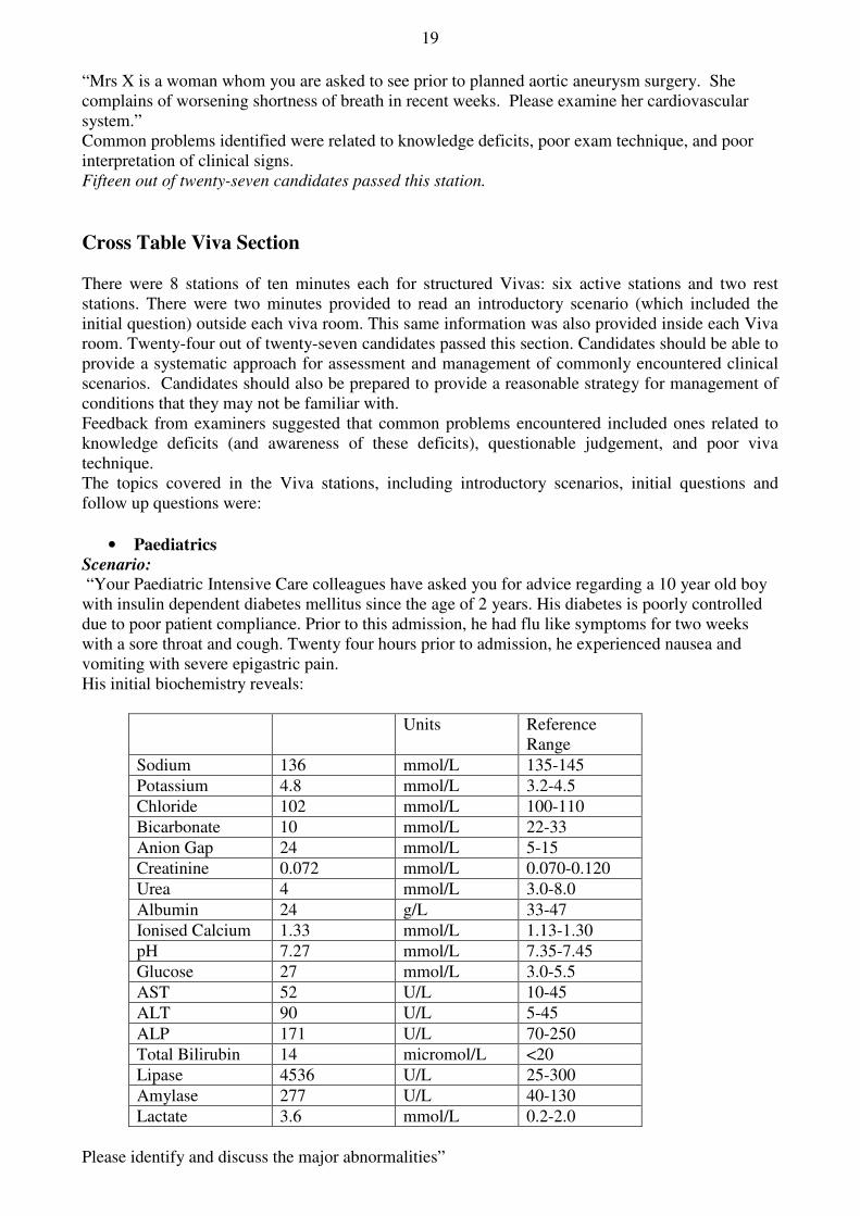
• Paediatrics Scenario: “Your Paediatric Intensive Care colleagues have asked you for advice regarding a 10 year old boy with insulin dependent diabetes mellitus since the age of 2 years. His diabetes is poorly controlled due to poor patient compliance. Prior to this admission, he had flu like symptoms for two weeks with a sore throat and cough. Twenty four hours prior to admission, he experienced nausea and vomiting with severe epigastric pain. His initial biochemistry reveals:
Units Reference Range
Sodium 136 mmol/L 135-145 Potassium 4.8 mmol/L 3.2-4.5 Chloride 102 mmol/L 100-110 Bicarbonate 10 mmol/L 22-33 Anion Gap 24 mmol/L 5-15 Creatinine 0.072 mmol/L 0.070-0.120 Urea 4 mmol/L 3.0-8.0 Albumin 24 g/L 33-47 Ionised Calcium 1.33 mmol/L 1.13-1.30 pH 7.27 mmol/L 7.35-7.45 Glucose 27 mmol/L 3.0-5.5 AST 52 U/L 10-45 ALT 90 U/L 5-45 ALP 171 U/L 70-250 Total Bilirubin 14 micromol/L <20 Lipase 4536 U/L 25-300 Amylase 277 U/L 40-130 Lactate 3.6 mmol/L 0.2-2.0
Please identify and discuss the major abnormalities”

20
Follow up question: “What are the possible causes of this presentation” Twenty-five out of twenty-seven candidates passes this station.
• Trauma Scenario: “A 27 year old man is admitted to hospital following a multiple vehicle crash on the freeway. He is awake and complaining of abdominal pain. There is an obvious open femoral fracture. Outline your initial assessment and management.” Follow up question: “He develops hypotension that responds to fluid therapy (4 litres of normal saline). How will you determine the source of this bleeding?” Twenty-five out of twenty-seven candidates passes this station.
• Neurological Scenario: “A 35 year old male with pneumococcal pneumonia and septicaemia has been in ICU for 4 days. His antibiotic treatment is intravenous penicillin G 2 million units 4 hourly, and he has been improving steadily. His septic shock has now resolved, his gas exchange has improved (FiO2 = 0.35), and he has begun to obey commands. However, he still has anuric renal failure, requiring bicarbonate based CVVHDF. While you are conducting the morning ward round he suddenly has a tonic - clonic convulsion, then commences another. What is your initial management of this patient?” Follow up question: “There are no lateralising neurological signs. He continues to fit intermittently, and is unresponsive between fits. What are the most likely causes to be considered at this point?” Common problems encountered included ones related to knowledge deficits, questionable judgement, and poor exam technique. Seventeen out of twenty-seven candidates passed this section.
• Haemato-oncology Scenario: “You have been called to the haematology ward to review a 55 year old man, who is 2 months post allogeneic bone marrow transplant for acute myeloid leukaemia which is thought to be in remission. In the last 72 hours since admission to hospital, he has become increasingly dyspnoeic, pyrexial to 38.3 C, and neutropenic. His chest X ray taken today shows increasing bilateral interstitial infiltrates. What is your differential diagnosis in this case?” Follow up question: “How would you differentiate between them?” Fourteen out of twenty-seven candidates passed this section.
• Renal Scenario: “A 76 year old, previously well, male is admitted to the ICU following an emergency abdominal aortic aneurysm repair. Immediate post operative ICU care has been exemplary, the patient receiving early and appropriate resuscitation. There has been improvement in the lactic acidosis, correction of coagulopathy and hypothermia. The patient however remains oliguric 4 hrs after the operation. List possible causes for the persistent oliguria?” Follow up question: “You suspect raised Intra-Abdominal Pressure and the Abdominal Compartment Syndrome, how do you measure the Intra-Abdominal Pressure?” Twenty-five out of twenty-seven candidates passed this section.
• Toxicology Scenario: “A 26 year old girl is admitted to your intensive care following a drug overdose. She admits to taking twenty-four tablets of paracetamol and twenty paracetamol-codeine tablets a few hours ago. She says that she recently broke up with her partner and has been feeling suicidal and depressed. She also admits to drinking heavily for the last few hours. On examination she appears drowsy and

21
has slurred speech and smells of alcohol. Outline your initial approach to management of this patient.” Follow up question: “What blood tests will you perform and why?” Common problems encountered including those related to knowledge deficits, inability to recognise the knowledge deficit, failure to recognise clinically significant issues, and inability o prioritise. Seventeen out of twenty-seven candidates passed this section. The Clinical Section: Hot cases The Clinical Section (comprising 2 hot cases) was conducted at the Alfred Hospital, Melbourne. Sixteen out of twenty-seven candidates passed the combined clinical section (seventeen out of twenty-seven passed the hot cases overall, and nineteen out of twenty-seven passed the cold cases overall). Candidates should listen carefully to the introduction given by the examiners and direct their examination accordingly. Patients were usually presented as problem solving exercises. For maximal marks, candidates should demonstrate a systematic approach to examination, clinical signs should be demonstrated, and a reasonable discussion regarding their findings should follow. The twenty minutes available for each case provides ample opportunity to discuss related investigations and plans of management. Some candidates waste valuable time at the start of the case by spending more than a couple of minutes around the bedside before they actually commence examining the patient. Exposing the patients should be limited to those areas that are necessary for that component of the examination, and in keeping with the modesty requirements of the patients. Candidates must show appropriate courtesy and respect to patients. Cases encountered as hot cases included patients with:
• Sepsis, psoas abscess • Multiple trauma after a motor vehicle crash with long bone fractures and a ruptured bladder • End-stage liver disease, acute renal failure and shock • Lung abscess and bronchopleural fistula • Respiratory distress and Guillain-Barre Syndrome • Faecal peritonitis and multiple organ dysfunction • Pancreatitis and multiple organ dysfunction
Introductory questions included: “This man presented with a gastrointestinal bleed. Can you work out why he may be jaundiced?” “This elderly woman has been in Intensive care with abdominal sepsis. She is becoming difficult to wean off the ventilator. Can you assess her to try and elucidate why she is being difficult to wean?” “This man has been in Intensive care for 8 weeks with an abdominal problem. Last night he spiked a temperature of 39°C. Can you examine him specifically to work out why he may have done so?” Comments documented at the time of the clinical examination suggested that common problems encountered related to poor examination technique (eg. slow to actually start examining the patient), and poor discussion. ________________________________________________________________________________ Dr Peter Morley Chairman, Court of Examiners, Chairman Examination Committee Circulation: Board of Joint Faculty Panel of Examiners Supervisors of Intensive Care Training Course Supervisors Regional Education Officers Registered Trainees