Embed Size (px)
Citation preview


1
General ManagerNebojsa Arsenijevic
Editor in Chief
Vladimir Jakovljevic
Co-EditorsNebojsa Arsenijevic, Slobodan Jankovic and Vladislav Volarevic
International Advisory Board
(Surnames are given in alphabetical order)Antovic J (Stockholm, Sweden), Bosnakovski D (Štip, FYR Macedonia), Chaldakov G (Varna, Bulgaria),
Conlon M (Ulster, UK), Dhalla NS (Winnipeg, Canada), Djuric D (Belgrade, Serbia),Fountoulakis N (Th essaloniki, Greece), Kusljic S (Melbourne, Australia), Lako M (Newcastle, UK),Mitrovic I (San Francisco, USA), Monos E (Budapest, Hungary), Muntean D (Timisoara, Romania),
Paessler S (Galvestone, USA), Pechanova O (Bratislava, Slovakia), Serra P (Rome, Italy),Strbak V (Bratislava, Slovakia), Svrakic D (St. Louis, USA), Tester R (Glasgow, UK),
Vlaisavljevic V (Maribor, Slovenia), Vujanovic N (Pittsburgh, USA), Vuckovic-Dekic Lj (Belgrade, Serbia)
Editorial Staff Gordana Radosavljevic, Marija Milovanovic, Jelena Pantic, Ivan Srejovic, Vladimir Zivkovic, Jovana Joksimovic
Management TeamNebojsa Arsenijevic, Ana Miloradovic, Milan Milojevic
Corrected byScientifi c Editing Service “American Journal Experts”
Design
PrstJezikIostaliPsi / Miljan Nedeljkovic
PrintFaculty of Medical Sciences,
University of Kragujevac
Indexed inEMBASE/Excerpta Medica, Index Copernicus, BioMedWorld, KoBSON, SCIndeks, Chemical Abstracts Service,
Cabell’s Directory, Celdes, CNKI Scholar (China National Knowledge Infrastructure), CNPIEC,EBSCO Discovery Service, Elsevier - SCOPUS, Google Scholar, J-Gate, Naviga (Softweco), Primo Central (ExLibris),
ReadCube, SCImago (SJR), Summon (Serials Solutions/ProQuest), TDOne (TDNet), WorldCat (OCLC)
Address:Serbian Journal of Experimental and Clinical Research, Faculty of Medical Sciences, University of Kragujevac
Svetozara Markovica 69, 34000 Kragujevac, PO Box 124Serbia
http://www.medf.kg.ac.rs/sjecr/index.php
SJECR is a member of WAME and COPE. SJECR is published four times circulation 250 issuesTh e Journal is fi nancially supported by Ministry for Science and Technological Development, Republic of Serbia
ISSN 1820 – 8665
2
Review Paper / Revijalni rad
STEM CELLS: NEW HOPE FOR SPINAL CORD INJURY
MATIČNE ĆELIJE: NOVA NADA ZA POVREDE KIČMENE MOŽDINE ...................................................................................................3
Original Scientific Paper / Originalni naucni rad
ST2 DEFICIENCY AMELIORATES HIGH FAT DIETINDUCED LIVER STEATOSIS IN BALB/C MICE
DELECIJA GENA ZA ST2 U BALB/C MIŠEVA UBLAŽAVA STEATOZU JETRE INDUKOVANU
DIJETOM SA VISOKIM SADRŽAJEM MASTI ......................................................................................................................................................9
Original Scientific Paper / Originalni naucni rad
SIMPLE AND COMPLEX COGNITIVE FUNCTIONS UNDER EXERTIONAL HEAT STRESS
UTICAJ TOPLOTNOG STRESA I FIZIČKE AKTIVNOSTI NA KOGNITIVNE SPOSOBNOSTI ..................................................21
Original Scientific Paper / Originalni naucni rad
PERIODONTAL DISEASE AND RISK FOR PRETERM BIRTH: A CASECONTROL STUDY
PARODONTOPATIJA I RIZIK ZA NASTAJANJE PREVREMENOG POROĐAJA: STUDIJA SLUČAJKONTROLA ...........27
Original Scientific Paper / Originalni naucni rad
EARLY CYTOKINE PROFILE CHANGES IN INTERSTITIAL AND NECROTIC FORMS OF ACUTE PANCREATITIS
RANE PROMENE NIVOA CITOKINA KOD INTERSTICIJALNIH I
NEKROTICNIH OBLIKA AKUTNOG PANKREATITISA ............................................................................................................................. 33
Original Scientific Paper / Originalni naucni rad
COMPARISON OF MEDIAL ARCHSUPPORTING INSOLES AND HEEL PADS
IN THE TREATMENT OF PLANTAR FASCIITIS
POREĐENJE ULOŽAKA ZA PODUPIRANJE MEDIJALNOG LUKA STOPALA I PETNIH ULOŽAKA
U LEČENJU PLANTARNOG FASITISA ................................................................................................................................................................. 39
Original Scientific Paper / Originalni naucni rad
MACROECONOMIC POLICY IMPACT ON ONCOLOGYRELATED PUBLIC EXPENDITURE
IN AN EMERGING EUROPEAN MARKET SIGNS OF EARLY RECOVERY
UTICAJ MAKROEKONOMSKE POLITIKE NA JAVNA IZDVAJANJA ZA
ONKOLOGIJU NA RASTUĆEM EVROPSKOM TRŽIŠTU ZNACI RANOG OPORAVKA .......................................................... 43
Case Report / Prikaz slučaja
ISOLATED CARPAL DISLOCATION OF THE TRAPEZIUM
IZOLOVANA KARPALNA DISLOKACIJA TRAPEZOIDNE KOSTI ..........................................................................................................51
Case Report / Prikaz slučaja
SECONDARY HAEMOPHAGOCYTIC LYMPHOHISTIOCYTOSIS
THE DIFFERENTIAL DIAGNOSIS DILEMMA IN PAEDIATRICS
SEKUNDARNA HEMOFAGOCITNA LIMFOHISTIOCITOZA
DIFERENCIJALNODIJAGNOSTIČKA DILEMA U PEDIJATRIJI .............................................................................................................55
Metodology Report / Metodološki prikaz
ENHANCEMENT OF DERMAL FIBROBLAST ISOLATION METHOD
UNAPREĐENJE METODE IZOLACIJE DERMALNIH FIBROBLASTA ....................................................................................................65
INSTRUCTION TO AUTHORS FOR MANUSCRIPT PREPARATION ......................................................................................................71
Table Of Contents

3Corresponding author: Marina Gazdic,Center for Molecular Medicine and Stem Cell Research,
Medical Faculty, University of Kragujevac, Serbia, [email protected]
UDK: 602.9:616.732-001.5-089.843 / Ser J Exp Clin Res 2015; 16 (1): 3-8
DOI: 10.1515/SJECR20150001
ABSTRACT
Stem cell therapy off ers several attractive strategies for spi-
nal cord repair. Th e regenerative potential of pluripotent stem
cells was confi rmed in an animal model of Spinal Cord Injury
(SCI); nevertheless, optimized growth and diff erentiation pro-
tocols along with reliable safety assays should be established
prior to the clinical application of hESCs and iPSCs. Th e
therapeutic eff ects of mesenchymal stem cells (MSCs) in SCI
result from neurotrophin secretion, angiogenesis, and antiin-
fl ammatory actions. Several preclinical SCI studies have re-
ported that the occurrence of axonal extension, remyelination
and neuroprotection occur after the transplantation of olfac-
tory ensheathing cells (OECs). Th e transplantation of neural
stem cells NSCs (NSCs) promotes partial functional improve-
ment after SCI because of their potential to diff erentiate into
neurons, oligodendrocytes, and astrocytes. Th e ideal source of
stem cells for safe and effi cient cell-based therapy for SCI re-
mains a challenging issue that requires further investigation.
SAŽETAK
Terapija matičnim ćelijama pruža nekoliko atraktivnih
mogućnosti za lečenje povreda kičmene moždine. Regenera-
tivni potencijal pluripotentnih matičnih ćelija je potvrdjen
u animalnim modelima povrede kičmene moždine, me-
djutim, protokoli za kultivaciju i diferencijaciju ovih ćelija
kao i testovi za potvrdu njihove bezbednosti tek moraju biti
ustanovljeni kako bi se hESCs i iPSCs primenile u kliničkoj
praksi. Terapijski efekat MSCs u povredi kičmene moždine se
zasniva na sposobnosti ovih ćelija da sekretuju neurotrofne
i antiinfl amatorne faktore, kao i da promovišu angiogenezu.
U nekoliko predkliničkih studija su pokazani rast aksona,
remijelinizacija i neuroprotektivno delovanje OECs. Tran-
splantacija NSCs doprinosi funkcionalnom oporavku nakon
povrede kičmene moždine diferencijacijom NSCs u neurone,
oligodendrocite i astrocite. Otkrivanje idealnog izvora ma-
tičnih ćelija za efi kasnu i bezbednu terapiju povrede kičmene
moždine i još uvek je izazov i zahteva dalja istraživanja.
STEM CELLS: NEW HOPE FOR SPINAL CORD INJURY
Marina Gazdic1, Vladislav Volarevic1 and Miodrag Stojkovic1,2
1 Center for Molecular Medicine and Stem Cell Research, Faculty of Medical Sciences, University of Kragujevac, Serbia;;2 Spebo Medical, Leskovac, Serbia
MATIČNE ĆELIJE: NOVA NADA ZA POVREDE KIČMENE MOŽDINE
Marina Gazdić1, Vladislav Volarević1 i Miodrag Stojković1,2
1 Centar za molekulsku medicinu i izučavanje matičnih ćelija, Fakultet medicinskih nauka, Univerzitet u Kragujevcu, Srbija;2 Spebo Medical, Leskovac, Srbija
Received / Primljen: 30. 09. 2014. Accepted / Prihvaćen: 12. 10. 2014.
REVIEW PAPER REVIJALNI RAD REVIEW PAPER REVIJALNI RAD REVIEW PAPER
INTRODUCTION
Spinal cord injury (SCI) is a devastating condition with
permanent lifelong consequences (1). Epidemiological data
show that the incidence of traumatic SCI in the US ranges
from 27 to 83 per million while in Europe it is approxi-
mately 10–30 new cases per million (1). SCI usually results
in sudden and long-lasting locomotor and sensory neuron
degeneration below the injury (2).
The pathophysiological processes that underlie SCI
comprise the primary and secondary phases of injury. Dur-
ing the primary phase, because of the direct mechanical
trauma of the spinal cord by fractured and displaced bone
fragments and disc material, there is massive axonal dam-
age as well as neuronal and glial cell losses (3, 4, 5). During
the secondary phase of injury, further tissue damage occurs
mostly from ischemia, electrolyte imbalance, inflammato-
ry response, oxidative stress and excitotoxicity (3). Despite
major advances in the medical and surgical care of SCI
patients, there are currently no effective therapies for the
treatment of traumatic SCI in humans (2). Stem cell thera-
py offers several attractive strategies for spinal cord repair.
Stem cells may play an important role in the replacement
of damaged neuronal and glial cells, axonal regeneration
and remyelination, the restoration of neuronal circuitry,
and the production of neurotrophic factors, anti-inflam-
matory cytokines, and other molecules that promote tissue
repair and neovascularization.
In this review, we will evaluate the therapeutic role of hu-
man embryonic stem cells (hESCs), induced pluripotent stem
cells (iPSCs), mesenchymal stem cells (MSCs), neural stem
cells (NSCs), and olfactory ensheathing cells (OECs) for treat-
ing SCI, and we will cover some of the clinical trials that aim to
translate laboratory stem cell research into clinical practice.
4
transplanted directly into the spinal cord of four ASIA A
patients with complete thoracic SCI. In 2011, Geron discon-
tinued this trial for financial reasons. The preliminary re-
sults indicated that GRNOPC1s do not cause any harm, but
the debate about the efficacy of these cells still continues.
Concerns about the transplantation of hESC-derived neu-
ral cells to treating SCI are related to the ethical issues of cell
derivation, the immune rejection of transplanted cells, the use
of differentiation protocols that still involve mediums, growth
factors, and supplements of animal origin, and the possibility
of teratoma formation from incomplete or aberrant differen-
tiation resulting in the formation of non-neural cells (6, 9).
Induced Pluripotent Stem Cells
iPSCs were originally generated by the ectopic expression
of four transcription factors called, namely Oct4, Sox2, Klf4,
and c-Myc in fibroblast cells by Takahashi and Yamanaka in
2006 (10). iPSCs show morphological, transcriptional, epige-
netic, and phenotypic similarity to hESCs and can differenti-
ate towards any cell in the human body including neurons,
glia, neural progenitor cells (NPCs), and motoneurons (11).
Human Embryonic Stem Cells
hESCs are derived from the inner cell mass of human
blastocysts; they have the ability to proliferate by maintain-
ing both their pluripotency and their ability to differentiate
into nearly all cell types, including neuronal and glial cells
(6). Improved protocols have been developed to differenti-
ate hESCs into motoneuron progenitors (MPs) and oligo-
dendrocyte progenitors (OPCs) (7, 8). The transplantation
of hESC-derived MPs and OPCs can efficiently recover
locomotor function in both contusion and transection ani-
mal models of SCI (7, 8). The regenerative mechanism of
hESC therapy for SCI depends on the potential of hESC-
derived OPCs and MPs to differentiate into neuronal and
glial cells and the immunomodulatory characteristics of
transplanted hESC-derived OPCs (7, 1).
Based on promising preclinical data from hESC-derived
OPC transplants in rodent SCI models, the Food and Drug
Administration (FDA) approved the first hESC clinical trial
in 2009. The Geron Company attempted to test the safety
of hESC-derived OPCs in human SCIs. Two million hESC-
derived OPC cells (GRNOPC1) within the acute phase were
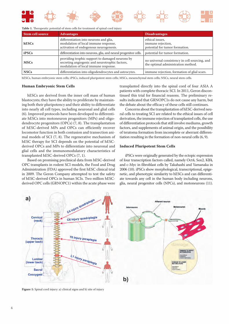
Figure 1: Spinal cord injury: a) clinical signs and b) site of injury
Table 1. Th erapeutic potential of stem cells for treatment of spinal cord injury
Stem cell source Advantages Disadvantages
hESCsdiff erentiation into neurons and glia, modulation of local immune response, activation of endogenous neurogenesis.
ethical issues, immune rejection, potential for tumor formation.
iPSCs diff erentiation into neurons, glia, and neural progenitor cells. potential for tumor formation.
MSCsproviding trophic support to damaged neurons by secreting angiogenic and neurotrophic factors, modulation of local immune response.
no universal consistency in cell sourcing, and the optimal administration method.
NSCs diff erentiation into oligodendrocytes and astrocytes. immune rejection, formation of glial scars.
hESCs, human embryonic stem cells; iPSCs, induced pluripotent stem cells; MSCs, mesenchymal stem cells; NSCs, neural stem cells.

5
Mesenchymal Stem Cells
MSCs are adult, self-renewable, multipotent cells that can
be found in almost all postnatal tissues (16). In addition to
their stem/progenitor properties, MSCs have been shown to
possess broad immunomodulatory abilities (16). The thera-
peutic effects of MSCs in SCI result from neurotrophin secre-
tion, angiogenesis, and antiinflammatory actions, rather than
direct translineage conversion to functional oligodendrocytes
or neurons (17, 18, 19, 20, 21). Engrafted MSCs act as neu-
roprotectors by secreting brain-derived neurotrophic factor
(BDNF), glia cell line-derived neurotrophic factor (GDNF),
Patient-specific iPSCs derived from somatic cells through the
ectopic expression of a defined set of factors do not present
ethical and immnunological concerns (11). The primary con-
cern about the use of these cells in clinical trials was with the
reprogramming technology that involved viral vectors and
their tumourigenicity (2). Some of the reprogramming issues
are solved by the deriving iPSCs bythrough nonviral meth-
ods such as mRNA or chemicals and small molecules (12, 13).
The regenerative potential of iPSCs was confirmed in a ro-
dent model of SCI (14, 15); nevertheless, optimized growth
and differentiation protocols and reliable safety assays should
be established prior to the clinical application of iPSCs.
Figure 3: Transplanted MSCs act as neuroprotectors in spinal cord injuries by producing growth factors and
anti-infl ammatory cytokines.
Figure 2: Method for derivation of iPSCs from adult somatic cell by introducing OCT4, SOX2, KLF4 and c-Myc.
iPSCs can diff erentiate toward neurons, oligodendrocytes and astrocytes.

6
nerve growth factor (NGF), vascular endothelial growth fac-
tor (VEGF), and hepatocyte growth factor (HGF) (22, 23).
The number of clinical trials that employ MSCs for SCI
treatment is increasing, indicating that despite several ques-
tions that still need to be addressed at pre-clinical levels, MSCs
are considered potentially beneficial for translational studies
(24). The pathological processes that occur at the lesion site in
SCIs evolve over time, from the acute to subacute to chronic
phases; therefore, transplantation at different times post-lesion
may have varied effects (25). Sykova et al. suggested that per-
forming MSC transplantation within a therapeutic window
of 3-4 weeks following SCI is critical for the success of MSC-
based therapy (26). Yoon et al. studied the effects of autologous
bone marrow MSC transplantation in combination with the
administration of granulocyte-macrophage colony-stimulating
factor to 35 patients with complete SCIs at acute, subacute and
chronic stages (27). No serious complications were reported,
and 30.4% of patients who received MSCs at acute and subacute
stages showed significant improvements in their ASIA scale
position (27). Few clinical studies have shown neurological im-
provements in MSC-treated patients who during the chronic
stage of SCI, when the glial scar is already present (28, 29, 30,
31, 32). In these studies, MSCs were transplanted directly into
the lesion intrathecally, intravenously and intrathecally (simul-
taneously) at once, and intraarterially.
Although the clinical study results are promising, there
are important issues that should be addressed to achieve
successful MSC-based therapy, that is, the universal consis-
tency in cell sourcing and culture conditions, the ideal cell
quantity and the optimal administration method (24, 1).
Olfactory Ensheathing Cells
OECs are a unique population of macroglia found in
the lamina propria of olfactory mucosa, around the olfac-
tory nerve fascicles and in the two outer layers of the olfac-
tory bulb. OECs have the dual nature of astroglial cells and
Schwann cells (33). Several preclinical SCI studies have re-
ported the occurrence of axonal extension, remyelination
and neuroprotection after OEC transplantation (34, 35, 36,
37, 38). OECs migrate to injured sites and secrete a large
number of factors that are necessary for the growth, devel-
opment, differentiation, and maturation of different types
of neurons and reduce astrocyte activity and glial scar for-
mation (39, 40). Spinal cord regeneration and functional
recovery depend on the nature and source of OECs, the
injury model, the graft cell preparation, the time of trans-
plantation, and the transplantation procedures (25).
Feron et al. performed a single-blind phase I clinical trial in
which three patients with SCI (chronic injuries) received au-
tologous OECs. The feasibility of the procedure and the safety
of these cells were reported, but there was no evidence of clini-
cal efficacy (41, 42). Lima et al. reported that the transplanta-
tion of minced olfactory mucosa in patients with chronic SCI
was not significantly efficient (44). By contrast, recent clini-
cal studies suggested that there was a neurological improve-
ment in SCI patients after OEC transplantation (44, 45).
Neural Stem Cells
NSCs are multipotent cells with the potential to dif-
ferentiate into neurons, oligodendrocytes, and astrocytes,
and they can be efficiently propagated in vitro (46).
NSCs can be found in the periventricular subependy-
mal layer, in the subgranular zone of the dentate gyrus, and
in the ependymal regions lining the central canal (47). The
activation of resident ependymal stem cells following SCI
is not sufficient to promote recovery because the cells dif-
ferentiate mostly into actrocytes and oligodendrocytes (48,
49). Several preclinical studies confirmed that the trans-
plantation of NSCs promotes a partial functional improve-
ment after SCI (50, 51). The transplantation of OPCs that
had differentiated from ependymal stem cells efficiently
recovered the locomotor function of an SCI animal model
(47). The source of NSCs, the methods of cell isolation
and preparation, the time of transplantation, the chosen
Figure 4: Potential uses of NSCs which were isolated from the adult brain and spinal cord as a source of neurons, oligodendrocytes, and astrocytes.

7
immunosuppression, and the type of injury (contusion vs.
transection) are important issues for achieving successful
NSC-based therapy after SCI (49). Human NSCs have been
isolated from foetal brains, and spinal cords have been iso-
lated from aborted foetuses. Unlike adult NSCs, foetal-
derived NSCs generate neurons in addition to glia in the
injured spinal cord (1).
Currently, two human trials involving allogeneic NSCs
for SCI are ongoing. The primary objectives of these stud-
ies are to determine the long term safety and preliminary
efficacy of NSC transplantation in subjects with thorac-
ic spinal cord trauma.
CONCLUSIONS
Numerous preclinical studies suggest that stem cells are
able to enhance recovery following SCI. However, the ideal
source of stem cells for the efficient and safe cell-based
therapy of SCI remains a challenging issue that requires
further investigation and continuous cooperation between
clinicians, researchers, and patients.
ACKNOWLEDGMENTS:
This study was supported by the Serbian Ministry of Sci-
ence (project numbers ON 175069 and ON175103). We high-
ly appreciate and acknowledge the generous assistance of Mr.
Milan Milojevic, who contributed to the creation of the figures
in this article. The authors declare no conflicts of interest.
REFERENCES
1. Volarevic V, Erceg S, Bhattacharya SS, Stojkovic
P, Horner P, Stojkovic M. Stem Cell-Based Therapy
for Spinal Cord Injury. Cell Transplantation 2013;
22(8):1309-23.
2. Lukovic D, Moreno Manzano V, Stojkovic M, Bhat-
tacharya SS, Erceg S. Concise review: human pluripo-
tent stem cells in the treatment of spinal cord injury.
Stem Cells 2012; 30(9):1787-92.
3. Rowland JW, Hawryluk GW, Kwon B, Fehlings MG.
Current status of acute spinal cord injury pathophysiol-
ogy and emerging therapies: Promise on the horizon.
Neurosurg. Focus 2008; 25:E2.
4. McTigue DM, Tani M, Krivacic K et al. Selective
chemokine mRNA accumulation in the rat spinal cord
after contusion injury. J Neurosci Res 1998;53:368–376.
5. Grossman SD, Rosenberg LJ, Wrathall JR. Temporal-
spatial pattern of acute neuronal and glial loss after spi-
nal cord contusion. Exp Neurol 2001;168:273–282.
6. Erceg S, Ronaghi M, Stojković M. Human embry-
onic stem cell differentiation toward regional specif-
ic neural precursors. Stem Cells 2009. 27(1):78-87.
7. Erceg S, Ronaghi M, Oria M, et al. Transplanted oli-
godendrocytes and motoneuron progenitors generated
from human embryonic stem cells promote locomotor
recovery after spinal cord transection. Stem Cells 2010;
28:1541–1549.
8. Nistor GI, Totoiu MO, Haque N, Carpenter MK, Kei-
rstead HS. Human embryonic stem cells differentiate
into oligodendrocytes in high purity and myelinate af-
ter spinal cord transplantation. Glia 2005; 49:385–396.
9. Mothe AJ, Tator CH. Advances in stem ell ther-
apy for spinal cord injury. J Clin Invest. 2012;
122(11):3824-34.
10. Takahashi K, Yamanaka S. Induction of pluripotent
stem cells from mouse embryonic and adult fibroblast
cultures by defined factors. Cell 2006; 126:663–676.
11. Lukovic D, Moreno-Manzano V, Klabusay M, Stojk-
ovic M, Bhattacharya SS, Erceg S. Non-coding RNAs
in pluripotency and neural differentiation of human
pluripotent stem cells. Front Genet. 2014; 14;5:132.
12. Warren L, Manos PD, Ahfeldt T, et al. Highly efficient
reprogramming to pluripotency and directed differen-
tiation of human cells with synthetic modified mRNA.
Cell Stem Cell 2010; 7:618–630.
13. Zhou H, Wu S, Joo JY, et al. Generation of induced
pluripotent stem cells using recombinant proteins. Cell
Stem cell 2009; 4:381–384.
14. Tsuji O, Miura K, Okada Y, et al. Therapeutic poten-
tial of appropriately evaluated safe-induced pluripotent
stem cells for spinal cord injury. Proc Natl Acad Sci
USA 2010; 107:12704–12709.
15. Nori S, Okada Y, Yasuda A, et al. Grafted human-induced
pluripotent stem-cell-derived neurospheres promote
motor functional recovery after spinal cord injury in
mice. Proc Natl Acad Sci USA 2011;108: 16825–16830.
16. Volarevic V, Al-Qahtani A, Arsenijevic N, et al. Inter-
leukin-1 receptor antagonist (IL-1Ra) and IL-1Ra pro-
ducing mesenchymal stem cells as modulators of dia-
betogenesis. Autoimmunity 2010; 43: 255–63.
17. Hawryluk GW, Mothe AJ, Chamankhah M, Wang J,
Tator C, Fehlings MG. In vitro characterization of
trophic factor expression in neural precursor cells.
Stem Cells Dev. 2012; 21(3):432–447.
18. Himes BT, Neuhuber B, Coleman C et al. Recovery of
function following grafting of human bone marrow-
derived stromal cells into the injured spinal cord. Neu-
rorehabil Neural Repair 2006; 20(2):278–296.
19. Hawryluk GW, Mothe A, Wang J, Wang S, Tator C, Fe-
hlings MG. An in vivo characterization of trophic fac-
tor production following neural precursor cell or bone
marrow stromal cell transplantation for spinal cord in-
jury. Stem Cells Dev. 2012; 21(12):2222–2238.
20. Caplan AI, Dennis JE. Mesenchymal stem cells as trophic
mediators. J Cell Biochem. 2006; 98(5):1076–1084.
21. Ruff CA, Wilcox JT, Fehlings MG. Cell-based trans-
plantation strategies to promote plasticity following
spinal cord injury. Exp. Neurol. 2012; 235:78–90.
22. Kim HJ, Lee HJ, Kim SH. Therapeutic effects of human
mesenchymal stem cells on traumatic brain injury in
rats: Secretion of neurotrophic factors and inhibition
of apoptosis. J. Neurotrauma 2010; 27:131–138.

8
23. Sasaki M, Radtke C, Tan AM, et al. BDNF hypersecret-
ing human mesenchymal stem cells promote functional
recovery, axonal sprouting, and protection of corti-
cospinal neurons after spinal cord injury. J. Neurosci.
2009; 29:14932–14941.
24. Martinez AM, Goulart CO, Ramalho Bdos S, Oliveira
JT, Almeida FM. Neurotrauma and mesenchymal stem
cells treatment: From experimental studies to clini-
cal trials. World J Stem Cells 2014; 6(2):179-94.
25. Li J, Lepski G. Cell transplantation for spinal cord injury: a
systematic review. Biomed Res Int. 2013; 2013:786475.
26. Syková E, Homola A, Mazanec R, et al. Autologous
bone marrow transplantation in patients with subacute
and chronic spinal cord injury. Cell Transplant. 2006;
15:675–687.
27. Yoon SH, Shim YS, Park YH, et al. Complete spinal cord
injury treatment using autologous bone marrow cell
transplantation and bone marrow stimulation with gran-
ulocyte macrophage-colony stimulating factor: phase I/
II clinical trial. Stem Cells 2007; 25:2066–2073.
28. Chernykh ER, Stupak VV, Muradov GM, et al. Applica-
tion of autologous bone marrow stem cells in the ther-
apy of spinal cord injury patients. Bull. Exp. Biol. Med.
2007; 143:543–547.
29. Kumar A, Kumar S, Narayanan R, Arul K, Baskaran
M. Autologous bone marrow derived mononuclear
cell therapy for spinal cord injury: A phase I/II clinical
safety and primary efficacy data. Exp. Clin. Transplant.
2009; 7:241– 248.
30. Callera F, do Nascimento RX. Delivery of autologous bone
marrow precursor cells into the spinal cord via lumbar
puncture technique in patients with spinal cord injury: A
preliminary safety study. Exp. Hematol. 2006; 34:130–13.
31. Cristante AF, Barros-Filho TE, Tatsui N, et al. Stem
cells in the treatment of chronic spinal cord injury:
Evaluation of somatosensitive evoked potentials in 39
patients. Spinal Cord 2009; 47:733–738.
32. Deda H, Inci MC, Kürekçi AE, et al. Treatment of chron-
ic spinal cord injured patients with autologous bone
marrowderived hematopoietic stem cell transplanta-
tion: 1-year follow-up. Cytotherapy 2008; 10:565–574.
33. Rao YJ, Zhu WX, Du ZQ, et al. Effectiveness of olfacto-
ry ensheathing cell transplantation for treatment of spi-
nal cord injury. Genet Mol Res. 2014; 13(2):4124-9.
34. García-Alias G, Lopez-Vales R, Fores J, Navarro X, Verdu
E. Acute transplantation of olfactory ensheathing cells or
Schwann cells promotes recovery after spinal cord injury
in the rat. J. Neurosci. Res. 2004; 75:632–641.
35. Kubasak MD, Jindrich DL, Zhong H, et al. OEG im-
plantation and step training enhance hindlimb-step-
ping ability in adult spinal transected rats. Brain 2008;
131:264–276.
36. Munoz-Quiles C, Santos-Benito FF, Llamusí MB, Ra-
mon-Cueto A. Chronic spinal injury repair by olfactory
bulb ensheathing glia and feasibility for autologous ther-
apy. J. Neuropathol. Exp. Neurol. 2009; 68:1294–1308.
37. Radtke C, Sasaki M, Lankford KL, Vogt PM, Kocsis JD.
Potential of olfactory ensheathing cells for cell-based
therapy in spinal cord injury. J. Rehabil. Res. Dev. 2008;
45:141–151.
38. Ramon-Cueto A, Cordero MI, Santos-Benito FF, Avi-
la J. Functional recovery of paraplegic rats and motor
axon regeneration in their spinal cords by olfactory en-
sheathing glia. Neuron 2000; 25:425–435.
39. Woodhall E, West AK, Chuah MI. Cultured olfactory
ensheathing cells express nerve growth factor, brain-
derived neurotrophic factor, glia cell line-derived neu-
rotrophic factor and their receptors. Brain Res Mol
Brain Res. 2001; 88(1-2):203-13.
40. Mayeur A, Duclos C, Honoré A, et al. Potential of olfac-
tory ensheathing cells from different sources for spi-
nal cord repair. PLoS One 2013; 8(4):e62860.
41. Feron F, Perry C, Cochrane J, et al. Autologous olfac-
tory ensheathing cell transplantation in human spinal
cord injury. Brain 2005; 128:2951–2960.
42. Mackay-Sim A, Feron F, Cochrane J, et al. Autolo-
gous olfactory ensheathing cell transplantation in
human paraplegia: a 3-year clinical trial. Brain 2008;
131:2376–2386.
43. Lima C, Pratas-Vital J, Escada P, Hasse-Ferreira A,
Capucho C, Peduzzi JD. Olfactory mucosa autografts
in human spinal cord injury: A pilot clinical study. J.
Spinal Cord Med 2006; 29:191–203.
44. Tabakow P, Jarmundowicz W, Czapiga B, et al. Trans-
plantation of autologous olfactory ensheathing cells in
complete human spinal cord injury. Cell Transplant.
2013; 22(9):1591-612.
45. Zheng Z, Liu G, Chen Y, Wei S. Olfactory ensheath-
ing cell transplantation improves sympathetic skin
responses in chronic spinal cord injury. Neural Regen
Res. 2013; 8(30):2849-55.
46. Hsu YC, Lee DC, Chiu IM. Neural stem cells, neural
progenitors, and neurotrophic factors. Cell Transplant.
2007; 16:133–150.
47. Moreno-Manzano V, Rodríguez-Jiménez, FJ, García-
Roselló M, et al. Activated spinal cord ependymal stem
cells rescue neurological function. Stem Cells 2009;
27:733–743.
48. Barnabe´-Heider F, Frisen J. Stem cells for spinal cord
repair. Cell Stem Cell 2008; 3:16–24.
49. Ronaghi M, Erceg S, Moreno-Manzano V, Stojkovic
M. Challenges of stem cell therapy for spinal cord in-
jury: human embryonic stem cells, endogenous neu-
ral stem cells, or induced pluripotent stem cells? Stem
Cells 2010; 28(1):93-9.
50. Iwanami A, Kaneko S, Nakamura M, et al. Transplanta-
tion of human neural stem cells for spinal cord injury in
primates. J. Neurosci. Res. 2005; 80:182–190.
51. Parr A. M, Kulbatski I, Zahir T, et al. (2008). Trans-
planted adult spinal cordderived neural stem/progeni-
tor cells promote early functional recovery after rat spi-
nal cord injury. Neuroscience 155:760–770.

9
ORIGINAL SCIENTIFIC PAPER ORIGINALNI NAUČNI RAD ORIGINAL SCIENTIFIC PAPER
ST2 DEFICIENCY AMELIORATES HIGH FAT DIETINDUCED LIVER STEATOSIS IN BALB/C MICE
Nemanja Jovicic 1,2,*, Ilija Jeftic 1,3*, Marina Miletic Kovacevic 2, Irena Tanaskovic 2, Nebojsa Arsenijevic 1, Miodrag L. Lukic 1, Nada Pejnovic 1,3*
1 Center for Molecular Medicine;2 Institute of Histology;
3 Institute of Pathophysiology, Faculty of Medical Sciences, University of Kragujevac, Kragujevac, Serbia
DELECIJA GENA ZA ST2 U BALB/C MIŠEVA UBLAŽAVASTEATOZU JETRE INDUKOVANU DIJETOM
SA VISOKIM SADRŽAJEM MASTINemanja Jovičić 1,2,*, Ilija Jeftić 1,3*, Marina Miletić Kovačević 2, Irena Tanasković 2, Nebojša Arsenijević 1, Miodrag L. Lukić 1, Nada Pejnović 1,3*
1 Centar za molekulsku medicinu;2 Institut za histologiju;
3 Institut za patofi ziologiju, Fakultet medicinskih nauka, Univerzitet u Kragujevcu, Kragujevac, Srbija
*Th e authors NJ and IJ contributed equally to this study.
ABSTRACT
Non-alcoholic fatty liver disease (NAFLD) is strongly as-
sociated with obesity, but the molecular mechanisms of liver
steatosis and its progression to non-alcoholic steatohepatitis
and fi brosis are incompletely understood. Immune reactiv-
ity plays an important role in the pathogenesis of NAFLD.
Th e IL-33/ST2 axis has a protective role in adiposity and
atherosclerosis, but its role in obesity-associated metabolic
disorders requires further clarifi cation. To investigate the
unresolved role of IL-33/ST2 signalling in NAFLD, we used
ST2-defi cient (ST2-/-) and wild type (WT) BALB/c mice
maintained on a high-fat diet (HFD) for 24 weeks. HFD-
fed ST2-/- mice exhibited increased weight gain, visceral
adipose tissue weight and triglyceridaemia and decreased
liver weight compared with diet-matched WT mice. Com-
pared with WT mice on an HFD, ST2 deletion signifi cantly
reduced hepatic steatosis, liver infl ammation and fi brosis
and downregulated the expression of genes related to lipid
metabolism in the liver. Th e frequency of innate immune
cells in the liver, including CD68+ macrophages and CD11c+
dendritic cells, was lower in HFD-fed ST2-/- mice, accom-
panied by lower TNFα serum levels compared with diet-
matched WT mice. Less collagen deposition in the livers of
ST2-/- mice on an HFD was associated with lower numbers
of profi brotic CD11b+Ly6clow monocytes and CD4+IL-17+ T
cells in the liver, lower hepatic gene expression of procolla-
gen, IL-33 and IL-13, and lower serum levels of IL-33 and
IL-13 compared with diet-matched WT mice.
Our fi ndings suggest that the IL-33/ST2 axis may have a
complex role in obesity-associated metabolic disorders. Al-
though it is protective in HFD-induced adiposity, the IL-33/
ST2 pathway promotes hepatic steatosis, infl ammation and
fi brosis.
Key words: Obesity, steatosis, non-alcoholic steatohepa-
titis, liver fi brosis, immune cells
SAŽETAK
Nealkoholna masna bolest jetre je najčešće udružena
sa gojaznošću, ali su molekularni mehanizmi razvoja stea-
toze i progresije u stetaohepatitis i fi brozu jetre nedovoljno
razjašnjeni. Imunski mehanizmi imaju važnu ulogu u razvo-
ju nealkoholne masne bolesti jetre. IL-33/ST2 signalni put
ima zaštitnu ulogu u gojaznosti i aterosklerozi, ali je njego-
va uloga u razvoju metaboličkih poremećaja udruženih sa
gojaznošću nedovoljno ispitana.
U ovom istraživanju ispitivali smo ulogu IL-33/ST2 sig-
nalnog puta u nealkoholnoj masnoj bolesti jetre na mišjem
modelu gojaznosti indukovane primenom dijete sa visokim
sadržajem masti u trajanju od 24 nedelje na ST2 defi cijent-
nim (ST2-/-) i miševima divljeg soja BALB/c.
ST2-/- miševi na dijeti sa visokim sadržajem masti su imali
veći prirast telesne težine, veću težinu visceralnog masnog tkiva
i više serumske nivoe triglicerida, dok je težina jetre bila manja
u pređenju sa miševima divljeg soja na istoj dijeti. Nadalje, de-
lecija ST2-/- gena je značajno smanjila steatozu jetre, infl amaci-
ju i fi brozu jetre što je bilo praćeno sniženom ekspresijom gena
uključenih u metabolizam lipida u jetri. Zastupljenost ćelija
prirodne imunosti u jetri, CD68+ makrofaga i CD11c+ dendrits-
kih ćelija i serumski nivo TNFα su bili niži kod ST2-/- miševa.
Manje izražena fi broza jetre u ST2-/- miševa je bila povezana sa
sniženom zastupljenošću profi brotskih CD11b+Ly6clow monocita
i CD4+IL-17+ T limfocita u jetri, sniženom ekspresijom gena za
prokolagen, IL-33 i IL-13 i sniženim serumskim nivoima IL-33 i
IL-13 u poredjenju sa miševima divljeg soja.
Dobijeni rezultati ukazuju na kompleksnu ulogu IL-
33/ST2 signalnog puta u metaboličkim poremećajima
udruženim sa gojaznošću. Iako protektivan za razvoj gojaz-
nosti, IL-33/ST2 signalni put pospešuje steatozu, infl amaciju
i fi brozu jetre.
Ključne reči: gojaznost, steatoza, nealkoholni steato-
hepatitis, fi broza jetre, imunske ćelije
Received / Primljen: 12. 02. 2015. Accepted / Prihvaćen: 24. 02. 2015.
Corresponding author: Nada Pejnovic, MD, PhD, Professor of Pathophysiology, Institute of Pathophysiology,
Faculty of Medical Sciences, University of Kragujevac, Svetozara Markovica 69, 34000 Kragujevac, Serbia
Tel: +381 34 306 800, Fax: +38134306800112, E-mail: [email protected]
UDK: 616.36-008.9:577.2 / Ser J Exp Clin Res 2015; 16 (1): 9-20
DOI: 10.1515/SJECR20150002

10
INTRODUCTION
Non-alcoholic fatty liver disease (NAFLD) is the most
common liver disease in developed countries and comprises
a wide spectrum of liver pathologies, from benign liver ste-
atosis to non-alcoholic steatohepatitis (NASH), eventually
causing liver cirrhosis that may lead to hepatocellular car-
cinoma (1). Approximately one third of the individuals with
simple steatosis develop NASH, and among them, up to 20%
will progress to liver cirrhosis over the period of years (2).
NAFLD is considered as a hepatic manifestation of met-
abolic syndrome, which is a cluster of interrelated metabolic
disorders, including obesity, hypertension and atheroscle-
rosis, insulin resistance and diabetes, and dyslipidemia and
fatty liver. The central features of metabolic syndrome are
related to lipotoxicity, glucotoxicity and chronic low-grade
inflammation leading to insulin resistance (3,4), for which
immune mechanisms and complex cytokine network coor-
dinate the inflammatory responses and metabolic distur-
bances (3,5). Genetic and environmental factors play a role
in the development of obesity (6,7), but the cellular and mo-
lecular mechanisms involved in obesity-associated meta-
bolic disorders are incompletely understood.
The hallmark of NAFLD is hepatocyte triglyceride
accumulation. Hepatic steatosis represents excessive fat
accumulation in hepatocytes and occurs as a result of
multiple metabolic pathways, including increased fat de-
livery, increased fat synthesis, reduced fat oxidation, and/
or reduced fat export in the form of VLDL (8). Increased
circulating fatty acids (FFAs) and de novo lipogenesis
from glucose are important determinants of hepatic ste-
atosis. Adipose tissue dysfunction in obesity is thought to
increase plasma FFAs, which are the major lipid provid-
ers in hepatic steatosis, and ectopic lipid accumulation in
liver and muscle tissues, leading to insulin resistance. The
mechanisms that lead to excessive plasma FFAs include in-
creased lipolysis in adipose tissue, increased dietary fatty
acids and newly synthesized fatty acids in the liver that are
esterified into triglycerides and either stored in hepatocyte
lipid droplets or secreted as plasma VLDLs (9).
The molecular mechanisms involved in the progression of
benign liver steatosis to liver inflammation and fibrosis in NA-
FLD are incompletely understood. Hepatic lipid accumulation
may promote the inflammatory response characterized by acti-
vated resident tissue macrophages (Kupffer cells), the increased
infiltration of myeloid and lymphoid cells within the liver and
the subsequent release of pro-inflammatory cytokines, includ-
ing TNF-α, IL-6 and IL-1β, all of which enhance the progres-
sion of NASH to fibrosis. Moreover, the most recent study
demonstrated that development of hepatic steatosis requires
IL-1 signalling, which promotes hepatic lipogenesis (10). Other
members of the IL-1 superfamily, including the IL-1 receptor
antagonist, IL-18 and IL-33, together with IL-1 have been im-
plicated in various pathological conditions, but their roles in
obesity-associated metabolic disorders are unclear.
IL-33 is a member of the IL-1 cytokine family, a multi-
functional cytokine involved in the pathogenesis of various
inflammatory and autoimmune diseases (11). IL-33 is a pleio-
tropic cytokine that binds to its plasma membrane receptor
complex comprising ST2 and the IL-1R accessory protein
(11) and generally promotes Th2-type immune responses. IL-
33 appears to exert protective metabolic effects in obesity and
atherosclerosis (12). IL-33 promotes liver fibrosis through the
activation and expansion of liver-resident innate lymphoid
cells, which produce profibrotic IL-13 (13). In the fibrotic
liver, IL-33 is present in activated hepatic stellate cells, which
are key cellular mediators of liver fibrosis (14).
The role of the IL-33/ST2 axis in obesity-associated
metabolic disorders requires further clarification. We
aimed to investigate the role of IL-33/ST2 signalling in the
development of hepatic steatosis, inflammation and fibro-
sis in a model of high fat diet (HFD)-induced obesity using
ST2-deficient (ST2-/-) mice on the BALB/c background.
MATERIALS AND METHODS
Experimental mice and study design
Eight-week-old, male mice were used in the experi-
ments. ST2-deficient mice (ST2-/- mice) on the BALB/c
background were generated by targeted disruption of the
ABBREVIATIONS
ABCA - ATP-binding cassette transporter
BSA - bovine serum albumin
CD - cluster of differentiation
CDNA - complementary DNA
FCS - foetal calf serum
FFAs - free fatty acids
Gal-3 - galectin 3
HFD - high fat diet
IFNγ - interferon-γ
IL - Interleukin
LXRα - Liver X receptor alpha
NAFLD - non-alcoholic fatty liver disease
NASH - non-alcoholic steatohepatitis
PBS - phosphate-buffered saline
PPARγ - peroxisome proliferator-activated receptor gamma
qRT-PCR - quantitative real-time polymerase chain reaction
RNA - Ribonucleic acid
TNFα - tumour necrosis factor alpha
VAT - visceral adipose tissue
VLDL - very low density lipoprotein
WT - wild type
α-SMA - alpha smooth muscle actin

11
mouse ST2 gene (15). ST2-/- mice were kindly provided
by Dr McKenzie (University of Cambridge, UK). ST2-de-
ficient (ST2-/-) and wild-type (WT) BALB/c mice were
accommodated in our animal facilities under standard
laboratory conditions in a temperature-controlled envi-
ronment with a 12-h light/dark cycle. Mice received wa-
ter and standard chow (10% calories from fat, Mucedola,
Milano, Italy) or a high fat diet (60% calories from fat,
Mucedola, Italy) ad libitum. Animals were sacrificed af-
ter 24 weeks of feeding, and blood samples and liver and
visceral adipose tissues were collected for further analy-
ses. All animal procedures were approved by the Ethical
committee of the Faculty of Medical Sciences, University
of Kragujevac.
Metabolic parameters
Body weights and fasting blood glucose levels were
measured periodically, every 4 weeks. Before the measure-
ments, mice were fasted for 4 h, and glucose levels (mmol/L)
were determined using the Accu-Chek Performa glucom-
eter (Roche Diagnostics, Mannheim, Germany). Serum
concentrations of total cholesterol, triglycerides, AST and
ALT were measured using the Olympus AU600 Chemistry
Immuno Analyzer (Olympus, Tokyo, Japan). Fasting insu-
lin levels in sera were measured using the Mouse Insulin
ELISA Kit (Alpco, Salem, NH, USA).
Liver histological analysis
Livers were excised, fixed in 10% buffered formalin and
embedded in paraffin. Tissue sections, 5-μm-thick, were
stained with haematoxylin and eosin and picrosirius red
as previously described (16). The quantification of red-
stained collagen in liver sections stained with picrosirius
red was performed on 10 fields of a section at 10X magni-
fication, as previously described (17).
We performed Oil Red O staining on 5-μm-thick liver
tissue cryosections. Tissue sections were fixed in para-
formaldehyde (10%), rinsed with 60% isopropanol and
stained with freshly prepared a working solution of Oil
Red O for 10 minutes. After rinsing with 60% isopropanol,
sections were counterstained with Mayer’s haematoxylin
and mounted using water-based mounting medium. The
quantification of red-stained lipids in mouse liver sections
stained with Oil Red O was performed on 10 fields of a
section at 100X magnification by digital image analyses, as
previously described (18).
The quantification of liver tissue inflammatory cell in-
filtration was performed in blinded fashion by two inde-
pendent observers. Analysis was performed on 10 fields
of a section at 10X magnification. Inflammatory cell infil-
tration was graded as follows: 0=no foci; 1=<2 foci/field;
2=2-4 foci per field; and 3=>4 foci per field. Then, a mean
score was calculated (19). Histological analysis was per-
formed on tissue sections using light microscope (BX51;
Olympus) equipped with a digital camera.
Immunohistochemistry
For immunohistochemical staining, we used paraffin-
embedded liver tissue sections (5-μm-thick). After per-
forming heat-mediated antigen retrieval in citrate buffer
(pH=6.0), deparaffinized tissue sections were incubated
with primary mouse anti-α-SMA antibody (ab7817, Ab-
cam, Cambridge, UK) or mouse anti-CD68 antibody
(ab49777, Abcam). Staining was visualized using the
Mouse-specific HRP/DAB Detection IHC Kit (ab64259,
Abcam), and sections were counterstained with Mayer’s
haematoxylin. Sections were photomicrographed with
a digital camera mounted on a light microscope (Olym-
pus BX51, Japan), digitized and analysed. Analysis was
performed on 10 fields of a section at 40X magnification.
The results are presented as the mean count of positively
stained cells per field.
Isolation of liver mononuclear cells
The mice were euthanized, and their livers were re-
moved, thoroughly dissected and passed through a 100-
μm nylon cell strainer (BD Biosciences); isolated cells were
then suspended in complete RPMI-1640 medium contain-
ing 10% foetal calf serum (FCS). Cell suspensions were
centrifuged at 507 rpm for 1 minute, and the supernatants
enriched for mononuclear cells were collected and centri-
fuged at 1500 rpm for 10 minutes, as previously described
(20). Cell pellets were then resuspended in complete RP-
MI-1640 medium.
Flow cytometry
Liver mononuclear cells were stained with combina-
tions of either fluorochrome-labelled primary Abs or iso-
type controls for 30 min at 4°C. For intracellular staining,
cells were activated with PMA/ionomycin and processed
as previously described (21). Cells were labelled with the
following fluorochrome-conjugated monoclonal antibod-
ies: anti-mouse CD3, CD11b, CD45, CD4, (BD Bioscienc-
es), F4/80, CD11c (BioLegend, San Diego, CA), and Ly-6C
(Life Technologies, Carlsbad, CA). The cells were analysed
using a FACSCalibur flow cytometer (BD Biosciences) and
FlowJo software (Tree Star).
Expression of genes related to lipid metabolism and
fibrosis in the liver
RNA was extracted from frozen mouse liver tissue us-
ing TRIzol (Invitrogen, Carlsbad, CA) according to the
manufacturer’s instructions. Total RNA (2 μg) was reverse-
transcribed to cDNA using the High Capacity cDNA Re-
verse Transcription Kit (Applied Biosystems, Foster City,
California, USA). qRT-PCR was performed using Power
SYBR MasterMix (Applied Biosystems) and miRNA-spe-
cific primers for procollagen, αSMA, IL-33, CD36, IL-13,
TGF-β, Abca-1, LXRα, and PPARγ as well as for β-actin, as

12
a housekeeping gene (Table 1). qPCR reactions were initi-
ated with a 10-minute incubation time at 95°C followed by
40 cycles of 95°C for 15 seconds and 60°C for 60 seconds
in a Mastercycler® ep realplex (Eppendorf, Hamburg, Ger-
many). The fold change of miRNA gene expression was
calculated by the equation 2−ΔΔCt, described by Livak and
Schmittgen (22), where Ct is the cycle threshold. ΔCt was
calculated by subtracting the Ct values of the endogenous
control from the Ct values of the miRNA of interest. ΔΔCt
was then calculated by subtracting ΔCt of the control from
ΔCt of the calibrator.
Cytokine measurements
Cytokine levels in sera were measured using mouse
Duoset ELISA kits for IL-6, IL-10, IL-13, IL-33, TGF-β,
IFN-γ and TNF-α (R&D Systems, Minneapolis, MN, USA)
according to the manufacturer’s instructions.
Statistical analysis
Statistical analysis was performed using SPSS 22.0.
Data are presented as the means ± SEM. Statistical signifi-
cance was assessed by the Mann-Whitney U test, and, if
appropriate, independent sample Student’s t test. Statisti-
cal significance was assumed at p<0.05.
RESULTS
Metabolic analysis in WT and ST2-/- BALB/c mice
exposed to an HFD
At the beginning of the experiment, WT and ST2-/-
BALB/c mice had similar body weights. After 24 weeks
of feeding with either chow or an HFD, no differences in
body weights were observed between WT and ST2-/- mice.
However, the weight gain and the weight gain expressed
as a percentage of the initial body weight were significant-
ly higher in ST2-/- mice on an HFD compared with diet-
matched WT mice (Fig. 1A).
The visceral adipose tissue (VAT) weight and the VAT
weight expressed as a percentage of total body weight were
significantly higher in HFD-fed mice of both genotypes
compared with chow-fed mice. The visceral adipose tis-
sue weight was significantly higher in HFD-fed ST2-/- mice
compared with HFD-fed WT mice (Fig. 1A). The liver
weight and the liver weight expressed as a percentage of the
total body weight were significantly lower in ST2-/- mice on
an HFD compared with HFD-fed WT mice (Fig. 1A). Fast-
ing blood glucose levels and the HbA1c percentage did not
differ in HFD-fed mice of both genotypes, whereas fasting
serum insulin levels were significantly lower in ST2-/- mice
compared with WT mice (Fig. 1B).
Serum triglycerides were significantly higher in HFD-
fed ST2-/- vs. WT mice as well as compared with chow-fed
ST2-/- mice. An HFD significantly increased total choles-
terol serum levels in both genotypes, with no significant
differences observed between ST2-/- and WT mice (Fig.
1C). ALT activity was significantly increased in HFD-fed
WT and ST2-/- mice compared with chow fed mice, with
no differences found between the genotypes (Fig. 1D). AST
levels did not differ between chow- or HFD-fed ST2-/- and
WT mice.
Liver steatosis, inflammation and fibrosis in WT
and ST2-/- BALB/c mice
Semiquantitative analysis of lipid deposition in liver
tissue sections stained with Oil Red O demonstrated that
HFD increased liver steatosis in WT and ST2-/- mice com-
pared with chow-fed animals. However, liver steatosis was
significantly lower in ST2-/- mice on an HFD compared
with diet-matched WT mice (Fig. 2A). Liver inflammation,
as evaluated by the inflammatory cell infiltrate score, was
significantly lower in ST2-/- mice on an HFD compared with
diet-matched WT mice (Fig. 2B). The degree of liver fi-
brosis, quantified by staining collagen with picrosirius red,
was significantly higher in both genotypes of mice fed an
HFD compared with chow-fed mice. Notably, the degree
of collagen deposition was significantly lower in HFD-fed
ST2-/- mice compared with HFD-fed WT mice (Fig. 2C).
Table 1: Primers used for qRT-PCR analysis
Sense (5’ to 3’) Antisense (5’ to 3’)
Procollagen GCTCCTCTTAGGGGCCACT CCACGTCTCACCATTGGGG
α-SMA ACTGGGACGACATGGAAAAG CATCTCCAGAGTCCAGCACA
IL-33 TCCTTGCTTGGCAGTATCCA TGCTCAATGTGTCAACAGACG
IL-13 CCTGGCTCTTGCTTGCCTT GGTCTTGTGTGATGTTGCTCA
TGF-β ATACAGGGCTTTCGATTCAGC GTCCAGGCTCCAAATATAGG
mLXR-α ATCGCCTTGCTGAAGACCTCTG GATGGGGTTGATGAACTCCACC
PPAR-γ CCCAATGGTTGCTGATTACAAA GAGGGTGTTAGAAGGTTCTTCATGA
Abca-1 CGCAGTGACCAGAAAACAATGTG TATCAATGAGGCAAGGGTGTGG
CD36 TCCAGCCAATGCCTTTGC TGGAGATTACTTTTTCAGTGCAGAA

13
Figure 1. Metabolic parameters
A. Body weight at the beginning of the experiment and after 24 weeks. Body weight gain as well as weight gain expressed as a percentage of the initial
body weight were signifi cantly higher in ST2-/- mice on a high-fat diet compared with diet-matched WT mice. Th e visceral adipose tissue weight and its
weight expressed as a percentage of the total body weight were signifi cantly higher in high-fat diet-fed vs. chow-fed groups of mice of both genotypes.
Th e visceral adipose tissue weight was signifi cantly higher in HFD-fed ST2-/- mice compared with HFD-fed WT mice. Th e liver weight and the liver
weight expressed as a percentage of the total body weight were signifi cantly lower in ST2-/- mice fed an HFD compared with diet-matched WT mice.
B. Th ere was no diff erence in fasting blood glucose levels between groups. Fasting serum insulin levels was signifi cantly lower in ST2-/- mice compared
with WT mice, both fed an HFD.
C. Total cholesterol levels in the sera were signifi cantly higher in the HFD-fed groups compared with the respective chow diet-fed groups. ST2-/- mice
fed an HFD had signifi cantly higher levels of serum triglycerides compared with ST2-/- as well as WT mice fed a chow diet.
D. ALT activity was signifi cantly increased in WT and ST2-/- mice fed an HFD compared with the respective chow-fed groups. Th ere were no diff er-
ences in AST activity between groups.

14
Figure 2. Liver steatosis, infl ammation and fi brosis
A. Signifi cantly higher liver steatosis was observed in WT mice fed an HFD compared with WT mice fed chow. Signifi cantly higher liver steatosis was
observed in ST2-/- mice fed an HFD compared with ST2-/- mice fed chow. Liver steatosis was signifi cantly lower in ST2-/- mice than in WT mice, both
fed an HFD.
B. Liver infl ammation, as evaluated by the infl ammatory cell infi ltrate score, was signifi cantly lower in HFD-fed ST2-/- mice compared with HFD-fed
WT mice.
C. Th e degree of liver fi brosis was signifi cantly higher in mice fed an HFD compared with mice fed a chow diet in both genotypes. Th e extent of liver
fi brosis was signifi cantly lower in ST2-/- mice fed an HFD compared with diet-matched WT mice.

15
HFD significantly increased the number of CD68+ mac-
rophages in livers in WT and ST2-/- mice. However, the
number of CD68+ macrophages was significantly lower in
chow- or HFD-fed ST2-/- mice compared with diet-matched
WT mice (Fig 3A). HFD feeding significantly increased the
number of αSMA-positive myofibroblasts in the livers of
ST2-/- mice only; there was no difference in the number of
αSMA-positive cells between the two genotypes of mice
fed chow or an HFD (Fig. 3B).
Immune cell composition in the livers of WT and
ST2-/- BALB/c mice
We analysed several populations of innate immune cell
and lymphocyte subpopulations. HFD feeding increased the
percentage of F4/80+ macrophages in the livers of WT mice;
in contrast, no significant differences in the proportion of
these cells were found between WT and ST2-/- mice on an
HFD. The percentage of CD11c+F4/80- dendritic cells (DCs)
was significantly higher in both genotypes fed an HFD com-
pared with chow-fed mice. CD11c+ DCs were significantly
lower in the livers of ST2-/- mice fed either chow or an HFD
compared with diet-matched WT mice (Fig. 4A).
HFD feeding significantly increased the percentage of
CD11b+Ly6Clow cells in WT mice compared with HFD-fed
ST2-/- mice. In contrast, HFD increased the percentages of
CD11b+Ly6Chigh cells in both genotypes, and the propor-
tion of these cells was significantly higher in both chow-
and HFD-fed ST2-/- mice compared with diet-matched
WT mice (Fig. 4B).
Figure 3. Immunohistochemical staining in liver
A. Number of αSMA positive cells was signifi cantly lower in ST2-/- mice on chow diet compared to ST2-/- mice on HFD, with no diff erences between
the genotypes of mice
B. Th e number of CD68+ macrophages was signifi cantly lower in ST2-/- mice on chow diet compared to ST2-/- mice on HFD as well as WT mice on
chow. ST2-/- mice on HFD had signifi cantly lower number of CD68+ macrophages compared to WT mice on HFD. Wild type mice on chow had
signifi cantly lower number of CD68+ macrophages compared to WT mice on HFD.
16
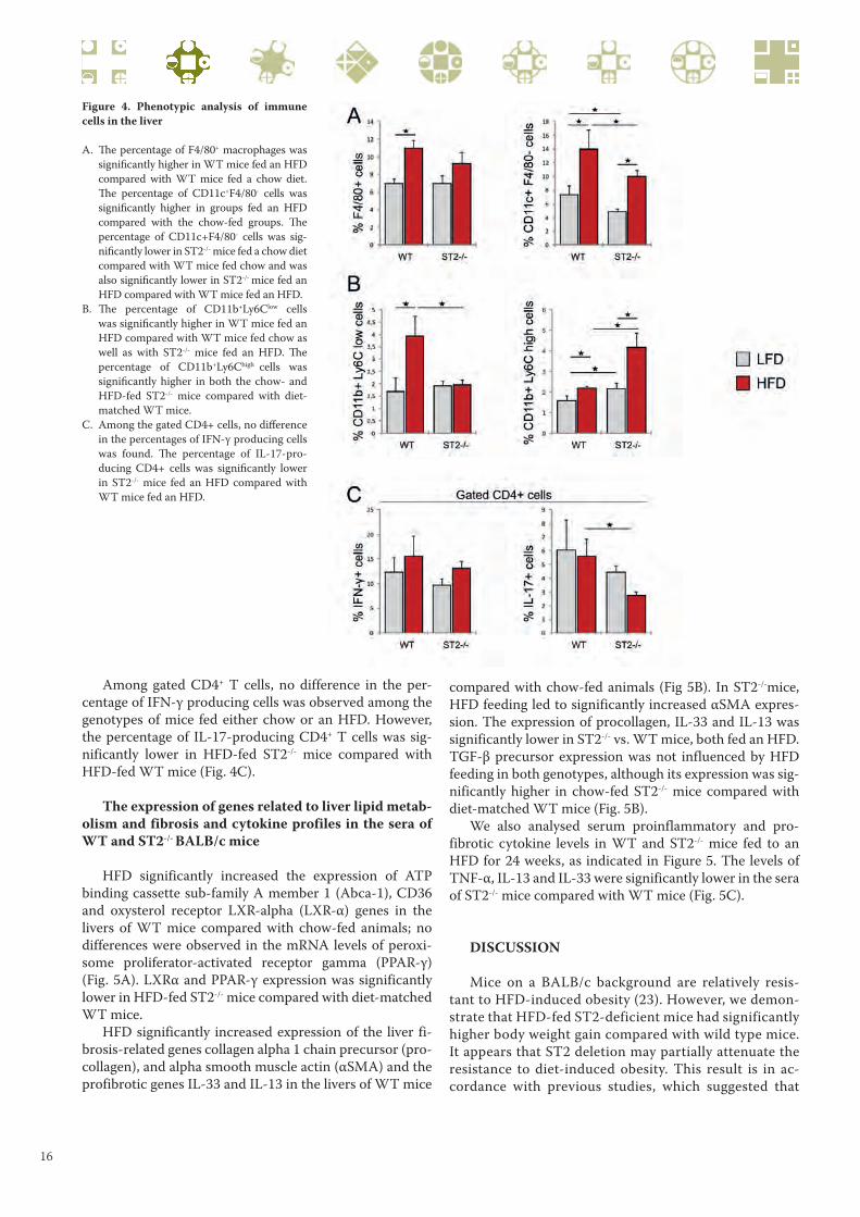
Figure 4. Phenotypic analysis of immune
cells in the liver
A. Th e percentage of F4/80+ macrophages was
signifi cantly higher in WT mice fed an HFD
compared with WT mice fed a chow diet.
Th e percentage of CD11c+F4/80- cells was
signifi cantly higher in groups fed an HFD
compared with the chow-fed groups. Th e
percentage of CD11c+F4/80- cells was sig-
nifi cantly lower in ST2-/- mice fed a chow diet
compared with WT mice fed chow and was
also signifi cantly lower in ST2-/- mice fed an
HFD compared with WT mice fed an HFD.
B. Th e percentage of CD11b+Ly6Clow cells
was signifi cantly higher in WT mice fed an
HFD compared with WT mice fed chow as
well as with ST2-/- mice fed an HFD. Th e
percentage of CD11b+Ly6Chigh cells was
signifi cantly higher in both the chow- and
HFD-fed ST2-/- mice compared with diet-
matched WT mice.
C. Among the gated CD4+ cells, no diff erence
in the percentages of IFN-γ producing cells
was found. Th e percentage of IL-17-pro-
ducing CD4+ cells was signifi cantly lower
in ST2-/- mice fed an HFD compared with
WT mice fed an HFD.
Among gated CD4+ T cells, no difference in the per-
centage of IFN-γ producing cells was observed among the
genotypes of mice fed either chow or an HFD. However,
the percentage of IL-17-producing CD4+ T cells was sig-
nificantly lower in HFD-fed ST2-/- mice compared with
HFD-fed WT mice (Fig. 4C).
The expression of genes related to liver lipid metab-
olism and fibrosis and cytokine profiles in the sera of
WT and ST2-/- BALB/c mice
HFD significantly increased the expression of ATP
binding cassette sub-family A member 1 (Abca-1), CD36
and oxysterol receptor LXR-alpha (LXR-α) genes in the
livers of WT mice compared with chow-fed animals; no
differences were observed in the mRNA levels of peroxi-
some proliferator-activated receptor gamma (PPAR-γ)
(Fig. 5A). LXRα and PPAR-γ expression was significantly
lower in HFD-fed ST2-/- mice compared with diet-matched
WT mice.
HFD significantly increased expression of the liver fi-
brosis-related genes collagen alpha 1 chain precursor (pro-
collagen), and alpha smooth muscle actin (αSMA) and the
profibrotic genes IL-33 and IL-13 in the livers of WT mice
compared with chow-fed animals (Fig 5B). In ST2-/-mice,
HFD feeding led to significantly increased αSMA expres-
sion. The expression of procollagen, IL-33 and IL-13 was
significantly lower in ST2-/- vs. WT mice, both fed an HFD.
TGF-β precursor expression was not influenced by HFD
feeding in both genotypes, although its expression was sig-
nificantly higher in chow-fed ST2-/- mice compared with
diet-matched WT mice (Fig. 5B).
We also analysed serum proinflammatory and pro-
fibrotic cytokine levels in WT and ST2-/- mice fed to an
HFD for 24 weeks, as indicated in Figure 5. The levels of
TNF-α, IL-13 and IL-33 were significantly lower in the sera
of ST2-/- mice compared with WT mice (Fig. 5C).
DISCUSSION
Mice on a BALB/c background are relatively resis-
tant to HFD-induced obesity (23). However, we demon-
strate that HFD-fed ST2-deficient mice had significantly
higher body weight gain compared with wild type mice.
It appears that ST2 deletion may partially attenuate the
resistance to diet-induced obesity. This result is in ac-
cordance with previous studies, which suggested that

17
Figure 5. Liver gene expression and cytokine profi les in sera
A. Th ere were no signifi cant diff erences in the expression of genes related to lipid metabolism in the livers of ST2-/- mice fed chow and an HFD. Sig-
nifi cantly increased expression of the Abca-1, CD36 and LXR-α genes was observed in W T mice fed an HFD compared with WT mice fed a chow
diet. Th e expression of CD36, LXRa and PPAR-γ was signifi cantly lower in ST2-/- mice fed an HFD compared with diet-matched WT mice.
B. Expression of the procollagen, alpha smooth muscle actin, IL-33 and IL-13 genes was signifi cantly increased in the livers of WT mice fed an HFD
compared with WT mice fed chow. Th e expression of αSMA was signifi cantly higher in the livers of ST2-/- mice fed an HFD compared with ST2-/-
mice fed chow. Th e expression of IL-33 and IL-13 was signifi cantly lower in ST2-/- mice fed an HFD compared with WT mice fed an HFD. Expres-
sion of the TGF-β precursor was signifi cantly higher in ST2-/- mice fed chow compared with diet-matched WT mice.
C. Th e levels of TNF-α, IL-13 and IL-33 were signifi cantly lower in the sera of ST2-/- mice fed an HFD compared with diet-matched WT mice.
IL-33 plays a protective role in obesity (12). Miller et al.
have previously demonstrated that exogenous IL-33 ex-
erted protective effects on adiposity and inflammation,
and Pantic et al. showed that ST2 deletion enhanced
visceral adiposity and inflammation in BALB/c mice. In
line with these studies (12,24) we also demonstrate the
higher amount of visceral adipose tissue in ST2-/- mice
maintained on an HFD. Our main objective in this study
was to investigate the unresolved role of the IL-33/ST2
axis in the development of hepatic steatosis. We demon-
strate here that HFD-induced steatosis was ameliorated
in ST2-/- mice compared with wild-type mice. Further-
more, the extent of liver steatosis was also significantly
lower in ST2-/- mice on a standard diet compared with
diet-matched WT mice. The lack of IL-33/ST2 signalling
resulted in increased visceral fat weight and hypertrig-
lyceridemia and attenuated liver steatosis in mice fed an
HFD. This finding was somewhat unexpected consider-
18
ing that enhanced lipolysis in enlarged adipose tissues in
obesity was shown to be the main contributing factor in
the development of liver steatosis (9, 25). Adipose tissue
lipolysis is the catabolic process leading to the break-
down of triglycerides stored in fat cells and the release
of fatty acids and glycerol (26). Our findings imply that
the protective effect of IL-33 in the obesity-associated
enlargement of visceral adipose tissue is not exerted on
hepatic steatosis. The role of IL-33/ST2 signalling in
hepatic steatosis has not been investigated. Recent re-
port suggests that IL-33/ST2 expression may promote
maternal lipolysis during pregnancy (27). It could be
speculated that the discrepancy between the protective
effects on HFD-induced adiposity and enhanced liver
steatosis may be related to the presumption that IL-33
promotes lipolysis and the “relocation” of fatty acids in
the liver. Furthermore, we demonstrated that WT mice
fed an HFD have increased expression of genes associ-
ated with lipid metabolism in the liver. The expression
of fatty acid translocase (CD36/FAT) was significantly
higher in WT mice than in ST2-/- mice, both fed an HFD.
When fatty acids are released from adipose tissue stores,
they enter the circulation as FFAs. CD36 is a molecule
involved in the uptake of fatty acids by cells (28). We
show markedly lower LXRα expression in HFD-fed
ST2-/- mice compared with HFD-fed WT mice. Liver X
receptor alpha (LXRα) is oxysterol-activated nuclear re-
ceptor that is expressed in the liver and in other tissues
and that regulates inflammation and lipogenesis. It has
been demonstrated that LXRα activation has potentially
deleterious effects by promoting hepatic steatosis and
insulin resistance (29,30). As opposed to ST2-/- mice,
WT mice fed an HFD had increased expression of per-
oxisome proliferator-activated receptor (PPARγ), which
has been associated with exacerbated steatosis when
overexpressed in hepatocytes (31).
Our findings point to an important role for IL-33/
ST2 signalling in obesity-associated changes in lipid
metabolism in the liver. A recent study showed that the
development of hepatic steatosis requires IL-1 signal-
ling (10), and considering that the IL-33/ST2 axis shares
similar downstream molecules with the IL-1 pathway,
IL-33/ST2 signalling should be further explored in he-
patic lipogenesis. Recently, the direct role of IL-17 in liv-
er steatosis and fibrosis has been demonstrated (32,33).
In accordance with this finding, we showed that IL-17-
producing CD4+ cells were less numerous in the livers of
HFD-fed ST2-/- mice compared with diet-matched WT
mice. Liver inflammation was attenuated in ST2-/- mice
fed an HFD, as evaluated by lower inflammatory scores
and lower numbers of CD68+ macrophages and percent-
ages of CD11c+ DCs in the livers of these mice compared
with diet-matched WT mice. Liver damage due to pro-
nounced steatosis in WT mice has been coupled with
on-going fibrosis. In contrast to HFD-fed ST2-/- mice,
higher gene expression of procollagen, IL-33 and profi-
brotic IL-13 observed in the livers of HFD-fed WT mice
supports the notion of the profibrotic role of IL-33/
ST2 signalling in the liver. This was accompanied by in-
creased collagen deposition in steatotic livers of HFD-fed
WT mice and increased sera levels of IL-33 and IL-13.
In addition, we also demonstrated that CD11b+Ly6clow
monocytes, which are cells with profibrotic or M2-type
functions in the liver (34,35,36), were more numerous
in the livers of HFD-fed WT mice compared with diet-
matched ST2-/- mice.
Conclusion
In summary, our findings are compatible with the no-
tion that the IL-33/ST2 (IL-33R) axis may play multiple
roles in obesity-associated metabolic disorders and NA-
FLD. IL-33/ST2 signalling attenuates adiposity and in-
flammation in visceral adipose tissue but promotes liver
steatosis, inflammation and fibrosis, most likely by modu-
lating cell trafficking and the metabolic pathways that link
adipose and liver tissues in obesity.
Acknowledgments
We would like to thank Ivan Jovanovic, Gordana Ra-
dosavljevic, Jelena Pantic, Aleksandar Ilic, Katerina Marti-
nova, Sandra Nikolic (Center for Molecular Medicine) and
Zoran Mitrovic (Institute of Pathology) for their technical
assistance.
Grants
This work was supported by grants from the Serbian
Ministry of Science and Technological Development
(175071 and 175069) (Belgrade, Serbia), a joint research
project (SCOPES, IZ73Z0_152407) and the Internal Proj-
ects of Faculty of Medical Sciences (JP 02-14, JP 03-14)
(Kragujevac, Serbia).
Disclosure
The authors declare that they have no competing inter-
ests or other interests that might be perceived to influence
the results and discussion reported in this paper.
REFERENCES
1. Cohen JC, Horton JD, Hobbs HH. Human fatty liv-
er disease: old questions and new insights. Science.
2011;332(6037):1519-23.
2. Henao-Mejia J, Elinav E, Jin C, Hao L, Mehal WZ,
Strowig T, et al. Inflammasome-mediated dysbiosis
regulates progression of NAFLD and obesity. Nature.
2012;482(7384):179-85.

19
3. Choi S, Diehl AM. Role of inflammation in nonalcohol-
ic steatohepatitis. Current opinion in gastroenterology.
2005;21(6):702-7.
4. Mouralidarane A, Soeda J, Visconti-Pugmire C, Samu-
elsson AM, Pombo J, Maragkoudaki X, et al. Maternal
obesity programs offspring nonalcoholic fatty liver dis-
ease by innate immune dysfunction in mice. Hepatol-
ogy. 2013;58(1):128-38.
5. Li Z, Soloski MJ, Diehl AM. Dietary factors alter he-
patic innate immune system in mice with nonalcoholic
fatty liver disease. Hepatology. 2005;42(4):880-5.
6. Grarup N, Sandholt CH, Hansen T, Pedersen O. Ge-
netic susceptibility to type 2 diabetes and obesity: from
genome-wide association studies to rare variants and
beyond. Diabetologia. 2014;57(8):1528-41.
7. Lin YC, Chang PF, Chang MH, Ni YH. Genetic variants
in GCKR and PNPLA3 confer susceptibility to nonal-
coholic fatty liver disease in obese individuals. Am J
Clin Nutr. 2014;99(4):869-74.
8. Postic C, Girard J. Contribution of de novo fatty acid
synthesis to hepatic steatosis and insulin resistance:
lessons from genetically engineered mice. J Clin Invest.
2008;118(3):829-38.
9. Ferre P, Foufelle F. Hepatic steatosis: a role for de novo
lipogenesis and the transcription factor SREBP-1c. Dia-
betes, obesity & metabolism. 2010;12 Suppl 2:83-92.
10. Negrin KA, Roth Flach RJ, DiStefano MT, Matevossian
A, Friedline RH, Jung D, et al. IL-1 signaling in obesity-
induced hepatic lipogenesis and steatosis. PLoS One.
2014;9(9):e107265.
11. Milovanovic M, Volarevic V, Radosavljevic G, Jovanovic
I, Pejnovic N, Arsenijevic N, et al. IL-33/ST2 axis in
inflammation and immunopathology. Immunol Res.
2012;52(1-2):89-99.
12. Miller AM, Asquith DL, Hueber AJ, Anderson LA, Hol-
mes WM, McKenzie AN, et al. Interleukin-33 induces
protective effects in adipose tissue inflammation dur-
ing obesity in mice. Circ Res. 2010;107(5):650-8.
13. Marvie P, Lisbonne M, L’Helgoualc’h A, Rauch M, Tur-
lin B, Preisser L, et al. Interleukin-33 overexpression is
associated with liver fibrosis in mice and humans. J Cell
Mol Med. 2010;14(6b):1726-39.
14. McHedlidze T, Waldner M, Zopf S, Walker J, Rankin
AL, Schuchmann M, et al. Interleukin-33-dependent
innate lymphoid cells mediate hepatic fibrosis. Immu-
nity. 2013;39(2):357-71.
15. Townsend MJ, Fallon PG, Matthews DJ, Jolin HE, McK-
enzie AN. T1/ST2-deficient mice demonstrate the im-
portance of T1/ST2 in developing primary T helper cell
type 2 responses. J Exp Med. 2000;191(6):1069-76.
16. Junqueira LC, Bignolas G, Brentani RR. Picrosirius
staining plus polarization microscopy, a specific meth-
od for collagen detection in tissue sections. Histochem
J. 1979;11(4):447-55.
17. Hadi AM, Mouchaers KT, Schalij I, Grunberg K,
Meijer GA, Vonk-Noordegraaf A, et al. Rapid quan-
tification of myocardial fibrosis: A new macro-
based automated analysis. Anal Cell Pathol (Amst).
2010;33(5):257-69.
18. Deutsch MJ, Schriever SC, Roscher AA, Ensenauer R.
Digital image analysis approach for lipid droplet size
quantitation of Oil Red O-stained cultured cells. Anal
Biochem. 2014;445:87-9.
19. Juluri R, Vuppalanchi R, Olson J, Unalp A, Van Natta
ML, Cummings OW, et al. Generalizability of the non-
alcoholic steatohepatitis Clinical Research Network
histologic scoring system for nonalcoholic fatty liver
disease. J Clin Gastroenterol. 2011;45(1):55-8.
20. Volarevic V, Mitrovic M, Milovanovic M, Zelen I, Nikol-
ic I, Mitrovic S, et al. Protective role of IL-33/ST2 axis
in Con A-induced hepatitis. J Hepatol. 2012;56(1):26-
33.
21. Foster B, Prussin C, Liu F, Whitmire JK, Whitton JL.
Detection of intracellular cytokines by flow cytometry.
Curr Protoc Immunol. 2007;Chapter 6:Unit 6.24.
22. Livak KJ, Schmittgen TD. Analysis of relative gene
expression data using real-time quantitative PCR
and the 2(-Delta Delta C(T)) Method. Methods.
2001;25(4):402-8.
23. Montgomery MK, Hallahan NL, Brown SH, Liu M,
Mitchell TW, Cooney GJ, et al. Mouse strain-dependent
variation in obesity and glucose homeostasis in response
to high-fat feeding. Diabetologia. 2013;56(5):1129-39.
24. Pantic JM, Pejnovic NN, Radosavljevic GD, Jovanovic
I.P, Djukic ALJ, Arsenijevic NN, Lukic ML. Lack of
ST2 enhances high - fat diet -induced visceral adi-
posity and inflammation in BALB/c mice [Delecija
gena za ST2 promoviše gojaznost i inflamaciju u
visceralnom adipoznom tkivu BALB/c miševa na di-
jeti sa visokim sadržajem masti]. Serb J Exp Clin Res
2013; 14(4): 155 -160.
25. Donnelly KL, Smith CI, Schwarzenberg SJ, Jessurun J,
Boldt MD, Parks EJ. Sources of fatty acids stored in liver
and secreted via lipoproteins in patients with nonalcohol-
ic fatty liver disease. J Clin Invest. 2005;115(5):1343-51.
26. Langin D. Adipose tissue lipolysis as a metabolic
pathway to define pharmacological strategies against
obesity and the metabolic syndrome. Pharmacol Res.
2006;53(6):482-91.
27. McKenna LA, Jordan F, Brown EA, Huda SS, Mac-
kay VA, Miller AM, et al. The role of interleukin-33
and its receptor ST2 in human pregnancy. Archives
of Disease in Childhood - Fetal and Neonatal Edition.
2011;96(Suppl 1):Fa98.
28. Su X, Abumrad NA. Cellular fatty acid uptake: a path-
way under construction. Trends Endocrinol Metab.
2009;20(2):72-7.
29. Grefhorst A, Parks EJ. Reduced insulin-mediated in-
hibition of VLDL secretion upon pharmacological
activation of the liver X receptor in mice. J Lipid Res.
2009;50(7):1374-83.
30. Beaven SW, Matveyenko A, Wroblewski K, Chao L,
Wilpitz D, Hsu TW, et al. Reciprocal regulation of
hepatic and adipose lipogenesis by liver X receptors

20
in obesity and insulin resistance. Cell metabolism.
2013;18(1):106-17.
31. Moran-Salvador E, Lopez-Parra M, Garcia-Alonso
V, Titos E, Martinez-Clemente M, Gonzalez-Periz A,
et al. Role for PPARgamma in obesity-induced he-
patic steatosis as determined by hepatocyte- and
macrophage-specific conditional knockouts. FASEB J.
2011;25(8):2538-50.
32. Tang Y, Bian Z, Zhao L, Liu Y, Liang S, Wang Q, et al.
Interleukin-17 exacerbates hepatic steatosis and in-
flammation in non-alcoholic fatty liver disease. Clin
Exp Immunol. 2011;166(2):281-90.
33. Tan Z, Qian X, Jiang R, Liu Q, Wang Y, Chen C, et al.
IL-17A plays a critical role in the pathogenesis of liver
fibrosis through hepatic stellate cell activation. J Immu-
nol. 2013;191(4):1835-44.
34. Lin SL, Castano AP, Nowlin BT, Lupher ML, Jr., Duf-
field JS. Bone marrow Ly6Chigh monocytes are se-
lectively recruited to injured kidney and differenti-
ate into functionally distinct populations. J Immunol.
2009;183(10):6733-43.
35. Karlmark KR, Weiskirchen R, Zimmermann HW,
Gassler N, Ginhoux F, Weber C, et al. Hepatic recruit-
ment of the inflammatory Gr1+ monocyte subset upon
liver injury promotes hepatic fibrosis. Hepatology.
2009;50(1):261-74.
36. Tacke F. Functional role of intrahepatic monocyte
subsets for the progression of liver inflammation and
liver fibrosis in vivo. Fibrogenesis & tissue repair.
2012;5(Suppl 1 Proceedings of Fibroproliferative disor-
ders: from biochemical analysis to targeted therapies-
Petro E Petrides and David Brenner):S27.

21
SIMPLE AND COMPLEX COGNITIVE FUNCTIONSUNDER EXERTIONAL HEAT STRESS
Jelena Maric 1, Milan Marjanovic 1, Dalibor Jovanovic 1, Filip Stojanovic 1, Djordje Vukmirovic 1, Vladimir Jakovljevic 2
1 Military Medical Academy, Belgrade, Serbia2 Faculty of Medical Sciences, University of Kragujevac, Kragujevac, Serbia
UTICAJ TOPLOTNOG STRESA I FIZIČKE AKTIVNOSTINA KOGNITIVNE SPOSOBNOSTI
Jelena Marić 1, Milan Marjanović 1, Dalibor Jovanović 1, Filip Stojanović 1, Đorđe Vukmirović 1, Vladimir Jakovljević 2
1 Vojnomedicinska akademija, Beograd, Srbija2 Fakultet medicinskih nauka, Univerzitet u Kragujevcu, Kragujevac, Srbija
ABSTRACT
Heat stress is a signifi cant problem in the military services.
Th is study investigated the eff ects of exertional heat stress on cog-
nitive performance.
Forty unacclimated male soldiers performed exertional heat
stress tests in cool (20 ºC) and hot environments (40 ºC). Cognitive
performance was assessed using a computerized battery before
and immediately after tests. Physical strain in cool conditions in-
duced mild but signifi cant defi cits in accuracy in complex tests.
Th e number of correct answers in the Matching to Sample Visual
Search was reduced (92,18% correct answers before vs. 88,64 af-
ter; p<0,05) and also in the spatial part of the Pattern and Spa-
tial Recognition Memory Test (85,25 vs. 8,75%; p<0,05). Th ese
decreases were more pronounced in hot conditions (92,38 vs.
84,31% in before and 84,21 vs. 73,42% in the latter test; ps<0,01
and <0,001, respectively). Exertional heat stress also impaired
more simple cognitive functions. A signifi cant decrease in accu-
racy (95,74 vs. 93,89%) and an increase in reaction time (300,32
vs. 315,00 ms) was observed in the Reaction Time test.
Strenuous physical activity in a hot environment induces mild
cognitive defi cits, especially in more complex tasks.
ORIGINAL SCIENTIFIC PAPER ORIGINALNI NAUČNI RAD ORIGINAL SCIENTIFIC PAPER
Received / Primljen: 12. 06. 2014. Accepted / Prihvaćen: 12. 07. 2014.
SAŽETAK
Toplotni stres predstavlja značajan problem u vojnoj
službi. U ovom istraživanju se ispituje uticaj toplotnog stresa
i fi zičke aktivnosti na kognitivne sposobnosti.
40 neaklimatizovanih vojnika muškog pola podvrgnuto
je testu toplotnog stresa i fi zičkog napora u termoneutralnoj
(20 ºC) i toploj sredini (40 ºC). Kognitivne funkcije su ispiti-
vane kompjuterizovanom baterijom neposredno pre i nakon
testa. U termoneutralnim uslovima, fi zički napor je izaz-
vao blagi pad preciznosti pri rešavanju složenih zadataka
u testu Matching to Sample Visual Search (92,18% vs. 88,64;
p<0,05), i prostornoj komponenti testa Pattern and Spatial
Recognition Memory (85,25 vs. 78,75%; p<0,05). Pad pre-
ciznosti je naglašeniji u toplim uslovima (92,38 vs. 84,31%;
p<0,01, odnosno 84,21 vs. 73,42%; p<0,001), gde je, osim
toga, došlo i do oštećenja jednostavnijih kognitivnih funkcija:
u testu Reaction Time zabeležen je pad preciznosti (95,74
vs. 93,89%), kao i produženje vremena reakcije (300,32 vs.
315,00 ms).
Intenzivan fi zički napor u toploj sredini dovodi do blagih
kognitivnih defi cita, pogotovo pri vršenju složenih zadataka.
INTRODUCTION
Heat stress is a significant problem in the military
services. Common preventive measures (e.g., restriction
of physical activity, removal of clothing and moving into
shade) are often impossible to achieve. Additional disad-
vantages include the burden of heavy equipment, ballistic
vests, and other heavy items that must be worn during
training and regular activities, and impermeable protec-
tive suits, which may inhibit evaporative cooling. Heat
stress can impair physical and mental performance (1,2).
Studies of cognitive performance under hot conditions are
Corresponding author: Jelena Maric, Institute of Public Health, Pancevo
Pasterova 2, Pancevo, e-mail: [email protected]
UDK: 612.59-057.36; 159.922.25.072-057.36 / Ser J Exp Clin Res 2015; 16 (1): 21-25
DOI: 10.1515/SJECR20150003
ABBREVIATIONS
CANTAB - Computerized Cognitive Attention Battery
EHS - Exertional Heat Stress
EHST - Exertional Heat Stress Test
HR - Heart Rate
Tty - Tympanic temperature
WBGT - Wet Bulb Globe Thermometer

22
difficult to evaluate because of variations in methodologi-
cal design, exposure duration, skills and acclimation level.
Human tolerance to heat stress also depends on fitness lev-
els. Individuals with higher aerobic capacity show better
adaptation to the combination of physical activity and hot
environments (3).
Computerized batteries are applied to minimize bias
in assessments of cognitive performance during physical
exertion and/or heat stress (4, 5). CANTAB (computerized
Cambridge Neuropsychological Test Automated Batteries)
is a set of neuropsychological tests that are specifically de-
signed for comparative assessments of cognition. The tests
are graded in nature, with standardized feedback and a de-
tailed recording of accuracy and speed.
Our study investigated the effects of strenuous physical
activity in combination with a hot environment on cogni-
tive performance in young fit male soldiers using comput-
erized test batteries.
SUBJECTS AND METHODS
The participants were 40 male soldiers, aged 20±0,9
years, with similar anthropometric and ergometric
characteristics (Table 1). Subjects signed an informed
consent form after a short briefing on the nature and
purpose of the experiment. The Ethical Committee of
the Military Medical Academy approved the experimen-
tal protocol. The procedures corresponded to interna-
tional standards of thermal strain evaluation (6, 7). The
subjects were randomly divided in two equal groups.
The experimental group (H group) performed the exer-
tional heat stress test (EHST) in a hot environment (40
ºC, 29 ºC WBGT), and the control group (C group) per-
formed the same test in a cool environment (20 ºC, 16
ºC WBGT). Trials were conducted in a climatic chamber
in the Military Medical Academy, Institute of Hygiene,
Department of Physiological Strain, during November
and December 2011. Parameters of heat stress (tym-
panic temperatures and heart rates) were automatically
monitored and recorded in real-time using a physiologi-
cal data monitoring system (MP150SKT100C, BIOPAC
Systems Inc., USA), and the Q4500 Exercise Test Moni-
tor (Quinton Instruments, USA) respectively. The test
and measurement protocols were described in detail in
our previous studies (8, 9). Tests were limited to 90 min-
utes because of the temperature conditions and level of
physical exertion. The following criteria were used for
early termination: Tty 39,5 ºC, HR 190 bpm, or intoler-
able subjective discomfort. Cognitive performance was
assessed using CANTAB, version 2,0, which consisted
of 5 different tests that measure selective, divided, and
sustained attention, information processing, working
memory and recognition (10). Tests were administered
using a computer with a touch-sensitive screen, and a
response key was used for reaction timing. Cognitive ex-
aminations were administered to each soldier before and
immediately after EHST, and each examination included
the following tests: Motor Screening (MOT), Reaction
Time (RTI), Matching to Sample Visual Search (MTS),
and Pattern and Spatial Recognition Memory, which in-
cludes visual and spatial components (PSRp and PSRs).
Accuracy (percentage of correct answers or number of
errors) and speed in ms (reaction time, movement time,
or attention time) were recorded for each test.
The Statistical Package for the Social Sciences (SPSS
20.0) was used to perform all statistical tests. Continuous
variables are presented as means ± standard deviation.
Normal distribution was tested using the Kolmogorov-
Smirnov test. Differences between performances before
and after EHST were tested using repeated measures t-
test. Significance was accepted at p<0,05.
Table 1. Anthropometric and ergometric parameters
in both investigated groups
H C p (H vs. C)
Body height (cm) 180,63 ± 5,71 179,55 ± 4,57 0,595081
Body weight (kg) 74,85 ± 9,66 78,13 ±5,32 0,656162
Body Mass Index (kg/m2) 22,9 ± 2,62 24,29 ± 1,95 0,803931
Percentage of body fat (%) 16,86 ± 3,62 17,06 ± 4,47 0,710612
VO2max
(ml/min/kg) 59,55 ± 10,64 56,60 ± 5,91 0,235727
VO2max
- Maximal oxygen consumption
Figure 1. Average tympanic temperatures during the tests in both inves-
tigated groups
Figure 2. Average heart rates during the tests in both investigated groups

23
RESULTS
Table 1 shows the anthropometric and ergometric char-
acteristics of the subjects. Both groups were similar in all
investigated features. No soldier showed any symptoms of
heat exhaustion or other severe heat illness during or after
EHST. All participants in the C group completed the tests,
but 3 soldiers in the H group terminated tests between 60
and 85 minutes because of subjective intolerable strain or
the reaching of the Tty limit. Figure 1 displays comparable
reviews of Tty values during EHST. The mean Tty was sig-
nificantly higher in the H group at the end of EHST (i.e.,
90th minute) (39,5±0,16 ºC vs. 36,78±0,11 ºC in the C group;
p=0,0006). The total increase in Tty values (average differ-
ence between Tty at the beginning and end of EHST) was
also significantly higher in the H group (1,56±0,26 ºC vs.
0,07±0,03 ºC in the C group; p=0,009). Figure 2 displays
comparable reviews of HR values during EHST. Mean HR
at the end of EHST was significantly higher in the H group
(148,90±5,90 vs. 120,50±7,60 bpm; p=0,0000). The limit of
180 bpm was never reached, and the maximum recorded
hart rate was 165 bpm. Table 2 summarises performance task
means. There was a significant decrease in the percentage of
correct answers in the C group in the MTS test after EHST
(88,64±6,95 after EHST vs. 92,18±5,14% before; p=0,038)
and the PSRs test (78,75±15,55 vs. 85,25±7,52%; p=0,037).
Decreases in the accuracy of both of these tests were more
significant in the H group (MTS 84.31±9,51 vs. 92,38±4,90%
and PSRs 73,42±10,01 vs. 84,21±8,54%; ps=0,002 and
0,0006, respectively). There was also a statistically signifi-
cant decrease in performance in the RTI test in the H group.
A decrease in the percentage of correct answers (93,89±3,43
vs. 95,74±3,52%; p=0,41) was accompanied by a significant
increase in reaction time (315,00±33,77 vs. 300,32±31,53
ms; p=0,048). There were no significant differences between
responses for latency or number of errors before and after
EHST in other tests, regardless of ambient conditions.
DISCUSSION
Heat strain impairs working efficiency and induces cog-
nitive deficits. These abilities are particularly important in
military services, where a dose-dependent relationship be-
tween heat stress and the number of errors in helicopter pi-
lots is well established in laboratory and terrain conditions
(11,12). Banta and Braun also found that Navy helicopter
pilots wearing individual cooling suits during the Gulf war
to mitigate the detrimental effect of heat stress on cognition
showed improved vigilance and accuracy (13). The possible
beneficial effects of microclimate cooling were also of inter-
est in the Army of Serbia. Jovanovic et al. demonstrated the
efficiency of individual microclimate cooling systems based
on phase-change materials, which alleviated the physical
Table 2. Cognitive performance in both investigated groups
Test Parameter H C
Before After Before After
MOT Latention(ms)
525,00±60,88
624,37±197,10
560,00±99,32
625,15±146,67
Errors(N)
27,00±7,17
26,42±8,19
29,75±13,52
25,35±11,76
RTI Correct answers(%)
95,74±3,52
93,89±3,43 *
96,77±3,59
96,82±3,04
Reaction time(ms)
300,32±31,53
315,00±33,77 *
308,70±50,15
308,9±35,52
Movement time(ms)
367,42±102,95
373,21±117,67
400,20±112,37
394,25±128,15
MTS Correct answers(%)
92,38±4,90
84,31±9,51 **
92,18±5,14
88,64±6,95 *
Latention(ms)
1564,47±507,41
1694,89±529,68
1604,80±526,77
1579±522,37
PSRp Correct answers(%)
90,56±5,71
87,94±9,70
93,54±5,14
90,98±7,41
Latention(ms)
1436,37±287,91
1371,42±312,63
1380,80±262,08
1325,75±254,29
PSRs Correct answers(%)
84,21±8,54
73,42±10,01 ***
85,25±7,52
78,75±15,55 *
Latention(ms)
1558,79±428,23
1653,79±444,71
1524,05±342,89
1481,65±380,81
* p<0,05 after vs. before
** p<0,01 after vs. before
*** p<0,001 after vs. before

24
exertion caused by uncompensable heat stress (8, 9). They
investigated male soldiers who performed EHST wearing
impermeable protective suits and various types of micro-
climate cooling vests in a simulated environment (climatic
chamber) and external conditions. They concluded that the
use of cooling vests under protective suits during physical
activity in hot conditions reduced sweating and alleviated
heat stress, which was measured by increased core tem-
perature and heart rate values. These cooling vest-induced
physiological changes directly improved heat tolerance and
hydration state, decreased the risk of heat illness, and ex-
tended the duration of soldiers’ exposure to hot conditions.
Dehydration itself profoundly influences cognitive perfor-
mance (14), and the use of microclimate cooling systems
may represent a promising solution to maintain vigilance
and accuracy in soldiers exposed to heat stress.
Our results indicate that physical strain itself induced
mild cognitive deficits, which was shown in the increased
number of errors during the performance of complex tasks
(i.e., MTS and the spatial part of the PSR test). These tests
are designed to investigate working memory and sustained
attention, and the latter test also investigates spatial ori-
entation, which is a very specific and advanced function
that is highly important in military services. The gener-
ally good performance of our soldiers in cool conditions is
consistent with the results reported by other authors, who
found that an acute boost of exercise may improve cogni-
tion (15). Mild deficits in our investigation may be caused
by the longer duration and more strenuous physical activ-
ity compared to the study cited above.
The combination of the same intensity of physical
strain and heat stress in our investigation led to more pro-
nounced cognitive impairments in the same complex test,
but it also imposed additional effect on attention, which
are tested in the RTI. Soldiers in hot conditions showed a
significant decrease in accuracy and a significant delay in
reaction time in the RTI. The only function that was unaf-
fected in this test was movement time. This mild cognitive
deficit can likely be attributed to heat stress.
Hancock and Vasmatzidis stated in their review (16) that
there are major difficulties in the interpretation of data on
the influence of heat stress on cognitive functions. These dif-
ficulties are the consequences of various testing procedures,
task demands and categorisation, duration of heat exposure,
and subjects’ skills and experience. These authors suggest
that simple mental tasks show little, if any, decrements fol-
lowing heat exposure, and these tasks are frequently en-
hanced during brief exposures. However, substantial heat
stress (30-33 ºC WBGT) may lead to impairments in the
performance of more complex tasks. The obtained results
are consistent with the high WBGT (29 ºC) in our trial.
Most of our subjects who performed tests in a hot envi-
ronment suffered intensive physical strain, which was record-
ed by the high values of core temperature (up to 39,5 ºC) and
heart rates for a substantial time. However, these subjects did
not show signs of serious cognitive deficits. Their relative re-
sistance to heat exertion may be attributed to their high aero-
bic capacity. This hypothesis is supported by the facts that
some aspects of heat stress-induced cognitive impairment
are explained by cerebral changes (17,18), blood oxidative
stress (19), and acute inflammatory reactions (20,21). Several
investigations (22,23) confirmed that regular aerobic activity
significantly induces beneficial adaptive effects on oxidative
stress responses and inflammation parameters to acute ex-
hausting endurance exercise in the heat, and our results may
be interpreted from this point of view.
CONCLUSIONS
This study demonstrated the effects of strenuous physi-
cal activity in a hot environment on physiological param-
eters, which showed higher values of tympanic temperature
and heart rate compared to cool conditions. This physiologi-
cal heat stress enhanced the cognitive impairment caused by
physical exertion itself. Performance deficits in a cool condi-
tion were mild and present only in complex tasks, but sim-
pler tasks were relatively unaffected. Performance deficits in
hot conditions were more pronounced and present in both
complex and simpler tasks, such as reaction time.
Acknowledgment
This investigation was performed as a part of the MNTR
2011-2014, project TR 34034, which was financially sup-
ported by Ministry of Education, Science and Technologi-
cal Development, Republic of Serbia.
REFERENCES
1. Amos D, Hansen R, Lau WM, Michalski JT: Physiologi-
cal and cognitive performance of soldiers conducting
routine patrol and reconnaissance operations in the
tropics. Milit Med 2000; 165 (12): 961-6.
2. Radakovic SS, Maric J, Surbatovic M, Radjen S, Filipovic
N, Stefanova E, et al. Effects of acclimation on cognitive
performance in soldiers during exertional heat stress.
Milit Med 2007; 172 (2):190-5
3. Selkirk GA, McLellan TM. Influence of aerobic fitness
and body fatness on tolerance to uncompensable heat
stress. Appl Physiol 2001;91(5):2055-63
4. Labelle V, Bosquet L, Mekary S, Bherer L. Decline in
executive control during acute bouts of exercise as a
function of exercise intensity and fitness level. Brain
Cognit 3001;81:10-7
5. Bandelow S, Maughan R, Shirreffs S, Ozgu K, Kurdak S,
Erso G, et al. The effects of exercise, heat, cooling and re-
hydration strategies on cognitive function in football play-
ers. Scand J Med Sci Sports 2010;20(Suppl.3):148-60
6. ISO 12894 (E): Ergonomics of the thermal environ-
ment – Medical supervision of individuals exposed to
extreme hot or cold environment. 2008 Sep,

25
7. ISO 9886: Ergonomics evaluation of thermal strain by
physiological measurements, 2008 Nov.
8. Jovanovic D, Karkalic R, Tomic Lj, Radakovic S, Velick-
ovic Z. Experimental investigations in the microclimate
body cooling systems based on phase change materials.
Thermal Science 2014; DOI: 10.2298/TSCI130216129J
9. Jovanovic D, Karkalic R, Zeba S, Pavlovic M, Rada-
kovic S. Physiological tolerance to uncompensated
heat stress in soldiers: effects of various types of body
cooling systems. Vojnosanit Pregl 2013; DOI: 10.2298/
VSP120731045D
10. Fray PJ, Robbins TW, and Sahakian BJ: Neuropsychiat-
ric applications of CANTAB. Int J Geriat Psych 1996;
11: 329-36.
11. Faerevik H, Reinertsen RE: Effects of wearing aeircrew
protective clothing on physiological and cognitive re-
sponses under various ambient conditions. Ergonomics
2003; 46(8): 780-99.
12. Froom P, Caine Y, Shochat I, Ribak J: Heat stress and
helicopter pilot errors. J Occup Med 1993; 35: 720-4.
13. Banta GR, Braun DE. Heat strain during at-sea helicop-
ter operations and the effect of passive microclimate
cooling. Aviat Space Environ Med 1997;68:126-31
14. Cian C, Barraud PA, Melin B, Raphel C: Effects of fluid
ingestion on cognitive function after heat stress or ex-
ercise-induced dehydration. Int J Psychophysiol 2001;
42: 243-51.
15. Tomporowski PD: Effects of acute bouts of exercise on
cognition. Acta Physhol 2003; 112: 297-324.
16. Hancock PA, Vasmatzidis I: Effects of heat stress on
cognitive performance: the current state of knowledge.
Int J Hyperthermia 2003; 19(3): 355-72.
17. Nielsen B, Nybo L: Cerebral changes during exercise in
the heat. Sports Med 2003; 33(1): 1-11.
18. Cheung SS, Sleivert GG: Multiple triggers for hyper-
thermic fatigue and exhaustion. Exerc Sport Sci Rev
2004; 32(3): 100-6.
19. Takahashi M, Suzuki K, Matoba H, Sakamoto S, Obara
S. Effects of different intensities of endurance exercise
on oxidative stress and antioxidant capacity. J Phys Fit-
ness sports Med 2012;1:183-9
20. Quindry J, Miller L, McGinnis G, Kliszczewiscz B,
Slivka D, Dunke C, et al. Environmental temperature
and exercise-induced blood oxidative stress. Int J Sport
Nutri Exerc Metab 2013;23:128-32
21. Mestre-Alfaro A, Ferrer MD, Banquells M, Riera J,
Drobnic F, Sureda A, et al. Body temperature modu-
lates the antioxidant and acute immune responses to
exercise. Free Radic Res 2012;46:799-808
22. Shanely RA, Nieman DC, Henson DA, Jin F, Knab AM,
Sha W. Inflammation and oxidative stress are lower in
physically fit and active adults. Scand J Med Sci Sports
2013;23:215-23
23. Akhtari-Shojaei E, Jafari A, Namdar H, Farajov A,
Khalili M. Comparison of inflammatory responses af-
ter acute moderate aerobic cycling in healthy young ac-
tive and inactive men. Biomed Int 2011;2:64-71
24. Chase B, Karwowski W, Benedict ME, Quesada PM,
Irwin-Chase HM. A study of computer-based task per-
formance under thermal stress. Int J Occup Saf Ergon
2003; 9(1): 5-15.
25. Hancock PA, Williams G, Manning CM, Miyake S.
Influence of task demand characteristics on workload
and performance. Int J Aviat Psychol 1995; 5: 63-86.

26

27
PERIODONTAL DISEASE AND RISK FOR PRETERM BIRTH:A CASECONTROL STUDY
Irena Andonova1, Vasil Iliev1, Nikica Živković1 1 Department of Gynaecology and Obstetrics, General Hospital, Šibenik, Croatia
PARODONTOPATIJA I RIZIK ZA NASTAJANJEPREVREMENOG POROĐAJA: STUDIJA SLUČAJKONTROLA
Irena Andonova1, Vasil Iliev1, Nikica Živković1
1 Odsek za ginekologiju i akušerstvo, Opšta bolnica, Šibenik, Hrvatska
ORIGINAL SCIENTIFIC PAPER ORIGINALNI NAUČNI RAD ORIGINAL SCIENTIFIC PAPER
ABSTRACT
Maternal periodontal infection has been recognizsed as a
risk factor for preterm and low birth weight infants. It is hy-
pothesized that pathogens causing periodontal disease might
translocate to the amniotic cavity and contribute to triggering
an adverse pregnancy outcome. Th e growing evidence that an
infection remote from the foetal-placental unit might have a
role in preterm delivery has led to an increased awareness of
the potential role of chronic bacterial infections in the body.
Th e aim of this study was to evaluate whether the presence of
chronic periodontitis might infl uence the incidence of preterm
labour and preterm birth.
Th is study was designed as a hospital-based case-control
study. Seventy pregnant women aged 18-40 years, with a sin-
gle live pregnancy were recruited from the Department of Gy-
naecology and Obstetrics of a general hospital in Sibenik, Croa-
tia, from March 2013 to March 2014.
Th e case group included: 30 pregnant women who were
hospitalized with signs of preterm labour. Th e control group
included 40 normal pregnancy patients, who were analysed
for up to 48 h after the delivery of a term baby having a birth
weight of more than 1500 g. A full-mouth periodontal ex-
amination was performed on all the patients. Information
was collected on the demographics, health behaviours, and
obstetric and systemic diseases that might have an infl uence
in preterm delivery.
Th e presence of chronic periodontitis tended to be high-
er in women with a preterm delivery (the case group), with
20 cases (66%), than in the women in the, control group, in
which chronic periodontitis was found in 14 cases (35%); this
diff erence reached statistical signifi cance (p≤0.01). Th e PTB
cases had a signifi cantly worse periodontal status than the
controls (p≤0.001). From the PTL group, 18 patients deliv-
ered preterm, and chronic periodontitis, found in 15 cases
(83%), was more prevalent than in the control group. Th e
risk of women having periodontitis or attachment loss ≥ 4
mm developing PTB showed an OR of 3.7 (95% CI: 1.91 to
4.86; P< 0.001).
Received / Primljen: 09. 06. 2014. Accepted / Prihvaćen: 18. 12. 2014.
SAŽETAK
Infekcija parodoncijuma majke se smatra faktorom rizika
za prevremeni porođaj i malu telesnu masu novorođenčeta.
Pretpostavlja se da patogeni koji uzrokuju parodontopatiju
mogu da dospeju u amnionsku šupljinu i da doprinesu po-
kretanju događaja koji za posledicu imaju naželjeni ishod
trudnoće. Sve veći broj dokaza koji ukazuje na činjenicu
da infekcije udaljene od feto-placentalne jedinice mogu da
imaju ulogu u nastajanju prevremenog porođaja su izazva-
li povećanje interesovanja za potencijalnu ulogu hroničnih
bakterijskih infekcija u organizmu. Cilj ove studije je procena
povezanosti hronične parodontopatije i incidencije prevre-
menog porođaja.
Studija je dizajnirana kao hospitalna slučaj-kontrola
studija. 70 trudnica, starosti od 18 do 40 godina, sa prvom
uspešnom trudnoćom, su ispitivane između marta 2013. i
marta 2015. godine, na Odeljenju ginekologije i akušerstva
Opšte bolnice u Šibeniku, Hrvatska.
Grupa „slučajeva“: Ovu grupu je činilo 30 trudnica hos-
pitalizovanih sa znacima prevremog porođaja. Kontrolna
grupa: Kontrolnu grupu su činile trudnice sa normalnom
trudnoćom, koje su ispitivane 48 h nakon porođaja, čije je
dete rođeno u terminu i imalo težinu na rođenju iznad 2500
g. Svim pacijentkinjama je izvršen pregled parodoncijuma
celih usta, a prikupljeni su i demografski podaci, informacije
o zdravstvenim navikama, akušerskim i sistemskim obolje-
njima koje mogu da utiču na porođaj.
Hronični parodontitis je bio više zastupljen kod žena koje su
imale prevremeni porođaj (grupa „slučajeva“) – 66% (20 žena)
u poređenju sa ženama koje su se porodile u teminu (kontrol-
na grupa) – 35% (14 žena), i ova razlika je statistički značajna
(p≤0,01). Žene u PTB grupi su imale značajno gore stanje pa-
rodoncijuma u odnosu na kontrole (p≤0,001). 18 žena iz PTL
grupe je rodilo nedonesenu decu, a hroinčni parodontitis je bio
češći – 83% (15 žena), u odnosu na kontrolnu grupu. Rizik za
nastajanje prveremenog porođaja za žene koje imaju parodon-
titis ili gubitak vezivnog tkiva ≥4 mm je pokazao OR (odds ra-
tio) od 3,7 (95%CI: 1,91 do 4,86; p<0,001)
Corresponding author: Irena Andonova, Department of Gynaecology and Obstetrics,
General Hospital, Šibenik, Croatia, [email protected]
UDK: 616.314.17-06-055.26; 618.39-02 / Ser J Exp Clin Res 2015; 16 (1): 27-32
DOI: 10.1515/SJECR20150004

28
INTRODUCTION
Oral health is an integral component of the general health
and well being of an individual. The knowledge regarding
the link between periodontal disease and systemic health is
increasing rapidly (1). Recently, many epidemiological, clini-
cal and laboratory studies have provided irrefutable evidence
that periodontitis is a risk factor for various systemic diseases,
such as cardiovascular diseases, atherosclerosis, diabetes mel-
litus, and pulmonary diseases (2, 3). Periodontal disease refers
to a group of endogenous polymicrobial infections that cause
inflammation and destruction of the supporting structures
of teeth. No overt pathogen has been identified; however, its
aetiology is strongly associated with anaerobic Gram-nega-
tive bacilli (4). In response to these gram-negative bacteria,
the human body activates its host immune response, and im-
mune cells secrete inflammatory mediators. Of particular im-
portance in periodontal disease are cytokines, prostaglandins
and matrix metalloproteinases. The destruction of periodon-
tal tissues, including the breakdown of the gingival connec-
tive tissue and alveolar bone, results mainly from activation of
the immune cells (5). The inflammatory mediators that have
been discussed in relationship to incidences of preterm birth
include cytokines, prostaglandins and matrix metalloprotei-
nases. Several cytokines play an important role in the initia-
tion and progression of periodontal disease. These cytokines
include interleukin-1 (IL-1), interleukin-6 (IL-6), interleu-
kin-8 (IL-8) and tumour necrosis factor (TNF-a). Prostaglan-
dins are responsible for most of the alveolar bone destruction
observed in periodontal disease, particularly prostaglandin
[E. sub. 2] (PGE 2). Matrix metalloproteinases (MMP) are a
family of enzymes that work together to destroy connective
tissue. It is istheorized that these inflammatory mediators
from subgingival plaque are able to enter the bloodstream
and travel to the maternal-foetal interface, thus contribut-
ing to preterm labour (6). During pregnancy, PGE 2 plays an
important role in regulating the onset of labour, contractions
and delivery. (PGE 2) levels rise throughout gestation, and
when a critical threshold level is reached, labour is induced
(7). These associations explain the basic theories for the link
between the presence of maternal periodontal disease and the
risk for adverse pregnancy outcomes.
However, accumulating evidence demonstrates that
oral bacteria might translocate directly into the uterus in-
during pregnancy, causing localized inflammation and an
adverse pregnancy outcome. Studies in humans and ani-
mals have demonstrated that oral bacteria could translo-
cate to the uterus in pregnancy through haematogenous
transmission. These recent discoveries shed new light on
our understanding of pregnancy complications. (8, 9) Al-
though infections of any type in pregnant women represent
a risk for adversities in the developing foetus, periodontal
disease might have a significant role in the pathogenesis of
adverse pregnancy outcomes.
The term periodontal disease encompasses a variety of
diseases ranging from gingivitis to periodontitis. Standard
diagnostic tools that are used to determine the presence
of periodontal disease include full- mouth periodontal ex-
aminations, measurements of the pocket depths (PD), the
clinical attachment levels (CAL), gingival recession (GR),
visible signs of gingival inflammation (such as bleeding on
probing (BOP)) and dental radiographs. No dental radio-
graphs were taken in this study because of the special con-
ditions of the patients.
OBSTRICAL OUTCOMES
Preterm birth (PTB) and low birth weight (LBW) are
leading perinatal problems worldwide and have evident
public health implications because their incidence does
not decrease in spite of the many attempts to prevent peri-
natal problems. Preterm birth, defined as birth before 37
weeks of gestation, is the leading cause of neonatal mortal-
ity, infant morbidity, and long-term sequelae. PTB is a ma-
jor medical, social, and economic concern. Preterm infants
are 75 times more likely to experience early death, and PTB,
the leading direct cause of neonatal death, is responsible
for 27 per cent of neonatal deaths worldwide, comprising
over one million deaths annually (10). The long-term dis-
abilities for surviving preterm infants include pulmonary
abnormalities, asthma, cerebral palsy, poor motor skills
and neurological or developmental disabilities (11). Hu-
man observational studies have identified a number of
risk factors for preterm delivery, and some are reversible,
whereas others are permanent (12). The risk factors occur
in combination, and, therefore, developing effective pre-
ventive strategies could be challenging.
Th e study shows a signifi cant association between peri-
odontal chronic disease and an adverse pregnancy outcome.
Periodontal disease represents a strong, independent risk
factor for preterm births, and periodontal prevention and
therapy should be a part of preventive prenatal care.
Keywords: periodontal disease, preterm labour, preterm
delivery
Studija pokazuje značajnu povezanost između hronične
parodontopatije i neželjenog ishoda trudnoće. Parodontopa-
tija predstavlja jak, nezavistan faktor rizika za prevremeni
porođaj. Usled toga prevencija parodontopatije i terapija bi
trebalo da budu deo prvencije prvermenog porođaja.
Ključne reči: parodontopatija, prevremeni porođaj,
prevremeno rođenje

29
Infections and inflammation, maternal or foetal stress,
uterine bleeding, and stretching of the uterus are isrecognized
as the most common reasons for PTB. Intrauterine infections
and maternal bacterial vaginosis are well known risk factors;
however, distant infections, even subclinical infections, could
induce preterm births. Periodontal disease is associated with
infections and inflammation. The presence of periodontal
disease and the maternal physical changes that occur during
pregnancy enable bacteria to enter the blood stream, leading
to “placental seeding” (13, 14). The significance of the rela-
tionship of periodontal disease and PTB is evident when the
number of affected women and children is considered. At least
20% of the women aged 30 to 50 years have periodontitis. In
addition, 12% of all the births worldwide are premature (de-
livered at less than 37 weeks of gestation); this problem affects
many women of child-bearing age in our country, with lasting
consequences to their past, present and future children.
The objective of this paper is to examine, from an
evidence-based perspective, whether periodontal disease
might contribute to the risk for PTB.
MATERIAL AND METHODS
A case control study was conducted in the Depart-
ment of Obstetrics and Gynaecology at General Hospital
Sibenik, R. Croatia, from March 2013 to March 2014. The
study was approved by the Institutional Ethics Committee
of General Hospital, Sibenik. Informed and written con-
sent was obtained from all of the subjects for this study.
Material
The study population included the following: a case
group of 30 pregnant women from 28 to 36+6 weeks of
gestation, with clinical signs of preterm labour, who were
hospitalized in our department; and a control group of 40
pregnant women with normal pregnancies, who delivered,
at term (37-42 g.w), a baby with a birth weight over 2500 gr.
The inclusion criteria included pregnant women, aged
18-40 years, with a single live pregnancy, who signed writ-
ten informed consent forms.
The Exclusion criteria included: multiple gestation,
polyhydramnios, uterine anomalies, a history of second
trimester abortion, a history of cervical surgery, cerclage
in the present pregnancy, a previous preterm delivery, sub-
stance abuse, smoking, an acute symptomatic vaginal in-
fection, and acute oral infection.
Methods
The gestational age of the subjects was determined
with the best obstetrical estimation methods using the de-
finitive menstrual history and ultrasonography performed
in the first trimester. The diagnosis of preterm labour is
generally based on the clinical criterion of regular pain-
ful uterine contractions (4 every 20 minutes or 8 every
60 minutes), accompanied by cervical changes, includ-
ing cervical effacement of at least 80 per cent or cervical
dilatation greater than 2 cm. All the subjects with signs of
preterm labour were followed and routinely treated in our
department, and they were managed according to the hos-
pital protocol for preterm labour.
One dentist performed a full-mouth periodontal exami-
nation on all of the patients at the time of their inclusion in
the study (the case group, at the time of hospitalization, and
the control group, within 2 days of delivery). The periodon-
tal examination was performed with the patient supine on a
hospital bed, using artificial light. The following data were
recorded: bleeding on probing (BOP), the probing depth
(PD) at the floor site per tooth, gingival recession and the
clinical attachment level (CAL) on all of the present teeth.
The patients were diagnosed with periodontal disease if they
exhibited a PD of 4 mm or greater in four or more teeth and
a CAL greater or equal to 3 mm at the same site. The BOP
was recorded; however, it was not used to determine a diag-
nosis of periodontal disease. Demographic, socioeconomic
and medical data on known risk factors were obtained from
the charts of the patients, and an examiner-administered
questionnaire was completed. This questionnaire was de-
signed to gather the demographic details, pregnancy and
medical history, health behaviour and dental condition. The
aim of the questionnaire was to collect information on as
many of the known risk factors for an adverse pregnancy
outcome as possible.
Statistical analysis:
The sample size was calculated using a statistical pack-
age (SPSS Version 10.0, SPSS, Inc., Chicago, IL, USA). Uni-
variate and bivariate analyses were performed. The chi-
square test or Fischer’s Exact test were used for analysing
the categorical variables. To compare the mean values, we
performed the t-test. The Spearman correlation coefficient
was used to reveal the correlation between the variables.
The unadjusted and adjusted ORs were calculated with
95% confidence intervals. Multivariate logistic regression
models were developed and performed stepwise to identi-
fy the risk factors for PTB; a p value < 0.05 was considered
as the cut-off point for the level of significance.
RESULTS
The main demographic and obstetrical data of all the
subjects are summarized in Table 1.
A total of 70 pregnant women were included 40 in the
control group and 30 in the preterm labour group (PTL).
In the case group (PTL), 18 patients did not respond to
therapy and delivered a preterm baby (PTB) in our hos-
pital. The age of the subjects was distributed normally,
without statistical significance between the mean values
(p= 0.72). Most of the women had secondary levels of edu-
cation. There was no significant difference in the marital
status or in the pre-natal care. None of the obstetrical risk
variables displayed a significant association with PTB in
this study.

30
The periodontal condition is presented in Table 2.
Periodontal disease was diagnosed in 66,7% (20 cases)
of the patients with preterm labour and in 35% (14 cases)
of the patients in the control group, and this difference
reached statistical significance (p ≤ 0.01). In the group of
18 patients who delivered preterm, chronic periodontitis
was more prevalent in 83% (15 cases) more cases than in
the control group (p ≤ 0.001), which is considered to be
very statistically significant. The risk for women having
periodontitis or an attachment loss ≥ 4 mm to develop a
PTB showed an OR of 3.7 (95% CI: 1.91 to 4.86; P< 0.001).
Additionally, more CAL was found in the PTB group than
in the control group. The only risk factor for preterm de-
livery that showed statistical significance at 95% CI was
periodontal disease.
DISCUSSION
The results of this study support the hypothesis that
chronic periodontal infection increases the risk of preterm
labour and delivery. The multivariate analysis showed a sig-
nificant association between PTB and periodontal disease.
The main levels of PD and CAL tended to be higher in the
preterm labour group (the case group) than in the term de-
livery group (the control group); the differences were sta-
tistically significantly higher in the women with preterm
births than in the woman with term deliveries, adjusting
for the baseline levels. This study showed a significant risk
of preterm delivery in the women with periodontal disease
than in the women without. Similar positive associations
between periodontal infection and preterm birth were re-
ported by Boggess et al (15). and Sharlene W. J. Afrika (16).
This finding suggests that pregnant woman with preterm
labour and chronic periodontal infection more often de-
livered a preterm baby. Gibbs et al (17). provided an excel-
lent outline of the possible association between infections
and adverse pregnancy outcomes in their review article. In
their hypothesis, microorganisms and their products enter
the uterine cavity during pregnancy by an ascending route
from the lower genital tract or by a blood-borne nongeni-
tal route, causing preterm birth. The growing evidence that
infection remote from the foetal-placental unit might have
a role in the preterm delivery of LBW infants has led to an
increased awareness of the potential role of chronic bac-
terial infections in the body. For approximately 15 years,
maternal periodontal disease has been implicated in a poor
pregnancy outcome. In 1996, Offenbacher and his group
reported a seven-fold increased risk of a mother with peri-
odontal disease delivering a PTLBW baby (18), and many
studies on the subject have been completed, with varying
results (19, 20). Lopez et al. confirmed periodontal disease
as an independent risk factor and observed that periodon-
tal therapy significantly reduces the incidence of preterm
birth with low birth weight in a population of women with
periodontal disease (21). A recent meta-analysis confirmed
a significant risk of preterm delivery for pregnant women
with periodontitis (overall risk ratio: 1.70) and a significant
risk for having a low birth weight infant (overall risk ratio:
2.11) (22).
Maternal periodontal infection could directly and/
or indirectly influence the health of the foetal-maternal
unit (23, 24, 25). In a study evaluating the relationship
between foetal inflammatory and immune responses to
oral pathogens and the risk for PTB, umbilical cord blood
specimens were examined for the presence of foetal im-
munoglobulin M (IgM) antibody against oral pathogens
and levels of C-reactive protein, IL-1, IL-6, TNF-alfa and
PGE2. The results showed that the presence of IgM an-
tibodies to oral pathogens and increased levels of TNF-
alfa were associated with increased rates of PTB and that
the combined effects of foetal IgM, C-reactive protein,
TNF-alfa and PGE2 resulted in a significantly increased
risk for PTB (26). Hill demonstrated that Fusobacterium
Table1. Demographic and obstetric information
in the control and case groups
Demographic and obstetricinformation
Control group(n=40)
Group with ptl (n= 30)
Subgroup ptb(n= 18)
Mean age ± SD 26.6 ± 6.0 25.9 ± 4.2 25.4 ± 1.6
First pregnancy [ n / (%) ] 16 ( 40%) 11(36,7%) 7 ( 38,8%)
Second pregnancy 18 ( 45%) 17 ( 56.6%) 10 ( 55,6%)
≥3 pregnancies 6 (15%) 2 ( 6,7%) 1 ( 5,6%)
Prenatal careNo pre-natal care 2 ( 5%) 0 0
< six visits 11( 27.5%) 7 ( 23,3%) 6 ( 33,4%)
Six + visits 27( 67,5%) 23 (76.7%) 12 (66,6%)
Marital status [n / (%) ]Single 3(7,5%) 2 (6,7%) 0
Married 22( 55%) 18(60%) 10( 55,5%)
Non-married couple 15(37,5%) 10( 33,3%) 8( 44,5%)
Education level [n / (%) ]Elementary school 2(5%) 3(10%) 2(11,1%)
Secondary school 33(82,5%) 24(80%) 14(77,8%)
University 5( 12,5%) 3( 10%) 2( 11,1%)
ptl- preterm labour; ptb -preterm birth
Table 2. Periodontal condition in patients with preterm labour and the
control group subjects
Periodontal conditionControl group (n=40)
Case group (n= 30)
P- Value
Healthy 26 ( 65%) 10 ( 33,3%) < 0.01
Chronic periodontitis 14 ( 35%) 20 (66,7%) <0.01
Clinical parameters (mean ± SD)
PD (mm) 3.1±0.06 4.1±0.09 <0.001
CAL ( mm) 2.9±0.04 4.0±0.08 <0.001
PD- probing depth, CAL- clinical attachment level

31
nucleatum could be isolated from amniotic fluid cultures
of women with preterm labour and intact membranes,
suggesting that the transient bacteraemia had originated
from the mouth via haematogenous spread and infection
of the amniotic fluid through the placenta (27). More re-
cently, Han et al. reported, by association, a term still-
birth case caused by F. nucleatum, and she had previously
demonstrated that F. nucleatum induced premature and
term stillbirths in pregnant mice (28, 29). The majority of
these species has been associated with pregnancy com-
plications in humans, although their sources of infection
were not previously known. The oral cavity might be a
significant yet previously overlooked source of infection
inside the womb.
Two meta-analyses of case-control studies reported
that periodontal diseases in pregnancy significantly in-
crease the risk of a subsequent preterm birth or LBW, and
the estimated OR was 1.78 (CI 95%: 1.58, 2.01) for preterm
births, 1.82 (CI 95%: 1.51, 1.20) for low birth weight infants
and 3.00 (CI 95%: 1.93, 4.68) for preterm low-birth weight
infants (30, 31). Studies on the effect of periodontal disease
treatment on the pregnancy outcome showed varying re-
sults. A study reported that there was a lack of association
of periodontitis and preterm birth in a multivariate analy-
sis, which supports the report of a meta-analysis showing
a lack of effect of periodontal disease treatment on the
preterm birth rate (32). Conflicting results were reported
from India showing a significant association of periodontal
disease treatment with preterm delivery (33).
Future studies should investigate these mechanisms to
understand the host susceptibility to oral-uterine transmis-
sion. Only when a thorough understanding of the mecha-
nism is achieved could meaningful intervention studies be
designed to utilize effective therapies, target the appropri-
ate populations, and measure the relevant outcomes.
CONCLUSION
It is urgent to determine the extent to which chronic
periodontitis affects pregnancy outcomes. Infection of
the gingiva and periodontium by gram-negative anaero-
bic bacteria provide a reservoir for microbial products and
sufficiently challenge the host to produce responses that
might be deleterious to the pregnant mother and foetus. Fu-
ture research should focus on the reasons why some women
develop adverse pregnancy outcomes because of an oral
inflammatory burden whereas others do not. In the future,
we hope to identify the women at risk for developing oral
bacteria-associated pregnancy complications so that pre-
ventive measurements could be taken to manage each case
individually. An in-depth knowledge of the disease mecha-
nism is the basis of ispersonalized medicine. Only if there
was a clear understanding of the causes would it be possible
to develop therapeutic and preventive measures to identify
the women at risk as well as to improve the birth outcome,
and, ultimately, the quality of lives.
REFERENCES
1. Cullinan MP, Ford PJ, Seymour G. Periodontal disease
and systemic health: curent status. Australian Dental
Jurnal, 2009; 54(1):62-69
2. Wu T, Trevisan M, Genco RJ, Dorn JP, Sempos CT. Pe-
riodontal disease and risk of cerebrovascular disease:
the first national health and nutrition survey and its fo-
llow- up study. Arch. Intern. Med. 2000;160:2749-2755
3. Willershausen B, Kasaj A, Willershausen I, Zahorka D,
Briseño B, Blettner M, Genth-Zotz S, Münzel T. Asso-
ciation between chronic dental infection and acute myo-
cardial infarction. J Endod. 2009 May; 35(5):626-30.
4. Spratt D. 4.1 Dental plaquae and bacterial colonization.
In: Medical biofilms. Jass J, Surman S, Walker J,editors,
John Wiley and Sons Ltd, 2003: 175-98.
5. Lee W, Aitken S, Sodek J, McCulloch CA. Evidence of
a direct relationship between neutrophil collagenase
activity and periodontal tissue destruction in vivo: role
of active enzyme in human periodontitis. J Periodontal
Res. 1995;30:23–33.
6. Klebanoff M, Searle K. The role of inflammation in
preterm birth – focus on periodontitis. BJOG. 2006
Dec;113 Suppl 3:43-5.
7. Goldenberg RL, Hauth JC, Andrews WW. Intraute-
rine infection and preterm delivery. N. Engl. J. Med.
2000;342(20), 1500–1507
8. Kramer MS. The epidemiology of adverse pregnancy
outcomes: an overview. J Nutr.2003; 133:1592S1596S.
9. Pretorius C, Jagatt A, Lamont RF. The relationship
between periodontal disease, bacterial vaginosis, and
preterm birth. J Perinat Med. 2007;35(2):93-9
10. Blencowe H, Cousens S, Oestergaard MZ, Chou D,
Moller AB, Narwal R, Adler A, Vera Garcia C, Rohde S,
Say L, Lawn JE. National, regional, and worldwide esti-
mates of preterm birth rates in the year 2010 with time
trends since 1990 for selected countries: a systematic
analysis and implications. Lancet 2012; 379:2162.
11. Mwaniki MK, Atieno M, Lawn JE, Newton CR. Long-term
neurodevelopmental outcomes after intrauterine and neo-
natal insults: a systematic review. Lancet 2012; 379:445.
12. Romero R, Espinoza J, Goncalves LF, Kusanovic JP, Friel
LA, Nien JK. 2006. Inflammation in preterm and term la-
bour and delivery. Semin Fetal Neonatal Med 11:317-326.
13. Marakoglu I, Gursoy UK, Marakoglu K, Cakmak H,
Ataoglu T. Periodontitis as a risk factor for preterm low
birth weight. Yonsei Med J.2008; 49:200-203.
14. Offenbacher S, Jared HL, O’Reilly PG, Wells SR, Salvi
GE, Lawrence HP. Potential pathogenic mechanisms of
periodontitis associated pregnancy complications. Ann
Periodontol 1998; 3:233.
15. Boggess KA, Moss K, Murtha A, Offenbacher S, Beck
JD. Antepartum vaginal bleeding, fetal exposure to oral
pathogens, and risk for preterm birth at <35 weeks of
gestation. Am J Obstet Gynecol 2006; 194:954.
16. Africa CW. Oral colonisation of gram negative ana-
erobes as a risk factor for preterm birth, Virulence
2011;2(6): 498-508;

32
17. Gibbs RS. The relationship between infections and ad-
verse pregnancy outcomes: an overview.Ann Periodon-
tol. 2001 Dec;6(1):153-63.
18. Offenbacher S, Katz V, Fertik G, Collins J, Boyd D,
Maynor G, McKaig R, Beck J. Periodontal infection as a
possible risk factor for preterm low birth weight. J Peri-
odontol 1996; 67:1103.
19. Offenbacher S, Lieff S, Boggess KA, Murtha AP, Madi-anos PN, Champagne CM. Maternal periodontitis and
prematurity. Part I: Obstetric outcome of prematurity
and growth restriction. Ann Periodontol 2001; 6:164.
20. Madianos P. N., Lieff S., Murtha A. P., Boggess K. A.,
Auten R. L., Beck J. D., Maternal periodontitis and pre-
maturity. Part II: maternal infection and fetal exposure.
Ann. Periodontol.2013;6 :175–182.
21. Lopez Nj, Da Silva i, Ipinza J, Gutierrez J. Periodontal
therapy reduces the rate of preterm low birth weight
in women with periodontal disease. J Periodontol.
2005;76:2144-53
22. Chambrone L, Guglielmetti MR, Pannuti CM, Cham-
brone LA. Evidence grade associating periodontitis to
preterm birth and/or low birth weight: I. A systematic
review of prospective cohort studies. J Clin Periodon-
tol. 2011;38:795-808
23. Han YW. Can oral bacteria cause pregnancy complica-
tions? Womens Health (Lond Engl) 2011; 7:401.
24. Goepfert AR, Jeffcoat MK, Andrews WW, Faye-Peter-
sen O. Periodontal disease and upper genital tract in-
flammation in early spontaneous preterm birth. Obstet
Gynecol 2004; 104:77
25. Fardini Y, Chung P, Dumm R, Joshi N, Han YW. Tran-
smission of oral bacteria to murine placenta: evidence
for the oral microbiome as a potential source of intraute-
rine infection. Infect. Immun.2010; 78(4), 1789–1796
26. Boggess KA, Moss K, Madianos P, Murtha AP, Beck J,
Offenbacher S. Fetal immune response to oral patho-
gens and risk of preterm birth. Am J Obstet Gynecol
2005; 193:1121.
27. Hill GB. Investigating the source of amniotic fluid iso-
lates of fusobacteria. Clin. Infect. Dis.1993;16(Suppl.
4), S423–S424 .
28. Han YW, Fardini Y, Chen C, Iacampo KJ, Peraino VA,
Shamonki JM. Term stillbirth caused by oral Fusobac-
terium nucleatum. Obstet.Gynecol.2010;115, 442–445
29. Han YW, Redline RW, Li M, Yin L, Hill GB. Fusobacte-
rium nucleatum induces premature and term stillbirths
in pregnant mice: implication of oral bacteria in pre-
term birth. Infect. Immun. 2004;72(4), 2272–2279
30. Khader YS, Taani Q. Periodontal disease and the risk of
preterm birth and low birth weight: A meta-analysis. J
Periodontol 2005; 76:161-65
31. Corbella S, Taschieri S, Francetti L, De Siena F, Del Fab-
bro M. Periodontal disease as a risk factor for adverse
pregnancy outcomes: A systematic review and meta-
analysis of case-control studies. Odontology2011; (
doi:10.1007/S10266-011-0036-z)
32. Polyzos NP, Polyzos IP, Zavos A, Valadis A, Mauri
D, Papanikolaou G.E, Tzioras S, Weber D. Obstetric
outcomes after treatment of periodontal disease dur-
ing pregnancy: Systematic review and meta-analysis.
BMJ2010; 341:c7017
33. Tarannum F, Faizuddin M. Effect of periodontal thera-
py on pregnancy outcome in woman affected by perio-
dontitis. J Periodontol 2007; 78:2095-2103

33
EARLY CYTOKINE PROFILE CHANGES IN INTERSTITIAL ANDNECROTIC FORMS OF ACUTE PANCREATITIS
Irena Kostic 1, Marko Spasic 2, Bojan Stojanovic 2, Milena Jurisevic 1, Dragce Radovanovic 2, Dragan Canović 2, Srdjan Stefanovic 1, Slobodan Jankovic 1
1 Faculty of Medical Sciences, University of Kragujevac, Kragujevac, Serbia 2 Department of General Surgery, Surgical Clinic, Clinical Center of Kragujevac, Kragujevac, Serbia
RANE PROMENE NIVOA CITOKINA KOD INTERSTICIJALNIH INEKROTIČNIH OBLIKA AKUTNOG PANKREATITISA
Irena Kostić 1, Marko Spasić 2, Bojan Stojanović 2, Milena Jurišević 1, Dragče Radovanović 2, Dragan Čanović 2, Srđan Stefanović 1, Slobodan Janković 1
1 Fakultet medicinskih nauka, Univerzitet u Kragujevcu, Kragujevac, Srbija 2 Odeljenje opšte hirurgije, Hirurška klinika, Klinički centar Kragujevac, Kragujevac, Srbija
Correspondence: Irena Kostic, Faculty of Medical Sciences, University of Kragujevac
Svetozara Markovica 69 Street, 34000, Kragujevac, Serbia
Tel./Fax. +381 64 2529750, E-mail: [email protected]
ABSTRACT
Acute pancreatitis (AP) is a common, potentially lethal,
acute infl ammatory process with a highly variable clinical
course. Th e aim of this study was to analyse early changes
in the serum concentrations of pro- and anti-infl ammatory
cytokines in the peripheral blood of patients with the inter-
stitial form of acute pancreatitis (IAP) and necrotic acute
pancreatitis (NAP), especially in those patients who had le-
thal outcomes.
Th e prospective study enrolled 52 patients who were di-
vided into IAP (65.38% of patients) and NAP (34.62% of
patients) groups. Th e serum levels of interleukins (IL) 6, 8
and 10, together with tumour necrosis factor (TNF)-alpha
were measured on the 1st and 3rd day of hospitalisation. Sig-
nifi cantly higher values of IL-6, IL-8 and IL-10 were found
on day 1 and 3 in NAP than in IAP. IL-6 was signifi cantly
higher on both days of measurement, whereas IL-10 on the
fi rst day and IL-8 on the third day were signifi cantly higher in
the group of patients who did not survive in comparison with
patients who had the interstitial form of AP.
In conclusion, the data from this study showed that im-
mune suppression and excessive immune stimulation in the
fi rst three days after admission could indicate the develop-
ment of NAP and a potentially lethal outcome.
Key words: cytokines; pancreatitis; acute; necrotising;
survivors
ORIGINAL SCIENTIFIC PAPER ORIGINALNI NAUČNI RAD ORIGINAL SCIENTIFIC PAPER
SAŽETAK
Akutni pancreatitis (AP) je učestao, potencijalno letalni
infl amatorni proces sa vrlo varijabilnim kliničkim tokom.
Cilj ovog istraživanja je bio da analizira rane promene u
serumskim koncentracijama pro- i anti- infl amatornih cito-
kina u perifernoj krvi bolesnika sa intersticijskom formom
akutnog pankreatitisa (IAP) i nekrotičnim oblikom akutnog
pankreatitisa (NAP), posebno kod onih bolesnika koji su
umrli u toku praćenja.
Prospektivna studija je uključila 52 bolesnika podeljenih
u grupe IAP (65.38%) i NAP (34.62%). Serumski nivoi inter-
leukina (IL) 6, 8 i 10, kao i tumorskog nekrotišućeg faktora
(TNF)-alfa određivani su u prvom i trećem danu hospitali-
zacije. Vrednosti IL-6, IL-8 i IL-10 bile su značajno više prvog
i trećeg dana u NAP nego u IAP grupi. IL-6 je bio značajno
povišen u oba dana merenja, dok su IL-10 prvog i IL-8 trećeg
dana dostigli značajno više vrednosti u grupi bolesnika sa
letalnim ishodom u poređenju sa grupom pacijenata koji su
imali intersticijsku formu AP.
U zaključku, podaci iz naše studije pokazuju da imun-
ska supresija i preterana imunska stimulacija u toku prva
tri dana posle prijema u bolnicu mogu da ukažu na razvoj
nekrotične forme akutnog pankreatitisa i potencijalno letal-
ni ishod.
Ključne reči: citokini; pankreatitis, akutni, nekrotični;
preživeli
UDK: 616.37-002.1-074:577.85 / Ser J Exp Clin Res 2015; 16 (1): 33-37
DOI: 10.1515/SJECR20150005
Received / Primljen: 04. 07. 2014. Accepted / Prihvaćen: 12. 01. 2015.
INTRODUCTION
Acute pancreatitis (AP) is a common, potentially lethal,
acute inflammatory process with a highly variable clinical
course. AP progresses to a severe form in approximately
10-20% of patients, resulting in systemic inflammatory
response syndrome (SIRS), multiple organ failure and a
prolonged hospitalisation with significant morbidity and
mortality (1).
Previously, AP was considered to be a disease of the pan-
creas. Currently, however, there is strong evidence for systemic
effects of the disease. Localised inflammation in the pancreas
is the body’s initial physiologic protective response, which is
generally strictly controlled at the site of injury. Loss of the local
control results in excessive uncontrolled activation of inflam-
matory cells and mediators, which is called SIRS (2).

34
Systemic inflammation in AP is concomitantly as-
sociated with rapidly strengthening compensatory anti-
inflammatory response syndrome (CARS) (3). There are
many mediators included in this interplay between SIRS
and CARS. In the clinical setting, early diagnosis and, if
possible, assessment of the prognosis of AP is a major in-
terest for the clinician.
The principal cells that modulate immune responses
are T helper (Th) lymphocytes, which orchestrate the
function of other immune cells by cytokine production.
Thus, the aim of this study was to analyse the early changes
in the serum concentrations of pro- and anti-inflammatory
cytokines in the peripheral blood of patients with necrotic
and interstitial AP, especially in those patients who had le-
thal outcomes.
PATIENTS AND METHODS
Patients
We conducted a prospective study that included 52
subjects who were admitted to the Surgical Intensive Care
Unit (SICU) of the Clinical Center of Kragujevac, Serbia
from October 2011 to July 2013. All ethical approvals were
obtained by the local Ethics Committee of the institution,
and the research was conducted in accordance with the
regulations governing Good Clinical and Laboratory Prac-
tices. Informed consent was obtained from all patients be-
fore enrolment in the study.
The diagnosis of AP was based on two of the following
three criteria: abdominal pain characteristic of AP, serum
amylase and/or lipase ≥ 3 times the upper limit of the nor-
mal values, and characteristic findings of AP on a CT/US
scan. Patients with underlying chronic pancreatitis, those
with acute postoperative pancreatitis, pregnant women
with acute pancreatitis, patients transferred from other
hospitals or other wards to the SICU of the Clinical Center
of Kragujevac after more than 48 hours from disease onset,
as well as those under 18 years of age were excluded from
this study.
Patients were recruited within 24 hours of the time
of hospital admission. Clinical data relating to the sever-
ity of the disease, the development of organ dysfunction
and/or septic complications were prospectively collected
in a standardised fashion according to the revised Atlanta
Classification (4). For the purpose of detection of (peri)
pancreatic necrosis, a single highly experienced radiolo-
gist evaluated and interpreted computerised tomography
(CT) scans in a blinded manner with respect to the disease
outcome, taking into consideration CT examinations that
had been performed at least three days after admission to
the SICU.
All patients were divided in two groups: intersti-
tial non-necrotic pancreatitis (IAP) and necrotising AP
(NAP). The diagnoses were verified by the findings of non-
enhancement in the pancreatic parenchyma after contrast
administration exceeding 3 cm in size or 30% of the total
gland area and/or by heterogeneous fluid collection within
the peripancreatic space containing solid material on the
abdominal CT scan.
METHODS
Blood samples were collected following patient enrol-
ment in the study within 30 hours of the onset of pain (1st
day of admission) and on the third day of the disease course.
Blood clots were cut and centrifuged to separate the serum,
and all serum samples were kept at -20°C before use. The
serum levels of cytokines were measured using sensitive
enzyme-linked immunosorbent assay (ELISA) kits specific
for humans (R&D Systems, Minneapolis, MN). The serum
levels of tumour necrosis factor (TNF)-alpha, interleukin
(IL)-6, IL-8 and IL-10 were determined at the Center for
Molecular Medicine and Stem Cell Research, Faculty of
Medical Sciences, University of Kragujevac.
Statistical analysis
The results are reported as the mean and standard er-
ror (SE) for continuous variables or as frequencies and
percentages for categorical data. The differences between
compared groups in the means of continuous variables
were analysed by an independent T test, a Kruskal-Wallis
test with Mann-Whitney U test, or a Wilcoxon test for
paired samples (depending on the actual data distribution
assessed by the Kolmogorov-Smirnov test for normality),
whereas the categorical variables were compared using a
Chi-squared test for frequencies. For all analyses, the level
of statistical significance was set at an alpha value of 0.05.
The statistical analyses were performed using SPSS 13.0
software.
RESULTS
Demographic and clinical features of the subjects
Fifty-two patients (34 male and 18 female) diagnosed
with AP were included in the study (table 1). Necrotic acute
pancreatitis (NAP) was observed in 18 (34.62%), patients
and the interstitial form of AP was observed in 34 (65.38%)
subjects enrolled in our study. There was no difference in
the mean age (58.667 ± 18.493 years for NAP and 58.618
± 14.6 years for IAP), whereas we observed male predomi-
nance in both forms of the disease.
During the disease course, 7 (13.46%) patients died, and
in 9 (17.308%) subjects, we observed infected pancreatic
necrosis as a consequence of a severe form of the disease.
Table 1. Sex distribution in diff erent forms of acute pancreatitis
Interstitial form (%) Necrotising form (%)
Male 22 12
Female 12 6
Total 34 (65.38) 18 (34.62)
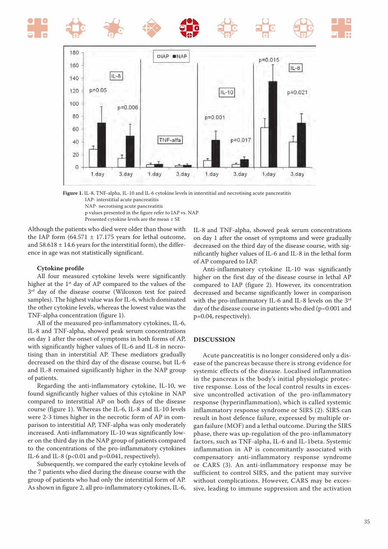
35
Although the patients who died were older than those with
the IAP form (64.571 ± 17.175 years for lethal outcome,
and 58.618 ± 14.6 years for the interstitial form), the differ-
ence in age was not statistically significant.
Cytokine profile
All four measured cytokine levels were significantly
higher at the 1st day of AP compared to the values of the
3rd day of the disease course (Wilcoxon test for paired
samples). The highest value was for IL-6, which dominated
the other cytokine levels, whereas the lowest value was the
TNF-alpha concentration (figure 1).
All of the measured pro-inflammatory cytokines, IL-6,
IL-8 and TNF-alpha, showed peak serum concentrations
on day 1 after the onset of symptoms in both forms of AP,
with significantly higher values of IL-6 and IL-8 in necro-
tising than in interstitial AP. These mediators gradually
decreased on the third day of the disease course, but IL-6
and IL-8 remained significantly higher in the NAP group
of patients.
Regarding the anti-inflammatory cytokine, IL-10, we
found significantly higher values of this cytokine in NAP
compared to interstitial AP on both days of the disease
course (figure 1). Whereas the IL-6, IL-8 and IL-10 levels
were 2-3 times higher in the necrotic form of AP in com-
parison to interstitial AP, TNF-alpha was only moderately
increased. Anti-inflammatory IL-10 was significantly low-
er on the third day in the NAP group of patients compared
to the concentrations of the pro-inflammatory cytokines
IL-6 and IL-8 (p<0.01 and p=0.041, respectively).
Subsequently, we compared the early cytokine levels of
the 7 patients who died during the disease course with the
group of patients who had only the interstitial form of AP.
As shown in figure 2, all pro-inflammatory cytokines, IL-6,
IL-8 and TNF-alpha, showed peak serum concentrations
on day 1 after the onset of symptoms and were gradually
decreased on the third day of the disease course, with sig-
nificantly higher values of IL-6 and IL-8 in the lethal form
of AP compared to IAP.
Anti-inflammatory cytokine IL-10 was significantly
higher on the first day of the disease course in lethal AP
compared to IAP (figure 2). However, its concentration
decreased and became significantly lower in comparison
with the pro-inflammatory IL-6 and IL-8 levels on the 3rd
day of the disease course in patients who died (p=0.001 and
p=0.04, respectively).
DISCUSSION
Acute pancreatitis is no longer considered only a dis-
ease of the pancreas because there is strong evidence for
systemic effects of the disease. Localised inflammation
in the pancreas is the body’s initial physiologic protec-
tive response. Loss of the local control results in exces-
sive uncontrolled activation of the pro-inflammatory
response (hyperinflammation), which is called systemic
inflammatory response syndrome or SIRS (2). SIRS can
result in host defence failure, expressed by multiple or-
gan failure (MOF) and a lethal outcome. During the SIRS
phase, there was up-regulation of the pro-inflammatory
factors, such as TNF-alpha, IL-6 and IL-1beta. Systemic
inflammation in AP is concomitantly associated with
compensatory anti-inflammatory response syndrome
or CARS (3). An anti-inflammatory response may be
sufficient to control SIRS, and the patient may survive
without complications. However, CARS may be exces-
sive, leading to immune suppression and the activation
Figure 1. IL-8, TNF-alpha, IL-10 and IL-6 cytokine levels in interstitial and necrotising acute pancreatitis
IAP- interstitial acute pancreatitis
NAP- necrotising acute pancreatitis
p values presented in the fi gure refer to IAP vs. NAP
Presented cytokine levels are the mean ± SE

36
of circulating cells of the immune system, including
monocytes and CD4+ T helper lymphocytes that shift
to Th2 response (5-8).
There are many pro- and anti-inflammatory mediators
included in this interaction between SIRS and CARS. In
this paper, we showed that early changes in the serum cy-
tokine profile could distinguish NAP from IAP and could
also indicate a lethal outcome. In the clinical setting, early
diagnosis and, if possible, assessment of the prognosis of
AP, is a major interest for the clinician. Indeed, early ag-
gressive treatment in patients who will develop the ne-
crotising form of the disease could potentially change the
outcome.
In our study, all examined cytokines were higher in the
necrotising compared to the interstitial form of AP. Their
levels gradually decreased on the third day of the disease
course, with significantly higher values of IL-6, IL-8 and
IL-10 in the NAP group (figure 1).
Our results are similar to the findings in patients with
acute alcoholic and severe biliary pancreatitis, in which the
serum IL-6 level reached its peak on admission and was
significantly higher in the severe form during the whole ob-
servational period (9,10). In the study conducted by Dam-
brauskas Z, et al. (11), a severe form of AP was associated
with a typical SIRS and a 2-5-fold increase in the expression
of pro-inflammatory cytokines (IL-6, IL-8, macrophage
inhibitory protein-MIF), together with the induction of
a compensatory and regulatory mechanism (IL-10). The
study also revealed that the serum IL-6 concentration is a
good predictor of the necrotising form of the disease and
systemic complications (SIRS, MOF). This marker can also
be utilised for the stratification of patients with necrotising
AP and those with a possible fatal outcome.
Regarding TNF-alpha, we did not find a significant dif-
ference between the necrotising and interstitial forms in
our AP patients. TNF-alpha plays a pivotal role in NAP,
acting early in the disease course, and is quickly cleared.
As a result of its rapid clearance, the TNF-alpha serum lev-
els are less useful than downstream cytokines (e.g. IL-6) as
biomarkers of early events (12).
We examined the early changes of another pro-in-
flammatory cytokine, IL-8, produced by macrophages
and epithelial cells, which exerts chemotactic activity on
neutrophils. In turn, activated neutrophils are a significant
source of IL-8 (13). In our study, patients with NAP had
significantly higher IL-8 levels on both days, whereas those
with a lethal outcome had significantly higher levels on the
third day of the disease course, indicating the persistence
of hyperinflammation (figures 1 and 2). This result was in
agreement with the previous studies that showed signifi-
cantly higher mean values of IL-8 and neutrophil elastase
in patients with complicated pancreatitis (10,14). In anoth-
er clinical study, the role of serum IL-8 in predicting lethal
AP was confirmed (15).
In the study conducted by Li, et al. (16), there was an in-
crease of Th2 cells (IL-4+) in the beginning of NAP, espe-
cially in the first week. They also showed decreased mono-
cyte surface expression of HLA-DR antigen in the first
week of the disease course, and this decreased expression
correlated positively with decreasing levels of TNF-alpha,
IL-6 and IL-10 during that period of NAP. IL-10, the most
potent anti-inflammatory cytokine, could be responsible
for the decreased monocyte HLA-DR expression in these
subjects with increased risk of secondary infections and
MODS (17). A high level of anti-inflammatory cytokine
IL-10 may follow the increase in the pro-inflammatory
factors TNF-alpha and IL-6, which might be a part of the
CARS (18). In our study, statistically higher levels of IL-10
and IL-6 on the admission day and at the third day of the
disease course were found in the NAP group as well as in
a group of patients who had lethal outcomes (figures 1 and
2), which confirms the previous results.
The network of various cytokines and other molecules
participating in the regulation of the inflammatory process-
Figure 2. IL-8, TNF-alpha, IL-10 and IL-6 cytokine levels in the IAP group of patients and AP patients with a lethal outcome
IAP-interstitial acute pancreatitis
p values presented in the fi gure refer to survivors vs. lethal outcome

37
4. Banks PA, Bollen TL, Dervenis C, et al. Acute Pancrea-
titis Classification Working Group. Classification of
acute pancreatitis-2012: revision of the Atlanta classifi-
cation and definitions by international consensus. Gut
2013; 62: 102-111
5. Ni Choileain N, Redmond HP. The immunological con-
sequences of injury. Surgeon 2006; 4: 23-31
6. Pietruczuk M, Dabrowska MI, Wereszczynska-Siemiat-
kowska U, Dabrowski A. Alteration of peripheral blood
lymphocyte subsets in acute pancreatitis. World J Gas-
troenterol 2006; 12: 5344-5351
7. Smith JW, Gamelli RL, Jones SB, Shankar R. Immuno-
logic responses to critical injury and sepsis. J Intensive
Care Med 2006; 21: 160-172
8. Kylanpaa ML, Repo H, Puolakkainen PA. Inflammation
and immunosuppression in severe acute pancreatitis.
World J Gastroenterol 2010; 16: 2867-2872
9. Panek J, Kusnierz-Cabala B, Dolecki M, Pietron J. Se-
rum proinflammatory cytokine levels and white blood
cell differential count in patients with different degrees
of severity of acute alcoholic pancreatitis. Pol Przeglad
Chirur 2012; 84: 230-237
10. Mayer J, Rau B, Gansauge F, Beger HG. Inflammatory
mediators in human acute pancreatitis: clinical and
pathophysiological implications. Gut 2000; 47: 546-
552
11. Dambrauskas Z, Giese N, Gulbinas A, et al. Different
profiles of cytokine expression during mild and severe
acute pancreatitis. World J Gastroenterol 2010; 16(15):
1845-1853
12. Malleo G, Mazzon E, Siriwardena AK, Cuzzocrea S.
Role of tumor necrosis factor-alfa in acute pancreatitis:
from biological basis to clinical evidence. Shock 2007;
28: 130-140
13. Strieter RM, Kosahava K, Allen RM, et al. Cytokine-
induced neutrophil-derived interleukin-8. Am J Pathol
1992; 141: 397-407
14. Gross V, Andreesen R, Leser HG, et al. Interleukin-8
and neutrophil activation in acute pancreatitis. Eur J
Clin Invest 1992; 22: 200-203
15. Rau B, Steinbach G, Gansauge F, Mayer J, Grunert
A, Beger HG. The potential role of procalcitonin and
interleukin 8 in the prediction of infected necrosis in
acute pancreatitis. Gut 1997; 41: 832-840
16. Li JP, Yang J, Huang JR, et al. Immunosuppression and
the infection in patients with early SAP. Frontiers in
Bioscience 2013; 18: 892-900
17. Fumeaux T, Pugin J. Role of interleukin-10 in the intra-
cellular sequestration of human leukocyte antigen-DR
in monocytes during septic shock. Am J Resp Crit Care
Med 2002; 166: 1475-1482
18. Smith JW, Gamelli RL, Jones SB, Shankar R. Immuno-
logic responses to critical injury and sepsis. J Intensive
Care Med 2006; 21: 161-172
es is very complex, and the precise timing of the release and
activation of these mediators is not known. SIRS and CARS
at a certain stage of the disease might even develop simul-
taneously (11). In our study, pro- and anti-inflammatory cy-
tokines were significantly higher on the 1st day of acute pan-
creatitis in the NAP group and in the group of patients with
lethal outcomes compared to the values in the IAP group
(figures 1 and 2). Anti-inflammatory IL-10 decreased more
than IL-6 and IL-8 and was significantly lower than pro-in-
flammatory cytokines on the 3rd day of the disease course in
the necrotising AP form and in the group of patients who
died. These findings could indicate that if SIRS and pro-in-
flammatory cytokines dominate the immune response, the
patients will develop the necrotising form of acute pancrea-
titis with a potentially lethal outcome.
This is the first study in which these cytokines (IL-6,
IL-8, IL-10 and TNF-alpha) were tested on the same group
of patients. We found that IL-6, IL-8 and IL-10 are good
indicators of the presence of necrosis and also of a poten-
tially lethal outcome.
More studies are needed to identify the immunological
phenomenon that may lead to the development of immu-
nomodulatory treatments in necrotising AP. A better un-
derstanding of the physiology of AP is necessary to accu-
rately identify those patients with necrotising AP in need
of monitoring and treatment in intensive care units.
AKNOWLEDGEMENTS
This study was partially financed by grant No III41010
(“Preclinical examination of bioactive substances”) given
by the Serbian Ministry of Education, Science and Techni-
cal Development, and also by project No 13/11, entitled
“The analysis of cytokine profile in patients with acute
pancreatitis treated in intensive care unit”, that is currently
being implemented in cooperation between the Faculty of
Medical Sciences, University of Kragujevac, and the Clini-
cal Center of Kragujevac.
REFERENCES
1. Isenmann R, Beger HG. Natural history of acute pan-
creatitis and the role of infection. Baillieres Best Pract
Res Clin 1999; 13: 291–301
2. Bone RC. Toward a theory regarding the pathogenesis
of the systemic inflammatory response syndrome: what
we do and do not know about cytokine regulation. Crit
Care Med 1996; 24: 163-172
3. Mentula P, Kylanpaa ML, Kemppainen E, et al. Plasma
antiinfiammatory cytokines and monocyte human leu-
cocyte antigen-DR expression in patients with acute
pancreatitis. Scan J Gastroenterol 2004; 39: 178-187

38

39
ORIGINAL SCIENTIFIC PAPER ORIGINALNI NAUČNI RAD ORIGINAL SCIENTIFIC PAPER
COMPARISON OF MEDIAL ARCHSUPPORTING INSOLESAND HEEL PADS IN THE TREATMENT OF PLANTAR FASCIITIS
Melih Malkoc, Ozgur Korkmaz, Adnan Kara, Ismail Oltulu, Ferhat Say
Medipol University Department of Orthopaedics and Traumatology Istanbul, Turkey
POREĐENJE ULOŽAKA ZA PODUPIRANJE MEDIJALNOG LUKASTOPALA I PETNIH ULOŽAKA U LEČENJU PLANTARNOG FASCITISA
Melih Malkoc, Oygur Korkmaz, Adnan Kara, Ismail Oltulu, Ferhat Say
Katedra za ortopediju i traumatologiju Univerziteta Medipol, Instabul, Turska
Correspondence: Ozgur Kormaz, Medipol University
Department of Orthopaedics and Traumatology, Istanbul, Turkey [email protected]
ABSTRACT
Plantar fasciitis is a disorder caused by infl ammation
of the insertion point of the plantar fascia over the medial
tubercle of the calcaneus. Foot orthotics are used to treat
plantar fasciitis. Heel pads medialise the centre of force,
whereas medial arch supporting insoles lateralise the force.
We assessed the clinical results of the treatment of plantar
fasciitis with silicone heel pads and medial arch-supported
silicone insoles.
We retrospectively reviewed 75 patients with heel pain.
A total of 35 patients in the fi rst group were treated with
medial arch supporting insoles, and 40 patients in the sec-
ond group were treated with heel pads. Th e patients were
evaluated with the Visual Analogue Scale (VAS) and the
Foot and Ankle Ability Measure (FAAM) at the fi rst and
last examinations.
Th e mean VAS score in the fi rst group was 8.6±1,2 (6-10);
the FAAM daily activity score was 66.2±16 (41.2-95.0),
and the sporting activity score was 45.4±24,4 (0.1-81) be-
fore treatment. At the last follow-up in this group, the mean
VAS score was 5.3±1,5 (0-9); the FAAM daily activity score
was 83,0±15,1 (55,9-100), and the sporting activity score
was 73,5±26,2 (25-100). Th e mean VAS score in the second
group was 8,6±0,9 (7-10); the FAAM daily activity score
was 66.4±17 (41.4-95.2), and the sporting activity score was
45.8±24,2 (0.8-81, 3) before the treatment. At the last follow-
up in this group, the mean VAS score was 5.5±1,2 (0-9); the
FAAM daily activity score was 83.4±14,9 (60, 2-100), and
the sporting activity score was 73.8±26 (28-100).
Th ere was no signifi cant diff erence in the clinical results of
both groups. Th e force distribution by the use of silicone heel
pads and medial arch-supported silicone insoles had no eff ect
on the clinical results of the treatment of plantar fasciitis.
Keywords: Plantar fasciitis, Medial arch-supporting in-
soles, Heel pads
SAŽETAK
Plantarni fascitis je poremećaj koji nastaje usled zapalje-
nja pripoja plantarne fascije za medijalni tuberkulum pet-
ne kosti. Za lečenje plantarnog fascitisa se koriste ortoze za
stopala. Petni ulošci pomeraju mesto opterećenja ka unutra,
dok ulošci za podupiranje medijalnog luka pomeraju mesto
opterećenja upolje. Klinički rezultati se bave ispitivanjem le-
čenja plantarnog fascitisa silikonskim petnim ulošcima i sili-
konskim ulošcima za podupiranje medijalnog luka stopala.
Retrospektivno je ispitano 75 pacijenata sa bolom u peti.
35 pacijenata u prvoj grupi su lečeni sa ulošcima za podupi-
ranje medijalnog luka stopala. 40 pacijenata u drugoj grupi
je lečeno petnim ulošcima. Pacijenti su ocenjivani pomoću
Visual Analogue Scale (VAS) i Foot and Ankle Ability Mea-
sure (FAAM) na prvom i na poslednjem pregledu.
Pre lečenja, srednja vrednost VAS skora u prvoj grupi je bila
8,6 ± 1,2 (6 - 10), FAAM skora adnevne aktivnosti 66,2 ± 16
(42,1 – 95,0) i skora sportske aktivnosti 45,4 ± 24,4 (0,1 - 81).
Srednje vrednosti na poslednjem pregledu VAS skora su iznosile
5,3 ± 1,5 (0 - 9), FAAM skora adnevne aktivnosti 83,0 ± 15,1
(55,9 – 100) i skora sportske aktivnosti 73,5 ± 26,2 (25 - 100).
Pre lečenja, srednja vrednost VAS skora u drugoj grupi je
iznosila 8,6 ± 0,9 (7 - 10), FAAM skora adnevne aktivnosti
66,4 ± 17 (41,4 – 95,2) i skora sportske aktivnosti 45,8 ± 24,2
(0,8 – 81,3). Na poslednjem pregledu srednja vrednost VAS
skora je bila 5,5 ± 1,2 (0 - 9), FAAM skora adnevne aktivnosti
83,4 ± 14,9 (60,2 - 100) i skora sportske aktivnosti 73,8 ± 26
(28 – 100).
Nema statistički značajne razlike u kliničkim rezultatima
između grupa. Raspodela sile pritiska upotrebom silikonskih
petnih uložaka ili silikonskih uložaka za podupiranje medi-
jalnog luka stopala nema uticaj na kliničke rezultate lečenja
plantarnog fascitisa.
Ključne reči: plantarni fascitis, ulošci za podupiranje
medijalnog luka stopala, petni ulošci
Received / Primljen: 09. 09. 2014. Accepted / Prihvaćen: 14. 12. 2014.
UDK: 617.586-007.2-76 / Ser J Exp Clin Res 2015; 16 (1): 39-42
DOI: 10.1515/SJECR20150006
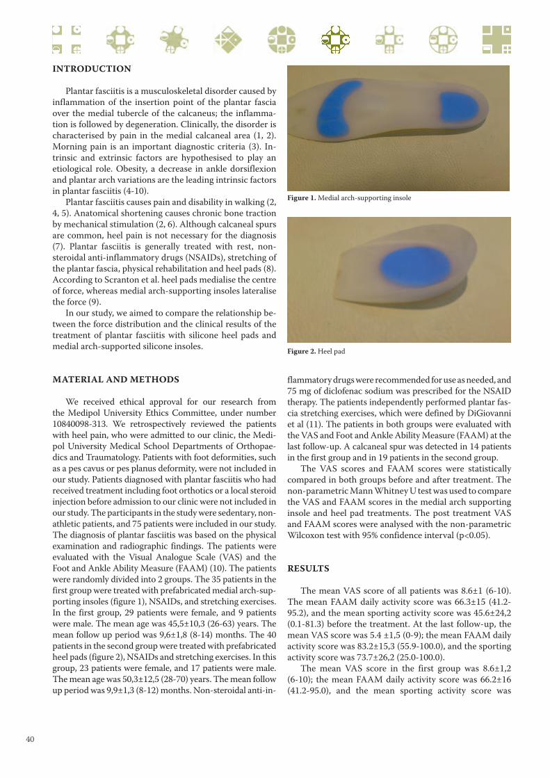
40
INTRODUCTION
Plantar fasciitis is a musculoskeletal disorder caused by
inflammation of the insertion point of the plantar fascia
over the medial tubercle of the calcaneus; the inflamma-
tion is followed by degeneration. Clinically, the disorder is
characterised by pain in the medial calcaneal area (1, 2).
Morning pain is an important diagnostic criteria (3). In-
trinsic and extrinsic factors are hypothesised to play an
etiological role. Obesity, a decrease in ankle dorsiflexion
and plantar arch variations are the leading intrinsic factors
in plantar fasciitis (4-10).
Plantar fasciitis causes pain and disability in walking (2,
4, 5). Anatomical shortening causes chronic bone traction
by mechanical stimulation (2, 6). Although calcaneal spurs
are common, heel pain is not necessary for the diagnosis
(7). Plantar fasciitis is generally treated with rest, non-
steroidal anti-inflammatory drugs (NSAIDs), stretching of
the plantar fascia, physical rehabilitation and heel pads (8).
According to Scranton et al. heel pads medialise the centre
of force, whereas medial arch-supporting insoles lateralise
the force (9).
In our study, we aimed to compare the relationship be-
tween the force distribution and the clinical results of the
treatment of plantar fasciitis with silicone heel pads and
medial arch-supported silicone insoles.
MATERIAL AND METHODS
We received ethical approval for our research from
the Medipol University Ethics Committee, under number
10840098-313. We retrospectively reviewed the patients
with heel pain, who were admitted to our clinic, the Medi-
pol University Medical School Departments of Orthopae-
dics and Traumatology. Patients with foot deformities, such
as a pes cavus or pes planus deformity, were not included in
our study. Patients diagnosed with plantar fasciitis who had
received treatment including foot orthotics or a local steroid
injection before admission to our clinic were not included in
our study. The participants in the study were sedentary, non-
athletic patients, and 75 patients were included in our study.
The diagnosis of plantar fasciitis was based on the physical
examination and radiographic findings. The patients were
evaluated with the Visual Analogue Scale (VAS) and the
Foot and Ankle Ability Measure (FAAM) (10). The patients
were randomly divided into 2 groups. The 35 patients in the
first group were treated with prefabricated medial arch-sup-
porting insoles (figure 1), NSAIDs, and stretching exercises.
In the first group, 29 patients were female, and 9 patients
were male. The mean age was 45,5±10,3 (26-63) years. The
mean follow up period was 9,6±1,8 (8-14) months. The 40
patients in the second group were treated with prefabricated
heel pads (figure 2), NSAIDs and stretching exercises. In this
group, 23 patients were female, and 17 patients were male.
The mean age was 50,3±12,5 (28-70) years. The mean follow
up period was 9,9±1,3 (8-12) months. Non-steroidal anti-in-
flammatory drugs were recommended for use as needed, and
75 mg of diclofenac sodium was prescribed for the NSAID
therapy. The patients independently performed plantar fas-
cia stretching exercises, which were defined by DiGiovanni
et al (11). The patients in both groups were evaluated with
the VAS and Foot and Ankle Ability Measure (FAAM) at the
last follow-up. A calcaneal spur was detected in 14 patients
in the first group and in 19 patients in the second group.
The VAS scores and FAAM scores were statistically
compared in both groups before and after treatment. The
non-parametric Mann Whitney U test was used to compare
the VAS and FAAM scores in the medial arch supporting
insole and heel pad treatments. The post treatment VAS
and FAAM scores were analysed with the non-parametric
Wilcoxon test with 95% confidence interval (p<0.05).
RESULTS
The mean VAS score of all patients was 8.6±1 (6-10).
The mean FAAM daily activity score was 66.3±15 (41.2-
95.2), and the mean sporting activity score was 45.6±24,2
(0.1-81.3) before the treatment. At the last follow-up, the
mean VAS score was 5.4 ±1,5 (0-9); the mean FAAM daily
activity score was 83.2±15,3 (55.9-100.0), and the sporting
activity score was 73.7±26,2 (25.0-100.0).
The mean VAS score in the first group was 8.6±1,2
(6-10); the mean FAAM daily activity score was 66.2±16
(41.2-95.0), and the mean sporting activity score was
Figure 1. Medial arch-supporting insole
Figure 2. Heel pad
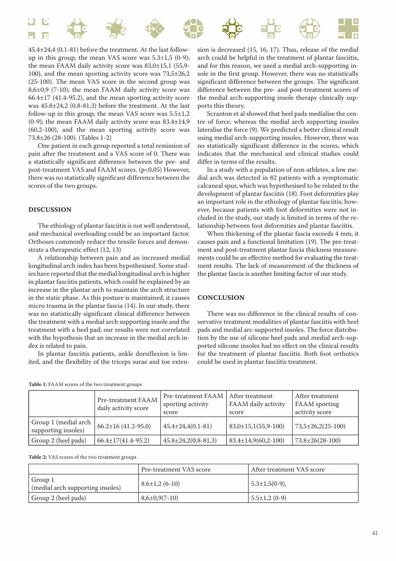
41
45.4±24,4 (0.1-81) before the treatment. At the last follow-
up in this group, the mean VAS score was 5.3±1,5 (0-9);
the mean FAAM daily activity score was 83,0±15,1 (55,9-
100), and the mean sporting activity score was 73,5±26,2
(25-100). The mean VAS score in the second group was
8,6±0,9 (7-10); the mean FAAM daily activity score was
66.4±17 (41.4-95.2), and the mean sporting activity score
was 45.8±24,2 (0.8-81,3) before the treatment. At the last
follow-up in this group, the mean VAS score was 5.5±1,2
(0-9); the mean FAAM daily activity score was 83.4±14,9
(60,2-100), and the mean sporting activity score was
73.8±26 (28-100). (Tables 1-2)
One patient in each group reported a total remission of
pain after the treatment and a VAS score of 0. There was
a statistically significant difference between the pre- and
post-treatment VAS and FAAM scores. (p<0,05) However,
there was no statistically significant difference between the
scores of the two groups.
DISCUSSION
The ethiology of plantar fasciitis is not well understood,
and mechanical overloading could be an important factor.
Orthoses commonly reduce the tensile forces and demon-
strate a therapeutic effect (12, 13)
A relationship between pain and an increased medial
longitudinal arch index has been hypothesised. Some stud-
ies have reported that the medial longitudinal arch is higher
in plantar fasciitis patients, which could be explained by an
increase in the plantar arch to maintain the arch structure
in the static phase. As this posture is maintained, it causes
micro trauma in the plantar fascia (14). In our study, there
was no statistically significant clinical difference between
the treatment with a medial arch supporting insole and the
treatment with a heel pad; our results were not correlated
with the hypothesis that an increase in the medial arch in-
dex is related to pain.
In plantar fasciitis patients, ankle dorsiflexion is lim-
ited, and the flexibility of the triceps surae and toe exten-
sion is decreased (15, 16, 17). Thus, release of the medial
arch could be helpful in the treatment of plantar fasciitis,
and for this reason, we used a medial arch-supporting in-
sole in the first group. However, there was no statistically
significant difference between the groups. The significant
difference between the pre- and post-treatment scores of
the medial arch-supporting insole therapy clinically sup-
ports this theory.
Scranton et al showed that heel pads medialise the cen-
tre of force, whereas the medial arch supporting insoles
lateralise the force (9). We predicted a better clinical result
using medial arch-supporting insoles. However, there was
no statistically significant difference in the scores, which
indicates that the mechanical and clinical studies could
differ in terms of the results.
In a study with a population of non-athletes, a low me-
dial arch was detected in 82 patients with a symptomatic
calcaneal spur, which was hypothesised to be related to the
development of plantar fasciitis (18). Foot deformities play
an important role in the ethiology of plantar fasciitis; how-
ever, because patients with foot deformities were not in-
cluded in the study, our study is limited in terms of the re-
lationship between foot deformities and plantar fasciitis.
When thickening of the plantar fascia exceeds 4 mm, it
causes pain and a functional limitation (19). The pre-treat-
ment and post-treatment plantar fascia thickness measure-
ments could be an effective method for evaluating the treat-
ment results. The lack of measurement of the thickness of
the plantar fascia is another limiting factor of our study.
CONCLUSION
There was no difference in the clinical results of con-
servative treatment modalities of plantar fasciitis with heel
pads and medial arc-supported insoles. The force distribu-
tion by the use of silicone heel pads and medial arch-sup-
ported silicone insoles had no effect on the clinical results
for the treatment of plantar fasciitis. Both foot orthotics
could be used in plantar fasciitis treatment.
Table 1: FAAM scores of the two treatment groups
Table 2: VAS scores of the two treatment groups
Pre-treatment FAAM
daily activity score
Pre-treatment FAAM
sporting activity
score
After treatment
FAAM daily activity
score
After treatment
FAAM sporting
activity score
Group 1 (medial arch
supporting insoles)66.2±16 (41.2-95.0) 45.4±24,4(0.1-81) 83,0±15,1(55,9-100) 73,5±26,2(25-100)
Group 2 (heel pads) 66.4±17(41.4-95.2) 45.8±24,2(0.8-81,3) 83.4±14,9(60,2-100) 73.8±26(28-100)
Pre-treatment VAS score After treatment VAS score
Group 1
(medial arch supporting insoles)8.6±1,2 (6-10) 5.3±1,5(0-9),
Group 2 (heel pads) 8,6±0,9(7-10) 5.5±1,2 (0-9)
42
REFERENCES
1. Kwong PK, Kay D, Voner RT, White MW. Plantar fascii-
tis: Mechanics and pathomechanics of treatment. Clin
Sports Med. 1987;7:119-26.
2. Greve JM, Grecco MV, Santos-Silva PR. Comparison of
radial shockwaves and conventional physiotherapy for
treating plantar fasciitis. Clinics. 2009;64:97-103.
3. Tisdel CL, Donley BG, Sferra JJ. Diagnosing and treat-
ing plantar fasciitis: A conservative approach to plantar
heel pain. Cleve Clin J Med. 1999;66:231-5.
4. Ogden JA, Alvarez RG, Levitt RL, Johnson JE, Marlow
ME. Electrohydraulic high-energy shock-wave treat-
ment for chronic plantar fasciitis. J Bone Joint Surg Am.
2004;86-A(10):2216-28.
5. Huang YC, Wang LY, Wang HC, Chang KL, Leong CP.
The relationship between the flexible flatfoot and plan-
tar fasciitis: ultrasonographic evaluation. Chang Gung
Med J. 2004 ;27(6):443-8.
6. Roxas M. Plantar fasciitis: diagnosis and therapeutic
considerations. Altern Med Rev. 2005 ;10(2):83-93.
Review.
7. Onwuanyi ON Calcaneal spurs and plantar heel pad
pain. The Foot 2000;10:182-5.
8. Buchbinder R Clinical practice. Plantar fasciitis. N Engl
J Med 2004;350:2159–66.
9. Scranton PE Jr, Pedegana LR, Whitesel JP. Gait analysis.
Alterations in support phase forces using supportive
devices. Am J Sports Med. 1982 ;10(1):6-11.
10. Martin RL, Irrgang JJ, Burdett RG, Conti SF, Van
Swearingen JM. Evidence of validity for the Foot
and Ankle Ability Measure (FAAM). Foot Ankle Int.
2005;26:968-983.
11. DiGiovanni BF1, Nawoczenski DA, Lintal ME, Moore
EA, Murray JC, Wilding GE, Baumhauer JF. Tissue-
specific plantar fascia-stretching exercise enhances
outcomes in patients with chronic heel pain. A pro-
spective, randomized study. J Bone Joint Surg Am.
2003;85-A(7):1270-7.
12. Kogel GF, Soromodinis SE, Paul JP. Biomechanics of
longitudinal arch support mechanics in foot orthoses
and their effect on plantar aponeurosis strain. Clin Bio-
mech 1998;11:243-252.
13. Kogler GF, Veer FB, Solomonidis SE, Paul JP. The infl
uence of medial and lateral placement of orthotic
wedges on loading of the plantar aponeurosis. J Bone
Joint Surg Am 1999;81:1403-13.
14. Imamura M, Imamura S, Carvalho AE, Mazagao RA,
Cassius DA, Fischer AA. Plantar fasciitis: A new treat-
ment approach. Arch Phys Med Rehabil. 2003;84: E4.
15. Riddle DL, Pulisic M, Pidcoe P, Johnson RE. Risk fac-
tors for Plantar fasciitis: A matched case-control study.
J Bone Joint Surg Am. 2003; 85-A:872-7.
16. Warren BL. Anatomical factors associated with pre-
dicting plantar fasciitis in long-distance runners. Med
Sci Sports Exerc. 1984;16: 60-3.
17. Allen RH, Gross MT. Toe flexors strength and passive
extension range of motion of the first metatarsophalan-
geal joint in individuals with plantar fasciitis. J Orthop
Sports Phys Ther. 2003;33:468-78.
18. Prichasuk S, Subhadrabandhu T. The relationship of
pes planus and calcaneal spurs to plantar heel pain.
Clin Orthop Relat Res. 1994;306:192-6.
19. Liang WH, Wang TG, Chen WS, Hou SM. Thinner
plantar fascia predicts decreased pain after shock wave
therapy. Clin Orthop and Relat Res. 2007;460:219-25.

43
MACROECONOMIC POLICY IMPACT ON ONCOLOGYRELATEDPUBLIC EXPENDITURE IN AN EMERGING EUROPEAN
MARKET SIGNS OF EARLY RECOVERYAleksandar Dagovic 1, Ana Zugic 2 and Mihajlo B Jakovljevic 3
1 Oncology and Radiotherapy Center, Clinical Center Kragujevac, Kragujevac, Serbia2 Faculty of Medical Sciences, University of Kragujevac, Kragujevac, Serbia
3 Department of Pharmacology and Toxicology, Faculty of Medical Sciences, University of Kragujevac, Kragujevac, Serbia
UTICAJ MAKROEKONOMSKE POLITIKE NA JAVNA IZDVAJANJAZA ONKOLOGIJU NA RASTUĆEM
EVROPSKOM TRŽIŠTU ZNACI RANOG OPORAVKAAleksandar Dagović 1, Ana Žugić 2 i Mihajlo B Jakovljević 3
1 Centar za onkologiju i radioterapiju, Klinički centar Kragujevac, Kragujevac, Srbija2 Fakultet medicinskih nauka, Univerzitet u Kragujevcu, Kragujevac, Srbija
3 Katedra za farmakologiju i toksikologiju, Fakultet medicinskih nauka, Univezitet u Kragujevcu, Kragujevac, Srbija
SAŽETAK
Cilj studije je analiza trendova u javnim izdvajanjima za
onkolosku zdravstvenu zaštitu u godinama duboke ekonom-
ske recesije u svetu i na Balkanu. Ostali ciljevi su utvrditi
fi nu strukturu troškova u ovoj kliničkoj disciplini kao i even-
tualno prisustvo korelacije obima potrošnje na dijagnostiku
i lečenje malignih neoplazmi sa makroekonomskim kretanji-
ma i zdravstvenom politikom u Srbiji.
Primenjena je retrospektivna studija slučaja, kojom je
obuhvaćen period od cetiri godine (2010-2013.) iz perspekti-
ve fi nansijera zdravstvene zastite i sa usvojenim vremenskim
horizontom od godinu dana. Studija je izvedena na osnovu
izvoda baze podataka o 37 978 epizoda bolničkog lecenja
i 12 505 pacijenata sa klinički potvrdjenim kancerom pri
regionalnom Centru za onkologiju i radioterapiju Kliničkog
Centra Kragujevac.
Ukupni direktni medicinski troskovi dijangostike i lecenja
kancera u posmatranom tercijernom centru su pali sa €7,
411, 446 u 2010 na €5, 715, 884 u 2012 i iznova snažno sko-
čili na €8, 536, 364 u 2013. Glavni domeni troškova koji su
najviše doprineli ukupnom obimu potrosnje su bili onkoloska
medicinska nega, radioterapija i lekovi.
Finansijski teret kancera u Srbiji je ogroman i nažalost
izgledno je da ce nastaviti da raste zahvaljujući nizu činilaca
poput starenja populacije, boljeg preživljavanja, naraslih oče-
kivanja građanstva o pravu na pristup naprednim metodama
lečenja kao i dugoročno izvesnog rasta pokrivenosti stanovniš-
tva zdravstvenom zaštitom. Posebno masivni faktori su širenje
dostupnosti metoda radioterapije i refundacija skupih biološ-
kih lekova. Obećavajući znaci oporavka nacionalnog tržista
će se, nadamo se, pretočiti u napore na poboljšanju pristupa i
priuštivosti onkološke nege običnom građaninu.
Ključne reči: Svetska ekonomska kriza; recesija; kancer;
troškovi; ekonomija; fi nansiranje zdravstva; zdravstvena po-
litika; refundacija; bolnice; Srbija
ABSTRACT
Healthcare fi nancing in Serbia has faced many challenges
over the past few decades. One of the most severe challenges
is a global macroeconomic recession whose far-reaching con-
sequences deserve particular attention from policymakers in
cases of the most demanding major prosperity diseases, such
as cancer. Th e objective of the study was to assess the precise
cost matrix of oncology medical care and its chronological
evolution during the key years of the macroeconomic reces-
sionary period during 2010-2013.
A retrospective database of hospital discharge invoices
was analysed, encompassing 37, 978 hospital admissions
and 12, 505 patients during a four-year period. Insight into
microeconomic patterns of consumption across groups of
medical services was provided. A payer’s perspective and
one-year time horizon have been adopted.
Total hospital direct medical costs of cancer diagnostics
and treatment in the observed tertiary care facility decreased
from €7, 411, 446 in 2010 to €5, 715, 884 in 2012 and then in-
creased to an extraordinary €8, 536, 364 in 2013. Th e costs of
oncology nursing care, imaging diagnostics and radiotherapy
have increased considerably while those of pharmaceuticals
and surgery have decreased radically - completely transform-
ing the resource allocation landscape of public cancer care.
Th e fi nancial burden of cancer in Serbia is considerable
and, unfortunately, expected to increase further in the coming
years. Worldwide economic recession and consecutive domes-
tic policy constraints of reimbursement limitations have heav-
ily aff ected the aff ordability of cancer treatment for ordinary
citizens. Promising signs of market recovery are clearly visible
in 2013, which will likely improve both access and equity of
medical care in Serbian oncology clinics.
Keywords: Worldwide Crisis; Recession; Cancer; Costs;
Economics; Health Financing; Health Policy; Reimburse-
ment; Hospital; Serbia
Received / Primljen: 20. 05. 2014. Accepted / Prihvaćen: 25. 06. 2014.
ORIGINAL SCIENTIFIC PAPER ORIGINALNI NAUČNI RAD ORIGINAL SCIENTIFIC PAPER
Correspondence: Mihajlo Jakovljevic, Department of Pharmacology and Toxicology,
Th e Faculty of Medical Sciences, University of Kragujevac, [email protected]
UDK: 330.322.3:614(497.11) / Ser J Exp Clin Res 2015; 16 (1): 43-50
DOI: 10.1515/SJECR20150007

44
INTRODUCTION
Cost-of-illness analyses of the key prosperity diseases
remain rather infrequent in Eastern Europe and the Bal-
kans region (1). Over the past decade, a few pioneering
assessments were published, laying the ground for the in-
formed decision making of local health policymakers (2).
These findings reflected the workload and financial bur-
den imposed by the diagnostics and treatment of chron-
ic obstructive pulmonary disease, community-acquired
pneumonia, alcohol dependence, diabetes mellitus, hepa-
titis C, risky pregnancies and others (3-8). Another set of
contributions revealed the considerable budgetary impact
of some key medical technologies, such as medical imaging
(9), radiotherapy (10) and monoclonal antibodies, applied
in oncology (11-12). These and further on-going efforts on
domestic, local health economic estimates are essential to
improve the financial efficiency of our health system (13).
This claim is supported by the well-known fact that similar
estimates from high-income markets are not straight- for-
wardly applicable to clinical settings across Eastern Europe
and the Balkans due to their substantially different histo-
ries, traditions and socioeconomic milieus (14-15).
Oncological morbidity deserves a particularly high
place among the leading noncommunicable prosperity dis-
eases. The diagnostics, treatment and rehabilitation associ-
ated with cancer are commonly much more demanding in
terms of medical technology use and physician consultation
time and frequency compared to other major illnesses. Ad-
ditionally, the clinical outcomes of these interventions are
far less predictable, with illness itself resulting in a heavily
reduced life expectancy, quality of life and working abil-
ity of an individual citizen. Cancer’s economic burden to
the community is enormous, and the issue of treatment af-
fordability remains high on the policy agendas of even the
richest countries worldwide (16). Pioneering assessments
in Serbia were published only recently and confirmed the
aforementioned facts evidenced elsewhere (17).
Unfortunately, both global and European cancer preva-
lence and incidence are increasing (18). Serbia exhibits
a slightly higher incidence compared to the EU average,
which remains somewhat lower compared to the Europe-
an average that includes the CIS countries and the Russian
Federation (19-20). Among the most frequently cited rea-
sons for higher oncological mortality in Serbia are poverty,
unhealthy lifestyles and ecological contamination due to
the Chernobyl disaster and wars in Yugoslavia during the
1990s (21-22). However, throughout Eastern Europe, poor
implementation of screening procedures (23) and limited
affordability of some innovative treatment technologies re-
main powerful contributors (24).
The global macroeconomic crisis has caused severe in-
stability among the Western Balkan economies bordering
the EU (25). The recession has compromised financial sus-
tainability within the health sector (27), and its far-reach-
ing consequences deserve particular attention from poli-
cymakers in cases of the most demanding major prosperity
diseases, such as cancer (28). The core research question
of this study was the assessment of the macroeconomic
recession’s impact on public medical spending mediated
by national policy (29). Early signs of economic recovery,
which have been present in Serbia since 2013, are likely to
improve access to and the affordability of medical care for
patients suffering from cancer. So far, targeted biological
therapy reimbursement has remained one of the hottest
domestic policy issues (30).
PATIENTS AND METHODS
To address the aforementioned research question, a
retrospective, bottom-up, case series study design over a
one-year horizon and payer’s perspective was implement-
ed (31). The tertiary care University of Kragujevac clinic
allowed selective examination of their electronic database
of discharge invoices. All patients whose cancer diagnosis
was confirmed by clinical, imaging, laboratory and patho-
histology findings and who were admitted and treated
at the regional Oncology and Radiotherapy Centre were
processed. Key cost drivers and determinants of resource
consumption during oncological inpatient care were iden-
tified. Personal data remained protected during the study
consistent with positive legislation on biomedical research
in human subjects in Serbia via anonymous handling of pa-
tient files. A fine cost matrix was produced through strati-
fication of the Republican Health Insurance Fund (RFZO)
“Blue Code Book” of all medical goods and services pro-
vided within the national health system.
The patient sample recruitment period was January
2010-December 2013 and included inhabitants of this cen-
tral Serbian region. Total sample size was 12, 505 patients or
37, 978 hospital admissions with assigned oncology treat-
ment protocols during the 2010-2013 period. These years
were selected because the 2010-2012 years were marked
by the heavy impact of worldwide economic crisis, while
2013 was a year of slow but steady recovery of the national
economy. To the authors’ best knowledge, this sampling
method and approach to longitudinal data is standard and
common in the discipline of health economics (32).
RESULTS
Total hospital direct medical costs of cancer diagnostics
and treatment at this tertiary university hospital fell from €7,
411, 446 in 2010 to €5, 715, 884 in 2012 and then increased
to an extraordinary €8, 536, 364 in 2013. Costs of oncology
nursing care, imaging diagnostics and radiotherapy have
greatly increased while pharmaceuticals and surgery fol-
lowed at a much slower pace, completely transforming the
resource allocation landscape of public cancer care.
Most major service groups follow this general pattern,
while pharmaceuticals and surgery differ from the domi-
nant trend. Drug acquisition represented 56, 4% of total
45
costs in 2010 and 53, 3% in 2012. Cytostatics and immu-
nosuppressants experienced a particularly steep decline in
value-based turnover of 55, 4%, decreasing from €1, 699,
164 in 2010 to €758, 490 in 2012. Antibiotics, antiemetics,
bone marrow stimulating factors and analgesics followed
the same pattern. Among the few drugs that increased dur-
ing the 2010-2012 period were monoclonal antibody costs,
which rose by 20, 3% (from €1, 350, 235 in 2010 to €1, 624,
245 in 2012).
Radiotherapy costs also increased during the 2010-
2012 period by 28, 1% (from €416, 193 to €533, 303), and
this increase is unfortunately mostly a consequence of a
higher workload produced by more frequent outpatient
visits and inpatient admissions.
Decreased surgery-related costs should be attributed to
the market decreases in the prices of consumables.
The radiology imaging, contrasts and films budget
impact changed substantially from 3, 515, 050 RSD (€33,
319) in 2010 to only 1, 168, 519 RSD (€10, 276) in 2012,
which is a nearly 70% decrease. Net savings acquired this
way should be attributed to the new information system
installed in the diagnostic services and clinic, which has
eliminated the need for traditional roentgen films in most
examination techniques. The value-based turnover across
major cost domains over these four years is shown in detail
in Table 1.
DISCUSSION
As indicated by the data above, some service groups
show a sudden and clear upward trend in 2013, while oth-
ers have followed at much slower pace. Thus, the big pic-
ture of resource allocation to cancer diagnostics and treat-
ment in large hospitals in Serbia has evolved according to
the macroeconomic landscape, market circumstances and
official policies of the national authorities (13, 15, 29).
The 2010 dominance of drug acquisition costs (pri-
marily conventional cytostatic, antiemetic, analgesic,
hormonal and antibiotic drugs) is overtaken in 2013 by
expanding radiotherapy, oncology nursing care and imag-
ing diagnostics. This pattern is consistent with previously
published evidence on the region stating that particularly
serious budget impacts were imposed by over-utilization
of high-tech radiology imaging procedures (11, 17, 24).
Pharmaceuticals were greatly influenced by novel, ex-
pensive biological treatments, such as monoclonal anti-
bodies (mAbs) and protease inhibitors. This pattern is only
small portion of far larger changes. One recently published
study analysed official annual data from the national medi-
cines agency (ALIMS) since 2004 (33). Total public expen-
diture on drugs with primary oncology-related indications
increased by approximately five times during the period
2004-2012. During that same decade, public consumption
of mAbs increased nearly 20 times due to the aforemen-
tioned societal and market changes (12). Although it is
favourable that many patients had access to these innova-
tive medicines, the cost-effectiveness of many mAbs is a
source of heated international debate (34, 35). The societal
affordability of expensive biologicals and willingness to pay
thresholds are rather low in Eastern Europe compared to
the West (13, 15). Contradictions such as shortages of ba-
sic, conventional cytostatics alongside reimbursement of
most expensive medicines occur frequently (11, 12).
The downward slope of cytostatic drug acquisition
costs from 2010 to 2012 in our regional sample has an
underlying cause, which is invisible in the presented data.
Unfortunately, due to pharmaceutical market disturbances
across Western Balkan economies, during the last quar-
ter of 2011 and the first five months of 2012, continuous
hospital supplies of these drugs were severely threatened.
These disturbances were mostly caused by delayed pay-
ments from state-owned health insurance funds to major
multinational manufacturers supplying the region (13,
15). One particularly sensitive issue was the lack of simple
5-fluorouracil, which is itself a quite inexpensive medicine
but is an essential part of many expensive and complex
treatment protocols that could not be provided for months
due to this shortage. Occasional shortages of cytostatic
medicines also occurred. Consequently, clinics have expe-
rienced sudden decreases in their need for drugs used to
treat the most common adverse effects of cytostatics, such
as antibiotics, bone marrow growth factors and antiemet-
ics used to treat febrile neutropenia, opportune infections
and vomiting (36-38).
Another core influence of the sudden decrease in
prescribing and dispensing of cytostatic drugs and their
costs is the restrictive reimbursement policy imposed by
the Republican Health Insurance Fund of Serbia (RHIFS),
which was mostly triggered by the macroeconomic reces-
sion (39). The common practice is financial coverage of
a particular medicine for select indications, such as nar-
rowly defined malignant tumour clinical types, grades
and stages, while the same drug might not be funded
for another malignancy. Policies of funding agencies to
prioritize interventions within their optimal clinical effi-
ciency and cost-effectiveness were in place in many major
markets around the globe (40-41).
The temporary decrease in oncology nursing care costs
from 2010 to 2012 occurred immediately following domes-
tic policy measures to allow contracted general practitio-
ners to prescribe and administer opioid analgesics were
implemented. This practice was uncommon within the na-
tional health system of Serbia. At approximately the same
time, an outpatient pain treatment service was founded
within this tertiary university clinic. Both measures ulti-
mately resulted in less frequent hospital admissions due
to severe metastatic pain, and patients used the opportu-
nity to resolve their symptoms on an outpatient basis and
preferred staying at home. That this strategy induces net
savings while improving patient satisfaction and quality of
life has already been observed elsewhere (42). The finan-
cial sustainability of home care in Serbia was recently an
objective of thorough consideration (43).

46
Table 1. Cost matrix of oncology related diagnostics and treatment within tertiary university clinic of Kragujevac 2010-2013
COST DOMAIN 2010 2011 2012 2013
Number of patients diagnosed with confi rmed malignancy
Number of hospital admissions due to cancer
Oncology related medical care
Hospital Admission 1,118,074 1,102,027 950,470 988,679
Physician Consultations 50,800 63,924 42,386 215,942
Clinical Pharmacology/ Pharmacist services 27 309 115
Rehabilitation services 355 2,408 1,462 1,153
Dialysis 868 493 373 754
Psychotherapy 83 12 8 709
Administrative expenses 138 154 135 295
All Other services (social care , transport, counselling, epidemiological 4,183 1,448 2,847 442,627
Total Cost of General - Oncology related medical care 1,174,533 1,170,776 997,796 1,650,161
Pharmaceuticals
Antineoplastic agents and immunosuppresants 1,691,948 1,200,255 758,490 572,908
Monoclonal antibodies 1,344,501 1,432,796 1,624,245 1,559,090
Analgesics NSAID, opioid, others - pain control medicines 14,268 10,891 7,587 6,000
Antibiotics, antimicotics, antiviral and antiprotozoal drugs 170,376 111,822 78,586 104,546
Antiemetics 190,435 138,018 100,743 110,606
Parenteral and enteral nutritive solutions and systems 118,386 92,833 86,628 91,349
Hematopoietic colony stimulating factors 150,539 79,288 65,552 101,069
Antiandrogens , antiestrogens – therapy of steroid dependent carcinoma 153 10
Blood and its derivatives – transfusions 155,917 111,031 109,201 60,997
All other drugs 345,498 306,273 218,122 264,412
Total cost for pharmaceuticals 4,182,025 3,483,217 3,049,154 2,870,977
Laboratory Analysis
Classical Biochemistry and hemathology 296,599 295,276 190,453 291,817
Targeted cancer prevention screenings 71 83 13 1,576
Tumor marker detection 1,010 2,042
Pathohistology tests and cytology examinations 125,808 131,191 105,862 119,742
Immunodiagnostics, genetics, cell culture techniques 77,689 66,720 51,570 73,677
Law medicine and forensic services 15,217 16,586 16,125 31,043
Total cost for laboratory analysis 515385 509,856 365,033 519,897
Surgery
Surgical Interventions 132,278 157,041 135,291 133,586
Nursing care and consumables 450,585 327,310 179,865 174,116
Total cost for surgery 582,863 484,351 315,156 307,702
Imaging diagnostics
Classical imaging diagnostics – Röntgen 5,764 6,177 4,568 6,498
Contrasts, fi lms and consumables intended for imaging diagnostics services 33,177 30,512 10,276 15,783
Ultrasound imaging examinations 6,577 7,369 7,295 9,032
Imaging diagnostics 221,555 231,005 174,272 358,947
Magnet resonance imaging 15,795 9,482 9,685 28,378
Nuclear medicine diagnostics and treatment 220,422 201,841 210,978 353,428
Total cost of imaging diagnostics 503,290 486,386 417,074 772,066
Interventional radiology
Interventional neuroradiology services (both diagnostic and treatment) 1,055 837 1,902 2,378
Cardial interventional radiology 1,476 3,241 3,222 3,296
Urological interventional radiology 2,621 1,474 948 2,005
Vascular interventional radiology 1,160 571 151 357
Interventional radiology -other methods (biopsies, cyst punctuations, nonvascular int. etc ) 206 356 13 366
Implants and consumables used in interventional radiology services (stents, tools etc) 32,407 35,032 32,131 44,944
Total cost of interventional radiology 38,925 41,511 38,367 53,346
Radiation treatment
Teleradiotherapy procedures in Oncology 393,010 406,776 440,524 1,544,853
Brachyradiotherapy (intracavitary) procedures in Oncology 21,416 139,496 92,779 817,362
Total cost of radiation treatment 414,425 546,272 533,303 2,362,215
Total hospital cost (RSD) 785,227,937 703,434,811 650,000,662 978,626,707
Total hospital cost per patient (€) 2,087 1,640 1,547
Total hospital cost (€) 7,411,447 6,722,370 5,715,884 8,536,364
47
Regardless of the weaknesses of these assessments in
the field, it is crucial for policymakers to notice the enor-
mous size and structure of public health expenditures on
hospital in- and outpatient cancer care peculiar to the
local setting (9-12, 17). Serbia will likely suffer from on-
going economic difficulties, such as increasing foreign
debt, currency devaluation, high unemployment rates and
inefficient healthcare financing (13, 15). A bold step for-
ward was the introduction of national health accounts in
the country based on similar World Health Organization
practices in other regions across the globe (44). National
health accounts (NHA) allow more precise knowledge of
financial flows within the health system and international
comparability of data (45). Economic and/or managerial
inefficiency recognized via the NHA system might be se-
lectively improved without affecting the availability or
quality of services elsewhere in the system (46).
The most effective strategy to cope with the increasing
burden of malignancies would likely be investment in pop-
ulation health education targeted to change risky health
behaviours (47). Another rewarding investment is broad
screening strategies whose cost-effectiveness has been well
established in other countries (23). These strategies are
particularly fruitful in some of the most prevalent carcino-
mas, which are curable by simple surgeries if discovered at
early stages of clinical evolution (48). The early discovery of
malignancies such as cervical, breast, colorectal, skin and
gastric carcinomas prevents serious, expensive morbidi-
ties (49, 50). The outcomes and success of late treatment of
advance disease forms, including surgery, complimentary
radiotherapy, cytostatic protocols and occasionally novel
biologicals, are highly unpredictable (24). Life expectancy
is usually low, and premature mortality has enormous ethi-
cal and economic consequences for the community. Un-
fortunately, we are losing not only elderly citizens but also
many people in their productive life stages (51).
Radiation treatments are major contributors to the total
costs of care (9-10). Insufficient equipment capacities are
common across the region (13, 15). Due to a poor network
of facilities across rural and remote regions of the Balkans
and difficulty accessing specialist care, many patients seek
treatment too late (24). Late treatment involves multiple
radiotherapy sessions with modest or poor success (52).
Providing palliative, end of life care for advanced stage,
metastatic disease is a more frequent practice compared to
Western European and high-income settings (53). Absen-
teeism, decreased working ability and premature death are
common (54-56).
It is crucial to emphasize that the aforementioned de-
creased cost of pharmaceuticals is not due to a decrease
in the underlying prevalence and incidence of cancer or to
successful public policy (17). Such savings are unfortunately
largely a consequence of reimbursement limitations im-
posed by the national health insurance fund due to the mac-
roeconomic recession (12, 13, 15). The considerable growth
of overall resource use in oncology clinical care in 2013 may
be a promising early sign of economic recovery (57).
Study limitations
Although representing a pioneering attempt in the field,
which is essential for Western Balkan health policymakers,
the study weakness slightly limit the generalizability gen-
eralisability of the conclusions. No indirect, absenteeism-
related costs were calculated in this trial. If Grossman’s hu-
man capital method was used, lost productivity, home care
and premature mortality costs would likely nearly double
current assessments (51, 58).
The retrospective approach used in this study was inevita-
ble to acquire a large sample (31). Patient data on resource use
(physician consultations, laboratory and imaging examinations,
interventional radiology methods, surgical interventions, phar-
maceuticals treatment, etc.) were acquired from clinical files.
Therefore, important data on patients’ clinical background was
lacking. These are more likely to be provided within a prospec-
tive framework, which would assume much smaller sample
(59). Further research should focus on the clinical outcomes of
cancer treatment and assessment of cost-effectiveness, espe-
cially of medical technologies (60). This was primarily a cost of
illness and budget impact estimate, and such efforts were well
outside the scope and budget of this study.
CONCLUSION
Serbian public health expenditure on cancer was severely
constrained by the reimbursement limitations imposed by au-
thorities due to national consequences of the global economic
recession. Slow but steady recovery is clearly visible according
to the large increase in oncology related public expenditure in
2013, which was evidenced in a large domestic tertiary care
university clinic. Under the assumption that GDP growth ac-
celerates to pre-recession levels, policymakers should dedi-
cate sufficient attention to improving the affordability and
timely delivery of medical care to patients suffering from can-
cer. This is a key issue in a country with sizeable private out-
of-pocket spending on healthcare. Properly targeted screen-
ing as well as efficient and accessible diagnostic and treatment
services would likely achieve better clinical outcomes, such as
improved patient longevity and quality of life. Health gains by
citizens in need will provide a return on investment to society
by enhancing national economic productivity.
Acknowledgements — The Ministry of Science and
Education of the Republic of Serbia has partially financed
this study through Grant N° 175014.
REFERENCES
1. Kim K, Hernlund E, Hernadi Z, Révész J, Pete I, Szánthó
M, et al. Treatment patterns, health care utilization, and
costs of ovarian cancer in Central and Eastern Europe
using a Delphi panel based on a retrospective chart re-
view. Int J Gynecol Cancer 2013; 23(5):823-32.

48
2. André N, Banavali, S, Snihur Y, Pasquier E. Has the
time come for metronomics in low-income and middle-
income countries? The Lancet Oncology 2013;14(6),
e239-48.
3. Lazić Z, Gajović O, Tanasković I, Milovanović D,
Atanasijević D, Jakovljević, M. GOLD Stage Impact
on COPD Direct Medical Costs in Elderly. Journal of
Health Behavior and Public Health 2012; 2(3):1-7.
4. Cupurdija, V., Lazic, Z., Petrovic, M., Mojsilovic, S.,
Cekerevac, I., Rancic, N., & Jakovljevic, M. (2015).
Community-acquired pneumonia: economics of inpa-
tient medical care vis-à-vis clinical severity*, *. J Bras
Pneumol, 41(1), 48-57.
5. Jovanović M, Jakovljević M. Inpatient detoxification
procedure and facilities: financing considerations from
an Eastern European perspective. Alcohol and Alco-
holism 2011; 46(3):364-5.
6. Biorac N, Jakovljević M, Stefanović D, Perović S,
Janković S. Assessment of diabetes mellitus type 2
treatment costs in the Republic of Serbia. Vojnosanit
Pregl 2009; 66(4):271-6.
7. Jakovljević M, Mijailović Z, Jovičić BP, Čanović P,
Gajović O, Jovanović et al.. Assessment of viral geno-
type impact to the cost-effectiveness and overall costs of
care for PEG-interferon-2α + ribavirine treated chronic
hepatitis C patients. Hepat Mon 2013;19:13(6):e6750.
8. Jakovljević M, Varjačić M, Janković SM. Cost-effective-
ness of ritodrine and fenoterol for treatment of preterm
labor in a low-middle-income country: a case study.
Value Health 2008;11(2):149-53.
9. Ranković A, Rančić N, Jovanovic M, Ivanovic M, Gajo-
vic O, Lazic Z. et al. Impact of imaging diagnostics on
the budget – Are we spending too much? Vojnosanit
Pregl 2013; 70(7): 709–11.
10. Jakovljević M, Ranković A, Rancic N, Jovanović M,
Ivanović M, Gajović O, et al. Radiology Services Costs
and Utilization Patterns estimates in Southeastern
Europe - A Retrospective Analysis from Serbia. Value
in Health Regional Issues CEEWAA 2013;10.1016/j.
vhri.2013.07.002.
11. Jakovljevic, M., Gutzwiller, F., Schwenkglenks, M.,
Milovanovic, O., Rancic, N., Varjacic, M., ... & Matter-
Walstra, K. (2014). Costs differences among mono-
clonal antibodies-based first-line oncology cancer
protocols for breast cancer, colorectal carcinoma and
non-Hodgkin’s lymphoma. JBUON, 19(3), 1111-1120.
12. Jakovljević MB. Oncology monoclonal antibodies ex-
penditure trends and reimbursement projections in the
emerging Balkan market. Farmeconomia. Health eco-
nomics and therapeutic pathways 2014;15(1):27-32.
13. Jakovljević M., Jovanović M, Lazić Z, Jakovljević V, Đukić
A, Veličković R et al. Current efforts and proposals to
reduce healthcare costs in Serbia. Serbian Journal of
Experimental and Clinical Research 2011;12(4):161-3.
14. Kaló Z, Landa K, Doležal T, Vokó Z. Transferability of
National Institute for Health and Clinical Excellence
recommendations for pharmaceutical therapies in on-
cology to Central-Eastern European countries. Eur J
Cancer Care 2012; 21(4): 442-9.
15. Jakovljević MB. Resource allocation strategies in South-
eastern European health policy. Eur J Health Econom-
ics: HEPAC: health economics in prevention and care.
2013; 14(2):153-9.
16. Malik NN. Controlling the cost of innovative can-
cer therapeutics. Nature reviews. Clinical Oncology
2009;6(9):550-2.
17. Radovanović A, Dagović A, Jakovljević M. Economics
of cancer related medical care: estimates worldwide
and available domestic evidence. Archive of Oncology,
2011:19(3-4):59-63.
18. Nanda A, Nossikov A, Prokhorskas R, Shabanah MH
Health in the central and eastern countries of the WHO
European Region: an overview. World health statistics
quarterly. Rapport trimestriel de statistiques sanitaires
mondiales 1993; 46(3):158-65.
19. Institute of public health of Serbia “Dr Milan Jovanovic
Batut”. Department for Prevention and Control of Non-
communicable Diseases. Cancer incidence and mor-
tality in central Serbia, Report No. XII (2012). http://
www.batut.org.rs/download/publikacije/Registar%20
za%20rak%20u%20Centralnoj%20Srbiji%202010.pdf
(Accessed 30 August 2013)
20. Institute of Public Health of Serbia, Belgrade “Dr
Milan Jovanovic Batut” Health Statistical Yearbook
of Republic of Serbia 2011. http://www.batut.org.rs/
download/publikacije/pub2011.pdf(2012). (Accessed
30 August 2013)
21. Chiesa F, Tradati N, Calabrese L, Gibelli B, Giugliano
G, Paganelli G, et al. Thyroid disease in northern Ital-
ian children born around the time of the Chernobyl
nuclear accident. Ann Oncol 2004; 15(12):1842-6.
22. Papathanasiou K, Gianoulis C, Tolikas A, Dovas D,
Koutsos J, Fragkedakis N, et al. Effect of depleted ura-
nium weapons used in the Balkan war on the incidence
of cervical intraepithelial neoplasia (CIN) and invasive
cancer of the cervix in Greece. Clinical and Experimen-
tal Obstetrics & Gynecology 2005;32(1):58-60.
23. Obradović M, Mrhar A, Kos M. Cost-effectiveness
analysis of HPV vaccination alongside cervical cancer
screening programme in Slovenia. European journal of
public health 2010; 20(4): 415-21.
24. Kovacevic A, Dragojevic-Simic V, Rancic N, Jurisevic
M, Gutzwiller F, Matter-Walstra K, Jakovljevic M, End-
of-life costs of medical care for advanced stage cancer
patients, Vojnosanit Pregl 2015; 72(4): 1–10.
25. Bechev D. The Periphery of the periphery: the Western
Balkans and the Euro Crisis. European Council on For-
eign Relations, 2012.
26. Panagiotou, Ritsa A. Effects of the global economic cri-
sis on South-east Europe. Journal of Balkan and Near
Eastern Studies 2010;12:187-94.
27. Stuckler, D., Basu, S., Suhrcke, M., Coutts, A., & McKee,
M. (2009). The public health effect of economic crises
and alternative policy responses in Europe: an empiri-

49
cal analysis. The Lancet, 374(9686), 315-323. Mazza R,
Lina M, De Marco C, Pozzi P, Boffi R. Prevention and
cancer care in Italy at the time of the world economic
crisis. Epidemiol Prev 2013;37(4-5):193
28. Jakovljevic M, Vukovic M, Antunović, M, Veličković R,
Siladji Djendji A, Janković, et al.( Do policy measures
impact cost consciousness of healthcare professionals?
Value in Health 2013; 16(7): A542.
29. Kentikelenis, A., Karanikolos, M., Papanicolas, I., Basu,
S., McKee, M., & Stuckler, D. (2011). Health effects of
financial crisis: omens of a Greek tragedy. The Lancet,
378(9801), 1457-1458.
30. Jakovljevic MB, Targeted immunotherapies overtaking
emerging oncology market value based growth, forth-
coming JBUON 2015; 20(1): 348-353 .
31. Motheral, B., Brooks, J., Clark, M.A., Crown, W.H.,
Davey, et al. A checklist for retrospective database
studies-report of the ISPOR Task Force on Retrospec-
tive Databases. Value in health. Journal of the Interna-
tional Society for Pharmacoeconomics and Outcomes
Research 2003; 6(2):90-7.
32. Puder KL, Wood LL, Sherrill A. Health economics with
retrospective data: selection bias issues. The Journal of
international medical research 1997; 25(1): 45-51.
33. Medicines and Medicals Devices Agency of Serbia. Prep-
aration of Professional Publications of the Agency. Bel-
grade, 2014. Available at: http://www.alims.gov.rs/eng/
about-agency/publication/ (last accessed February 2014).
34. Wild F. Increases in pharmaceutical expenditures of
PHI by monoclonal antibodies. Versicherungsmedizin
2013; 65(2), 91-3.
35. Jakovljević MB, Nakazono S, Ogura S. Contemporary
generic market in Japan - key conditions to success-
ful evolution. Expert Rev Pharmacoecon Outcomes
Res Expert Rev Pharmacoecon Outcomes Res. 2014
Apr;14(2):181-94.
36. Witteveen PO, van Groenestijn MA, Blijham GH,
Schrijvers AJ. Use of resources and costs of palliative
care with parenteral fluids and analgesics in the home
setting for patients with end-stage cancer. Ann Oncol
1999; 10(2):161-5.
37. Kovačević A, Tarabar D, Jakovljević M, Dragojević-
Simić V. Colorectal carcinoma chemotherapy: cur-
rent status and future directions, Pharmaca Serbica
2011;3(3-4):20-5.
38. Viale PH, Grande C, Moore S. Efficacy and cost: avoid-
ing undertreatment of chemotherapy-induced nausea
and vomiting. Clinical journal of oncology nursing,
2012;16(4):e133-141.
39. The Republic Fund of Health Insurance. Republic of Ser-
bia. http://www.eng.rfzo.rs Accessed 30 August 2013.
40. Kolodziej M, Hoverman JR. Value-based reimburse-
ment in oncology. Am J Manag Care 2012;18(3 Spec
No.):SP124-6.
41. Ward JC. Oncology reimbursement in the era of
personalized medicine and big data. J Oncol Pract.
2014;10(2):83-6.
42. Tanneberger S, Pannuti F, Mirri R, Panetta A, Mari-
ano P, Giordani S, et al. Home hospital for advanced
stage cancer patients: costs and benefits. Zeitschrift
für ärztliche Fortbildung und Qualitätssicherung 1997;
91(2):117-23.
43. Konstantinović D, Lazarević V, Milovanović V, Lapcević
M, Konstantinović V, Vuković M. Financial sustainabil-
ity of home care in the health system of the Republic of
Serbia. Srp Arh Celok Lek 2013;141(3-4):214-8.
44. Gajić-Stevanović M, Teodorović N, Dimitrijević S,
Jovanović D. Assessment of financial flow in the health
system of Serbia in a period 2003-2006. Vojnosanit
Pregl. 2010;67(5):397-402.
45. World Health Organization. Guide to producing na-
tional health accounts: with special applications for
low-income and middle-income countries. 2003.
46. Berman P. A. National health accounts in developing
countries: appropriate methods and recent applica-
tions. Health Economics 1997; 6(1), 11-30.
47. Yamada, T., Chen, C. C., Yamada, T., Fahs, M., & Fu-
kawa, T. (2006). Behavioral analysis of the choice of
community-based formal home care, informal home
care and nursing home care in Japan. The Geneva
Papers on Risk and Insurance-Issues and Practice,
31(4), 600-632.
48. Barfar E, Rashidian A, Hosseini H, Nosratnejad S,
Barooti E, Zendehdel K.Cost-effectiveness of mam-
mography screening for breast cancer in a low socio-
economic group of Iranian women. Arch Iran Med.
2014;17(4):241-5.
49. Taplin SH, Barlow W, Urban N, Mandelson MT, Timlin
DJ, Ichikawa L. Stage, age, comorbidity, and direct costs
of colon, prostate, and breast cancer care. J Natl Cancer
Inst 1995; 87(6): 417-26.
50. Zavras A, Andreopoulos N, Katsikeris N, Zavras D,
Cartsos V, Vamvakidis A. Oral cancer treatment costs
in Greece and the effect of advanced disease. BMC
Public Health 2002;2:12
51. Macioch T, Hermanowski T. The indirect costs of can-
cer-related absenteeism in the workplace in Poland.
Journal of occupational and environmental medicine
2011; 53(12):1472-7.
52. Macklis R, Lasher J. Palliative radiotherapy for skeletal
metastases: cost-substitution analyses and economic
impact. J Oncol Manag 1999;8(2):17-22.
53. Larkin PJ, Dierckx de Casterlé B, Schotsmans P. Transi-
tion towards end of life in palliative care: an exploration
of its meaning for advanced cancer patients in Europe. J
Palliat Care 2007;23(2):69-79.
54. Jakovljevic M, Zugic A, Rankovic A, Dagovic A. Radia-
tion Therapy Remains The Key Cost Driver Of Oncol-
ogy Inpatient Treatment. J Med Econ. 2014 Sep 30:1-
15. doi:10.3111/13696998.2014.971162.
55. Barton MB, Gebski V, Manderson C, Langlands AO.
Radiation therapy: are we getting value for money?
Clinical oncology (Royal College of Radiologists (Great
Britain1995; 7(5): 287-92.

50
56. Van Loon J, Grutters J, Macbeth F. Evaluation of novel
radiotherapy technologies: what evidence is needed
to assess their clinical and cost effectiveness, and how
should we get it? The Lancet Oncology 2012; 13(4):
e169-77.
57. Rescigno P, Imbevaro S, Jirillo A. The economic crisis
and cancer chemotherapy: the role of the oncologist.
Tumori 2012; 98(4): 532-3.
58. Zweifel P. The Grossman model after 40 years. The Eu-
ropean journal of health economics : HEPAC : health
economics in prevention and care, 2012; 13(6); 677-82.
59. Husereau D, Drummond M, Petrou S, Carswell C, Mo-
her D, Greenberg D. ISPOR Health Economic Evaluation
Publication Guidelines-CHEERS Good Reporting Prac-
tices Task Force. Consolidated Health Economic Evalu-
ation Reporting Standards (CHEERS)--explanation and
elaboration: a report of the ISPOR Health Economic
Evaluation Publication Guidelines Good Reporting
Practices Task Force. Value Health. 2013;16(2):231-50.
60. Uyl-de Groot CA. (2006) Economic evaluation of can-
cer therapies: More and better studies will lead to better
choices in cancer care. Eur J Cancer; 42(17), 2862-6.

51
CASE REPORT PRIKAZ SLUČAJA CASE REPORT PRIKAZ SLUČAJA CASE REPORT
ISOLATED CARPAL DISLOCATION OF THE TRAPEZIUMBekir Eray Kilinc, Adnan Kara, Mehmet Mesut Sonmez, Yunus Oc, Savas Camur
Igdir State Hospital, Igdir, Turkey
IZOLOVANA KARPALNA DISLOKACIJA TRAPEZOIDNE KOSTIBekir Eray Kilinc, Adnan Kara, Mehmet Mesut Sonmez, Yunus Oc, Savas Camur
Državna bolnica u Igdiru, Igdir, Turska
Correspondence: Bekir Eray Kilinc, Igdir State Hospital, Igdir, Turkey [email protected]
ABSTRACT
Trapezium fractures and dislocations of the trapezi-
um are both extremely rare injuries whether they occured
with or without fractures of the surrounding bones. Spe-
cific radiological images can be difficult to help for the
diagnosis. CT scan may be necessary for the diagnosis and
adequate treatment. We are presenting an unusual case of
volar and radial isolated trapezium dislocation concomi-
tant second metacarpal basis fracture in which is treated
by using open reduction and Kirschner wire fixation. In
our case, isolated dislocation of trapezium was a result of
violent and direct trauma. Different techniques have been
proposed to achieve a stable fixation and the treatment
outcomes. In our case, open reduction, Kirschner wire
fixation and intercarpal ligament repair through dorsal
approach are recommended for satisfactory outcomes in
similiar cases.
SAŽETAK
Frakture i dislokacije trapezoidne kosti su izuzetno retke
povrede bilo da se javljaju udruženo sa prelomima okolnih ko-
stiju ili izolovano. Radiološki snimci su uglavnom nedovoljni za
postavljanje dijagnoze, tako da je često neophodno uraditi CT
skeniranje za postavljanje dijagnoze i adekvatnu terapiju. U
ovom radu je predstavljen neobičan slučaj volarne i radijalne
izolovane dislokacije trapezoidne kosti sa pridruženim prelo-
mom baze druge metakarpalne kosti, koji je lečen otvorenim
nameštanjem i fi ksacijom Kiršnerovim žicama. U slučaju
koji smo predstavili, izolovana dislokacija trapezoidne kosti
je nastala kao posledica nasilne i direktne traume. Predlažu
se različite tehnike za postizanje stabilne fi ksacije preloma i
adekvatnog ishoda lečenja. U ovom slučaju, kao i u sličnim
slučajevima, otvoreno nameštanje, fi ksacija Kiršnerovim
žicama i reparacija interkarpalnog ligamenta dorzalnim
pristupom je preporučljuva za pozitivan ishod lečenja.
Received / Primljen: 24. 09. 2014. Accepted / Prihvaćen: 14. 12. 2014.
UDK: 617.576-001.5-073 / Ser J Exp Clin Res 2015; 16 (1): 51-54
DOI: 10.1515/SJECR20150008
INTRODUCTION
Trapezium fractures and dislocations of the trapezium
are both extremely rare injuries, whether they occur with or
without fractures of the surrounding bones (1, 2). However,
they are very important traumas to detect and treat early,
given the importance of the trapezium in the carpometa-
carpal joint in actions such as grip and pinch. Occasionally,
there may also be associated ligament damage (anterior
oblique ligament, dorsoradial ligament, intermetacarpal lig-
ament, posterior oblique ligament) (3). Specific radiological
images help only slightly to obtain a diagnosis. A CT scan
may be necessary for diagnosis and adequate treatment (4,
5). We present an unusual case of volar and radial isolated
trapezium dislocation concomitant with a fracture of the
second metacarpal basis. The case was treated with open
reduction and Kirschner wire fixation.
CASE PRESENTATION
A 21-year-old right-handed man, a pastry worker, was
seen in our emergency unit for pain, diffuse oedema and
functional impairment of the wrist and thumb. An isolated
volar and radial dislocation of the trapezium and a fracture
of the second metacarpal basis were diagnosed on radio-
graphic images and confirmed by a CT scan (Figure 1a, 1b,
2a, 2b).
A closed reduction of the trapezium was attempted
in the emergency clinic. Post-reduction radiographs re-
vealed that the trapezium remained dislocated. The wrist
was immobilized in a splint with the thumb in an overly-
ing thumb position, and the patient was admitted to the
clinic for surgery.
Under general anaesthesia, a dorsal longitudinal inci-
sion was made. A volar and radial dislocation of the tra-
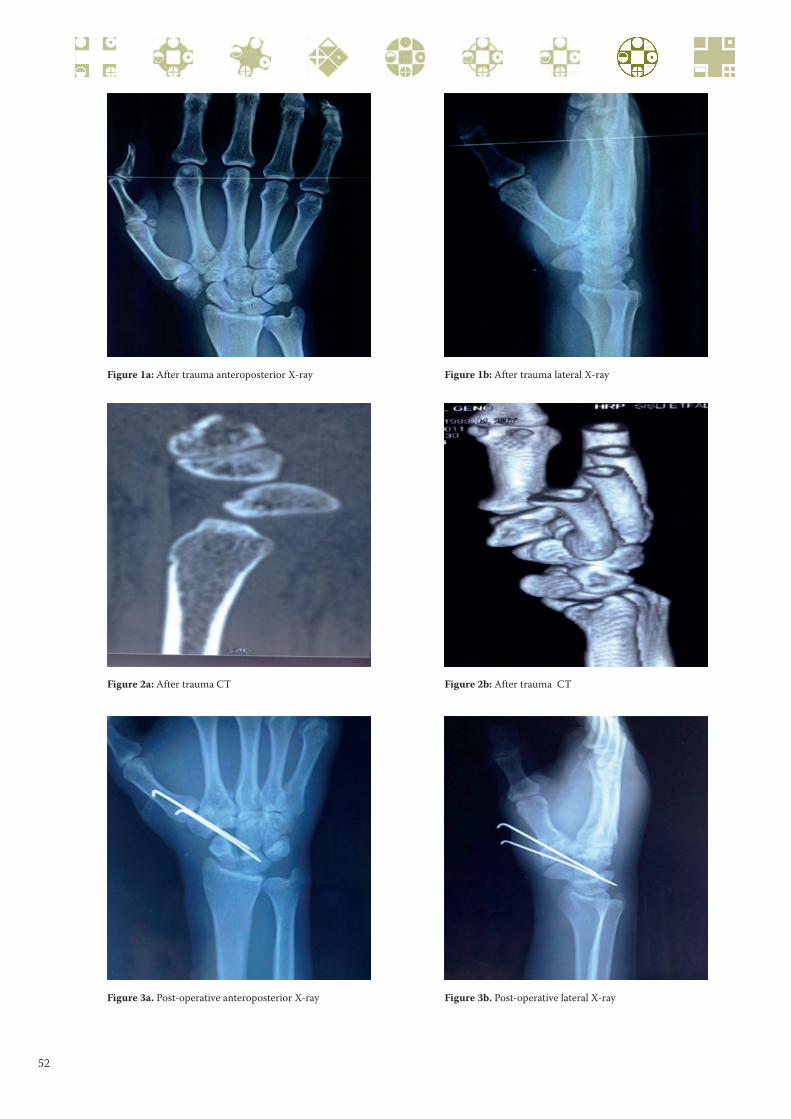
52
Figure 1b: After trauma lateral X-ray
Figure 2b: After trauma CT
Figure 3b. Post-operative lateral X-ray
Figure 1a: After trauma anteroposterior X-ray
Figure 2a: After trauma CT
Figure 3a. Post-operative anteroposterior X-ray

53
Figure 4b: Lateral view at 36 months
Figure 5b: Extension at 36 ay
Figure 4a : Antero-posterior view at 36 months
Figure 5a: Flexion at 36 months
pezium were observed. The intercarpal ligaments between
the trapezium and surrounding bones were ruptured.
Open reduction was performed through the incision, and
the bone was fixed with two Kirschner wires. The opera-
tion was completed after the capsule and ligament were
repaired (Figure 3a, 3b). The wrist was immobilized in a
splint including the proximal phalanx of the thumb. After
four weeks, before the free mobilization began, the Kirsch-
ner wires were removed. The patient returned to work af-
ter 45 days and was satisfied with the result.
At 36 months follow-up, the patient had a painless range
of motion. The active and passive motions of his wrist were
the same compared with the uninjured side, with normal
appositional and oppositional pinch strength. There was
no avascular necrosis and no radiological signs of arthrosis
(Figure 4a, 4b, 5a, 5b).
DISCUSSION
Dislocation of carpal bones is uncommon and generally
occurs as a result of a high-energy injury. The mechanism of
injury usually involves either direct dorsoradial impaction
or indirect axial loading. Indirect trauma that is transmit-
ted by the thumb may produce an incomplete dislocation
of the trapezium. Isolated dislocations and fractures of the
carpal bones are rare and are usually associated with other
hand or wrist injuries (Bennett’s fracture, Rolando’s fracture,
fracture of the scaphoid, hook of hamate, distal radius and
carpometacarpal dislocation) (6, 7). The clinical presenta-
tion can be quite variable depending on the displacement
of the fracture and the involvement of the carpometacarpal
joint. Some patients only complain of minor pain at the base
of the thumb without any gross swelling or deformity. Es-

54
pecially in cases with associated dislocation, rupture of the
surrounding ligaments and the dorsal joint capsule may re-
sult in instability. Once appropriately stabilized, these cases
may require repair. Reconstruction of the inter-metacarpal
and capsular structures, such as the inter-metacarpal abduc-
tor pollicis longus augmentation described by Brunelli et al.
may be required, especially in isolated dislocations (3).
Trapezium injuries are likely to be missed on routine
radiographs. A CT should be performed if the patient with
localized pain has tenderness in the region even if the re-
sults of routine radiographs appear to be normal (4, 5).
Occult injuries could be identified by using a special view
such as a true anteroposterior radiograph (Robert’s view)
that is excellent for identifying the trapezium and the base
of the metacarpal.
Anatomical reduction is recommended because of the
importance of the trapeziometacarpal joint of the thumb
function. Surgical treatments have been proposed in the
literature. Peterson recommended excision of the trape-
zium following complete dislocation because of the like-
lihood of avascular necrosis (8). Brunelli reported recon-
struction of the intermetacarpal and capsular structures in
isolated dislocations (9).
In our case, isolated dislocation of trapezium was a
result of violent and direct trauma. Different techniques
have been proposed to achieve a stable fixation and the
treatment outcomes. In our case, open reduction, Kirsch-
ner wire fixation and intercarpal ligament repair through
a dorsal approach are recommended for satisfactory out-
comes in similar cases.
REFERENCES
1. L.D. McKie, L.G. Rocke, T.C. Taylor. Isolated disloca-
tion of the trapezium Archives of Emergency Medicine,
1988, 5, 38-40.
2. Peterson CL. Dislocation of the Multangulum Majus or
Trapezium (And its Treatment in Two Cases with Ex-
tirpation). Arch Chir Neerlandicum 1950, 2:369-376.
3. Brunelli G, Monini L, Brunelli F. Stabilisation of the
trapezio-metacarpal joint. J Hand Surgery Br 1989,
14:209-212.
4. Iston N, Pimpalnerkar AL, Arafa MA. Isolated fracture
of the trapezium: an easily missed injury. J hand Surg.
1997 28(7):485-488.
5. Horch R: A new method for treating isolated fractures
of theos trapezium. Arch Orthop Trauma Surg 1998,
117:180-182.
6. Barbier O, Nguyen L, Ollat D, Versier G. Fracture: dis-
location of the trapezium: a case report and review of
the literature. European Journal of Orthopaedic Sur-
gery & Traumatology 2012, 22(4):333-336.
7. Garavaglia G, Bianchi S, Santa DD, Fusetti C. Trans-
trapezium carpo-metacarpal dislocation of the thumb.
Arch Orthop Trauma Surg 2004, 124(1):67-68.
8. Peterson CL. Dislocation of the multangulum majus or
trapezium and its treatment in 2 cases with extirpation.
Arch Chir Neerl 1950 (4):369-376.
9. Brunelli GA. Brunelli GR. A new surgical technique for
carpal instability with scapho-lunar dislocation. Ann
Chir Main Memb Super 1995, 14(4-5):207-213.

55
CASE REPORT PRIKAZ SLUČAJA CASE REPORT PRIKAZ SLUČAJA CASE REPORT
SECONDARY HAEMOPHAGOCYTIC LYMPHOHISTIOCYTOSIS THE DIFFERENTIAL DIAGNOSIS DILEMMA
IN PAEDIATRICSRasa Medovic1, Biljana Vuletic1,2, Zorica Raskovic1,2, Aleksandra Simovic1,2, Jelena Nestorovic–Tanaskovic1,2, Zoran Igrutinovic1,2
1Pediatric Clinic, Clinical Center Kragujevac2Department of Pediatrics, Faculty of Medical Sciences, University of Kragujevac
SEKUNDARNA HEMOFAGOCITNA LIMFOHISTIOCITOZA DIFERENCIJALNODIJAGNOSTIČKA DILEMA
U PEDIJATRIJIRaša Medović1, Biljana Vuletić1,2, Zorica Rašković1,2, Aleksandra Simović1,2, Jelena Nestorović–Tanasković1,2, Zoran Igrutinović1,2
1Klinika za pedijatriju, Klinički centar Kragujevac2Katedra za pedijatriju, Fakultet medicinskih nauka, Univerzitet u Kragujevcu
Correspondence: Dr Raša Medović, Pediatric Clinic, Clinical Center Kragujevac, Zmaj Jovina 30, 34000 Kragujevac
Tel: 0692772927, 034/505174; E-mail: [email protected]
ABSTRACT
Secondary haemophagocytic lymphohistiocytosis (SHFLH)
is a rare, potentially fatal disorder, most commonly caused by
the Epstein–Barr virus. It is characterized by neoplastic pro-
liferation of cells that belong to the monocyte–macrophage
system and by varied clinical expression.
A girl aged 3 years and 7 months was hospitalized due to
continuing high febricity, yellow skin colouring, hepatosple-
nomegaly and cytopenia in a complete blood count (CBC).
Four weeks before hospitalization, she had a lacunar angina
and lymphadenopathy.
A low number of erythrocytes, leukocytes and thrombocytes
were noted in CBC, with anaemia and the presence of virocytes
in a peripheral blood smear. Biochemical blood analyses indi-
cated hyperbilirubinaemia, increased values of transaminases,
lactic dehydrogenase, ferritin, triglycerides, D-dimer, accelera-
tion of the activated partial thromboplastin time and decreased
values of fi brinogen, with increased values of C-reactive protein
and procalcitonin. Using an ultrasound examination of the ab-
domen, hepatosplenomegaly was perceived; using echocardio-
graphic examination, pericardium layering was noticed; and
using a roentgen graphic picture of the lungs, the presence of
pleural eff usion was detected. In a bone marrow biopsy, the per-
centage of blasts did not exceed 25%, and rare chemophagocytes
were noticed. Using serologic tests, positivity to Epstein-Barr vi-
rus in IgM class was demonstrated.
According to the criteria by Histiocyte Society, there were
suffi cient criteria to establish a diagnosis of SHFLH. With the
exception of symptomatic therapies, according to the proto-
col for SHFLH treatment, a double antibiotic therapy and IV
immunoglobulins were given, to which the patient responded
with a clinical and laboratory recovery. Th erefore, there was
no demand for a treatment protocol with cytostatics or bone
marrow transplantation.
To resolve a diff erential diagnosis dilemma in solving
cases of uncertain febrile neutropenia.
Key words: haemophagocytic lymphohistiocytosis, Ep-
stein–Barr virus, children
SAŽETAK
Sekundarna hemofagocitna limfohistiocitoza (SHFLH)
je redak, potencijalno fatalan poremećaj, pokrenut najčešće
Epstein–Barr virusom. Karakteriše se neoplastičnom prolife-
racijom ćelija monocitno–makrofagnog sistema i različitim
kliničkim ispoljavanjem.
Devojčica uzrasta 3 godine i 7 meseci hospitalizovana
zbog dugotrajne visoke febrilnosti, žute prebojenosti kože,
hepatosplenomegalije i citopenije u kompletnoj krvnoj slici
(KKS). Četiri nedelje pre hospitalizacije imala je lakunarnu
anginu i limfadenopatiju.
U KKS je uočen nizak broj eritrocita, leukocita i trombo-
cita, uz anemiju i prisustvo virocita u perifernom razmazu
krvi. Od biohemijskih analiza krvi detektovana je hiper-
bilirubinemija, povećane vrednosti transaminaza, laktične
dehidrogenaze, feritina, triglicerida, ubrzanje aktiviranog
parcijalnog tromboplastinskog vremena, D–dimera i sni-
žene vrednosti fi brinogena, uz povećane vrednosti parame-
tara infl amacije (C–reaktivnog proteina i prokalcitonina).
Ultrazvučnim pregledom abdomena uočena je hepatosple-
nomegalija, ehokardiografskim pregledom perikardno ra-
slojavanje, a rentgenografskim snimkom pluća postojanje
pleuralnog izliva. U bioptatu kostne srži procenat blasta
nije prelazio 25%, a uočeni su ređi hemofagociti. Serološ-
kim testovima dokazana je pozitivnost na Ebstein–Barr
virus u IgM klasi.
Prema kriterijumima udruženja Histiocyte Society,
postojalo je dovoljno kriterijuma za postavljanje dijagno-
ze SHFLH. Pored simptomatske terapije, prema protokolu
za lečenje SHFLH ordinirana je dvojna antibiotska tera-
pija i i.v. imunoglobulini, na koje je pacijentkinja odrea-
govala kliničkim i laboratorijskim oporavkom. Nije zah-
tevala lečenje protokolima citostatika ili transplantaciju
kostne srži.
Diferencijalno–dijagnostička dilema u slučaju rešavanja
nerazjašnjenih febrilnih neutropenija.
Ključne reči: hemofagocitna limfohistiocitoza, Ebstein–
Barr virus, deca
Received / Primljen: 1. 10. 2014. Accepted / Prihvaćen: 29. 12. 2014.
UDK: 616.15:616.988 / Ser J Exp Clin Res 2015; 16 (1): 55-63
DOI: 10.1515/SJECR20150009

56
The criteria for making a diagnosis of haemophago-
cytic lymphohistiocytosis are (presence of 5 to 8 criteria is
necessary) (1, 4, 5, 27–30):
1. Temperature >38°C for five days, resistant to the appli-
cation of antipyretics
2. Splenomegaly according to echosonographic criteria,
depending on age and sex (31)
3. Cytopenia ≥ 2 cell lines
• Haemoglobin (Hb) level < 90 g/l (newborns < 4 weeks,
Hb<100 g/l)
• Number of neutrophils (Neu) < 1 x10⁹/l
• Number of thrombocytes (Tr) < 100 x109/l
4. Hypertriglyceridaemia and/or hypofibrinogenaemia
• Triglycerides≥ 3 mmol/l
• Fibrinogen < 1,5 g/l
5. Hyperferritinaemia – Ferritin ≥ 500 μg/l (32, 33)
6. Serum concentration CD25 (receptor for IL–2) ≥ 2400
U/ml (32, 34, 35)
7. Decreased or absent activity of NK cells (35)
8. Haemophagocytosis seen through a microscope in a pe-
ripheral blood smear, bone marrow aspiration, cytologi-
cal examination of liquor or biopsy of lymph node (36)
Disease prognosis is uncertain, and the treatment must
be started as soon as possible, as the goal is to prevent in-
creased inflammatory response (1, 4, 27, 30, 37).
CASE REPORT
A girl aged 3 years and 7 months was directed to the
Pediatric Clinic, Clinical Centre Kragujevac because she
had an increased temperature of 40ºC, which lasted for
two days before admission and rarely responded to the
application of antipyretics (Paracetamol). Her symptoms
were fatigue, loss of appetite, sickness and vomiting of un-
digested food content 2–3 times per day, as well as watery
stools (2–3 times a day), that were light and had visible mu-
cus. Her parents provided information that her urine was
darker than normal and had a brown colour. Basic labo-
INTRODUCTION
Histiocytoses are disorders that are characterized by
unknown pathophysiological mechanisms of emergence
and by neoplastic proliferation and accumulation of cells
from the monocyte–macrophage system (1, 2). There are
two groups of immunological cells: histiocytes, which are
dendritic cells with primary antigen–presenting cell func-
tion, and macrophages, with primary phagocytic function
(1). Classification of these diseases is difficult, and current-
ly, the most reliable method is histopathological classifica-
tion according to the international Histiocyte Society. The
classification is accepted by the World Health Organiza-
tion (1, 3). (Table 1)
Haemophagocytic lymphohistiocytosis belongs to the
second class of histiocytoses and is divided into primary
(familial or hereditary) and secondary form. There are no
clinical or laboratory differences between these two forms
of the disease. They are differentiated according to genetic
examinations (4, 5).
The familial type of the disease is often comorbid with
some immunodeficient conditions, such as Chédiak–Hi-
gashi syndrome, Griscelli syndrome, and X–linked lym-
phoproliferative syndrome. It is autosomal recessive and
found in 1 in every 30 to 50 thousand newborns. Genes
that encode perforin are established, and they are consid-
ered to be responsible for the expression of the primary
form. These genes include PRF1, UNC13D, STX11, STX-
BP2 and RAB27A, which are located on the second arm of
chromosome 9 and 10 (4–11).
Emergence of secondary haemophagocytic lympho-
histiocytosis is primarily attributed to viruses. In approxi-
mately 70% of cases it is caused by the Epstein–Barr virus
and less commonly Cytomegalovirus, Humane Herpesvi-
rus, HIV, Parvovirus, viruses of Hepatitis A, B or C, and
extremely rarely bacteria (Salmonella, Staphylococcus,
Mycobacterium tuberculosis, Brucella, Leptospirosis,
Rickettsia prowazekii), fungi (Candida albicans) or para-
sites (Leishmania). The causes of the disease can be malign,
rheumatologic and autoimmune diseases (4, 5, 12–26).
Table 1. Classifi cation of histiocytosis
Class Syndrome Cell type Diagnostic characteristics of cells
I
Histiocytosis of Langerhans cells
(eosinophilic granuloma, Hand Schuller Christian
disease, Letterer–Siwe disease)
Langerhans
(dendritic) cells
Birbeck granules under electronic
microscope, CD1 positive cells
II
Haemophagocytic lymphohistiocytosis
primary (familial)- secondary-
Mononuclear phagocytesNegative Birbeck granules and CD1,
positive nonspecifi c esterase
III
Malign diseases of histiocytes
Acute monocytic leukaemia (FAB M5)
Real histiocytic lymphoma
Malign cells of
monocytic–macrophagic
composition
Malign morphologic characteristics
of cells, with cell features from class II
IV
Other histiocytic syndromes (benign)
Sinus histiocytosis with massive lymphadenopathy
(Rosai–Dorfman disease)
Juvenile Xanthogranuloma
Reticulohistiocytoma
Mononuclear phagocytesNegative Birbeck granules and CD1,
positive nonspecifi c esterase

57
μmol/l), values of transaminases [aspartate aminotrans-
ferase (AST) and alanine aminotransferase (ALT)] and
values of lactic dehydrogenase (LDH) were increased dur-
ing the hospitalization (AST 123...141 U/l, ALT 296...168
U/l, LDH 1364...1270 U/l). Additionally, we found the ac-
celeration of activated partial thromboplastin time (aPTT
32,4...34,7 s). Fibrinogen levels were decreased, whereas
there were high levels of D-dimer (fibrinogen 2,017...1,673
g/l, D–dimer 3884,49 ng/ml). Ferritin values were consid-
erably increased, 2409 g/l. Cholesterol values, including
HDL and LDL fractions, were within the limits of normal
values. However, hypertriglyceridaemia was determined
(4,08 mmol/l). Additionally, hypoalbuminaemia was deter-
mined (albumins 22 g/l). Values of glycaemia, urea, creati-
nine and uric acid were in the referential scope.
Increased values of the following inflammation param-
eters were detected that persisted in the following days:
sedimentation of erythrocytes (SE) 15 mm/lh, C–reactive
protein (CRP) 50,2...22,2 mg/l and procalcitonin (PCT)
2,910...1,780 ng/ml [which demanded detailed bacterio-
logical swabs of pharynx and nose, antistreptolysin titre
(ASOT), two blood cultures and urinoculture] and viro-
logical examinations (Cytomegalovirus, Epstein–Barr vi-
rus, HIV, Toxoplasma gondii, Hepatitis A, B and C, and
Micoplasma pneumoniae).
Considering the diagnosis of febrile neutropenia, it was
necessary to include a double empirical antibiotic therapy.
Therefore, cephalosporin of the third generation was ap-
plied – Ceftazidime, with Aminoglicozide and Amikacin
(38–40).
Values of immunoglobulin fractions (IgA, IgM, IgG), as
well as of the component complements (C3 and C4) were
within the limits of referential values for that age.
Using an ultrasound examination of the abdomen, the
presence of hepatomegaly (anteroposterior diameter of
right lobe 116 mm) and splenomegaly (cranio–caudal di-
ameter 106 mm) were determined, according to the table
of normal dimensions for the patient’s age and sex (31).
Bone marrow aspiration was performed, and using the
microscopic examination of aspirate, cellularity of III–IV
degree was determined along with evident productive
megakaryocytes and all forms in the development of red
and white blood cells. The percentage of blast did not ex-
ceed 25%. After 3 days of hospitalization, bone marrow
aspiration was repeated and a detailed microscopic exami-
nation showed rare haemophagoctes in several places (Pic-
tures 1A. and 1B.). Both peripheral blood and bone mar-
row aspirate samples were collected for flow cytometry,
which was not performed due to technical reasons.
Although the therapy was applied, during the first 3
days, the girl was still highly febrile (39,5°C), inactive, had
a loss of appetite, and her skin, visible mucus, urine and
stool were slightly discoloured. Furthermore, she suffered
from tachydyspnea, with a number of respirations reach-
ing 36/min. She developed a dry and sensitive cough, in
bases with weak, quiet breathing sound during ausculta-
tions more to the left side, and low oxygen saturations,
ratory analyses were performed. An examination of urine
sediment, conducted using a qualitative method, albumins,
urobilinogens and bilirubins were detected. Moreover, in a
complete blood count, anaemia (Hb 108 g/l), leucopenia
(Le 1,45 x10⁹/l), and thrombocytopenia (Tr 56 x109/l) were
noticed. Apart from antipyretics whilst she was highly fe-
brile, she did not use any medications in the 3 weeks before
admission.
Personal anamnesis of the child was without any speci-
fications. Four weeks before the appearance of symptoms,
the child was treated with penicillin for a period of 7 days
due to the lacunar angina and the swelling of lymphatic
nodes on her neck. In the family anamnesis, apart from
the data regarding members of her family suffering from
hypertension and diabetes mellitus type II, the presence of
other heredodegenerative diseases was negative.
During the physical examination, it was determined
that the girl, body weight 16 kg, body height 106 cm and
body mass index 14,29 kg/m2, was conscious, highly febrile
(38,8ºC), and considerably adynamic, with a subicteric tint
of her skin, which showed a decrease in turgor and elas-
ticity. Additionally, she had icteric sclera, coated tongue,
slightly hyperemic pharynx, and the lymph nodes of her
neck had normal dimensions for her age. She had no signs
of haemorrhagic syndrome. Auscultatory findings on the
heart and lungs were normal, SaO2 96%, number of respi-
rations was 24/min, TA was 95/55 mmHg, and pulse was
132 beats/min. Her abdomen was not painfully sensitive
during palpation. Her liver was palpated 4 cm below the rib
arch and her spleen on the rib arch. Neurological findings,
besides mild somnolence, were normal for her age.
Due to the serious clinical outlook and laboratory pa-
rameters, especially the decreased number of neutrophils,
the girl was hospitalized in the intensive care unit in the
isolation department because she was thought to have a
septic condition. There, her vital functions were regularly
monitored, intravenous rehydration was started, anti-
pyretic therapy was applied every 6 hours, and to correct
the intestinal flora, an antidiarrhoeal diet was introduced
with a probiotic preparation.
Upon admission, initial laboratory examinations were
performed. She had normal electrolyte and acid–base sta-
tus. According to the complete blood count that was re-
peated daily during hospitalization, among pathological
values, anaemia was determined (Hb 102...98...94 g/l), with
a reduced number of erythrocytes (Er 3,72...3,67...3,52
x1012/l), perceived hypochromia Er in the peripheral blood
smear, normal percentage of reticulocytes (Rtc 0,9%) and
with negative direct and indirect Coombs tests. Distinct
leucopenia was persistent (Le 1,4...1,4...1,9 x109/l), with
predominance of lymphocytes in the leukocyte formula (Ly
1,1...1,2...1,4 x109/l, with 10–20% of atypical lymphocytes
in the peripheral blood smear), neutropenia to agranu-
locytosis (neutrophils 0,25...0,18...0,34 x109/l), as well as
thrombocytopenia (Tr 44...35...46 x109/l).
Among biochemical analyses of the blood, values of total
(TBill) and conjugated (DBill) bilirubin (72 μmol/l and 44,2

58
perechogenicity of the pericardium were determined, with
minimal layering of the visceral and parietal leaf of 3 mm.
After placement onto the surface, the results of sterile
blood cultures and urinoculture were ready. The presence
of normal flora in the nose and pharynx swab was deter-
mined, and ASOT were in the limits of referential values.
However, virological analyses proved seronegativity to all
considered viruses, except for a highly positive titre to Ep-
stein–Barr virus in the IgM class and a less positive one
in the IgG class. Therefore, it was confirmed that infec-
tion 4 weeks before hospitalization, which was regarded
as lacunar angina and treated with beta lactam antibiot-
ics, was actually infective mononucleosis. These findings
led to a suspicion that the girl suffers from secondary hae-
mophagocytic lymphohistiocytosis (SHFLH), which was
caused by the Epstein–Barr virus.
According to the treatment protocol for SHFLH (43),
IV immunoglobulins were introduced in the therapy.
The girl was directed to the referential institution, Insti-
tute of Mother and Child Care “Dr Vukan Čupić” in Novi
Beograd, due to additional medical diagnoses and possible
treatment with cytostatic protocol and/or a possible need
for bone marrow transplantation.
SaO2 91–92%. Therfore, roentgenography of the lungs
(RTG) was conducted. Paramediastinally zones of con-
solidations of lung parenchyma with a tendency to merge
were noticed on images. A shadow of effusion in the left
costophrenic angle up to the level of the fifth rib was also
noticed. A repeated recording of the following day showed
that the condition was progressing (Pictures 2A and 2B).
In light of this, considering the possibility of the emer-
gence of more serious nosocomial infections, current an-
tibiotic therapy was changed, and secondary antibiotics
were given according to the protocol– Meropenem and
Vancomycin, with antimycotic Fluconazole, and consid-
ering an atypical cause of pneumonia, Azithromycin was
also applied (38–42).
Moreover, due to the quiet heart tones during auscul-
tation, electrocardiographic examination (the finding was
normal for her age), and laboratory observation of heart
enzymes [troponin I, creatine kinase (CK) and CK MB,
and N-terminal beta natriuretic peptide (NTproBNP) were
all within the limits of referential values) were performed.
However, at the echocardiographic examination, besides
normal heart morphology and contractility, and an ejec-
tion fraction of 66%, light tricuspid regurgitation and hy-
Pictures 1A and 1B. Haemophagocytosis in the preparation of bone marrow aspirate
Picture 2A. RTG lung 04.09.2013. Picture 2B. RTG lung 05.09.2013.

59
the application of therapy. Treatment is identical for both
forms, which are potentially fatal diseases, and poor prog-
nosis and higher mortality rate are typical of the primary
form (1, 29, 30, 48–51).
The Epstein–Barr virus is thought to be responsible for
the emergence of secondary haemophagocytic lymphohis-
tiocytosis in approximately 70% of cases. The mechanism
of disorder in the proliferation of cytotoxic and helper T
lymphocytes is unknown. However, a similar monoclonal
proliferation of T lymphocytes was determined in those
patients suffering from secondary haemophagocytic lym-
phohistiocytosis and in EBV+ T cell lymphoma (52). The
consequence is a hyper production of proinflammatory
cytokines, especially interferon γ, cell factors, tumour ne-
crosis factor α and of various interleukins (IL – 1, 6, 16, 18),
decreased inactivation of NK cell functions and to hyper
activation and accumulation of macrophages (12, 29, 30,
38, 48–50, 53).
Clinical signs that may indicate the presence of hae-
mophagocytic lymphohistiocytosis are nonspecific. Com-
monly, high temperature >38ºC and fever (in 80% of pa-
tients), with a considerably bad general state (fatigue,
sleepiness, refusal to eat) are evident. The highly febrile
state lasts for several days and is resistant to antipyretics,
which was the case with our patient. Sickness, vomiting,
and watery stools before hospitalization can be attributed
to the general bad condition of the child, to the increase in
intra–abdominal pressure due to hepatosplenomegaly and
to the influence of mediator released from the monocyte-
macrophage system to the cell function of gastrointestinal
tract. The girl did not have a generalized lymphadenopa-
thy, that is, besides splenomegaly (in 60%) and hepatomeg-
aly (50%), described in 40% of cases as one of the signs of
the diseases (1, 12, 27–30, 37, 50, 51, 54, 55). A common
sign that is mentioned is rash, with visible marks on the
skin (35–65%), as well as neurological symptomatology in
the sense of somnolence, meningismus, ataxia, and even
convulsions and epileptic attacks (30%) (56, 57).
Yellow colouring of the skin, a visible mucous mem-
brane, lighter stool and dark coloured urine, and the pres-
ence of urobilinogens and bilirubins in the urine indicated
hyperbilirubinaemia, which was confirmed in laboratory.
The origin of hyperbilirubinaemia and comorbid hepatos-
plenomegaly was examined. Firstly, a possibility of the ex-
istence of a stronger haemolysis of erythrocytes in the en-
larged spleen was eliminated by determining the number of
Er in CBC, the percentage of reticules in peripheral blood,
as well as, direct and indirect Coombs tests. Increased val-
ues of transaminases and lactic dehydrogenase indirectly
indicated a necrosis of hepatocytes, whilst acceleration of
the activated partial thromboplastin time and decreased
values of fibrinogens with high values of D–dimer pointed
to the decreased synthetic liver function.
In addition to anaemia and thrombocytopenia, in CBC,
leucopenia persisted, with a predominance of lymphocytes
in leukocyte count, with 10–15% of atypical lymphocytes
(so called virocytes), which indicated that it is a viral in-
During hospitalization at the Institute, there was a
spontaneous, steady, both clinical and laboratory recovery
of the child, with the application of only symptomatic ther-
apy (the continuation of antibiotic therapy started in Clini-
cal Center Kragujevac and IV rehydration). Therefore, IV
immunoglobulin therapy, to which the girl adequately re-
sponded, was most likely sufficient. She became afebrile, in
better general state, regained her appetite, and there was
no more colouring of the skin and mucus. The number of
cell lines in CBC was normalized, as well as the biochemi-
cal analyses of the blood. With such a condition of the
child, bone marrow aspiration was not repeated. On the
roentgen graphic picture of lungs it was determined that
the finding is in considerable regression with no signs of
pulmonary parenchymal consolidation, but with the per-
sistence of a small shadow of pleural effusion in the right
costophrenic angle. In the ECHO examination of the heart,
the presence of an increased amount of pericardium fluid
was not determined. Moreover, in the ECHO examination
of abdomen, persistence of splenomegaly was determined,
with considerably decreased values (cranial–caudal di-
ameter of 96 mm) and no hepatomegaly (anteroposterior
diameter of the right lobe 103 mm). A parameter that is
also in favour of a SHFLH diagnosis is soluble anti CD25,
whose values were increased over 2400 U/ml.
Due to technical reasons, the activity of NK cells were
not determined, and genetic examinations of the primary
form of the disease were not undertaken because these
analyses are not performed in the Republic of Serbia.
Since then, the girl has been in a good general state with
regular physical findings and normal laboratory analyses.
She is submitted for regular examinations by the haema-
tologist in charge.
DISCUSSION
Haemophagocytic lymphohistiocytosis was first de-
scribed in 1939 as histiocytic medullary reticulosis (44). In
1952, a familial type of the disease in two newborn twins
was described (45), and 1965 after the simultaneous de-
velopment of the disease in father and son, it was thought
that infection can be important in the pathogenesis of the
disease (46). The hereditary form usually emerges until 4
years of age, and according to some authors even until the
second year of age. In the cases of anamnestically known
and/or proven infection 3–4 weeks before the beginning of
the disease, the secondary form should be considered. In
the cases where there are relatives with the same disease,
the familial form should be considered. However, it is often
the case that the hereditary form is initiated by some viral
influences (47). The primary form is found together with
some immunodeficient conditions, whilst it is typical for
the secondary form to emerge in children with a normal
immunological status, which was the case with our female
patient. The secondary form may develop into a spontane-
ous remission, whilst the primary form always necessitates
60
eter of the disease). Hypertriglyceridaemia and hypoalbu-
minaemia cannot be considered as the cause of pleural and
pericardium effusion, with the emergence of chylothorax
in the first, or the decrease in colloid–osmotic pressure of
plasma in the second case, but the effusion is most surely a
consequence of aseptic serositis, i.e., influence of mediator
to epithelial cells of pleura and pericardium and their in-
creased secretion (62–64). Moreover, bone marrow aspira-
tion was performed once more, and a detailed and targeted
microscopic search of biopsies revealed rare haemophago-
cytes in several places.
The clinical and laboratory criteria are summarized
in table 2. According to international Histiocyte Society,
these criteria as well as the auxiliary diagnostic criteria that
we also followed in solving differential diagnosis dilemmas
are used to diagnose SHFLH (1, 4, 5, 27–30).
Prognosis of the disease is uncertain, and it depends
on the age and immunological status of the patient, degree
of organs affected, dissemination of the disease, speed of
establishing a diagnosis and the response to therapy. The
disease can end with a spontaneous recovery, adequate cu-
rative treatment, or it could also result in rapid and pro-
gressive death, regardless of the applied therapy. Sponta-
neous recovery is expected in 20–30% of the patients. With
an adequate symptomatic and immunosuppressive treat-
ment, recovery is expected in 60–70% of the patients. That
percentage is higher after bone marrow transplantation.
The mortality rate, depending on the studies, is approxi-
mately 25–60% of patients (1, 4, 12, 13, 50, 51, 65–68).
Apart from the symptomatic therapy (IV rehydration
with the correction of electrolytic and acid–base status,
antipyretics, oxygen therapy, adequate care and diet, trans-
fusion of deplasmatisized erythrocytes and concentrated
thrombocytes, antibiotics and antimycotics), immunosup-
pressive and immunomodulatory therapy are performed
(corticosteroids, immunoglobulins, antithymocytic globu-
lin, cyclosporine A) as well as various protocols with cyto-
statics (initial cytostatic therapy – Etoposide, intrathecal
methotrexate, with corticosteroids, and continuous cyto-
static therapy – same medications in smaller dosages, ac-
cording to the treatment protocol suggested by Histiocyte
society). As a final measure, allogenic transplantation of
bone marrow is suggested (27, 30, 37–43, 68–71). Some
fection, as well as neutropenia to agranulocytosis. Anam-
nestic data on non–usage of drugs or on the exposition to
toxic materials in the last 3 weeks excluded a possibility of
bone marrow aplasia caused by these substances (58).
To exclude the possibility of the emerging malignant
haemopathies, in a peripheral blood smear, as well in a mi-
croscopic examination of bone marrow biopsy, over 25%
developing forms of such cells were not found (59). More-
over, as tumour markers, values of ferritin were increased.
However, besides the role of a tumour marker, ferritin was,
in this case, considered as an index of inflammation and
liver function (60). Additionally, normal levels of uric acid,
which is a marker of increased cell degradation of tumour
cells (59), were detected. Using an ultrasound examination
of the abdomen, the existence of retroperitoneum enlarged
lymph cords was not determined. Unfortunately, flow cy-
tometry was not performed due to technical reasons.
Because the girl was regularly vaccinated and had a
BCG mark of 3 mm, normal immunological status, was
from a good socioeconomic environment, with no evi-
dence of living in a collective group or a possible contact
with tuberculosis, the possibility of tuberculosis pleural
effusions was not considered. Before and during hospital-
ization, there was no neurological symptomatology in the
female patient apart from light sleepiness that is a conse-
quence of a complete bad condition and high temperature.
Therefore, there was no need for diagnostic lumbar punc-
ture, which is also in the diagnostic protocol for SHFLH
(30, 37, 50, 51, 56, 57).
The results of sterile blood cultures and urinoculture,
which revealed normal flora from the swab of the nose
and pharynx and ASOT that was in the limits of referen-
tial values, excluded a possibility of a septic state caused
by bacterial infection. With a confirmed seronegativity to
all considered viruses, except for highly positive titre to
Epstein–Barr virus in the IgM class, it was thought that
the girl had a case of haemophagocytic lymphohistiocyto-
sis (SHFLH).
To establish a final diagnosis, values of triglycerides
that were increased were considered, as well as the values
of albumins (according to Huang et al (61), decreased val-
ues of albumin will be another criteria for establishing a
diagnosis for SHFLH, and it is a good predicative param-
Table 2. Clinical and laboratory criteria for establishing the diagnosis
Criteria for establishing the diagnosis (7 out of 8) Auxiliary criteria
Temperature above 38,5°C for 9 days1.
Splenomegaly (106 mm)2.
Cytopenia ≥ 2 cell lines3.
Haemoglobin 98 g/l
Neutrophils 0,25 x 10⁹/l
Th rombocytes 45 x 109/l
Hypertriglyceridaemia and/or hypofi brinogenaemia4.
Triglycerides 4,08 mmol/l
Ferritin 2409 μg/l5.
Haemophagocytosis in bone marrow6.
Soluble anti CD25 (receptor for IL–2) ≥ 2400 U/ml7.
IgM EBV positive1.
Pleuritis, pericarditis2.
Decreased values of fi brinogen3.
Increased values of transaminases4.
Increased values of bilirubin5.
Accelerated aPTT6.
Increased values of D–dimer7.
Increased values of LDH8.
Hypoalbuminaemia9.
Hepatomegaly10.

61
11. Kogawa K, Lee SM, Villanueva J, Marmer D, Sumegi
J, Filipovich AH. Perforin expression in cytotoxic lym-
phocytes from patients with hemophagocytic lympho-
histiocytosis and their family members. Blood. 2002;
99(1): 61–6
12. Ansuini V, Rigante D, Esposito S. Debate around infec-
tion–dependent hemophagocytic syndrome in pediat-
rics. BMC Infect Dis. 2013; (13): 13–15.
13. Fisman DN. Hemophagocytic syndromes and infec-
tion. Emerg Infect Dis. 2000; 6(6): 601–8.
14. Janka G, Imashuku S, Elinder G, Schneider M, Henter
JI. Infection– and malignancy–associated hemophago-
cytic syndromes. Secondary hemophagocytic lympho-
histiocytosis. Hematol Oncol Clin North Am. 1998;
12(2): 435–44.
15. Danish EH, Dahms BB, Kumar ML. Cytomegalovirus–
associated hemophagocytic syndrome. Pediatrics.
1985; 75(2): 280-3.
16. Bhatia S, Bauer F, Bilgrami SA. Candidiasis–associated
hemophagocytic lymphohistiocytosis in a patient in-
fected with human immunodeficiency virus. Clin In-
fect Dis. 2003; 37(11): 161–6.
17. Watanabe T, Okazaki E, Shibuya H. Influenza A virus–
associated encephalopathy with haemophagocytic syn-
drome. Eur J Pediatr. 2003; 162(11): 799–800.
18. Kaya Z, Oztürk G, Gürsel T, Bozdayi G. Spontaneous
resolution of hemophagocytic syndrome and dissemi-
nated intravascular coagulation associated with Parvo-
virus B19 infection in a previously healthy child. Jpn J
Infect Dis. 2005; 58(3): 149–51.
19. Tuon FF, Gomes VS et al. Hemophagocytic syn-
drome associated with hepatitis A: case report and
literature review. Rev Inst Med Trop Sao Paulo. 2008;
50(2): 123–7.
20. Brastianos PK, Swanson JW, Torbenson M, Sperati J,
Karakousis PC. Tuberculosis–associated haemophago-
cytic syndrome. Lancet Infect Dis. 2006; 6(7): 447–54.
21. Cascio A, Giordano S, Dones P, Venezia S, Iaria C, Zii-
no O. Haemophagocytic syndrome and rickettsial dis-
eases. J Med Microbiol. 2011; 60(Pt 4): 537–42.
22. Gosh JB, Roy M, Bala A. Infection associated with He-
mophagocytic Lymphohistiocytosis triggered by noso-
comial infection. Oman Med J. 2009; 24(3): 223–5.
23. Cascio A, Pernice LM et al. Secondary hemophago-
cytic lymphohistiocytosis in zoonoses. A systematic
review. Eur Rev Med Pharmacol Sci. 2012; 16(10):
1324–37.
24. Gagnaire MH, Galambrun C, Stéphan JL. Hemophago-
cytic syndrome: a misleading complication of visceral
leishmaniasis in children – a series of 12 cases. Pediat-
rics. 2000; 106(4): E58.
25. Onishi R, Namiuchi S. Hemophagocytic syndrome in
a patient with rheumatoid arthritis. Intern Med. 1994;
33(10): 607–11.
26. Wong KF, Hui PK, Chan JK, Chan YW, Ha SY. The
acute lupus hemophagocytic syndrome. Ann Intern
Med. 1991; 114(5): 387–90.
studies have shown efficiency in the application of biologic
therapy (72). Antiviral medications (ege.g., Acyclovir) do
not have significance in the treatment (12, 13, 27, 30).
In our example, besides the applied symptomatic and
antibacterial therapy, a therapy with IV immunoglobulins
was sufficient, and it is described in the works of other au-
thors (43, 66–68).
Based on the facts reported herein, we can conclude
that although hemophagocytic lymphohistiocytosis is a
rare disease, one should be aware of it, especially in clini-
cally uncertain conditions, such as in cases of febrile cy-
topenia, when the patients do not respond to the applica-
tion of standard symptomatic therapy. Timely diagnosis
offers higher chances for survival, with a timely beginning
of immunosuppressive, immunomodulatory therapy and
possible bone marrow transplantation.
REFERENCES
1. Lipton JM, Arceci RJ. Histiocytic disorders. In: Hoff-
man R, Edward B, eds. Hematology: basic principles
and practice. 4th ed. Philadelphia: Churchill Living-
stone. 2005; 857–67.
2. Ladisch S. Histiocytosis syndrome in childhood. In:
Behrman ER, Kliegman MR, Jenson BH. NELSON
Textbook of pediatrics, vol 2, 17th ed. Belgrade: Saun-
ders, Bard 2009; 1727–30. (translated)
3. Favara BE, Feller AC, Pauli M et al. Contemporary clas-
sification of histiocytic disorders. The WHO Commit-
tee On Histiocytic/Reticulum Cell Proliferations. Re-
classification Working Group of the Histiocyte Society.
Med Pediatr Oncol. 1997; 29(3): 157–66.
4. Verbsky JW, Grossman WJ. Hemophagocytic lympho-
histiocytosis: diagnosis, pathophysiology, treatment,
and future perspectives. Ann Med. 2006; 38(1): 20–31.
5. Janka GE. Familial and acquired hemophagocytic lym-
phohistiocytosis. Annu Rev Med. 2012; (63): 233–46.
6. Feldmann J, Le Deist F, Ouachée–Chardin M et al.
Functional consequences of perforin gene mutations in
22 patients with familial haemophagocytic lymphohis-
tiocytosis. Br J Haematol. 2002; 117(4): 965–72.
7. Feldmann J, Callebaut I, Raposo G et al. Munc 13–4 is
essential for cytolytic granules fusion and is mutated in
a form of familial hemophagocytic lymphohistiocytosis
(FHL3). Cell. 2003; 115(4): 461–73.
8. Ohadi M, Lalloz MR, Sham P et al. Localization of a
gene for familial hemophagocytic lymphohistiocytosis
at chromosome 9q21.3–22 by homozygosity mapping.
Am J Hum Genet. 1999; 64(1): 165–71.
9. Dufourcq–Lagelouse R, Jabado N, Le Deist F et al. Link-
age of familial hemophagocytic lymphohistiocytosis to
10q21–22 and evidence for heterogeneity. Am J Hum
Genet. 1999 Jan; 64(1): 172–9.
10. Stepp SE, Dufourcq–Lagelouse R, Le Deist F et al. Per-
forin gene defects in familial hemophagocytic lympho-
histiocytosis. Science. 1999; 286(5446): 1957–9.

62
27. Henter JI, Horhe A, Arico M et al. HLH–2004: Diagnostic
an therapeutic guidelines for hemophagocytic lympho-
histiocytosis. Pediatr Blood Pediatr Blood Cancer. 2007;
48(2): 124–31.
28. Henter JI, Elinder G, Ost A. Diagnostic guidelines for
hemophagocytic lymphohistiocytosis. The FHL Study
Group of the Histiocyte Society. Semin Oncol. 1991;
18(1): 29–33.
29. Rosado FG, Kim AS. Hemophagocytic lymphohistiocy-
tosis: an update on diagnosis and pathogenesis. Am J
Clin Pathol. 2013; 139(6):713–27.
30. Tang YM, Xu XJ. Advances in hemophagocytic lym-
phohistiocytosis: pathogenesis, early diagnosis/differ-
ential diagnosis, and treatment. Scientific World Jour-
nal. 2011; 11: 697–708.
31. Konuş OL, Ozdemir A, Akkaya A, Erbaş G, Celik H,
Işik S. Normal liver, spleen and kidney dimensions in
neonates, infants, and children: evaluation with sonog-
raphy. AJR Am J Roentgenol. 1998; 171(6): 1693–8.
32. Rademacher C, Hartmann D, Spiethoff A, Jakobs R.
Ferritin and soluble interleukin–2–receptor in the diag-
nosis of fever of unknown origin. Dtsch Med Wochen-
schr. 2014; 139(1-2): 23–7.
33. Lehmberg K, McClain KL, Janka GE, Allen CE. Deter-
mination of an appropriate cut-off value for ferritin in
the diagnosis of hemophagocytic lymphohistiocytosis.
Pediatr Blood Cancer. 2014 Apr 21. [Epub ahead of
print] PubMed PMID: 24753034.
34. Imashuku S, Hibi S et al. Soluble interleukin-2 receptor:
a useful prognostic factor for patients with hemophago-
cytic lymphohistiocytosis. Blood. 1995; 86(12): 4706–7.
35. Wang LL, Hu YX, Chen WF et al. Significance of soluble
interleukin-2 receptor and NK cell activity in patients
with hemophagocytic lymphohistiocytosis. Zhongguo
Shi Yan Xue Ye XueZaZhi. 2012; 20(2): 401–4.
36. Sloma I, Vincent H, Addebbous A, Rivoisy C, Turhan
GA, Michot JM. Haemophagocytic histiocyte in a pe-
ripheral blood film. Br J Haematol. 2014; 165(2): 163.
37. Jabado N, McCusker C, BasileGde S. Pediatric hemophago-
cytic syndromes: a diagnostic and therapeutic challenge.
Allergy Asthma Clin Immunol. 2005; 1(4): 142–60.
38. Kobayashi S, Ito M, Sano H et al. Clinical analysis of
combination therapy for febrile neutropenic patients in
childhood cancer. Pediatr Int. 2013; 55(1): 65–71.
39. Raghavendra M, Hoeg RT, Bottner WA, Agger WA.
Management of neutropenic fever during a transition
from traditional hematology/oncology service to hos-
pitalist care. WMJ. 2014; 113(2): 53–8.
40. Hung KC, Chiu HH, Tseng YC et al. Monotherapy with
meropenem versus combination therapy with ceftazi-
dime plus amikacin as empirical therapy for neutro-
penic fever in children with malignancy. J Microbiol
Immunol Infect. 2003; 36(4): 254–9.
41. Biondi E, McCulloh R, Alverson B, Klein A, Dixon A,
Ralston S. Treatment of Mycoplasma Pneumonia: A
Systematic Review. Pediatrics. 2014; 133(6):1081–90.
42. Aytaç S, Yildirim I, Ceyhan M et al. Risks and outcome
of fungal infection in neutropenic children with hema-
tologic diseases. Turk J Pediatr. 2010; 52(2): 121–5.
43. Rajajee S, Ashok I, Manwani N, Rajkumar J, Gowris-
hankar K, Subbiah E. Profile of Hemophagocytic Lym-
phohistiocytosis; Efficacy of Intravenous Immunoglob-
ulin Therapy. Indian J Pediatr. 2014 May 9. [Epub ahead
of print] PubMed PMID: 24806152.
44. Scott R, Robb-Smith A. Histiocytic medullary reticulo-
sis. Lancet. 1939; (2): 194–8.
45. Farquhar J, Claireaux A. Familial haemophagocytic re-
ticulosis. Arch Dis Child. 1952; 27(136): 519–525.
46. Boake WC; Card WH, Kimmey JF. Histiocytic Medul-
lary Reticulosis, Concurrence in Father and Son. Arch
Intern Med. 1965; 116(2): 245–52.
47. Henter JI, Ehrnst A, Andersson J, Elinder G. Familial
hemophagocytic lymphohistiocytosis and viral infec-
tions. Acta Paediatr. 1993; 82(4): 369–372.
48. Vaiselbuh SR, Bryceson YT, Allen CE, Whitlock JA,
Abla O. Updates on histiocytic disorders. Pediatr Blood
Cancer. 2014; 61(7): 1329–35.
49. Janka GE, Lehmberg K. Hemophagocytic lymphohisti-
ocytosis: pathogenesis and treatment. Hematology Am
Soc Hematol Educ Program. 2013; 2013: 605–11.
50. Chandrakasan S, Filipovich AH. Hemophagocytic lym-
phohistiocytosis: advances in pathophysiology, diagno-
sis and treatment. J Pediatr. 2013; 163(5): 1253–9.
51. Balwierz W, Czogała M, Pawińska–Wasikowska K,
Cwiklińska M, Walicka–Soja K. Hemophagocytic lym-
phohistiocytosis: diagnostic problems in pediatrics.
Przegl Lek. 2010; 67(6): 417–24.
52. Real E, Gomez A, Alcaraz MJ, Saez AI, Pastor E, Grau
E. Fulminant hemophagocytic syndrome as presenting
feature of T–cell lymphoma and Epstein–Barr virus in-
fection. Haematologica. 2000; 85(4): 439–40.
53. Usmani GN, Woda BA, Newburger PE. Advances in
understanding the pathogenesis of HLH. Br J Haema-
tol. 2013; 161(5): 609–22.
54. Guo X, Li Q, Zhou CY, Zhao YN. Clinical analysis of
Epstein–Barr virus–associated hemophagocytic syn-
drome in children. Zhongguo Shi Yan Xue Ye XueZa-
Zhi. 2013; 21(2): 460–4.
55. Liapis K, Apostolidis J, Delimpasis S. EBV–associat-
ed hemophagocytic syndrome. Am J Hematol. 2011;
86(5): 422.
56. Henter JI, Nennesmo I. Neuropathologic findings and
neurologic symptoms in twenty–three children with
hemophagocytic lymphohistiocytosis. J Pediatr. 1997;
130(3): 358–65.
57. Haddad E, Sulis ML, Jabado N, Blanche S, Fischer A,
Tardieu M. Frequency and severity of central nervous
system lesions in hemophagocytic lymphohistiocyto-
sis. Blood. 1997; 89(3): 794–800.
58. Janković G. Aplastic anemia. In: D. Manojlovic. Inter-
nal Medicine, 5th ed. Belgrade: The Institute for Text-
books, 2009; 1019–21.

63
59. Čolović M. Leucemias. In: D. Manojlovic. Internal
Medicine, 5th ed. Belgrade: The Institute for Textbooks,
2009; 1031–43.
60. Aulbert E, Fromm H, Hornemann H. Ferritin in acute
leukemia. Serum ferritin concentration as a nonspecific
tumor marker for M1 and M2 myeloid leukemia. Med
Klin (Munich). 1991; 86(6): 297–304.
61. Huang SC, Chen JS, Cheng CN, Yang YJ. Hypoalbumin-
aemia is an independent predictor for hemophagocytic
lymphohistiocytosis in childhood Epstein–Barr virus–
associated infectious mononucleosis. Eur J Haematol.
2012; 89(5): 417–22.
62. Eid A, Keddissi JI, Kinasewitz GT. Hypoalbuminemia
as a cause of pleural effusions. Chest 1999; 115(4):
1066-9.
63. Tutor JD. Chylothorax in infants and children. Pediat-
rics. 2014; 133(4):722–33.
64. Popović G. Pleural effusions: a special review of pleural
effusions in extrapulmonary disease. Pneumon 2010;
47(1-2): 73–80.
65. Kogawa K, Sato H, Asano T et al. Prognostic factors of
Epstein–Barr virus–associated hemophagocytic lym-
phohistiocytosis in children: report of the Japan His-
tiocytosis Study Group. Pediatr Blood Cancer. 2014;
61(7): 1257–62.
66. Ishii E, Ohga S et al. Nationwide survey of hemophago-
cytic lymphohistiocytosis in Japan. Int J Hematol. 2007;
86(1): 58–65.
67. Chen CJ, Huang YC et al. Hemophagocytic syndrome:
a review of 18 pediatric cases. J Microbiol Immunol In-
fect. 2004; 37(3): 157–63.
68. Bakhshi S, Pautu JL. EBV associated hemophagocytic
lymphohistiocytosis with spontaneous regression. In-
dian Pediatr. 2005; 42(12): 1253–5.
69. Imashuku S. Treatment of Epstein–Barr virus–related he-
mophagocytic lymphohistiocytosis (EBV–HLH); update
2010. J Pediatr Hematol Oncol. 2011; 33(1): 35–9.
70. Marsh RA, Jordan MB, Filipovich AH. Reduced–inten-
sity conditioning haematopoetic cell transplantation for
haemophagocytic lymphohistiocytosis: an important
step forward. Br J Haematol. 2011; 154(5): 556–63.
71. Shiraishi A, Ohga S, Doi T et al. Treatment choice of im-
munotherapy or further chemotherapy for Epstein–Barr
virus–associated hemophagocytic lymphohistiocytosis.
Pediatr Blood Cancer. 2012; 59(2): 265–70.
72. Balamuth NJ, Nichols KE, Paessler M, Teachey DT. Use
of rituximab in conjunction with immunosuppressive
chemotherapy as a novel therapy for Epstein Barr vi-
rus–associated hemophagocytic lymphohistiocytosis. J
Pediatr Hematol Oncol. 2007; 29(8): 569–73.

64
65Corresponding author: Prof. Dr. Zoran Milosavljevic, Faculty of Medical Sciences Kragujevac, Institute for Histology and Embryology,
Svetozara Markovića 69; 34000 Kragujevac, Serbia
Phone: +381 641247461; Fax: +381 34 306800; E-mail: [email protected]
UDK: 575.853.085 / Ser J Exp Clin Res 2015; 16 (1): 65-69
DOI: 10.1515/SJECR20150010
ENHANCEMENT OF DERMAL FIBROBLAST ISOLATION METHOD
Milan Zaric1, Ivana Nikolic1, Ivanka Zelen1, Marina Mitrovic1 and Zoran Milosavljevic2
1 Department for Biochemistry, Faculty of Medical Sciences, University of Kragujevac, Serbia,2 Department of Histology and Embriology, Faculty of Medical Sciences, University of Kragujevac, Serbia
UNAPREĐENJE METODE IZOLACIJE DERMALNIH FIBROBLASTA
Milan Zarić1, Ivana Nikolić1, Ivanka Zelen1, Marina Mitrović1 i Zoran Milosavljević2
1 Institut za biohemiju, Fakultet medicinskih nauka, Univerzitet u Kragujevcu, Kragujevac, Srbija2 Institut za histologiju i embriologiju, Fakultet medicinskih nauka, Univerzitet u Kragujevcu, Kragujevac, Srbija
Received / Primljen: 28. 06. 2014. Accepted / Prihvaćen: 01. 09. 2014.
METHODOLOGY REPORT METODOLOŠKI PRIKAZ METHODOLOGY REPORT
ABSTRACT
Cultivated fi broblasts have been widely used in a large
number of in vitro studies. Although they readily proliferate
under cell culture conditions, improvements in methods for
their isolation are necessary. Here, we present our modifi ed
enzyme digestion method and compare its effi ciency with
commonly used techniques.
Th ree foreskin samples from young, middle-aged and old
donors were used. Th e classical explant, standard enzyme
digestion method with collagenase and our improved enzyme
digestion method were compared for effi ciency of fi broblast
isolation and the time needed to achieve 95% confl uence in
a 30-mm Petri dish.
Th e explant method was the slowest to achieve fi bro-
blast confl uence, especially with the tissues from the older
donors (up to 23 days). With the standard enzyme digestion
method, the skin tissue was partially digested, but the fi bro-
blasts reached confl uence much faster (the younger donor
cells needed approximately 7 days to reach confl uence). Our
modifi ed “mixed” enzyme digestion method was the fastest
(the fi broblasts from the younger donors needed up to 5 days
to reach confl uence).
For studies requiring the primary isolation and cultiva-
tion of dermal fi broblasts, the best method to achieve this
goal is the tissue digestion method with the multiple enzyme
solution.
Keywords: dermal fi broblast, isolation, cultivation, en-
zyme digestion
SAŽETAK
Kultivisani fi broblasti se često upotrebaljavaju u brojnim
„in vitro“ studijama. Iako oni relativno lako proliferišu u uslo-
vima ćelijskih kultura, standarne metode primarne izolacija
fi broblasta nisu dovoljno efi kasne. U ovom radu mi prikazuje-
mo modifi kovanu metodu enzimske digestije tkiva i upoređu-
jemo njene rezultate sa standardnim metodama.
U eksperimenu su korišćena tri uzorka dobijena nakon
cirkumcizije kod mladih, sredovečnih i starih pacijeneta.
Upoređivana je efi kasnost primarne izolacije fi broblasta ko-
rišćenjem eksplant metode, standardne enzimske metode uz
korišćenje kolagenaze i naše modifi kovane metode enzimske
digestije tkiva. Upoređivano je vreme neophodno za dostiza-
nje 95% konfl uencije fi broblasta u 30mm Petri šoljama.
Eksplant metoda je najsporija kada je u pitanju dostiza-
nje konfl uencije i to posebno kod starih donora (do 23 dana).
Standardna metoda enzimske digestije dovodi do nepotpune
disocijacije tkiva humane kože, ali je dostizanje konfl uencije
fi broblasta bilo znatno brže nego kod eksplanta (kod mladih
donora polovinom 7. dana). Naša, modifi kovana enzimska
metoda sa mešavinom enzima je najbrže dovela do konfl u-
encije ćelija (kod mladih donora 5. dana).
Kada dizajn neke studije zahteva efi kasnu izolaciju der-
malnih fi broblasta i visok procenat vijabilnosti ćelija, najbo-
lja metoda je digestija tkiva sa mešavinom enzima.
Ključne reči: dermalni fi rboblasti, izolacija, kultivacija,
enzimska digestija

66
INTRODUCTION
Fibroblasts are the main cell population in the dermal
part of the skin. They play a crucial role in the maintenance
of the normal histological organisation of the dermis and
the repairing of wounded connective tissue. In the dermis
of the skin, there are two different layers of connective
tissue (1), including the papillary layer, which is mainly
composed of loose connective tissue with a large number
of biologically active fibroblasts, and the reticular layer,
which is largely comprised of dense irregular, connective
tissue that contains less fibroblasts with weaker mitotic
activity (2, 3). In the cell culture system, fibroblasts read-
ily proliferate in the presence of bovine sera and even a
small amount of nutrients in the medium and frequently
overgrow other cells in cultivation dishes; thus, they are
commonly referred to as “weeds” of cell cultures (4). Con-
sidering that cultivated fibroblasts have been widely used
in a great number of in vitro studies, ranging from exami-
nations of metabolic pathways, cell-to-cell interactions,
and drug effects to tissue engineering and genetic inves-
tigations, determining a reliable and quick method for the
isolation and establishment of healthy primary fibroblast
cultures is important (5-10). Although most studies have
been performed on fibroblast cell lines purchased from
cell factories, experiments with laboratory animals fre-
quently require primary isolation for the confirmation of
results obtained from cell cultures. There are few studies
in the literature providing data on the improvement of
isolation protocols, especially with regard to the short-
ening of the time necessary to establish healthy primary
cultures and improvements in total cell yields (11, 12).
Authors have agreed that the main success-limiting fac-
tors are the tissue type and the age of the donor. Younger
and especially embryonic connective tissues are potent
sources of fast-growing fibroblasts. In contrast, dense
tissues from older donors may present significant prob-
lems for successful fibroblast culture establishment (13).
In research laboratories, two major methods of fibroblast
isolation are typically used, tissue explant and enzyme di-
gestion with different types of matrix metalloproteinases,
particularly collagenase (10-16). The main disadvantage
of the explant method is the fact that it generates cells
quite slowly (15). On the other hand, enzyme digestion
can generate a great number of fibroblasts quickly, but
problems that are mainly due to incomplete tissue diges-
tion may occur when a small amount of tissue is to be
processed (17, 18). Here, we present an improved proto-
col for fibroblast isolation and compare its efficiency with
those of the standard explant and enzymatic methods.
MATERIALS AND METHODS
Foreskin samples from the three donors were used. All
experiments were performed according to the EU (86/609/
EEC) and local ethical guidelines with consent from the
donors. The ages of the donors were 18, 34 and 68 years
for samples one, two and three, respectively. All donors
underwent a circumcision procedure, and their skin was
found to be absent of pathological changes. The skin sam-
ples were kept in sterile saline solution with antibiotics,
and all samples were processed within three hours. Af-
ter the removal of excess tissue, the samples were trans-
ferred to a sterile dispase II (20 mg/ml, Sigma-Aldrich,
USA) solution overnight at 8°C. Then, the epidermis was
peeled off with tweezers. The remaining tissue was finely
minced with scissors to obtain small dermal pieces of ap-
proximately 1 mm in diameter. The skin tissue was then
measured, and 18 grams of tissue from each sample were
divided into three equal parts to be used for each isola-
tion method so that the same amount of skin tissue would
be evaluated in each case. Three different methods for the
isolation of dermal fibroblasts were used as follows: the
explant technique, the standard enzymatic dissociation
procedure with collagenase II (10 mg/ml (Gibco, USA),
overnight at 37°C) and our isolation procedure with a
“mixed” enzyme solution (10 mg/ml collagenase II, 5 mg/
ml dispase II, 1 mg/ml DNase (Invitrogen, USA), and 10
mM calcium-chloride (Sigma-Aldrich, USA), overnight at
40°C). For the explant method, pieces of tissue from each
donor were transferred into three 30-mm Petri dishes, and
cell culture medium was added. Tissue lysates obtained
by enzyme digestion were inspected for the degree of di-
gestion and cell viability using a fluorescence microscope
with an acridine orange/ethidium bromide fluorescent
dye. The cells were then isolated by washing in the buf-
fer solution and centrifugation. Isolated fibroblasts from
each donor were seeded in six Petri dishes. All cells were
cultivated in low-glucose DMEM medium (Invitrogen,
USA). The medium was changed every other day, and the
time needed for the cells to reach over 95% confluence
in the 30-mm Petri dishes was then measured. Near the
end of the experiment, transparent millimeter paper was
attached under the bottom of the Petri dish and used to
assess the degree of confluence. All values were expressed
as the mean ± standard deviation (SD). Commercial SPSS
version 20.0 for Windows (SPSS version 20.0; SPSS Inc.,
Chicago, IL, USA) was used for statistical analysis. The
data distributions were evaluated for normality using the
Shapiro-Wilk test. Statistical evaluation was performed
by Student’s t-test for paired observations or one-way
ANOVA, depending on the data distribution. P values of
less than 0.05 were considered significant.
RESULTS
The results of the three different fibroblast isolation
methods are described as follows. With the explant meth-
od, the fibroblasts started to show outgrowth after 4-9
days. The earliest outgrowth was observed with the tissue
obtained from sample 1 (mean value of 102±19 hours),
while sample 2 showed the same results, but with a delay
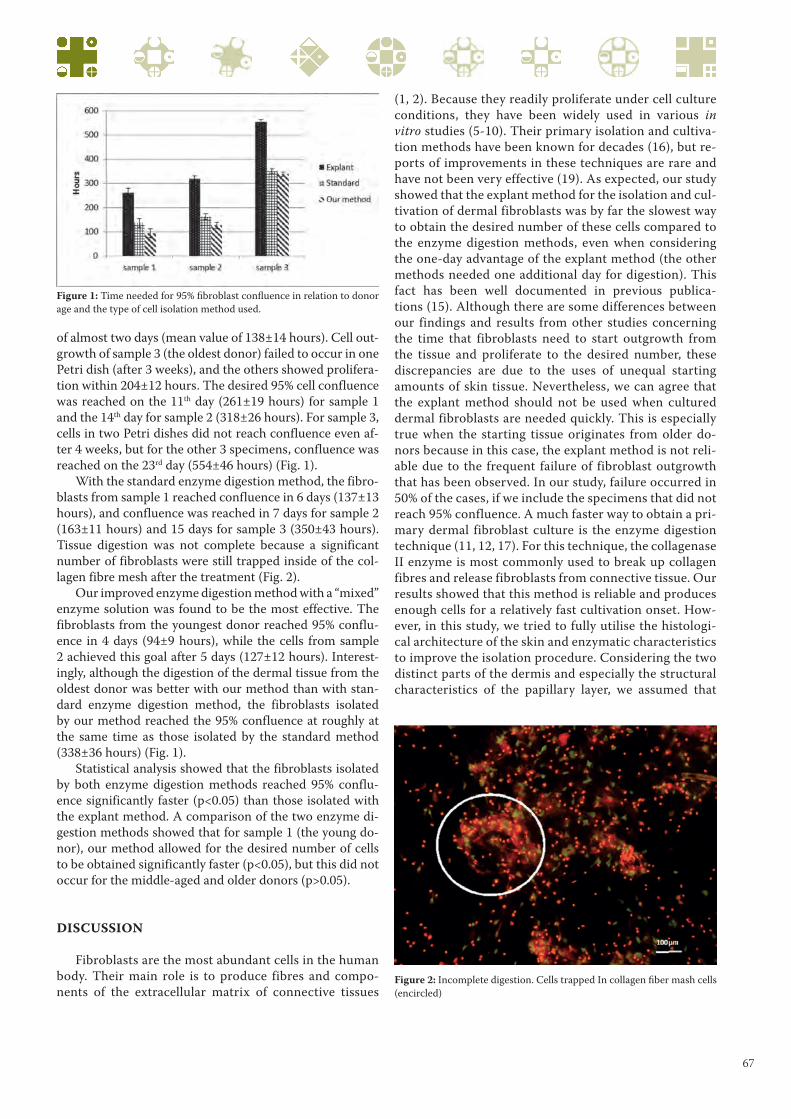
67
of almost two days (mean value of 138±14 hours). Cell out-
growth of sample 3 (the oldest donor) failed to occur in one
Petri dish (after 3 weeks), and the others showed prolifera-
tion within 204±12 hours. The desired 95% cell confluence
was reached on the 11th day (261±19 hours) for sample 1
and the 14th day for sample 2 (318±26 hours). For sample 3,
cells in two Petri dishes did not reach confluence even af-
ter 4 weeks, but for the other 3 specimens, confluence was
reached on the 23rd day (554±46 hours) (Fig. 1).
With the standard enzyme digestion method, the fibro-
blasts from sample 1 reached confluence in 6 days (137±13
hours), and confluence was reached in 7 days for sample 2
(163±11 hours) and 15 days for sample 3 (350±43 hours).
Tissue digestion was not complete because a significant
number of fibroblasts were still trapped inside of the col-
lagen fibre mesh after the treatment (Fig. 2).
Our improved enzyme digestion method with a “mixed”
enzyme solution was found to be the most effective. The
fibroblasts from the youngest donor reached 95% conflu-
ence in 4 days (94±9 hours), while the cells from sample
2 achieved this goal after 5 days (127±12 hours). Interest-
ingly, although the digestion of the dermal tissue from the
oldest donor was better with our method than with stan-
dard enzyme digestion method, the fibroblasts isolated
by our method reached the 95% confluence at roughly at
the same time as those isolated by the standard method
(338±36 hours) (Fig. 1).
Statistical analysis showed that the fibroblasts isolated
by both enzyme digestion methods reached 95% conflu-
ence significantly faster (p<0.05) than those isolated with
the explant method. A comparison of the two enzyme di-
gestion methods showed that for sample 1 (the young do-
nor), our method allowed for the desired number of cells
to be obtained significantly faster (p<0.05), but this did not
occur for the middle-aged and older donors (p>0.05).
DISCUSSION
Fibroblasts are the most abundant cells in the human
body. Their main role is to produce fibres and compo-
nents of the extracellular matrix of connective tissues
(1, 2). Because they readily proliferate under cell culture
conditions, they have been widely used in various in
vitro studies (5-10). Their primary isolation and cultiva-
tion methods have been known for decades (16), but re-
ports of improvements in these techniques are rare and
have not been very effective (19). As expected, our study
showed that the explant method for the isolation and cul-
tivation of dermal fibroblasts was by far the slowest way
to obtain the desired number of these cells compared to
the enzyme digestion methods, even when considering
the one-day advantage of the explant method (the other
methods needed one additional day for digestion). This
fact has been well documented in previous publica-
tions (15). Although there are some differences between
our findings and results from other studies concerning
the time that fibroblasts need to start outgrowth from
the tissue and proliferate to the desired number, these
discrepancies are due to the uses of unequal starting
amounts of skin tissue. Nevertheless, we can agree that
the explant method should not be used when cultured
dermal fibroblasts are needed quickly. This is especially
true when the starting tissue originates from older do-
nors because in this case, the explant method is not reli-
able due to the frequent failure of fibroblast outgrowth
that has been observed. In our study, failure occurred in
50% of the cases, if we include the specimens that did not
reach 95% confluence. A much faster way to obtain a pri-
mary dermal fibroblast culture is the enzyme digestion
technique (11, 12, 17). For this technique, the collagenase
II enzyme is most commonly used to break up collagen
fibres and release fibroblasts from connective tissue. Our
results showed that this method is reliable and produces
enough cells for a relatively fast cultivation onset. How-
ever, in this study, we tried to fully utilise the histologi-
cal architecture of the skin and enzymatic characteristics
to improve the isolation procedure. Considering the two
distinct parts of the dermis and especially the structural
characteristics of the papillary layer, we assumed that
Figure 1: Time needed for 95% fi broblast confl uence in relation to donor
age and the type of cell isolation method used.
Figure 2: Incomplete digestion. Cells trapped In collagen fi ber mash cells
(encircled)
68
proteases, such as dispase II, would enhance cell release
by breaking down the attachment of the fibroblast to the
collagen fibres because this enzyme hydrolyses peptides
bound to non-polar amino acids, which are often found
in collagen (20, 21). The results proved that this assump-
tion was correct. The digestion of the young dermal tis-
sue was better with the “mixed” enzyme solution, and
the tissue specimens treated with our solution gave rise
to more fibroblasts that reached 95% confluence more
rapidly than the ones obtained with the standard enzyme
digestion method. Furthermore, our method included an
elevated calcium concentration and a higher temperature
than usual because calcium ions are important for the abil-
ity of collagenase to bind to collagen fibres and start diges-
tion, and the activity of this enzyme is higher at slightly
elevated temperatures (22, 23). The only major exception
occurred when tissue samples from older donors were in-
vestigated. In these cases, the “mixed” enzyme solution
failed to produce more cells than the standard one, and the
fibroblasts reached confluence at virtually the same time.
This is surely because of the atrophic changes in the skin of
elderly individuals, including reduced cellularity and a sig-
nificant increase in collagen content (13). Older dermal tis-
sue is denser and less responsive to enzymatic treatment.
In addition, fibroblasts lose mitotic potential with age;
thus, the overall effect of the digestion solution was dimin-
ished. We were somewhat puzzled by the results concern-
ing the middle-aged donors. We expected results similar to
those obtained with the younger tissue e.g., the significant
improvement of cell isolation, but tissue digestion with the
combined enzyme solution was not as complete compared
with the young donor. There was some difference in favour
of the “mixed” enzyme solution compared to the standard
one, but it was not significant. This is probably also due
to the structural changes of the skin; therefore, we are in
agreement with some authors who have stated that the ag-
ing process of the human skin is already apparent during
the fourth decade of life (24, 25).
REFERENCES
1. M. Ross , W. Pawlina. Histology of the skin. In Histol-
ogy: A Text and Atlas 6th edition, Lippincott Williams
& Wilkins 2010; 493-501.
2. M. Ross , W. Pawlina. Histology of the skin. In Histol-
ogy: A Text and Atlas 6th edition, Lippincott Williams
& Wilkins 2010; 183-185.
3. Boo S, Dagnino L. Integrins as Modulators of Trans-
forming Growth Factor Beta Signaling in Dermal Fi-
broblasts During Skin Regeneration After Injury. Adv.
Wound Care 2013; 2(5): 238-246.
4. Miron-Mendoza M, Lin X, Ma L, Ririe P, Petroll WM.
Individual versus collective fibroblast spreading and
migration: regulation by matrix composition in 3D cul-
ture. Exp Eye Res. 2012; 99: 36-44.
5. Bojesen KB, Clausen O, Rohde K, Christensen C,
Zhang L, Li S, Kohler L, Nielbo S, Nielsen J, Gjorlund
MD, Poulsen FM, Bock E, Berezin V. Nectin-1 binds
and signals through the fibroblast growth factor recep-
tor. J Biol Chem. 2012; 26; 287(44): 37420-33.
6. Chung B, Hinek A, Keating S, Weksberg R, Shah V,
Blaser S, Hawkins C, Chitayat D Overgrowth with
increased proliferation of fibroblast and matrix met-
alloproteinase activity related to reduced TIMP1: a
newly recognized syndrome? Am J Med Genet A. 2012;
158A(10): 2373-81.
7. Miron-Mendoza M, Lin X, Ma L, Ririe P, Petroll WM.
Individual versus collective fibroblast spreading and
migration: regulation by matrix composition in 3D cul-
ture. Exp Eye Res. 2012; 99: 36-44.
8. Erisken C, Zhang X, Moffat KL, Levine WN, Lu HH.
Scaffold fiber diameter regulates human tendon fibro-
blast growth and differentiation. Tissue Eng Part A.
2013; 19(3-4): 519-28.
9. Itoh N, Ornitz DM. Fibroblast growth factors: from
molecular evolution to roles in development, metabo-
lism and disease. J Biochem. 2011; 149(2): 121-30.
10. H. Jiang and F. Grinnell. Cell–Matrix Entanglement
and Mechanical Anchorage of Fibroblasts in Threedi-
mensional Collagen Matrices. Molecular Biology of the
Cell. 2005; 16: 5070–5076.
11. Huang HI, Wu CZ. Isolation and differentiation poten-
tial of fibroblast-like stromal cells derived from human
skin. Methods Mol Biol. 2012; 879:465-70.
12. Park JC, Kim YB, Kim HJ, Jang HS, Kim HS, Kim BO,
Han KY. Isolation and characterization of cultured
human periodental ligament fibroblast-specific cD-
NAs. Biochem Biophys Res Commun. 2001; 20;
282(5): 1145-53.
13. A. Tiganescu, W Parish, E. Walker, M. Cooper, G. Lav-
ery, P. Stewart. Reversal of age-induced dermal atrophy
in 11β-hydroxysteroid dehydrogenase type 1-null mice.
Endocrine Abstracts 2012; 28, P302.
14. McElreavey KD, Irvine AI, Ennis KT, McLean WH.
Isolation, culture and characterisation of fibroblastlike
cells derived from the Wharton’s jelly portion of human
umbilical cord. Biochem Soc Trans. 1991; 19(1): 29S.
15. Goldschmidt E, Hem S, Ajler P, Ielpi M, Loresi M,
Giunta D, Carrizo A, Yampolsky C, Argibay P. A new
model for dura mater healing: human dural fibroblast
culture. Neurol Res. 2013; 35(3): 300-7.
16. Hentzer B, Kobayasi T. Enzymatic liberation of vi-
able cells of human skin. Acta Derm Venereol.
1978;58(3):197-202.
17. De Falco E1, Scafetta G, Napoletano C, Puca R, Vingolo
EM, Ragona G, Iorio O, Frati G. Cell Tissue Bank. 2013;
14(2): 277-87. Epub 2012; 21. A standardized labora-
tory and surgical method for in vitro culture isolation
and expansion of primary human Tenon’s fibroblasts.
18. McFarland KL, Glaser K, Hahn JM, Boyce ST, Supp
DM. Culture medium and cell density impact gene ex-

69
pression in normal skin and abnormal scar-derived fi-
broblasts. J Burn Care Res. 2011; 32(4): 498-508.
19. Wang H, Van Blitterswijk CA, Bertrand-De Haas M,
Schuurman AH, Lamme EN. Improved enzymatic isola-
tion of fibroblasts for the creation of autologous skin substi-
tutes. in Vitro Cell Dev Biol Anim. 2004; 40(8-9): 268-77.
20. http://www.lifetechnologies.com/order/catalog/
product/17105041.
21. Fields, Gregg B. Interstitial collagen catabolism. Journal
of Biological Chemistry 288.13 2013; 8785-8793.
22. Tezvergil-Mutluay A, Agee KA, Hoshika T, Carrilho
M, Breschi L, Tjaderhane L, Nishitani Y, Carvalho
RM, Looney S, Tay FR, Pashley DH. The requirement
of zinc and calcium ions for functional MMP activity
in demineralized dentin matrices. Dent Mater. 2010;
26(11): 1059-6.
23. Shrinivas, D. and G. R. Naik. Characterization of al-
kaline thermostable keratinolytic protease from ther-
moalkalophilic Bacillus halodurans JB 99 exhibiting
dehairing activity. International Biodeterioration &
Biodegradation 65.1 (2011): 29-35.
24. Zouboulis, Christos C. and Evgenia Makrantonaki.
Clinical aspects and molecular diagnostics of skin ag-
ing. Clinics in dermatology 29.1, 2011; 3-14.
25. Longo, Caterina, et al. Skin aging: in vivo microscopic
assessment of epidermal and dermal changes by means
of confocal microscopy. Journal of the American Acad-
emy of Dermatology 68.3, 2013; e73-e82.

70

71
Serbian Journal of Experimental and Clinical Research
is a peer-reviewed, general biomedical journal. It publishes
original basic and clinical research, clinical practice arti-
cles, critical reviews, case reports, evaluations of scientific
methods, works dealing with ethical and social aspects of
biomedicine as well as letters to the editor, reports of asso-
ciation activities, book reviews, news in biomedicine, and
any other article and information concerned with practice
and research in biomedicine, written in the English.
Original manuscripts will be accepted with the under-
standing that they are solely contributed to the Journal.
The papers will be not accepted if they contain the mate-
rial that has already been published or has been submitted
or accepted for publication elsewhere, except of prelimi-
nary reports, such as an abstract, poster or press report
presented at a professional or scientific meetings and not
exceeding 400 words. Any previous publication in such
form must be disclosed in a footnote. In rare exceptions
a secondary publication will acceptable, but authors are
required to contact Editor-in-chief before submission of
such manuscript. the Journal is devoted to the Guidelines
on Good Publication Practice as established by Commit-
tee on Publication Ethics-COPE (posted at www.publica-
tionethics.org.uk).
Manuscripts are prepared in accordance with „Uni-
form Requirements for Manuscripts submitted to Biomedi-
cal Journals“ developed by the International Committee of
Medical Journal Editors. Consult a current version of the
instructions, which has been published in several journals
(for example: Ann Intern Med 1997;126:36-47) and posted at
www.icmje.org, and a recent issue of the Journal in prepar-
ing your manuscript. For articles of randomized controlled
trials authors should refer to the „Consort statement“ (www.
consort-statement.org). Manuscripts must be accompanied
by a cover letter, signed by all authors, with a statement that
the manuscript has been read and approved by them, and not
published, submitted or accepted elsewhere. Manuscripts,
which are accepted for publication in the Journal, become the
property of the Journal, and may not be published anywhere
else without written permission from the publisher.
Serbian Journal of Experimental and Clinical Research
is owned and published by Medical Faculty University of
Kragujevac. However, Editors have full academic freedom
and authority for determining the content of the journal,
according to their scientific, professional and ethical judg-
ment. Editorial policy and decision making follow proce-
dures which are endeavoring to ensure scientific credibility
of published content, confidentiality and integrity of auth
ors, reviewers, and review process, protection of patients’
rights to privacy and disclosing of conflict of interests. For
difficulties which might appear in the Journal content such
as errors in published articles or scientific concerns about
research findings, appropriate handling is provided. The re-
quirements for the content, which appears on the Journal
internet site or Supplements, are, in general, the same as for
the master version. Advertising which appears in the Jour-
nal or its internet site is not allowed to influence editorial
decisions.
MA NU SCRIPT
Manuscripts for Serbian Journal of Experimental and
Clinical Research are available for submission through the
Editorial Manager System http://www.editorialmanager.
com/sjecr/.
For papers that are accepted, Serbian Journal of Experi-
mental and Clinical Research obligatory requires authors
to provide an identical, electronic copy in appropriate tex-
tual and graphic format.
The ma nu script of original, scinetific articles should
be ar ran ged as fol lowing: Ti tle pa ge, Ab stract, In tro duc-
tion, Pa ti ents and met hods/Ma te rial and met hods, Re-
sults, Di scus sion, Ac know led ge ments, Re fe ren ces, Ta bles,
Fi gu re le gends and Fi gu res. The sections of other papers
should be arranged according to the type of the article.
Each ma nu script com po nent (The Ti tle pa ge, etc.)
should be gins on a se pa ra te pa ge. All pa ges should be num-
be red con se cu ti vely be gin ning with the ti tle pa ge.
INSTRUCTION TO AUTHORSFOR MANUSCRIPT PREPARATION

72
All me a su re ments, ex cept blood pres su re, should be re-
por ted in the System In ter na ti o nal (SI) units and, if ne ces-
sary, in con ven ti o nal units, too (in pa rent he ses). Ge ne ric
na mes should be used for drugs. Brand na mes may be in-
ser ted in pa rent he ses.
Aut hors are advi sed to re tain ex tra co pi es of the ma nu-
script. Serbian Journal of Experimental and Clinical Research
is not re spon si ble for the loss of ma nu scripts in the mail.
TI TLE PA GE
The Ti tle pa ge con ta ins the ti tle, full na mes of all the
aut hors, na mes and full lo ca tion of the de part ment and in-
sti tu tion whe re work was per for med, ab bre vi a ti ons used,
and the na me of cor re spon ding aut hor.
The ti tle of the ar tic le should be con ci se but in for ma-
ti ve, and in clu de ani mal spe ci es if ap pro pri a te. A sub ti tle
could be ad ded if ne ces sary.
A list of ab bre vi a ti ons used in the pa per, if any, should be
in clu ded. The ab bre vi a ti ons should be listed alp ha be ti cally,
and fol lo wed by an ex pla na ti on of what they stand for. In ge-
ne ral, the use of ab bre vi a ti ons is di sco u ra ged un less they are
es sen tial for im pro ving the re a da bi lity of the text.
The na me, te lep ho ne num ber, fax num ber, and exact po-
stal ad dress of the aut hor to whom com mu ni ca ti ons and re-
prints sho uld be sent are typed et the end of the ti tle pa ge.
AB STRACT
An ab stract of less than 250 words should con ci sely sta-
te the ob jec ti ve, fin dings, and con clu si ons of the stu di es
de scri bed in the ma nu script. The ab stract do es not con-
tain ab bre vi a ti ons, fo ot no tes or re fe ren ces.
Be low the ab stract, 3 to 8 keywords or short phra ses
are pro vi ded for in de xing pur po ses. The use of words from
Medline thesaurus is recommended.
IN TRO DUC TION
The in tro duc tion is con ci se, and sta tes the re a son and
spe ci fic pur po se of the study.
PA TI ENTS AND MET HODS/MA TE RIAL
AND MET HODS
The se lec tion of pa ti ents or ex pe ri men tal ani mals, in-
clu ding con trols, should be de scri bed. Pa ti ents’ na mes and
ho spi tal num bers are not used.
Met hods should be de scri bed in suf fi ci ent de tail to per mit
eva lu a tion and du pli ca tion of the work by ot her in ve sti ga tors.
When re por ting ex pe ri ments on hu man su bjects, it
should be in di ca ted whet her the pro ce du res fol lo wed
we re in ac cor dan ce with et hi cal stan dards of the Com-
mit tee on hu man ex pe ri men ta ti on (or Ethics Commit-
tee) of the in sti tu tion in which they we re do ne and in
ac cor dan ce with the Hel sin ki Dec la ra tion. Ha zar do us
pro ce du res or che mi cals, if used, should be de scri bed
in de ta ils, in clu ding the sa fety pre ca u ti ons ob ser ved.
When ap pro pri a te, a sta te ment should be in clu ded ve-
rifying that the ca re of la bo ra tory ani mals fol lo wed ac-
cep ted stan dards.
Sta ti sti cal met hods used should be outli ned.
RE SULTS
Re sults should be cle ar and con ci se, and in clu de a mi-
ni mum num ber of ta bles and fi gu res ne ces sary for pro per
pre sen ta tion.
DI SCUS SION
An ex ha u sti ve re vi ew of li te ra tu re is not ne ces sary. The
ma jor fin dings sho uld be di scus sed in re la tion to ot her pu-
blis hed work. At tempts sho uld be ma de to ex pla in dif fe-
ren ces bet we en the re sults of the pre sent study and tho se
of the ot hers. The hypot he sis and spe cu la ti ve sta te ments
sho uld be cle arly iden ti fied. The Di scus sion sec tion sho uld
not be a re sta te ment of re sults, and new re sults sho uld not
be in tro du ced in the di scus sion.
ACKNOWLEDGMENTS
This section gives possibility to list all persons who con-
tributed to the work or prepared the manuscript, but did not
meet the criteria for authorship. Financial and material sup-
port, if existed, could be also emphasized in this section.
RE FE REN CES
Re fe ren ces should be iden ti fied in the text by Ara bic
nu me rals in pa rent he ses. They should be num be red con-
se cu ti vely, as they ap pe ared in the text. Per so nal com mu ni-
ca ti ons and un pu blis hed ob ser va ti ons should not be ci ted
in the re fe ren ce list, but may be men ti o ned in the text in
pa rent he ses. Ab bre vi a ti ons of jo ur nals should con form to
tho se in In dex Serbian Journal of Experimental and Clini-
cal Research. The style and pun ctu a tion should con form to
the Serbian Journal of Experimental and Clinical Research
style re qu i re ments. The fol lo wing are exam ples:
1. Introduction
This document describes standards for preparing the ref-
erences in the APA style. The following sections give detailed
instructions on citing books, journal articles, newspaper ar-
ticles, conference papers, theses, webpages and others.

73
Please provide all the required elements in the refer-
ences to your paper. Please pay particular attention to
spelling, capitalization and punctuation. Accuracy and
completeness of references are the responsibilities of the
author. Before submitting your article, please ensure you
have checked your paper for any relevant references you
may have missed.
A complete reference should give the reader enough
information to find the relevant article. And most impor-
tantly, complete and correct references may allow auto-
matic creation of active links by the MetaPress technol-
ogy that we use for making the electronic version of our
journal. Active reference linking is regarded as the greatest
benefit of electronic publishing and it adds a lot of value to
your publication.
2.Book
a. Book (one author)
Format:Author. (Year of publication). Book title. Place of publi-
cation: Publisher.
Example:Baxter, R. (1982). Exactly Solvable Models in Statistical
Mechanics. New York: Academic Press.
b. Book (two or more authors)
Format:Author1, Author2 & Author3. (Year of publication).
Book title. Place of publication: Publisher.
Example:Kleiner, F.S., Mamiya C.J. & Tansey R.G. (2001). Gard-
ner’s art through the ages (11th ed.). Fort Worth, USA: Har-
court College Publishers.
c. Book chapter or article in an edited book
Format:Author(s) of chapter. (Year of publication). Chapter ti-
tle. In Editors of the book (Eds.), Book title (Chapter page
range). Place of publication: Publisher.
Example:Roll, W.P. (1976). ESP and memory. In J.M.O. Wheatley
& H.L. Edge (Eds.), Philosophical dimensions of parapsy-
chology (pp. 154-184). Springfield, IL: American Psychiat-
ric Press.
d. Proceedings from a conference
Format:Author(s). (Year of publication). Title. In Conference
name, Date (Page range). Place of publication: Publisher.
Example:Field, G. (2001). Rethinking reference rethought. In
Revelling in Reference: Reference and Information Ser-
vices Section Symposium, 12-14 October 2001 (pp. 59-64).
Melbourne, Victoria, Australia: Australian Library and In-
formation Association.
e. ebook
Format:Author(s). (Year of publication). Title. Publisher. Retrie-
ving date, http address. DOI.
Example:Johnson, A. (2000). Abstract Computing Machines.
Springer Berlin Heidelberg. Retrieved March 30, 2006, from
SpringerLink http://springerlink.com/content/w25154. DOI:
10.1007/b138965.
f. Thesis
Format:Author(s). (Year of publication). Title. Information, Pla-
ce of publication.
Example:Begg, M. M. (2001). Dairy farm women in the Waikato
1946-1996: Fifty years of social and structural change. Un-
published doctoral dissertation, University of Waikato,
Hamilton, New Zealand.
g. Report
Format:Author(s). (Year of publication). Title. Place of publica-
tion: Publisher. (Report number)
Example:Osgood, D. W., & Wilson, J. K. (1990). Covariation of
adolescent health problems. Lincoln: University of Nebras-
ka. (NTIS No. PB 91-154 377/AS)
h. Government publication
Format:Institution name. (Year of publication). Title. Place of
publication: Publisher.
Example:Ministerial Council on Drug Strategy. (1997). The na-
tional drug strategy: Mapping the future. Canberra: Au-
stralian Government Publishing Service.
3. Article
a. Journal Article (one author)
Format:Author. (Year of publication). Article title. Journal Title.
Volume (issue), range of pages. DOI.
Example:Nikora, V. (2006). Hydrodynamics of aquatic eco-
systems: spatial-averaging perspective. Acta Geophysica,
55(1), 3-10. DOI: 10.2478/s11600-006-0043-6.
b. Journal Article (two or more authors)
Format:Author1, Author2 & Author3. (Year of publication). Ar-
ticle title. Journal Title. Volume (issue), range of pages. DOI.
Example:Cudak, M. & Karcz J. (2006). Momentum transfer in
an agitated vessel with off-centred impellers. Chem. Pap.
60(5), 375-380. DOI: 10.2478/s11696-006-0068-y.

74
c. Journal article from an online database
Format:Author(s). (Year of publication). Article title [Electronic
version]. Journal Title. Volume (issue), range of pages. Re-
trieved date of access, from name of database. DOI.
Example:Czajgucki Z., Zimecki M. & Andruszkiewicz R. (2006,
December). The immunoregulatory effects of edeine ana-
logues in mice [Abstract]. Cell. Mol. Biol. Lett. 12(3), 149-
161. Retrieved December 6, 2006, from PubMed database
on the World Wide Web: http://www.pubmed.gov. DOI:
10.2478/s11658-006-0061-z.
d. Newspaper article (no author)
Format:Article title. (Publication date). Journal Title. page.
Example:Amazing Amazon region. (1989, January 12). New York
Times, p. D11.
e. Encyclopedia article
Format:Author. (Year of publication). Article title. In Encyclo-
pedia title (volume number, pages). Place of publication:
Encyclopedia name.
Example:Bergmann, P. G. (1993). Relativity. In The new encyclo-
pedia britannica (Vol. 26, pp. 501-508). Chicago: Encyclo-
pedia Britannica.
4. Other formats
a. Web page
Format:Author/Sponsor. (last update or copyright date). Title.
Retrieved date of access, from URL.
Example:Walker, J. (1996, August). APA-style citations of elec-
tronic resources. Retrieved November 21, 2001, from http://
www.cas.usf.edu/english/walker/apa.html
b. Lecture note
Format:Author(s). (Date of presentation). Lecture title. Lecture
notes distributed in the unit, at the name of the teaching
organisation, the location.
Example:Liffers, M. (2006, August 30). Finding information in
the library. Lecture notes distributed in the unit Functio-
nal Anatomy and Sports Performance 1102, University of
Western Australia, Crawley, Western Australia.
c. Patent
Format:Author. (Year). Patent number. The location. Issue
body.
Example:Smith, I. M. (1988). U.S. Patent No. 123,445. Washing-
ton, D.C.: U.S. Patent and Trademark Office.
d. Standard
Format:Issue body. (Year). Standard name. Standard number.
The location.
Example:Standards Association of Australia. (1997). Australian
standard: Pressure equipment manufacture. AS4458-1997.
North Sydney.
e. Video
Format:Producer, P. P. (Producer), & Director, D.D. (Director).
(Date of publication). Title of motion picture [Motion pic-
ture]. Country of origin: Studio or distributor.
Example:Zhang, Y. (Producer/Director). (2000). Not one less [Mo-
tion Picture].China: Columbia Pictures Industries, Inc.
f. Audio recording
Format:Songwriter, W. W. (Date of copyright). Title of song
[Recorded by artist if different from song writer]. On Title
of album [Medium of recording]. Location: Label. (Recor-
ding date if different from copyright date).
Example:Taupin, B. (1975). Someone saved my life tonight [Re-
corded by Elton John]. On Captain fantastic and the brown
dirt cowboy [CD]. London: Big Pig Music Limited.
g. Mailing listFormat:Author. (Exact date of posting). Subject line of mes-
sage. Message posted to followed by name of mailing list,
archived at followed by address for the archived version of
the message
Example:Hammond, T. (2000, November 20). YAHC: Handle
Parameters, DOI Genres, etc. Message posted to Ref-Links
electronic mailing list, archived at http://www.doi.org/
mail-archive/ref-link/msg00088.html
h. Computer softwareFormat:Author(s). (Year). Title [computer software]. The loca-
tion: Company.
Example:Ludwig, T. (2002). PsychInquiry [computer software].
New York: Worth.

75
MOST COMMON REFERENCE STYLES
MetaPress can capture data from every style of references, but using one of the listed will increase the number of active links in the references. Once you have chosen one of the styles, please do not change it.
APA style1
Article in a journal:Lippke, S., & Ziegelmann, J. (2006). Understanding and modelling health behaviour change: The multi-stage model of health behaviour change. Journal of Health Psychology,11(1), 37-50,DOI:10.2478/s11533-007-0023-3.
Book:Jones, E., Farina, A., Hastorf, A., Markus, H., Miller, D., & Scott, R. (1984). Social stigma: The psychology of marked relationships. New York: W. H. Freeman.
Chicago style2
Article in a journal:Spitzer, Steven. Review of The Limits of Law Enforcement, by Hans Zeisel. American Journal of Sociology 91 (1985): 726-29; DOI:10.2478/s11533-007-0023-3.
Book:Lloyd, Donald A., and Harry R. Warfel. American English and Its Cultural Setting. New York: Alfred A. Knopf, 1956.
Harvard style3
Article in a journal:Conley, TG & Galenson, DW 1998, ‘Nativity and wealth in mid-nineteenth century cities’, Journal of Economic History, vol. 58, no. 2, pp. 468-493, DOI:10.2478/s11533-007-0023-3.
Book:Hodgson, A 1998, Accounting theory, John Wiley & Sons, Brisbane.
Oxford style4
Article in a journal:KHOO, G.K. Accounting for leases. The Chartered Accountant in Australia, 46(5): Nov. 1975: 19-23: DOI:10.2478/s11533-007-0023-3.1 Read more: http://www.library.uwa.edu.au/education_training___
and___support/guides/how_to_cite_your_sources/apa_style2 Read more: http://www.wisc.edu/writing/Handbook/DocChiWork-
sCited.html3 Read more: http://www.library.uwa.edu.au/education_training___
and___support/guides/how_to_cite_your_sources/citing_your_sourc-
es_-_harvard_style#Reference4 Read more: http://www.usq.edu.au/library/help/ehelp/ref_guides/
oxford.htm
Book:GIBBS, Graham. Teaching students to learn: a student-centred approach. Milton Keynes, Open University Press, 1981.
MLA style5
Article in a journal:Joyce, Michael. “On the Birthday of the Stranger (in Memory of John Hawkes).” Evergreen Review 5 Mar. 1999. 12 May 1999 http://www.evergreenreview.com/102/evexcite/joyce/nojoyce.html. DOI:10.2478/s11533-007-0023-3.
Book:Bird, Isabella L. A Lady’s Life in the Rocky Mountains. New York, 1881. Victorian Women Writers Project. Ed. Perry Willett. 27 May 1999. Indiana U. 4 Oct. 1999
IEE style6
Article in a journal:I.E. Sutherland, R.F. Sproull, and R.A. Schumaker, “A Characterization of 10 Hidden-Surface Algorithms,” ACM Computing Surveys, Mar. 1974, pp. 1-55, DOI:10.2478/s11533-007-0023-3.
Book:W.M. Newman and R.F. Sproull, Principles of Interactive Computer Graphics, McGraw-Hill, 1979, p. 402.
Vancouver style7
Article in a journal:You CH, Lee KY, Chey WY, Menguy R. Electrogastrographic study of patients with unexplained nausea, bloating and vomiting. Gastroenterology 1980;79:311-4; DOI:10.2478/s11533-007-0023-3.
Book:Eisen HN. Immunology: an introduction to molecular and cellular principles of the immune response. 5th ed. New York: Harper and Row; 1974.
TA BLES
Ta bles should be typed on se pa ra te she ets with ta ble
num bers (Ara bic) and ti tle abo ve the ta ble and ex pla na tory
no tes, if any, be low the ta ble.5 Read more: http://www.bedfordstmartins.com/online/cite5.html6 Read more: http://www.computer.org/portal/site/ieeecs/menuitem.
c5efb9b8ade9096b8a9ca0108bcd45f3/index.jsp?&pName=ieeecs_
level1&path=ieeecs/publications/author/style&fi le=refer.
xml&xsl=generic.xsl&7 Read more: http://www.library.uwa.edu.au/education_training___
and___support/guides/how_to_cite_your_sources/citing_your_sourc-
es_-_vancouver_style

76
FI GU RES AND FI GU RE LE GENDS
All il lu stra ti ons (pho to graphs, graphs, di a grams) will be
con si de red as fi gu res, and num be red con se cu ti vely in Ara-
bic nu me rals. The num ber of fi gu res in clu ded should be the
le ast re qu i red to con vey the mes sa ge of the pa per, and no fi-
gu re should du pli ca te the da ta pre sented in the ta bles or text.
Fi gu res should not ha ve ti tles. Let ters, nu me rals and sym-
bols must be cle ar, in pro por tion to each ot her, and lar ge
eno ugh to be readable when re du ced for pu bli ca tion. Fi gu-
res should be sub mit ted as ne ar to the ir prin ted si ze as pos-
si ble. Fi gu res are re pro du ced in one of the fol lo wing width
si zes: 8 cm, 12 cm or 17 cm, and with a ma xi mal length of 20
cm. Le gends for fi gu res sho uld be gi ven on se pa ra te pa ges.
If mag ni fi ca tion is sig ni fi cant (pho to mic ro graphs)
it should be in di ca ted by a ca li bra tion bar on the print,
not by a mag ni fi ca tion fac tor in the fi gu re le gend. The
length of the bar should be in di ca ted on the fi gu re or in
the fi gu re le gend.
Two com ple te sets of high qu a lity un mo un ted glossy
prints should be sub mit ted in two se pa ra te en ve lo pes, and
shi el ded by an ap pro pri a te card bo ard. The backs of sin-
gle or gro u ped il lu stra ti ons (pla tes) should be ar the first
aut hors last na me, fi gu re num ber, and an ar row in di ca ting
the top. This in for ma tion should be pen ci led in lightly or
pla ced on a typed self-ad he si ve la bel in or der to pre vent
mar king the front sur fa ce of the il lu stra tion.
Pho to graphs of iden ti fi a ble pa ti ents must be ac com pa-
nied by writ ten per mis sion from the pa ti ent.
For fi gu res pu blis hed pre vi o usly the ori gi nal so ur ce
should be ac know led ged, and writ ten per mis sion from the
copyright hol der to re pro du ce it sub mit ted.
Co lor prints are ava i la ble by re qu est at the aut hors
ex pen se.
LET TERS TO THE EDI TOR
Both let ters con cer ning and tho se not con cer ning
the ar tic les that ha ve been pu blis hed in Serbian Journal
of Experimental and Clinical Research will be con si de-
red for pu bli ca tion. They may con tain one ta ble or fi gu-
re and up to fi ve re fe ren ces.
PRO OFS
All ma nu scripts will be ca re fully re vi sed by the pu blis her
desk edi tor. Only in ca se of ex ten si ve cor rec ti ons will the
ma nu script be re tur ned to the aut hors for fi nal ap pro val. In
or der to speed up pu bli ca tion no pro of will be sent to the
aut hors, but will be read by the edi tor and the desk edi tor.

77

78
CIP - Каталогизација у публикацији
Народна библиотека Србије, Београд
61
SERBIAN Journal of Experimental and Clinical Research
editor-in-chief Vladimir Jakovljević.
- Vol. 9, N° 1 (April 2008) -
- Kragujevac (Svetozara Markovića 69) :
Medical Faculty, 2008 - (Kragujevac : Medical Faculty). - 29 cm
Je nastavak: Medicus (Kragujevac) = ISSN 1450-7994
ISSN 1820-8665 = Serbian Journal of
Experimental and Clinical Research
COBISS.SR-ID 149695244