Embed Size (px)
Citation preview
10/25/2013
1
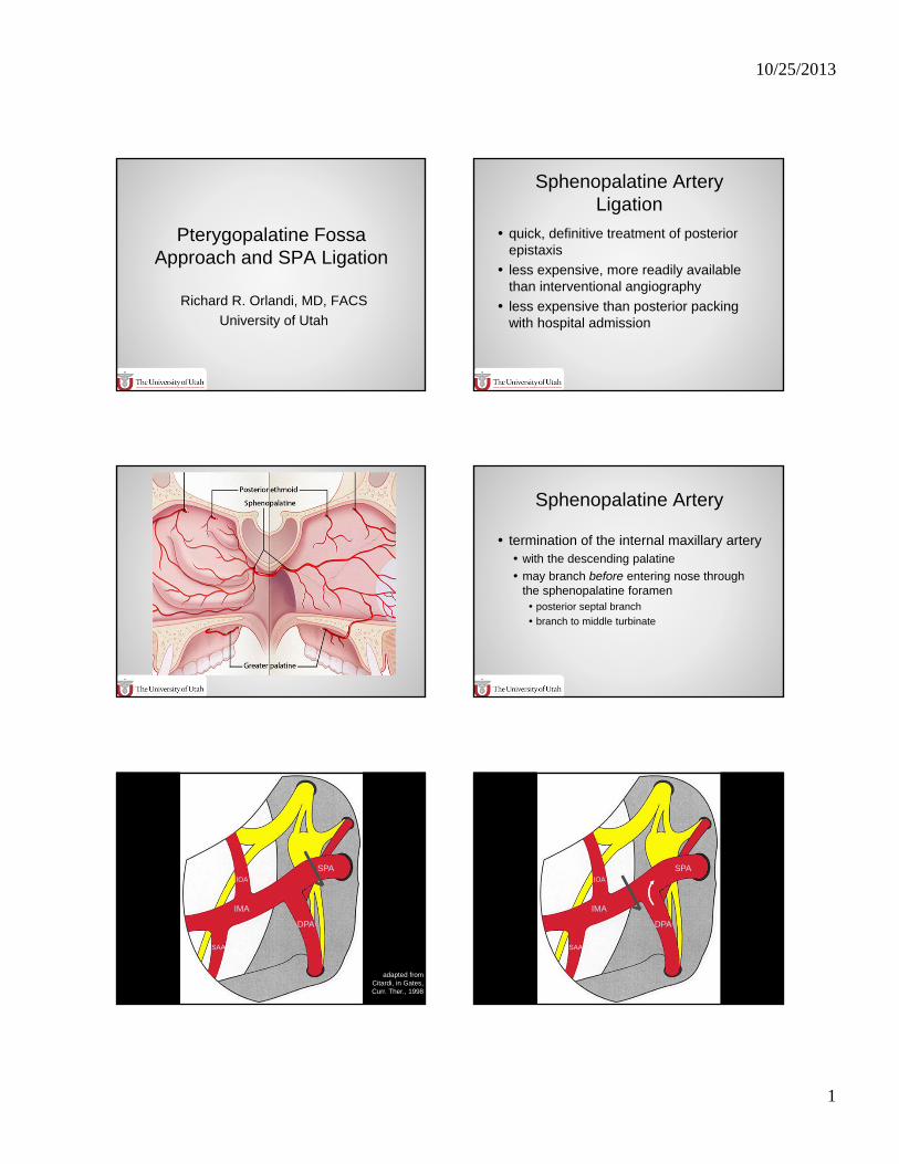
Pterygopalatine Fossa Approach and SPA Ligation
Richard R. Orlandi, MD, FACS
University of Utah
Sphenopalatine Artery Ligation
quick, definitive treatment of posterior epistaxis
less expensive, more readily available than interventional angiography
less expensive than posterior packing with hospital admission
Sphenopalatine Artery Sphenopalatine Artery
termination of the internal maxillary artery with the descending palatine
may branch before entering nose through the sphenopalatine foramen posterior septal branch
branch to middle turbinate
adapted from Citardi, in Gates, Curr. Ther., 1998
SPAIOA
SAA
DPA
IMA
SPAIOA
SAA
DPA
IMA
10/25/2013
2
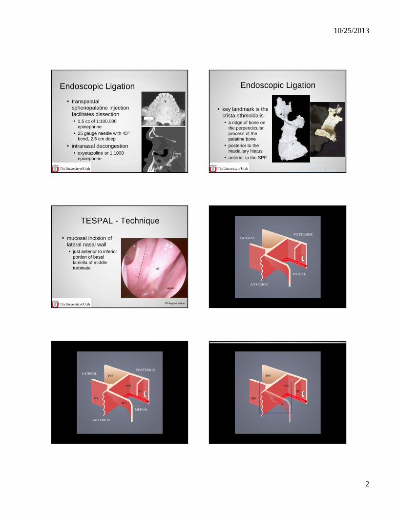
Endoscopic Ligation
transpalatalsphenopalatine injection facilitates dissection 1.5 cc of 1:100,000
epinephrine
25 gauge needle with 45ºbend, 2.5 cm deep
intranasal decongestion oxyetazoline or 1:1000
epinephrine
Rohen, Color Atlas of Anatomy; Bolger, AJR, 1999
Endoscopic Ligation
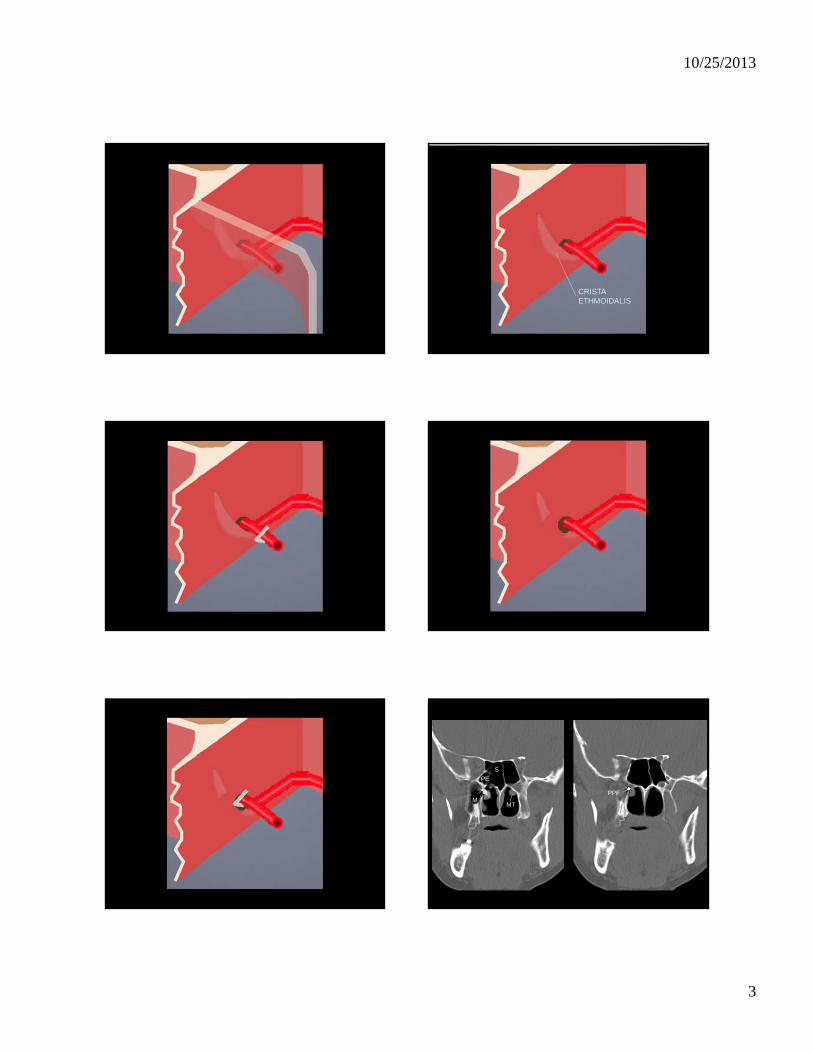
key landmark is the crista ethmoidalis a ridge of bone on
the perpendicular process of the palatine bone
posterior to the maxiallary hiatus
anterior to the SPF
TESPAL - Technique
mucosal incision of lateral nasal wall just anterior to inferior
portion of basal lamella of middle turbinate MT
septum
30 degree scope
ANTERIOR
LATERAL
MEDIAL
POSTERIOR
MT
PES
MS
PPF
ST
ANTERIOR
LATERAL
MEDIAL
POSTERIOR
PES
MS
PPF
ST
10/25/2013
3
CRISTAETHMOIDALIS
M
S
MT
PE
PPF
10/25/2013
4
NPN
SPACE
flap elevatedmedially
suction
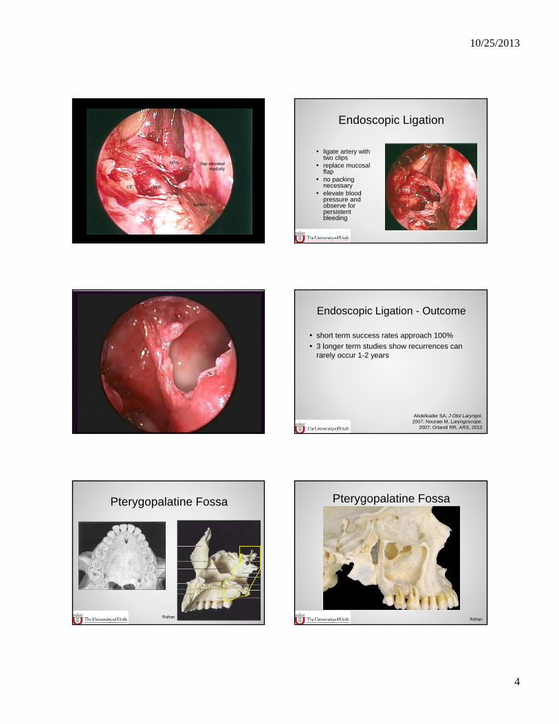
Endoscopic Ligation
ligate artery with two clips
replace mucosal flap
no packing necessary
elevate blood pressure and observe for persistent bleeding
short term success rates approach 100%
3 longer term studies show recurrences can rarely occur 1-2 years
Endoscopic Ligation - Outcome
Abdelkader SA, J Otol Laryngol, 2007; Nouraei M, Laryngoscope,
2007; Orlandi RR, ARS, 2010
Pterygopalatine Fossa
Rohan
Pterygopalatine Fossa
Rohan
10/25/2013
5
Pterygopalatine Fossa
body ofsphenoid bone
pterygoidprocess
posterior wall ofmaxillary sinus
Rohan
Pterygopalatine Fossasphenopalatineforamen
Rohan
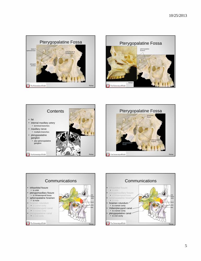
Contents
fat
internal maxillary artery terminal branches
maxillary nerve multiple branches
pterygopalatineganglion aka sphenopalatine
ganglion
Rohan
Pterygopalatine Fossa
sphenopalatineforamen
infraorbitalfissure
pterygomaxillaryfissure
Rohan
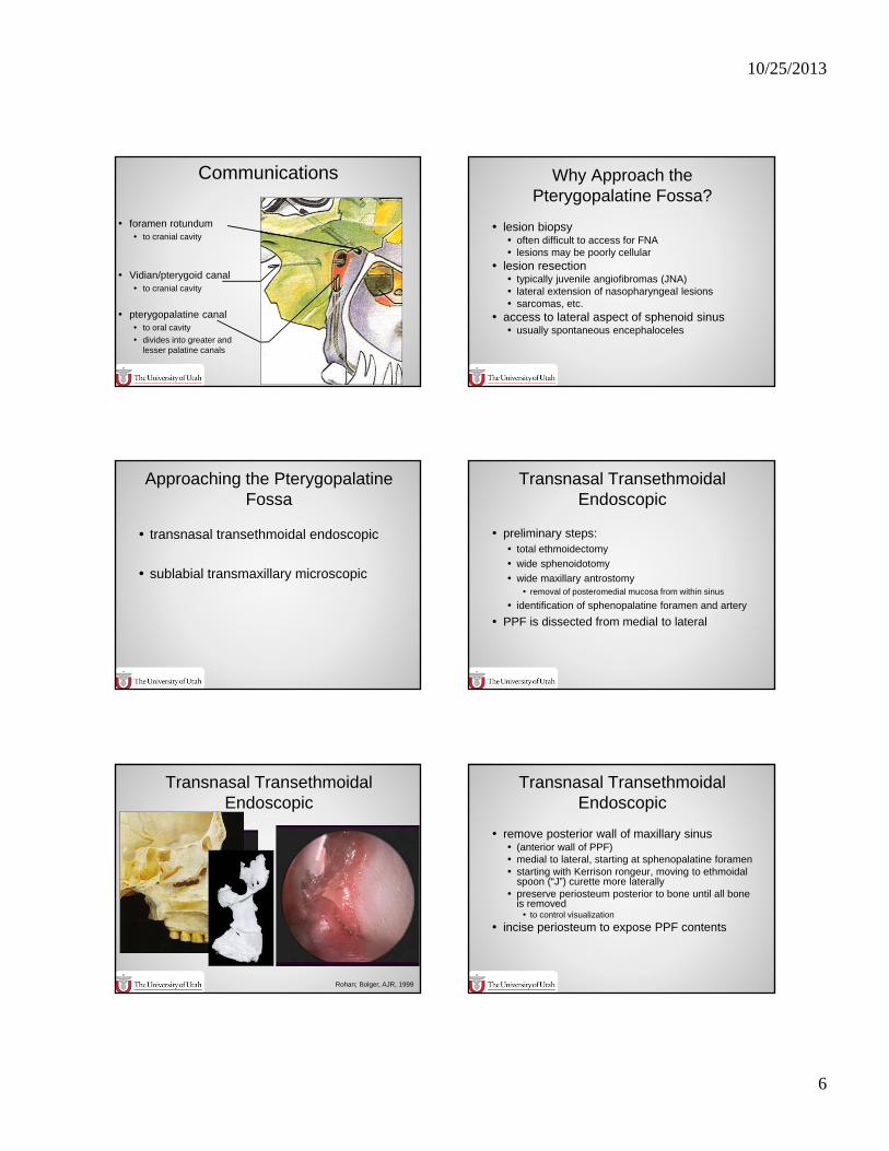
Communications
infraorbital fissure to orbit
pterygomaxillary fissure to infratemporal fossa
sphenopalatine foramen to nose
foramen rotundum to cranial cavity
Vidian/pterygoid canal to cranial cavity
pterygopalatine canal to oral cavity
MH
ION
NLD
PEA/AEA
ON
Rohan
Communications
infraorbital fissure to orbit
pterygomaxillary fissure to infratemporal fossa
sphenopalatine foramen to nose
foramen rotundum to cranial cavity
Vidian/pterygoid canal to cranial cavity
pterygopalatine canal to oral cavity
MH
ION
NLD
PEA/AEA
ON
Rohan
10/25/2013
6
Communications
foramen rotundum to cranial cavity
Vidian/pterygoid canal to cranial cavity
pterygopalatine canal to oral cavity
divides into greater and lesser palatine canals
Why Approach the Pterygopalatine Fossa?
lesion biopsy often difficult to access for FNA lesions may be poorly cellular
lesion resection typically juvenile angiofibromas (JNA) lateral extension of nasopharyngeal lesions sarcomas, etc.
access to lateral aspect of sphenoid sinus usually spontaneous encephaloceles
Approaching the Pterygopalatine Fossa
transnasal transethmoidal endoscopic
sublabial transmaxillary microscopic
Transnasal Transethmoidal Endoscopic
preliminary steps: total ethmoidectomy
wide sphenoidotomy
wide maxillary antrostomy removal of posteromedial mucosa from within sinus
identification of sphenopalatine foramen and artery
PPF is dissected from medial to lateral
Transnasal Transethmoidal Endoscopic
Rohan; Bolger, AJR, 1999
Transnasal Transethmoidal Endoscopic
remove posterior wall of maxillary sinus (anterior wall of PPF) medial to lateral, starting at sphenopalatine foramen starting with Kerrison rongeur, moving to ethmoidal
spoon (“J”) curette more laterally preserve periosteum posterior to bone until all bone
is removed to control visualization
incise periosteum to expose PPF contents
10/25/2013
7
Transnasal Transethmoidal Endoscopic
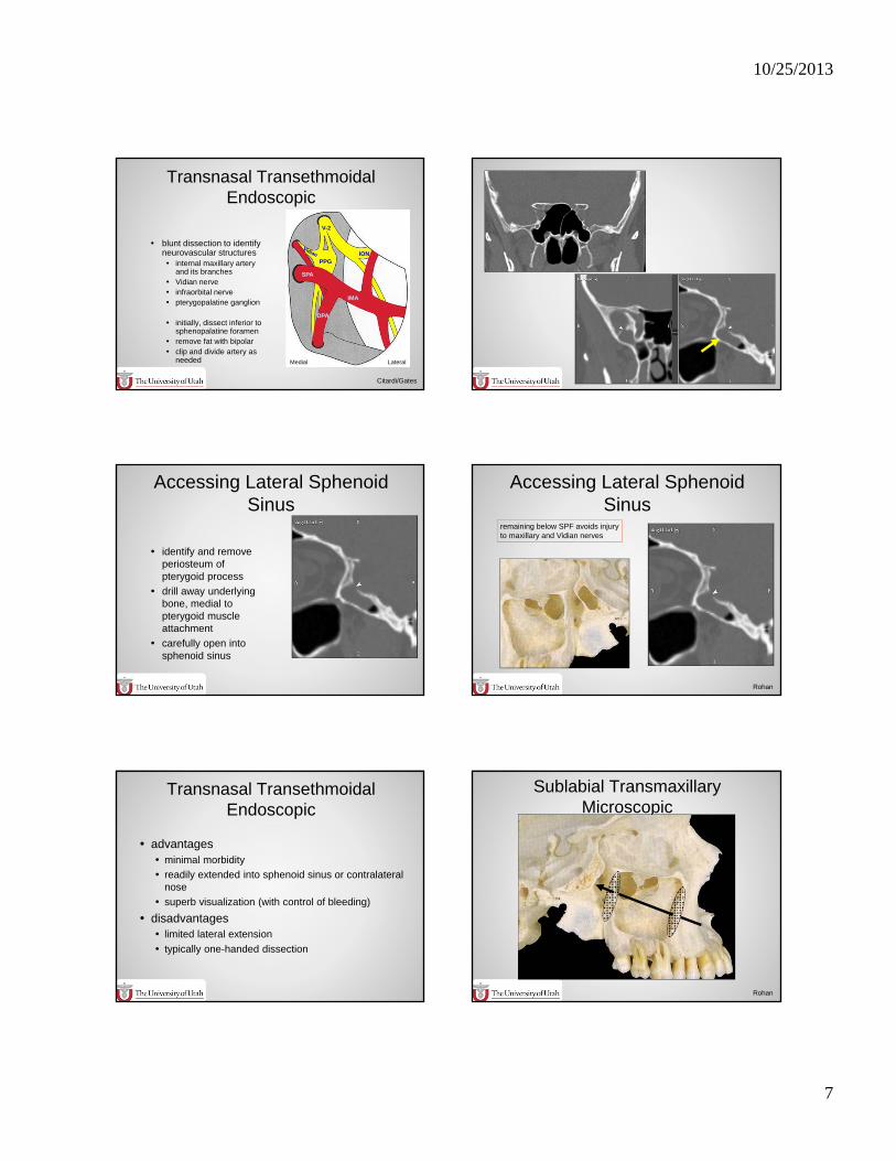
blunt dissection to identify neurovascular structures internal maxillary artery
and its branches Vidian nerve infraorbital nerve pterygopalatine ganglion
initially, dissect inferior to sphenopalatine foramen
remove fat with bipolar clip and divide artery as
needed
IMA
PPG
DPA
SPA
V-2
ION
LateralMedial
Citardi/Gates
Accessing Lateral Sphenoid Sinus
identify and remove periosteum of pterygoid process
drill away underlying bone, medial to pterygoid muscle attachment
carefully open into sphenoid sinus
Accessing Lateral Sphenoid Sinus
remaining below SPF avoids injuryto maxillary and Vidian nerves
Rohan
Transnasal Transethmoidal Endoscopic
advantages minimal morbidity
readily extended into sphenoid sinus or contralateral nose
superb visualization (with control of bleeding)
disadvantages limited lateral extension
typically one-handed dissection
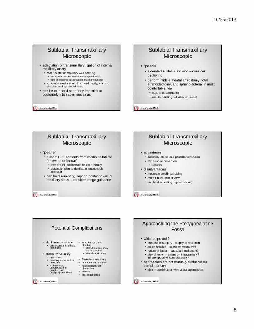
Sublabial Transmaxillary Microscopic
Rohan
10/25/2013
8
Sublabial Transmaxillary Microscopic
adaptation of transmaxillary ligation of internal maxillary artery wider posterior maxillary wall opening
can extend into the medial infratemporal fossa care to preserve posterolateral maxillary buttress
extension medially into the nasal cavity, ethmoid sinuses, and sphenoid sinus
can be extended superiorly into orbit or posteriorly into cavernous sinus
Sublabial Transmaxillary Microscopic
“pearls” extended sublabial incision – consider
degloving
perform middle meatal antrostomy, total ethmoidectomy, and sphenoidotomy in most comfortable way (e.g., endoscopically)
prior to initiating sublabial approach
Sublabial Transmaxillary Microscopic
“pearls” dissect PPF contents from medial to lateral
(known to unknown) start at SPF and remain below it initially dissection plan is identical to endoscopic
approach
can be disorienting beyond posterior wall of maxillary sinus – consider image guidance
Sublabial Transmaxillary Microscopic
advantages superior, lateral, and posterior extension
two handed dissection suctioning
disadvantages moderate swelling/bruising
more limited field of view
can be disorienting superomedially
Potential Complications
skull base penetration cerebrospinal fluid leak,
meningitis
cranial nerve injury optic nerve maxillary nerve and its
branches Vidian nerve,
pterygopalatine ganglion, and postganglionic fibers
vascular injury and bleeding internal maxillary artery
and its branches internal carotid artery
Eustachian tube injury mucocele and sinusitis nasolacrimal duct
obstruction trismus oral-antral fistula
Approaching the Pterygopalatine Fossa
which approach? purpose of surgery – biopsy or resection lesion location – lateral or medial PPF nature of lesion – vascular? malignant? size of lesion – extension intracranially?
infratemporally? contralaterally?
approaches are not mutually exclusive but complimentary also in combination with lateral approaches