Embed Size (px)
Citation preview

1
RESPIRATORY ASSESSMENT
Beth Klements, MS, PCPNP-BC, AE-CAsthma Clinical Nurse Specialist
Pediatric Nurse Practitioner
Anatomy of the Respiratory SystemUpper Respiratory Tract:
• Nostrils, nasopharynx, Eustachian tubes, sinuses, larynx and upper trachea
Lower Respiratory Tract:
• Lower trachea
• Bronchi
• Right & Left Lung
Lower Respiratory Tract
• Three right lobes separated by a minor fissure
• Right main stem bronchus is shorter and wider
• More common to find foreign body • Two left lobes separated by a major fissure• Diaphragm is main muscle of respiration• Accessory muscles: intercostal, sternocleidomastoid, spinal, neck and abdominal muscles

2
Anatomic & Physiologic Differences within the Pediatric Respiratory
Tract
Factors Placing a Child at Risk for Respiratory Disease
Premature birth
Smoking within the home
Maternal smoking during pregnancy
Ill contacts (like Day Care or School)
Altered immune mechanisms
Chronic illnesses
Congenital defects with respiratory association
Pertinent History
Onset of symptoms
What day and time
Key signs & symptoms
Shortness of breath
Noisy breathing
Cough
Type
Sputum production

3
History Intensity/Progression of the symptoms
Worsening
Acute (less than three weeks)
Chronic (greater than 3 weeks)
Recurrent (symptom free at least 2 days in past weeks)
Associated symptoms
stomach ache
ear ache
illnesses in past
History
Treatment currently being used
Any Over the Counter medications
Prescriptions
Home remedies
History
Environmental factorsSmoking in the home (Keep in mind adolescents)Pets Other new exposures
Seasonal allergiesExercise related symptomsBirth/ Family History

4
Physical Examination
Vital Signs
respiratory rate -- key indicator of lower respiratory involvement
Remember – lowering temp may bring other vital signs into normal range
Normal Respiratory Rates
Age (years) Respiratory (breaths/min)
0-1 25-40
1-5 20-30
5-10 15-25
10-16 15-20

5
Respiratory Distress
Resting respiratory rate > 60 breaths/min in infants < 2 months of age
Resting respiratory rate of greater than 50 breaths/min in infants 2-12 months of age
Resting respiratory rate of greater than 40 breaths/min in children 1-5 years
General Appearance
Nasal flaring
Position of comfort
Level of anxiety
Affect
Color
Inspection
Thoracic deformities or asymmetry
Nasal flaring, mouth breathing
Use of accessory muscles
Retractions

6
Inspection of other body areas
Respiratory Rate: Note depth, ease and rhythm
Fingernails and nail beds for cyanosis
Clubbing of fingers…related to decreased perfusion
Palpation
Paranasal & frontal sinuses
Chest & Abdominal configuration
Areas of tenderness over the chest wall
Lymph nodes for any enlargement
Transmitted Voice
Tactile Fremitus….Ask the child to say “99” as you use the bony part of your palm to palpate the vibrations it makes through the anterior/posterior lung fields
Decreased sensation with asthma
Often absent in a collapsed lung
Increased over a consolidated area
7
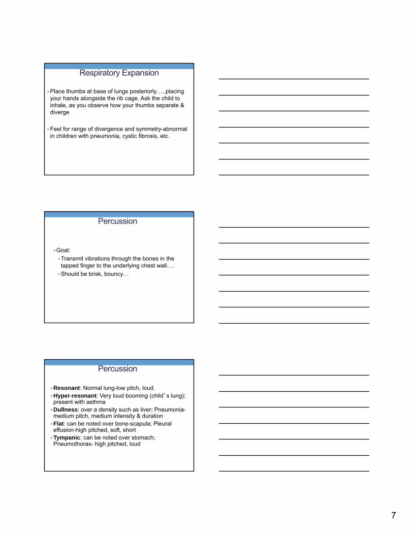
Respiratory Expansion
Place thumbs at base of lungs posteriorly…..placing your hands alongside the rib cage. Ask the child to inhale, as you observe how your thumbs separate & diverge
Feel for range of divergence and symmetry-abnormal in children with pneumonia, cystic fibrosis, etc.
Percussion
Goal:
Transmit vibrations through the bones in the tapped finger to the underlying chest wall….
Should be brisk, bouncy…
Percussion
Resonant: Normal lung-low pitch, loud.Hyper-resonant: Very loud booming (child’s lung); present with asthmaDullness: over a density such as liver; Pneumonia-medium pitch, medium intensity & durationFlat: can be noted over bone-scapula; Pleural effusion-high pitched, soft, shortTympanic: can be noted over stomach; Pneumothorax- high pitched, loud

8
Auscultation
Use of stethoscope
Diaphragm: High pitched sounds ( better in measuring normal and abnormal lung sounds)
Bell: Low pitched sounds
Auscultation
Correct Technique• Placing bell or diaphragm against chest wall.• Keeping noise level down.• Attempting to have child upright to access all areas for auscultation
Errors • Listening to breath sounds over clothing• Auscultating in a noisy room• Auscultating only convenient areas
Auscultation
Note:
Symmetry of breath sounds
Normal ?
Increased ?
Decreased ?
Adventitious sounds = extra sounds

9
Cough
Become familiar with the characteristics of the various types of coughs
Ask the child to cough during the examination
Different kinds of cough associated with common and not so common respiratory conditions
Abnormal Inspiratory/Expiratory Ratio
Expiration is prolonged in asthma due to air trapping (called hyperinflation)
As a result, the inspiratory/expiratory ratio is often �1:2 (expiration is more than twice as long as inspiration.)
Abnormal or Adventitious Lung Sounds
Crackles:
Intermittent, brief, non-musical type sounds
Fine Crackles: soft, high pitched & brief
Coarse Crackles: louder, lower in pitch &
not very brief
10
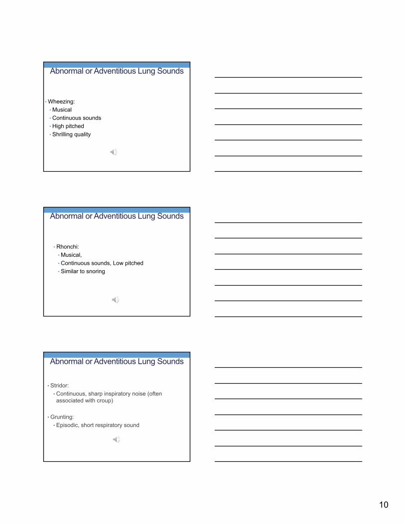
Abnormal or Adventitious Lung Sounds
• Wheezing:
• Musical
• Continuous sounds
• High pitched
• Shrilling quality
Abnormal or Adventitious Lung Sounds
• Rhonchi:
• Musical,
• Continuous sounds, Low pitched
• Similar to snoring
Abnormal or Adventitious Lung Sounds
• Stridor:
• Continuous, sharp inspiratory noise (often associated with croup)
• Grunting:
• Episodic, short respiratory sound

11
Clinical implications
• Crackles: May suggest pneumonia, bronchitis or congestive heart failure
• Wheezing: Suggests constricted or narrow airways as in asthma or bronchiolitis
• Rhonchi: indicate secretions within the large airway (upper respiratory congestion)
Coughs
• Sinusitis: Productive, night and day, worse when first laying down and on awakening
• Bronchitis: Dry, then loose & rattling cough
• Pneumonia: Often loose cough
• Asthma: Dry, tight, occasionally wheezy
• Pertussis: Spasmodic, choking, repetitive (no inspiration during coughing spasm)
• Croup: Seal-like bark
Common Respiratory Illnesses
• Upper Respiratory Tract
• Upper respiratory infection
• Sinusitis
• Foreign body of the airway
• Pharyngitis (viral, bacterial) & Tonsillitis
• Pertussis
• Lower Respiratory Tract
• Bronchitis
• Pneumonia
• Asthma

12
Airway Obstruction
Airway Obstruction - Symptoms
• Wheezing
• Repetitive hacking cough, often ineffective
• Dyspnea
• Post-tussive vomiting
• Cyanosis
Obstruction – Physical Findings
• Diminished chest excursion
• Prolonged expiratory phase
• Increased respiratory rate
• Increased airway resistance; over-inflation of lungs; barrel chest
• Percussion over inflated chest hyper-resonance
• Increased use of accessory muscles
13
Allergic Rhinitis
Allergic Rhinitis
• Can significantly interfere with Quality of Life• Affects important social interactions• Affects school performances
-Decreased attention-Increased difficulty with cognitive skills due to combination of medications and symptoms
Allergic Rhinitis - Treatment
• Allergen identification
• Environmental controls
• Stepwise approach similar to asthma (modify based on symptoms)
• Intranasal steroids are most effective
• Antihistamines
• Immunotherapy if indicated

14
Sinusitis
Sinus Development
• Maxillary – 3rd to 4th month fetal gestation
• Ethmoid – birth• Sphenoid – 5 years• Frontal – 7-8 years (fully developed by age 20)
Risk Factors
• Frequent viral infections
• Environmental allergies
• Allergic rhinitis
• Smoke exposure
• Day care
• Gastroesophageal Reflux

15
Sinusitis - Characteristics
• Inflammation of mucous membranes that line the sinuses
• Interferes with normal sinus drainage
• Air and mucous become trapped
• Bacteria multiplies
Symptoms
• “Cold” lasting more than 2 weeks
• Thick yellow/green discharge
• Post nasal drip
• Sore throat especially in AM
Symptoms
• Usually no headache/toothache
• No facial tenderness
• Swelling around eyes
• Appearance of conjunctivitis
• Irritability or fatigue

16
Microbiology
• Principal pathogens
• Streptococcus Pneumoniae 30%
• Haemophilus influenzae 20%
• Moraxella Catarrhalis 20%
Treatments
• Imaging studies not recommended <6years
• CT scan sometimes necessary for severe disease
Prevention
• Vaccinations
• Hib
• Prevnar and/or Pneumovax
• Environmental Controls
• Nasal Saline Washes/sprays
• Nasal steroids

17
Asthma
Asthma -- Characteristics
• A Chronic inflammatory disease of the airways
• Characterized by
• Episodic wheezing
• Episodic coughing
• Chronicity
• Hyper-responsiveness of airways to a variety of stimuli
• Largely reversible obstruction of the airways
Asthma - Physical Findings
• Dyspnea, labored breathing
• Retractions
• Cough, restlessness, apprehension and fatigue
• Cyanosis of lips, nail beds, gums
• Tachycardia
18
Asthma - Physical Findings
• Physical Signs
• Prolonged expiratory phase
• Wheezing & Rhonchi
• Increased Obstruction: High pitched; breath sounds diminished
• In severe obstruction, breath sounds may be silent because of poor air exchange
Asthma - Physical Findings
• Dullness to percussion over the area of consolidation
• Decreased breath sounds
• Tactile Fremitus increased
• Crackles suggest pneumonia occasionally wheezing
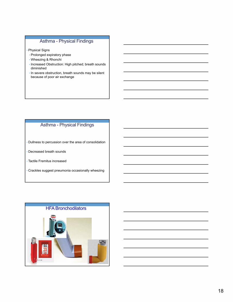
HFA Bronchodilators

19
Current Albuterol Formulations
• Ventolin HFA – with built-in dose counter
• Proventil HFA
• ProAir HFA
• Combivent – (Albuterol and Atrovent)
• Xopenex HFA - Levalbuterol
Long-Term Control
• Taken daily, over a long period of time
• Used to reduce inflammation, relax airway muscles, and improve symptoms and pulmonary function
• Inhaled corticosteroids
• Long-acting beta2-agonists
• Leukotriene modifiers
Inhaled Corticosteroid and Combination Therapies in MDIs

20
Long Term Control: Inhaled Corticosteroids
Fluticasone (Flovent)3 different strengths, all orange
Budesonide (Pulmicort)Ciclesonide (Alvesco)Mometasone (Asmanex)Beclomethasone (QVAR)Flunisolide (Aerospan) New in 2014Fluticasone furoate (Arnuity Ellipta) New in 2015

21
Long-acting Beta2 Agonists
Relax bronchial smooth muscle
Available as DPI
12 hour duration of action
Most effective when in combination with inhaled steroids (not mono therapy)
Effective control for nocturnal symptoms
Combination Inhaled Corticosteroids and Long Acting Beta Agonists
Advairo Fluticasone/salmeterolo MDI or Discuso Approved for 6 years and olderSymbicorto Budesonide/Formoterolo MDI –requires a spacero Approved for ≥ 12 y.oDulerao Mometasone/Formoterolo MDI –requires a spacero Approved for ≥ 12 y.oBreoo Fluticasone/Vilanterolo Approved for ≥ 18 y.o.
Leukotriene Modifiers Montelukast (Singulair) – Once daily tablet, chewable,
& sprinkles Approved for ages ≥ 1 year
4 mg, 5 mg, 10 mg
Black Box Warning
Zafirlukast (Accolate) – Twice daily tablet Ages ≥ 5 years
Requires routine monitoring of Liver function
Zileuton (Zyflo) – BID or QID ≥ 12 years
Requires routine monitoring of Liver function
22
Omalizumab (Xolair)
Indications for Xolair
• 12 years or older
• Moderate or Severe Persistent Asthma
• Positive skin test
• Poor control on inhaled steroids
• IgE levels are 30-700 IU/ml
Mepolizumab (Nucala)
• Add-on maintenance treatment
• ≥ 12 years old, with severe asthma
• Reduces blood eosinophils, which may contribute to asthma
• 100 mg subcutaneous injection every 4 weeks

23
©2017
Reslizumab (Cinqair)
Add‐on maintenance treatment
≥ 18 years old, with severe asthma
Reduces blood eosinophils, which may contribute to asthma
IV infusion, 3 mg/kg, every 4 weeks, over 25‐50 minutes
Reasons for Poor Control
• Wrong Diagnosis
• Poor adherence to recommended Treatment
• Under estimation of Asthma Severity
• Co-morbidities
• Obesity
• Gastroesophogeal reflux
• Mental illness
Follow up
• Visits every 2-6 weeks until control is achieved
• Are adjustments necessary?
• Before increasing meds, consider
• Environment
• Adherence
• Co-morbidities
• Step up if not controlled
• If poor control, consider increasing by 2 steps, oral corticosteroids, or both

24
Follow up
• When follow up is achieved, contact every 3 – 6 months
General Guidelines for Referral to an Asthma Specialist
• Patient has had a life-threatening asthma exacerbation.
• Patient is not meeting the goals ofasthma therapy.
• Signs and symptoms are atypical.
• Other conditions complicate asthma.
25
Asthma Action Plans
• Describe regular medications and measures
• Describe actions to take when asthma worsens
• Asthma severity classification
• School Nurse permission form
Website for 2007 NHLBI guidelines
• www.nhlbi.nih.gov/guidelines/asthma
Asthma/Allergy Connection
• Treatment of one disease improves the other
• Allergen immunotherapy lowers risk of asthma

26
Asthma Management
• #1- Education of patient / family
• Pharmacological approaches
• Identify and eliminate or decrease exposure to allergens
• Allergen immunotherapy
Guideline for Management of Ambulatory Asthma Patients
Patient (2-25 years) presents in clinicwith wheezing or SOB
Triage assessment by RN/NP/MD
Is patient in Severe Respiratory Distress?(Emergent)•Color pale to cyanotic•Severe Dyspnea•Inaudible breath sounds•Altered mental status
Provide supplemental oxygen
Immediate transport to ED via wheel chair by RN/NP/MD or call Code Blue
ED charge nurse (5-1800) and expect line (5-2170) called by another clinician
Is patient in Moderate Respiratory distress/Urgent?•Moderate Dyspnea •Inspiratory &/or expiratory wheezes •Moderate retractions
Neb #1 – Albuterol 0.5%, 0.5ml in 2ml NS
Steroids (prednisone/prednisolone) if incomplete response to 1st Neb or sooner if clinically indicated
(Patients ≤ 40 kg: 2 mg/kg; Patients > 40 kg: 40- 80 mg x 1 dose)
Neb #2 – Albuterol 0.5%, 0.5mL in 2 mL NS or combination of Albuterol and Ipratropium 0.02% neb (infants and children less than 12 y/o: 250 mcg; children >/=12 y/o 500 mcg)
Is patient in Moderate Respiratory Distress after receiving ≥ 2 Nebs?
Call ED Charge nurse (5-1800) and expect line (5-2170)
Transport Patient to ED
Discharge home
Is patient in Mild Respiratory distress/Non-Urgent?•Mild Dyspnea•None or end expiratory wheezes• None or mild retractions•O2sat (RA)≥ 95%
Yes
Yes
No
No
Yes
•Consider Albuterol MDI with spacer or neb
No
© Updated May 2013
Continue to monitor or transfer - per provider’s
clinical judgment
or
Risk Factors for Asthma Deaths
• Previous life-threatening asthma, respiratory arrest• Hospitalization or emergency room visit for asthma within the past year
• Use of two or more Beta agonist, metered dose inhalers (Albuterol) each month
• Poor perception of hypoxia or airway obstruction• Psychosocial disturbances, obesity, drug
abuse
27
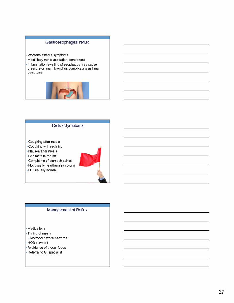
Gastroesophageal reflux
• Worsens asthma symptoms
• Most likely minor aspiration component
• Inflammation/swelling of esophagus may cause pressure on main bronchus complicating asthma symptoms
Reflux Symptoms
• Coughing after meals
• Coughing with reclining
• Nausea after meals
• Bad taste in mouth
• Complaints of stomach aches
• Not usually heartburn symptoms
• UGI usually normal
Management of Reflux
• Medications
• Timing of meals
• No food before bedtime
• HOB elevated
• Avoidance of trigger foods
• Referral to GI specialist
28
Pneumonia
• Caused by infectious agents which invade the lungs, creating an inflammatory response….loss of air and consolidation
Pneumonia - Characteristics
• Cough
• Day & night
• Often productive-yet not noticed since children swallow mucus
Pneumonia – Physical findings
• Chest pain (often a complaint of older children)
• Tachypnea
• Retractions
• Nasal Flaring
• Cyanosis
29
Cystic Fibrosis - Characteristics
• Autosomal recessive genetic disorder
• Inadequate salt and water secretion on the cellular level
• Inability to clear mucoid secretions
• Inability to clear secretions from endo-bronchial spaces-leads to colonization by bacteria and results in chronic inflammation
Cystic Fibrosis - Physical Findings
• Increase cough
• Increase sputum production
• Decreased breath sounds
• Inspiratory and Expiratory crackles
• Shortness of breath
• Fatigue
• Tachypnea
Bronchitis - Characteristics
• Inflammation of the bronchus main stem-combined with concurrent or lower respiratory tract infection
• Usually preceded by a viral URI

30
Physical Exam Findings
• Low grade or no fever
• Nasopharyngeal infection-conjunctivitis and rhinitis
• Coarse breath sounds
• Rhonchi-high pitched and resemble wheezes
Pertussis
• Commonly known as “whooping cough”
• High-pitched inspiratory whoop
• Highly contagious in young children
• Caused by Bordetella pertussis, Bordetella parapertussis, Bordetella bronchiseptica
• Transmitted via aerosol droplets from close contact
Pertussis - Physical Findings
• Catarrhal Stage- 1-3 weeks
• Mild cough, sneezing and fever

31
Pertussis - Physical Findings
• Paroxysmal Stage- 2-4 weeks
• Persistent staccato, paroxysmal cough ending with inspiratory whoop
• Vomiting at end of paroxysmal cough and whoop
• Cyanosis, sweating, exhaustion after coughing
Pertussis - Physical Findings
• Convalescent Stage- 2-3 weeks
• Waning of paroxysmal coughing episodes
Deformities of the Thorax

32
Pectus ExcavatumDepression in the lower sternum; “Funnel Chest”. Usually only cosmetic…..but if very pronounced, it can restrict lung capacity
Pectus Carinatum: Upper ribs bending inward and the sternum is being thrust outward (more anteriorly)…also
called pigeon breast.
Scoliosis – Curving of the spineOne shoulder may seem higher than the other
33
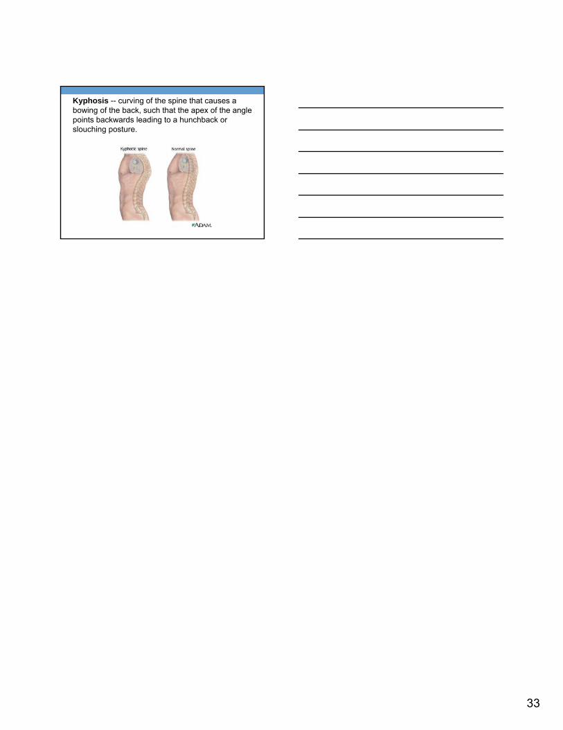
Kyphosis -- curving of the spine that causes a bowing of the back, such that the apex of the angle points backwards leading to a hunchback or slouching posture.