Embed Size (px)
Citation preview

3,350+OPEN ACCESS BOOKS
108,000+INTERNATIONAL
AUTHORS AND EDITORS114+ MILLION
DOWNLOADS
BOOKSDELIVERED TO
151 COUNTRIES
AUTHORS AMONG
TOP 1%MOST CITED SCIENTIST
12.2%AUTHORS AND EDITORS
FROM TOP 500 UNIVERSITIES
Selection of our books indexed in theBook Citation Index in Web of Science™
Core Collection (BKCI)
Chapter from the book Spine SurgeryDownloaded from: http://www.intechopen.com/books/spine-surgery
PUBLISHED BY
World's largest Science,Technology & Medicine
Open Access book publisher
Interested in publishing with IntechOpen?Contact us at [email protected]
10
Autologous Macrophages Genetically Modified by Ex Vivo Electroporation and Inserted by
Lumbar Puncture Migrate and Concentrate in Damaged Spinal Cord Tissue: A Safe and Easy
Gene Transfer Method for the Treatment of Spinal Cord Injury
Tadanori Ogata, Tadao Morino, Hideki Horiuchi, Masayuki Hino, Gotaro Yamaoka and Hiromasa Miura
Department of Orthopaedic Surgery, Ehime University School of Medicine Japan
1. Introduction
Spinal cord injury is one of the most serious conditions in the field of orthopedic surgery.
Several pharmacological trials have been performed for the treatment of traumatic spinal
cord injury. However, only high-dose steroid therapy has been established as an effective
treatment for this condition. It is necessary to develop an effective treatment to inhibit
secondary neuronal damage and to promote neuronal regeneration after the spinal cord
injury.
Recently, direct delivery of neurotrophic factors, such as nerve growth factor (NGF), glial-
derived neurotrophic factor (GDNF), and brain-derived neurotrophic factor (BDNF), has
been demonstrated to provide neuroprotection and counteract lesion-induced atrophy after
traumatic injury to the central nervous system (CNS) (Kobayashi et al., 1997; Houle et al.,
1999; Bregman et al., 2002; Cao et al., 2002; Zhou, et al., 2006).
Intravenous applications of neurotrophic factors, such as GDNF and BDNF, are also
possible therapeutic methods. In the injured spinal cord, however, blood flow in the
nervous tissue decreases remarkably (Hamamoto et al., 2007). When neuroprotective
substances are added intravenously, only a small amount of the substances reach the injured
portion of the spinal cord. In addition, the half lives of most proteins in vivo are relatively
short. Therefore, systemic intravenous administration of neurotrophic protein may not be an
efficient way to treat damaged spinal cord tissue.
Direct infusion of neurotrophic proteins into the neural parenchyma using pumps is another approach to treatment. However, several limitations should be considered as follows: 1) The spread of the proteins throughout the neural parenchyma is often limited. 2) The chronic implantation of the canula in the parenchyma results in the formation of a
www.intechopen.com
Spine Surgery
138
neural scar at the insertion site. 3) The implanted canula may induce inflammation or clogging of the infusion device. 4) Continuous outflow of liquid can cause additional damage at the insertion site.
Several trials of gene transfer into the CNS have been conducted both in vivo and in vitro. Adenovirus including the target genes has been successfully used to achieve gene transfer in the CNS (Fink et al., 2000; Miagkov et al., 2004; Kwon et al., 2007). However, viral infection of the CNS may be too dangerous for clinical use because of the risk of meningitis (Driesse et al., 2000).
To develop a novel system for substance delivery to damaged ischemic tissue, we focused on the tissue-migration ability of macrophages. Macrophages migrate into damaged tissue or inflammatory tissue. After spinal cord injury, the appearance of macrophages in the damaged tissue has been reported (Dusart et al., 1994; Morino et al., 2003). For this study, gene transfer by ex vivo electroporation, a non-viral gene transfer method, was performed on autologous macrophages, and the cells were injected into the subarachnoid space. It is believed that if the cells migrate and concentrate in the damaged spinal cord, it will provide a safe and effective method of substance delivery to the damaged spinal cord parenchyma.
2. Experimental procedures
2.1 Animals
A total of 56 male Wistar rats (350 g-weight, purchased from Japan Clea Co., Japan) were used for this experiment (in vivo 50, in vitro 6). The research protocol was accepted by the ethical committee for animal experiments at Ehime University (Ehime, Japan).
2.2 Collection of autologous macrophages and GFP gene incorporation
Intraperitoneal macrophages were easily collected from rats. The intraperitoneal space was rinsed with 30 ml of Dulbecco's modified Eagle medium (DMEM, Gibco, Grand Island, NY) via midline incision of the abdomen. The DMEM, which contained a large concentration of macrophages, was collected and centrifuged, and cells were re-suspended in cell permeabilization buffer (140 mM KCl, 5 mM NaCl, 10 mM glucose, 0.5 mM EGTA, 10 mM HEPES, pH 7.2). To identify gene-transfected cells, we used pEGFPLuc Vector (Clontech Inc. USA). This vector was constructed by inserting the GFP gene for over-expression of GFP
protein. 200 l of the cells (1 x 10⁶ cells /ml) were mixed with 20 l of GFP-containing vector in disposable cuvette-electrodes (2 mm gap, BTZ 620, BTX Inc., USA). The final
concentration of the vector was 0.1 g/l. Then, six 20 ms electric pulses of 20 V were applied by electroporator (CUY 21, NEPA GENE Co., Japan). The treated cells were then cultured (in vitro) or returned to the animals by intrathecal application (in vivo).
2.3 Cell culturing and observation of GFP protein expression
The gene-transfected macrophages were cultured in 6-well culture plates (Nunc, Naperville, IL) at a concentration of 5 x 10⁴ cells/well with DMEM containing 15% fetal calf serum. Culturing was performed in a humidified 5% CO2 atmosphere at 37°C. Seven days after culturing, the cells were observed under a fluorescent microscope.
www.intechopen.com

Autologous Macrophages Genetically Modified by Ex Vivo Electroporation and Inserted by Lumbar Puncture Migrate...
139
2.4 Spinal cord injury model (SCI model)
Under general anesthesia using halothane, the rat spinal cord was carefully exposed by removing the vertebral lamina at the 11th vertebra. Spinal cord impact injury was performed using a MASCIS Impactor (New Jersey, USA). The impact weight was dropped from a height of 25 mm. In one group of rats, a laminectomy of the 11th vertebra was performed without spinal cord injury (sham).
2.5 Macrophage transplantation into the spinal cord by lumbar puncture
100 l of a liquid suspension of the gene-transfected macrophages (1 x 10⁵ cells) was injected into the subarachnoid space at the 4-5th lumbar intervertebral level just after the spinal cord injury.
2.6 Histological examination
Rats were sacrificed for histological study by deep anesthesia using diethyl ether and their spinal cords were taken out immediately. Horizontal or axial frozen sections with a
thickness of 20 m were produced, and autofluorescence was observed under fluorescence microscopy. To quantify the number of migrated gene-transfected macrophages, photographs at an area peripheral to the center of the SCI were taken and the number of GFP-positive cells were counted by three individuals who did not know any information about the pictures. For the first 96 hours after the SCI, the GFP autofluorescence was weak, and therefore, the cells were hardly distinguishable from the background until after 96 hours had elapsed. The counts were averaged and data were expressed as the number of GFP-positive cells per 1 mm2.
To confirm the expression of transferred GFP, some sections were subjected to
immunostaining by anti-GFP antibody according to avidin-biotin-complex (ABC) method
using Vectastain ABC kit (Vector labo. Inc., CA). The sections were fixed on glass slides
with 4% paraformaldehyde in phosphate-buffered saline (PBS) for 5 minutes. Then, after
washing twice with PBS, endogenous peroxidase was blocked by treatment with 3%
H2O2/H2O for 5 minutes. Slices were exposed to anti-GFP antibody (1:1000 in 1% horse
serum/PBS; MBL Co., Ltd., Nagoya, Japan) overnight at 4C. Sections were then washed
by PBS and exposed to biotin-conjugated anti-rabbit IgG for 1 hour at room temperature.
After washing the antibody, sections were treated by ABC method according to the assay
protocol of the kit, and finally colour developed with 3,3'-diaminobenzidine
tetrahydrochloride (DAB, Wako Chemicals Ltd.) substrate (0.02% H2O2 plus 0.1% DAB in
0.1M Tris-buffer) for 5 minutes and washed immediately with water for 20 minutes.
Sections were dehydrated through graded alcohols and xylene and were then mounted in
HSR solution (Yoshitomi co., Osaka, Japan).
To clarify which kinds of cells had been transplanted, some sections were subjected to immunostaining by OX-42, a marker of macrophages. Frozen sections were prepared according to the method mentioned above. Then, after washing twice with PBS, slices on slides were exposed to anti-OX42 antibody (Immunotech. Co. Marseille, France) overnight
at 4C, and rhodamine-conjugated anti-mouse IgG antibody (Chemicon International Co. CA, USA) for 1 hour. The sections were then observed under fluorescent microscopy.
www.intechopen.com
Spine Surgery
140
2.7 Evaluation of motor function
Motor function was assessed with the Basso, Beattie and Bresnahan (BBB) scoring scale (Basso et al., 1996). The BBB scale is a 21-point scale that ranks no locomotion as 0 points and a normal gait as 21 points. The BBB scale is one of the most widely used methods for evaluating hind-limb motor function in rats and mice. Hind-limb motor function was evaluated at 2, 3, 4, 7, 14, 21, 28, and 56 days after SCI. The evaluation of BBB scores was done by three individuals who were unaware of the treatments that the rats had received. The average of the three observers’ scores was employed as data in this study.
3. Results
3.1 Expression of green fluorescent protein (GFP) protein in cultured gene-transfected macrophages
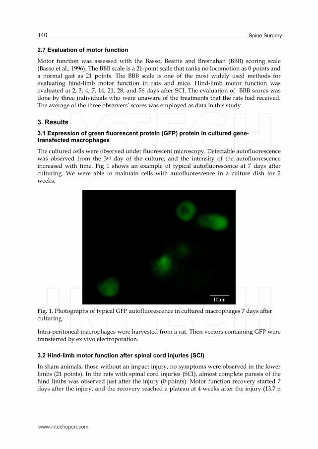
The cultured cells were observed under fluorescent microscopy. Detectable autofluorescence was observed from the 3rd day of the culture, and the intensity of the autofluorescence increased with time. Fig 1 shows an example of typical autofluorescence at 7 days after culturing. We were able to maintain cells with autofluorescence in a culture dish for 2 weeks.
Fig. 1. Photographs of typical GFP autofluorescence in cultured macrophages 7 days after culturing.
Intra-peritoneal macrophages were harvested from a rat. Then vectors containing GFP were transferred by ex vivo electroporation.
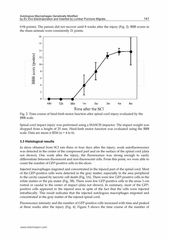
3.2 Hind-limb motor function after spinal cord injuries (SCI)
In sham animals, those without an impact injury, no symptoms were observed in the lower limbs (21 points). In the rats with spinal cord injuries (SCI), almost complete paresis of the hind limbs was observed just after the injury (0 points). Motor function recovery started 7 days after the injury, and the recovery reached a plateau at 4 weeks after the injury (13.7 ±
www.intechopen.com
Autologous Macrophages Genetically Modified by Ex Vivo Electroporation and Inserted by Lumbar Puncture Migrate...
141
0.94 points). The paresis did not recover until 8 weeks after the injury (Fig. 2). BBB scores in the sham animals were consistently 21 points.
Fig. 2. Time course of hind-limb motor function after spinal cord injury evaluated by the BBB scale.
Spinal cord impact injury was performed using a MASCIS impactor. The impact weight was dropped from a height of 25 mm. Hind-limb motor function was evaluated using the BBB scale. Data are mean ± SEM (n = 4 to 6).
3.3 Histological results
In slices obtained from SCI rats three or four days after the injury, weak autofluorescence was detected in the center of the compressed part and on the surface of the spinal cord (data not shown). One week after the injury, the fluorescence was strong enough to easily differentiate between fluorescent and non-fluorescent cells. From this point, we were able to count the number of GFP-positive cells in the slices.
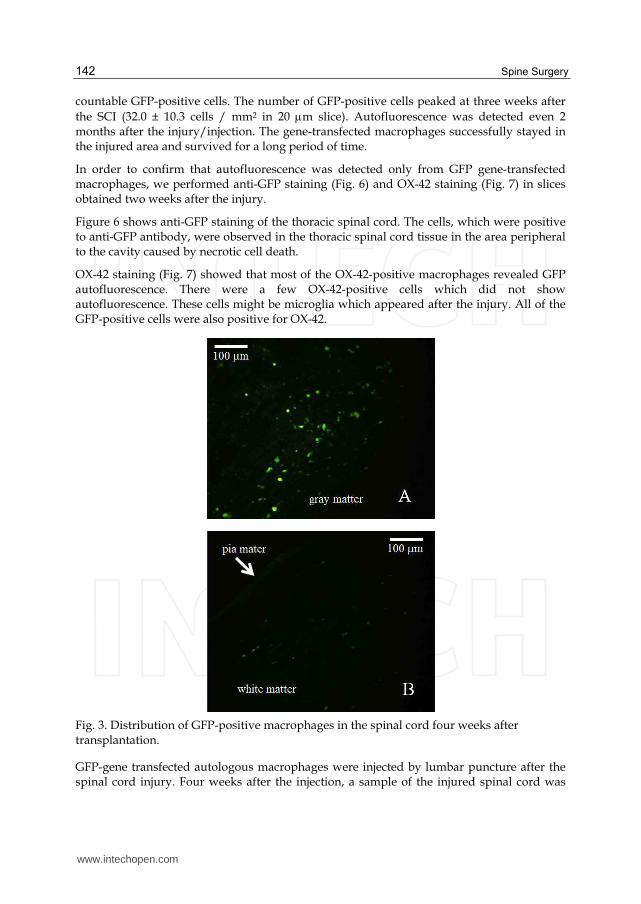
Injected macrophages migrated and concentrated in the injured part of the spinal cord. Most of the GFP-positive cells were detected in the gray matter, especially in the area peripheral to the cavity caused by necrotic cell death (Fig. 3A). There were few GFP-positive cells in the white matter or the pia mater (Fig. 3B). There were few GFP-positive cells in the areas 1-cm rostral or caudal to the center of impact (data not shown). In summary, most of the GFP-positive cells appeared in the injured area in spite of the fact that the cells were injected intrathecally. This result indicates that the injected autologous macrophages migrated and concentrated in the gray matter of the injured spinal cord.
Fluorescence intensity and the number of GFP-positive cells increased with time and peaked at three weeks after the injury (Fig. 4). Figure 5 shows the time course of the number of
www.intechopen.com
Spine Surgery
142
countable GFP-positive cells. The number of GFP-positive cells peaked at three weeks after
the SCI (32.0 ± 10.3 cells / mm2 in 20 m slice). Autofluorescence was detected even 2 months after the injury/injection. The gene-transfected macrophages successfully stayed in the injured area and survived for a long period of time.
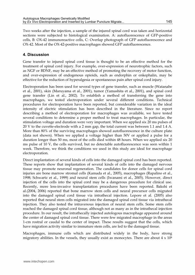
In order to confirm that autofluorescence was detected only from GFP gene-transfected macrophages, we performed anti-GFP staining (Fig. 6) and OX-42 staining (Fig. 7) in slices obtained two weeks after the injury.
Figure 6 shows anti-GFP staining of the thoracic spinal cord. The cells, which were positive to anti-GFP antibody, were observed in the thoracic spinal cord tissue in the area peripheral to the cavity caused by necrotic cell death.
OX-42 staining (Fig. 7) showed that most of the OX-42-positive macrophages revealed GFP autofluorescence. There were a few OX-42-positive cells which did not show autofluorescence. These cells might be microglia which appeared after the injury. All of the GFP-positive cells were also positive for OX-42.
Fig. 3. Distribution of GFP-positive macrophages in the spinal cord four weeks after transplantation.
GFP-gene transfected autologous macrophages were injected by lumbar puncture after the spinal cord injury. Four weeks after the injection, a sample of the injured spinal cord was
www.intechopen.com

Autologous Macrophages Genetically Modified by Ex Vivo Electroporation and Inserted by Lumbar Puncture Migrate...
143
taken and axial sections were subjected to histological examination. Most of the GFP-positive cells were detected in the gray matter (A). There were few GFP-positive cells in the white matter or in the pia mater (B).
Fig. 4. Autofluorescence of GFP-positive macrophages in the gray matter of the injured spinal cord. GFP-gene transfected autologous macrophages were injected by lumbar puncture after the spinal cord injury. One (A), two (B), three (C) and four (D) weeks after the injury/injection, a sample of the injured spinal cord was taken and horizontal sections were subjected to histological examination. The pictures show the areas peripheral to the center of the impact injury.
Fig. 5. Number of GFP-positive cells in the center of the damaged spinal cord tissue.
At the indicated time point after the injury/injection, a sample of the injured spinal cord was taken and horizontal sections were subjected to histological examination. The cells,
www.intechopen.com

Spine Surgery
144
which revealed significant autofluorescence in the area peripheral to the center of impact injury, were counted. Data are shown in mean ± SEM (n = 4 to 6).
Fig. 6. Anti-GFP staining of the spinal cord two weeks after transplantation.
Two weeks after the injection, a sample of the injured spinal cord was taken and horizontal sections were subjected to histological examination. Horizontal sections in the thoracic spinal cord were stained by anti-GFP antibody. Gene-transferred autologous macrophages migrated into the spinal cord tissue (arrows).
Fig. 7. Co-localization of OX-42 and GFP protein in the cells in the injured portion of spinal cord.
www.intechopen.com
Autologous Macrophages Genetically Modified by Ex Vivo Electroporation and Inserted by Lumbar Puncture Migrate...
145
Two weeks after the injection, a sample of the injured spinal cord was taken and horizontal sections were subjected to histological examination. A: autofluorescence of GFP-positive cells, B: OX-42 immunoreactive cells, C: Overlap photograph of GFP autofluorescence and OX-42. Most of the OX-42-positive macrophages showed GFP autofluorescence.
4. Discussion
Gene transfer to injured spinal cord tissue is thought to be an effective method for the treatment of spinal cord injury. For example, over-expression of neurotrophic factors, such as NGF or BDNF, may be an effective method of promoting the recovery of motor function, and over-expression of endogenous opioids, such as endorphin or enkephalin, may be effective for the reduction of hyperalgesia or spontaneous pain after spinal cord injury.
Electroporation has been used for several types of gene transfer, such as muscle (Watanabe et al., 2001), skin (Maruyama et al., 2001), tumor (Yamashita et al., 2001), and spinal cord gene transfer (Lin et al., 2002). To establish a method of transferring the gene into macrophages, we tested electroporation under several different conditions. Technical procedures for electroporation have been reported, but considerable variation in the ideal intensity of electric stimulation has been described in the literature. Since no report describing a method of electroporation for macrophages was available, we have tested several conditions to determine a proper method to treat macrophages. In particular, the stimulation voltage and duration were very important. When we applied six 20 ms pulses of 20 V to the cuvette-electrode with a 2 mm gap, the total current was between 1.1 and 1.4 A. More than 80% of the surviving macrophages showed autofluorescence in the culture plate (data not shown). When we applied a voltage higher than 50V or applied a pulse for a duration longer than 50 ms, most of the cells died within 48 hours. When we applied six 20 ms pulse of 10 V, the cells survived, but no detectable autofluorescence was seen within 1 week. Therefore, we think the conditions we used in this study are ideal for macrophage electroporation.
Direct implantation of several kinds of cells into the damaged spinal cord has been reported. These reports show that implantation of several kinds of cells into the damaged nervous tissue may promote neuronal regeneration. The candidates for donor cells for spinal cord injuries are bone marrow stromal cells (Kamada et al., 2005), macrophages (Rapalino et al., 1998; Schwartz et al., 1999) and neural stem cells (Iwanami et al., 2005). However, direct injection of the cells into the spinal cord may be a dangerous procedure for clinical use. Recently, more less-invasive transplantation procedures have been reported. Bakshi et al.(2004, 2006) reported that bone marrow stem cells and neural precursor cells migrated into the damaged spinal cord tissue via intrathecal injection. Lepore et al. (2005) also reported that neural stem cells migrated into the damaged spinal cord tissue via intrathecal injection. They also tested the intravenous injection of neural stem cells. Some stem cells reached the damaged spinal cord tissue, although not as many as in the intrathecal injection procedure. In our result, the intrathecally injected autologous macrophage appeared around the center of damaged spinal cord tissue. There were few migrated macrophage in the areas 1-cm rostral or caudal to the center of impact. These results suggest that the cells, which have migration activity similar to immature stem cells, are led to the damaged tissue.
Macrophages, immune cells which are distributed widely in the body, have strong migratory abilities. In the vessels, they usually exist as monocytes. There are about 4 x 10⁸
www.intechopen.com

Spine Surgery
146
cells per 1 liter of blood. When inflammation occurs, they migrate into the damaged tissue and change into macrophages. Macrophages are available in injured nervous tissue after spinal cord or peripheral nerve injuries (Leskovar et al., 2000; Morino et al., 2003). Inflammation after an injury may be an inducer of monocyte/macrophage migration. The hypothesis of this study was that if autologous macrophages exist in the subarachnoid space after a spinal cord injury, they may migrate and concentrate in the center of the inflamed or damaged area. This hypothesis has proven to be correct. In the present study, macrophages were collected from rats, and plasmids including the target gene were transferred into the cells by ex vivo electroporation. Then, the gene-transfected autologous macrophages were returned to the subarachnoid space. We successfully implanted the gene-transfected autologous macrophages into the injured part of the spinal cord by intrathecal injection. This method is much safer than direct injection of the cells into the injured area.
Neuroprotective therapies using neurotrophic factors may be effective not only for acute
spinal cord injuries, but also for chronic stages after the injury. Kwon et al. (2002) reported
that rubrospinal neurons, whose axons had been cut in the cervical spinal cord 1 year before,
have regenerative capacity and that massive atrophy of rubrospinal neurons can be reversed
by applying BDNF.
Therefore, we should develop therapeutic methods for the treatment of both the acute and
chronic phase of the neurodegenerative and regenerative processes. For the treatment of the
acute phase of spinal cord injury, we previously reported that hypothermic treatment
(Ogata et al., 2000) and intrathecal application of SB203580, a selective inhibitor of p38
mitogen-activated protein kinase (Horiuchi et al., 2003) effectively induced motor function
recovery after SCI. For treatment of more chronic stages of SCI, continuous delivery of
neuroprotective or neuroregenerative substances, such as neurotrophic proteins, should be
required for at least several months. In the present study, we demonstrated that implanted
autologous macrophages survived for a long period of time (more than 2 months) and GFP
expression continued from one week to 2 months after the injection (Fig. 5). If this procedure
of gene transfer by a single injection is performed every two months, substance delivery can
be achieved continuously for one year or more.
Since an enormous amount of intra-peritoneal macrophages can be easily collected from rats
or mice, we used intra-peritoneal macrophages in the present study. If this procedure is
used in humans, monocytes, the precursor cells of macrophages, could be collected from
peripheral blood and used instead of macrophages. Since a considerable amount of
monocytes can be easily collected from peripheral blood, repetitive cell transplantation via
lumbar puncture may be carried out easily.
In summary, we successfully transferred the GFP gene into the damaged spinal cord via
gene-transfected autologous macrophages by intrathecal injection. This method may be a
useful substance-delivery system for the treatment of spinal cord injury.
5. Conclusion
In the present study, we successfully transferred the GFP gene into the damaged spinal cord via gene-transfected autologous macrophages by intrathecal injection. This method may be a useful substance-delivery system for the treatment of spinal cord injury.
www.intechopen.com

Autologous Macrophages Genetically Modified by Ex Vivo Electroporation and Inserted by Lumbar Puncture Migrate...
147
6. References
Bakshi, A., Barshinger, A.L., Swanger, S.A., Madhavani, V., Shumsky, J.S., Neuhuber, B. & Fischer, I. (2006). Lumbar puncture delivery of bone marrow stromal cells in spinal cord contusion: a novel method for minimally invasive cell transplantation. J Neurotrauma. 23: 55-65.
Bakshi, A., Hunter, C., Swanger, S., Lepore, A. & Fischer, I. (2004) Minimally invasive delivery of stem cells for spinal cord injury: advantages of the lumbar puncture technique. J. Neurosurg. Spine 1: 330-337.
Basso, D.M., Beattie, M.S. & Bresnahan, J.C. (1996) Graded histological and locomotor outcomes after spinal cord contusion using the NYU weight-drop device versus transection. Exp. Neurol. 139: 244-256
Bregman, B.S., Coumans, J.V., Dai, H.N., Kuhn, P.L., Lynskey, J., McAtee, M. & Sandhu, F. (2002) Transplants and neurotrophic factors increase regeneration and recovery of function after spinal cord injury. Prog. Brain Res. 137: 257-273.
Cao, X., Tang, C. & Luo, Y. (2002) Effect of nerve growth factor on neuronal apoptosis after spinal cord injury in rats. Chin. J. Traumatol. 5: 131-135.
Driesse, M.J., Esandi, M.C., Kros, J.M., Avezaat, C.J., Vecht, C., Zurcher, C., van der Velde, I., Valerio, D., Bout, A. & Sillevis Smitt, P.A. (2000) Intra-CSF administered recombinant adenovirus causes an immune response-mediated toxicity. Gene Ther. 7: 1401-1409.
Dusart, I. & Schwab, M.E. (1994) Secondary cell death and the inflammatory reaction after dorsal hemisection of the rat spinal cord. Eur. J. Neurosci. 6: 712-724.
Fink, S.L., Ho, D.Y., McLaughlin, J. & Sapolsky, R.M. (2000) An adenoviral vector expressing the glucose transporter protects cultured striatal neurons from 3-nitropropionic acid. Brain Res. 859:21-25.
Hamamoto, Y., Ogata, T., Morino, T., Hino, M. & Yamamoto, H. (2007) Real-time direct measurement of spinal cord blood flow at the site of compression: relationship between blood flow recovery and motor deficiency in spinal cord injury. Spine 32: 1955-1962.
Horiuchi, H., Ogata, T., Morino, T., Chuai, M. & Yamamoto, H. (2003) Continuous intrathecal infusion of SB203580, a selective inhibitor of p38 mitogen-activated protein kinase, reduces the damage of hind-limb function after thoracic spinal cord injury in rat. Neurosci. Res. 47: 209-217.
Houle, J.D. & Ye, J.H. (1999) Survival of chronically-injured neurons can be prolonged by treatment with neurotrophic factors. Neuroscience 94: 929-936.
Iwanami, A., Kaneko, S., Nakamura, M., Kanemura, Y., Mori, H., Kobayashi, S., Yamasaki, M., Momoshima, S., Ishii, H., Ando, K., Tanioka, Y., Tamaoki, N., Nomura, T., Toyama, Y. & Okano, H. (2005) Transplantation of human neural stem cells for spinal cord injury in primates. J. Neurosci. Res. 80: 182-190.
Kamada, T., Koda, M., Dezawa, M., Yoshinaga, K., Hashimoto, M., Koshizuka, S., Nishio, Y., Moriya, H. & Yamazaki, M. (2005) Transplantation of bone marrow stromal cell-derived Schwann cells promotes axonal regeneration and functional recovery after complete transection of adult rat spinal cord. J Neuropathol. Exp. Neurol. 64: 37-45.
Kobayashi, N.R., Fan, D.P., Giehl, K.M., Bedard, A.M., Wiegand, S.J. & Tetzlaff, W. (1997) BDNF and NT-4/5 prevent atrophy of rat rubrospinal neurons after cervical axotomy, stimulate GAP-43 and Talpha1-tubulin mRNA expression, and promote axonal regeneration. J. Neurosci. 17: 9583-9595.
www.intechopen.com

Spine Surgery
148
Kwon, B.K., Liu, J., Lam, C., Plunet, W., Oschipok, L.W., Hauswirth, W., Di Polo, A., Blesch, A. & Tetzlaff, W. (2007) Brain-derived neurotrophic factor gene transfer with adeno-associated viral and lentiviral vectors prevents rubrospinal neuronal atrophy and stimulates regeneration-associated gene expression after acute cervical spinal cord injury. Spine 32: 1164-1173.
Kwon, B.K., Liu, J., Messerer, C., Kobayashi, N.R., McGraw, J., Oschipok, L. & Tetzlaff, W. (2002) Survival and regeneration of rubrospinal neurons 1 year after spinal cord injury. Proc. Natl. Acad. Sci. U S A. 99: 3246-3251.
Lepore, A.C., Bakshi, A., Swanger, S.A., Rao, M.S. & Fischer, I. (2005) Neural precursor cells can be delivered into the injured cervical spinal cord by intrathecal injection at the lumbar cord. Brain Res. 1045: 206-216.
Leskovar, A., Moriarty, L.J., Turek, J.J., Schoenlein, I.A. & Borgens, R.B. (2000) The macrophage in acute neural injury: changes in cell numbers over time and levels of cytokine production in mammalian central and peripheral nervous systems. J. Exp. Biol. 203: 1783-1795.
Lin, C.R., Tai, M.H., Cheng, J.T., Chou, A.K., Wang, J.J., Tan, P.H., Marsala, M. & Yang, L.C. (2002) Electroporation for direct spinal gene transfer in rats. Neurosci. Lett. 317: 1-4.
Maruyama, H., Ataka, K., Higuchi, N., Sakamoto, F., Gejyo, F. & Miyazaki, J. (2001) Skin-targeted gene transfer using in vivo electroporation. Gene Ther. 8: 1808-1812.
Miagkov, A., Turchan, J., Nath, A. & Drachman, D.B. (2004) Gene transfer of baculoviral p35 by adenoviral vector protects human cerebral neurons from apoptosis. DNA Cell Biol. 23: 496-501.
Morino, T., Ogata, T., Horiuchi, H., Takeba, J., Okumura, H., Miyazaki, T. & Yamamoto, H. (2003) Delayed neuronal damage related to microglia proliferation after mild spinal cord compression injury. Neurosci. Res. 46: 309-318.
Ogata, T., Morino, T., Takeba, J., Matsuda, Y., Okumura, H., Shibata, T., Schubert, P. & Kataoka, K. (2000) Mild hypothermia amelioration of damage during rat spinal cord injury: inhibition of pathological microglial proliferation and improvement of hind-limb motor function, in Hayashi N (ed.), Brain Hypothermia, Springer-Verlag, Tokyo, pp. 47-54.
Rapalino, O., Lazarov-Spiegler, O., Agranov, E., Velan, G.J., Yoles, E., Fraidakis, M., Solomon, A., Gepstein, R., Katz, A., Belkin, M., Hadani, M. & Schwartz, M. (1998). Implantation of stimulated homologous macrophages results in partial recovery of paraplegic rats. Nat. Med. 4: 814-821.
Schwartz, M., Lazarov-Spiegler, O., Rapalino, O., Agranov, I., Velan, G. & Hadani, M. (1999) Potential repair of rat spinal cord injuries using stimulated homologous macrophages. Neurosurg. 44: 1041-1045.
Watanabe, K., Nakazawa, M., Fuse, K., Hanawa, H., Kodama, M., Aizawa, Y., Ohnuki, T., Gejyo, F., Maruyama, H. & Miyazaki, J. (2001) Protection against autoimmune myocarditis by gene transfer of interleukin-10 by electroporation. Circulation 104: 1098-1100.
Yamashita, Y.I., Shimada, M., Hasegawa, H., Minagawa, R., Rikimaru, T., Hamatsu, T., Tanaka, S., Shirabe, K., Miyazaki, J.I. & Sugimachi, K. (2001) Electroporation-mediated interleukin-12 gene therapy for hepatocellular carcinoma in the mice model. Cancer Res. 61: 1005-1012.
Zhou, L.H. & Wu, W. (2006) Survival of injured spinal motoneurons in adult rat upon treatment with glial cell line-derived neurotrophic factor at 2 weeks but not at 4 weeks after root avulsion. J. Neurotrauma 23: 920-927.
www.intechopen.com
Spine SurgeryEdited by Dr. Kook Jin Chung
ISBN 978-953-51-0469-8Hard cover, 148 pagesPublisher InTechPublished online 28, March, 2012Published in print edition March, 2012
InTech EuropeUniversity Campus STeP Ri Slavka Krautzeka 83/A 51000 Rijeka, Croatia Phone: +385 (51) 770 447 Fax: +385 (51) 686 166www.intechopen.com
InTech ChinaUnit 405, Office Block, Hotel Equatorial Shanghai No.65, Yan An Road (West), Shanghai, 200040, China
Phone: +86-21-62489820 Fax: +86-21-62489821
"Spine Surgery" is an authoritative and didactic textbook on the various fields of spine. It is written by manyauthors, internationally honorable experts to share their opinions with you. The chapters cover from anatomyof spine, spinal imaging technique, biology of spine, bone graft substitute, minimally invasive spinal surgery toeven spinal deformity. It has many up to date results to help readers including university graduate students,medical instrumentation developers, and medical professionals including orthopaedic and neurosurgeons,rehabilitative professionals. The readers are provided with precious information and valuable guide in yourdaily practice.
How to referenceIn order to correctly reference this scholarly work, feel free to copy and paste the following:
Tadanori Ogata, Tadao Morino, Hideki Horiuchi, Masayuki Hino, Gotaro Yamaoka and Hiromasa Miura (2012).Autologous Macrophages Genetically Modified by Ex Vivo Electroporation and Inserted by Lumbar PunctureMigrate and Concentrate in Damaged Spinal Cord Tissue: A Safe and Easy Gene Transfer Method for theTreatment of Spinal Cord Injury, Spine Surgery, Dr. Kook Jin Chung (Ed.), ISBN: 978-953-51-0469-8, InTech,Available from: http://www.intechopen.com/books/spine-surgery/autologous-macrophages-genetically-modified-by-ex-vivo-electroporation-and-inserted-by-lumbar-punctu