Embed Size (px)
Citation preview

PHILIPP J OPHTHALMOL VOL 33 NO. 2 JULY - DECEMBER 2008 59PHILIPPINE ACADEMY OF OPHTHALMOLOGY
ORIGINAL ARTICLE
PHILIPPINE JOURNAL OF
Ophthalmology JULY – DECEMBER 2008VOL. 33 • NO. 2
ABSTRACTObjective
This study determined the daily rate of change of endothelial-cell density(ECD) and corneal thickness (CT) in donor eyes stored in Optisol GS corneal-storage medium. Correlation between ECD and CT was also determined.
MethodsTwenty-eight corneas from 15 donors (mean age, 38 ± 13.78 years; range, 4
to 78 years) were harvested and preserved in Optisol GS. The corneas wereserially examined for ECD and CT using a Konan keratoanalyzer. Readingswere performed upon harvest and then daily on the first to fourth postharvestdays. Statistical analyses included student’s t-test and Pearson’s correlation.
ResultsThere were no statistically significant changes in ECD compared with
baseline (all p values > 0.05) up to the fourth postharvest day. There werestatistically significant decreases in CT in all readings compared with baseline(all p values < 0.05). There was a weak negative relationship between CT andECD (r = –0.15).
ConclusionsECD appeared to be stable in donor corneas stored for up to 4 days in
Optisol GS. The corneas also became significantly thinner. Definite correlationbetween CT and ECD could not be established.
Keywords: Optisol GS, Corneal endothelial-cell density, Corneal thickness
PHILIPP J OPHTHALMOL 2008; 33(2): 59-62 © PHILIPPINE ACADEMY OF OPHTHALMOLOGY
Melville M. Martires, MDSantiago Antonio B. Sibayan, MD, PhDMa. Elizabeth T. Concepcion, MDMa. Dominga B. Padilla, MD
Department of OphthalmologyCarlos L. Sevilla Eye CenterMakati Medical CenterMakati City, Philippines
Correspondence toSantiago Antonio B. Sibayan, MD, PhD
Suite 326
Makati Medical Center
2 Amorsolo St.
1229 Makati City, Philippines
Telephone : +63-2-8151316
Fax : +63-2-8928686
E-mail : [email protected]
No financial assistance was received for this study.
The authors have no proprietary or financial interest in
any product used or cited in this study.
Serial endothelial-cell-densityand corneal-thickness
measurements in corneaspreserved in Optisol GS

60 PHILIPP J OPHTHALMOL VOL 33 NO. 2 JULY - DECEMBER 2008 PHILIPPINE ACADEMY OF OPHTHALMOLOGY
MILLIONS of people worldwide suffer from opacitiesand deformities of the cornea, resulting in deficient lighttransmission and blindness. Penetrating keratoplasty(PKP) is an operation that has enabled cornea-blind indi-viduals to improve or regain their vision. In this proce-dure, diseased corneal tissue is removed and replaced bycadaveric donor corneal material. These donor corneasare harvested from the eyes of recently deceased individu-als and transferred to nutrient solution until transplantedto a recipient.1
Endothelial cells line the inner surface of the corneaand are responsible for corneal nutrition and clarity. Thesecells do not divide and have a finite population. As ageneral rule, the lower the endothelial-cell density (ECD),the less viable is the cornea. ECD is, therefore, used as anindicator of corneal viability.2-3
Although there is no established relationship betweencorneal thickness (CT) and graft survival, minimizationof corneal swelling following preservation is ideal. Thisallows for better immediate postoperative visual results.Optisol GS (Bausch & Lomb, Irvine, CA, USA) is a cornealstorage medium supplemented with dehydrating agents(dextran and chondroitin sulfate) that lessen swelling andmaintain corneal clarity.4-5
Corneas remain viable in a storage medium for a finiteperiod due to endothelial-cell loss over time. A recentliterature search showed no available studies on the dailyrate of ECD loss of donor corneas. Such data would behelpful in prognosticating the outcomes of those whounderwent PKP.5-6 Thus, this study determined the dailyECD change in Filipino donor corneas stored in OptisolGS. The change in CT was also evaluated.
METHODSThis is a prospective cohort study. A total of 28 donor
corneas from 15 donors (mean age, 38 ± 13.78 years;range, 4 to 78 years) were evaluated at the Santa LuciaInternational Eye Bank of Manila. All donor tissues metthe inclusion and exclusion criteria for human cornealtransplantation (Table 1). After obtaining consent fromthe donor’s family, corneal tissue was retrieved by trainedeye-bank personnel either by whole-globe enucleation orby in situ removal. Corneoscleral rims were then trans-ferred to Optisol GS.
Specular microscopy and pachymetry of the centralcorneal endothelium was performed daily in triplicatefrom preservation time to day 4 using a Konan Kerato-analyzer (Konan Medical, Hyogo, Japan). These readingswere then averaged and subjected to statistical analysis.All measurements were performed by a single examiner(MMM). As corneas are generally shipped for use by thefifth day, no further readings were performed.
Statistical analysis of ECD and CT was performed using
Figure 1. Line graph representing endothelial cell density (ECD) at various periods following
corneal harvest. There were no statistically significant changes in ECD compared to
baseline (p values indicated below line) or to the previous day’s readings (p values indicated
above line). Error bars = 1 standard error of the mean.
Table 1. Inclusion and exclusion criteria for obtaining donor corneal
tissues.
Inclusion Criteria
Corneas of deceased individuals of any age with a known cause of death.
Proper consent from the donor and/or relatives must have been obtained.
Corneas must have been harvested within 12 hours from time of death.
Corneas must be undamaged or minimally damaged.
Eyes with no known ocular surgery.
Exclusion Criteria
CMV infection
Congenital rubella
Conjunctivitis
Creutzfeld-Jacob disease
Death from CNS disease of unknown etiology
Death of unknown cause
Dementia
Encephalitis
Hepatitis (active)
High risk for blood-borne diseases
HIV infection
Hodgkin’s disease
Leukemia
Lymphosarcoma
Multifocal leukoencephalopathy
Pneumonia
Post-refractive surgery (e.g., LASIK, EPILASIK) and/or intraocular surgery
Pulmonary TB (active)
Rabies
Reye’s syndrome
Septicemia
Subacute sclerosing panencephalitis
Syphilis (active)
student’s t-test. P values ≤ 0.05 were considered statisticallysignificant. The relationship between CT and ECD wasdetermined using Pearson’s correlation coefficient (r).
Endothelial Cell Density
0.07 0.30 0.45 0.38
0.07
0.42
0.06
0.18
0 1 2 3 4
4000
3500
3000
2500
2000
1500
Storage Time (Days)
EC
D (
cell
s/s
qu
are
mm
)
PHILIPP J OPHTHALMOL VOL 33 NO. 2 JULY - DECEMBER 2008 61PHILIPPINE ACADEMY OF OPHTHALMOLOGY
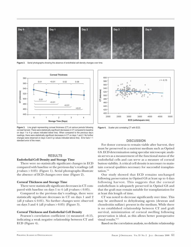
Figure 2. Serial photographs showing the absence of endothelial-cell-density changes over time.
Figure 4. Scatter plot correlating CT with ECD.Figure 3. Line graph representing corneal thickness (CT) at various periods following
corneal harvest. There were statistically significant decreases in CT compared to baseline
on days 1 to 4 (p values indicated below line). When compared to the previous day’s
readings, there were statistically significant decreases in CT on days 1 and 2. No further
changes were observed on days 3 and 4 (p values indicated above line). Error bars = 1
standard error of the mean.
RESULTSEndothelial-Cell Density and Storage Time
There were no statistically significant changes in ECDcompared with baseline or the previous day’s readings (allp values > 0.05) (Figure 1). Serial photographs illustratethe absence of ECD changes over time (Figure 2).
Corneal Thickness and Storage TimeThere were statistically significant decreases in CT com-
pared with baseline on days 1 to 4 (all p values < 0.05).Compared to the previous day’s readings, there were
statistically significant decreases in CT on days 1 and 2(all p values ≤ 0.01). No further changes were observedon days 3 and 4 (all p values > 0.05) (Figure 3).
Corneal Thickness and Endothelial Cell DensityPearson’s correlation coefficient (r) measured –0.15,
indicating a weak negative relationship between CT andECD (Figure 4).
DISCUSSIONFor donor corneas to remain viable after harvest, they
must be preserved in a nutrient medium such as OptisolGS. ECD determination using specular microscopic analy-sis serves as a measurement of the functional status of theendothelial cells and can serve as a measure of cornealbutton viability. A critical cell density is necessary to main-tain corneal qualities necessary for successful transplan-tation.7-8
Our study showed that ECD remains unchangedfollowing preservation in Optisol GS at least up to 4 daysfollowing harvest. This suggests that the cornealendothelium is adequately preserved in Optisol GS andthat the graft may remain suitable for transplantation forat least this length of time.
CT was noted to decrease significantly over time. Thismay be attributed to dehydrating agents (dextran andchondroitin sulfate) present in the medium. While thereis no established relationship between CT and graftsurvival, minimization of corneal swelling followingpreservation is ideal, as this allows better postoperativevisual results.9-11
Based on the correlation analysis, no definite relationship
Day 0 Day 1 Day 2 Day 3 Day 4
580
560
540
520
500
480
460
440
CT
(m
icro
ns
)
Storage Time (Days)
0 1 2 3 4
0.01 0.02 0.58<0.01
0.01
<0.01
<0.01
<0.01
Corneal Thickness
ECD (cells/square mm)
CT
(m
icro
ns)
600
580
560
540
520
500
480
460
440
420
400
2000 2500 3000 3500 4000 4500 5000
r = -0.15

62 PHILIPP J OPHTHALMOL VOL 33 NO. 2 JULY - DECEMBER 2008 PHILIPPINE ACADEMY OF OPHTHALMOLOGY
References
1. Thuret G, Chiquet C, Bernal F, el al. Prospective, randomized clinical and
endothelial evaluation of 2 storage times for corneal donor tissue in organ culture
at 31 degrees centigrade. Arch Ophthalmol 2003; 121: 442-450.
2. Amman J, Holley GP, Lee SB, et al. Increased endothelial cell density in the
paracentral and peripheral regions of the human cornea. Am J Ophthalmol 2003;
135: 584-590.
3. Ing JJ, Ing HH, Nelson LR, et al. Ten year prospective results of penetrating
keratoplasty. Ophthalmology 1998; 105: 1855-1865.
could be established between ECD and CT. This impliesthat ECD and CT are independent of each other.
In conclusion, over a period of 4 days of storage inOptisol GS, ECD remained stable and CT decreased.These findings suggest that corneas preserved in OptisolGS remain viable and do not deteriorate for at least thislength of time.
4. Sperling S. Endothelial cell density in donor corneas. Arch Ophthalmol 1980; 58:
278-282.
5. Bourne WM, Nelson LR, Maguire LJ, et al. Comparison of Chen medium and Optisol
GS for human corneal preservation at 4 degrees centigrade. Cornea 2001; 20:
683-686.
6. Mannis MJ, Holland EJ, Beck RW, et al. Clinical profile and early surgical
complications in the cornea donor study. Cornea 2006; 25: 164-170.
7. Naor J, Slomovic AR, Chipman M, et al. A randomized, double masked clinical trial
of Optisol GS vs. Chen Medium for human corneal storage. Arch Ophthalmol 2002;
120: 1280-1285.
8. Benetz BA, Gal RL, Ruedy KJ, et al. Specular microscopy ancillary study methods
for donor endothelial cell density determination of cornea donor study images. Curr
Eye Res 2006; 31: 319-327.
9. Lass JH, Gal RL, Ruedy KJ, et al. An evaluation of image quality and accuracy of
eye bank measurement of donor cornea endothelial cell density in the specular
microscopy ancillary study. Ophthalmology 2005; 112: 431-440.
10. Amann J, Holley GP, Lee SB, et al. Increased endothelial cell density in the
paracentral and peripheral lesions of the human cornea. Am J Ophthalmol 2003;
135: 584-590.
11. Saggau DD, Bourne WM. A comparison of two preservation media (CSM and K-
Sol) by scanning electron microscopy of preserved corneal endothelium. Arch
Ophthalmol 1989; 107: 429-432.