Embed Size (px)
Citation preview
Putting it together in the resus room
CARDIAC ARREST
Justin BowraCritical Care Ultrasound Course
2
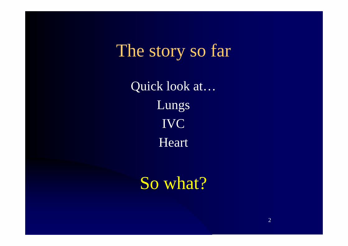
The story so far
Quick look at…LungsIVCHeart
So what?

3
The arrested patient
Adapted from Lichtenstein's SESAME protocol
(with permission)
4
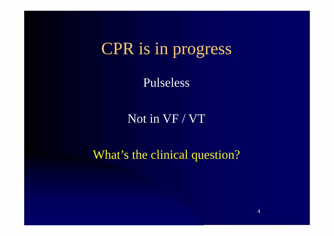
CPR is in progress
Pulseless
Not in VF / VT
What’s the clinical question?
Formulate a question

6
Formulate the question
a. Is there a reversible cause?
b. Where’s the ETT?

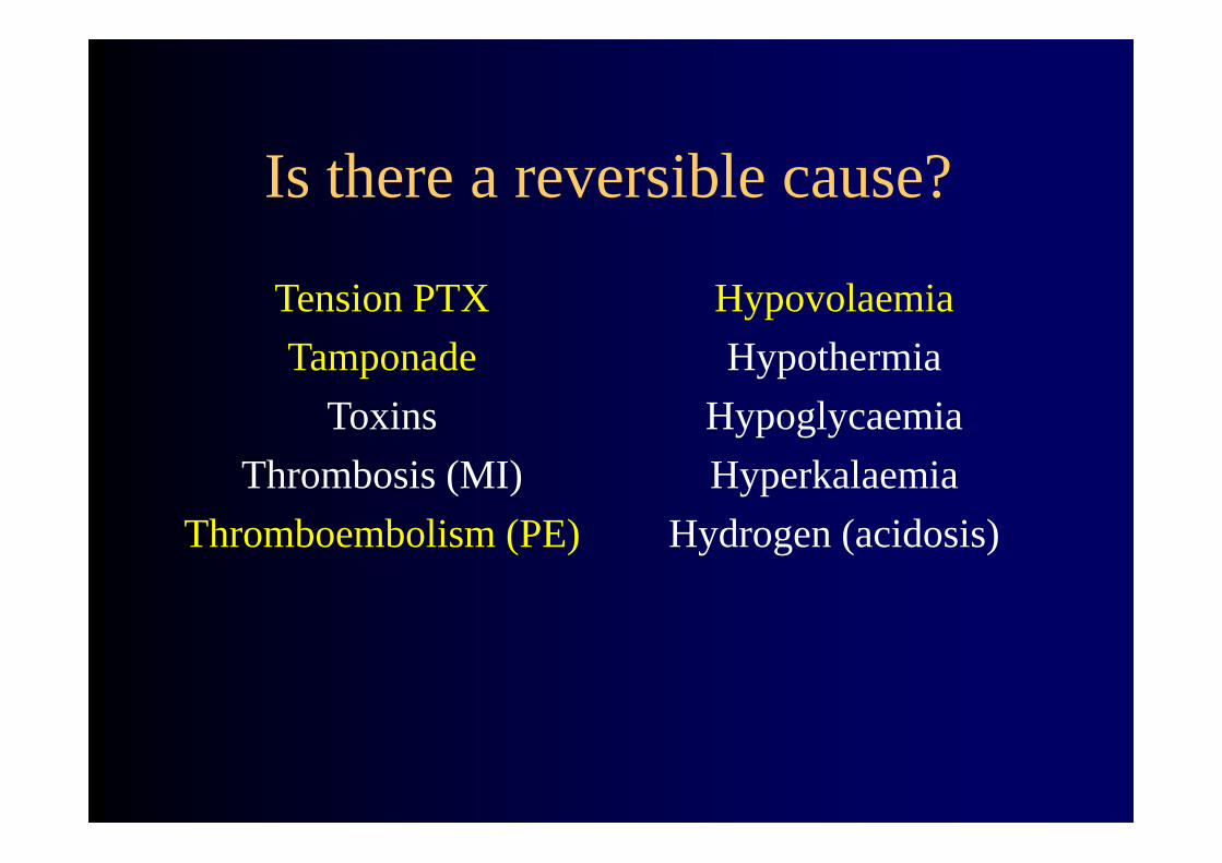
Is there a reversible cause?
Tension PTXTamponade
ToxinsThrombosis (MI)
Thromboembolism (PE)
HypovolaemiaHypothermia
HypoglycaemiaHyperkalaemia
Hydrogen (acidosis)
Is there a reversible cause?
Tension PTXTamponade
ToxinsThrombosis (MI)
Thromboembolism (PE)
HypovolaemiaHypothermia
HypoglycaemiaHyperkalaemia
Hydrogen (acidosis)

Where’s the ETT?
Bilateral pleural sliding = both lungs ventilated

Where’s the ETT?
Bilateral pleural sliding = both lungs ventilated
Unilateral sliding = 1 lung ventilation / PTX
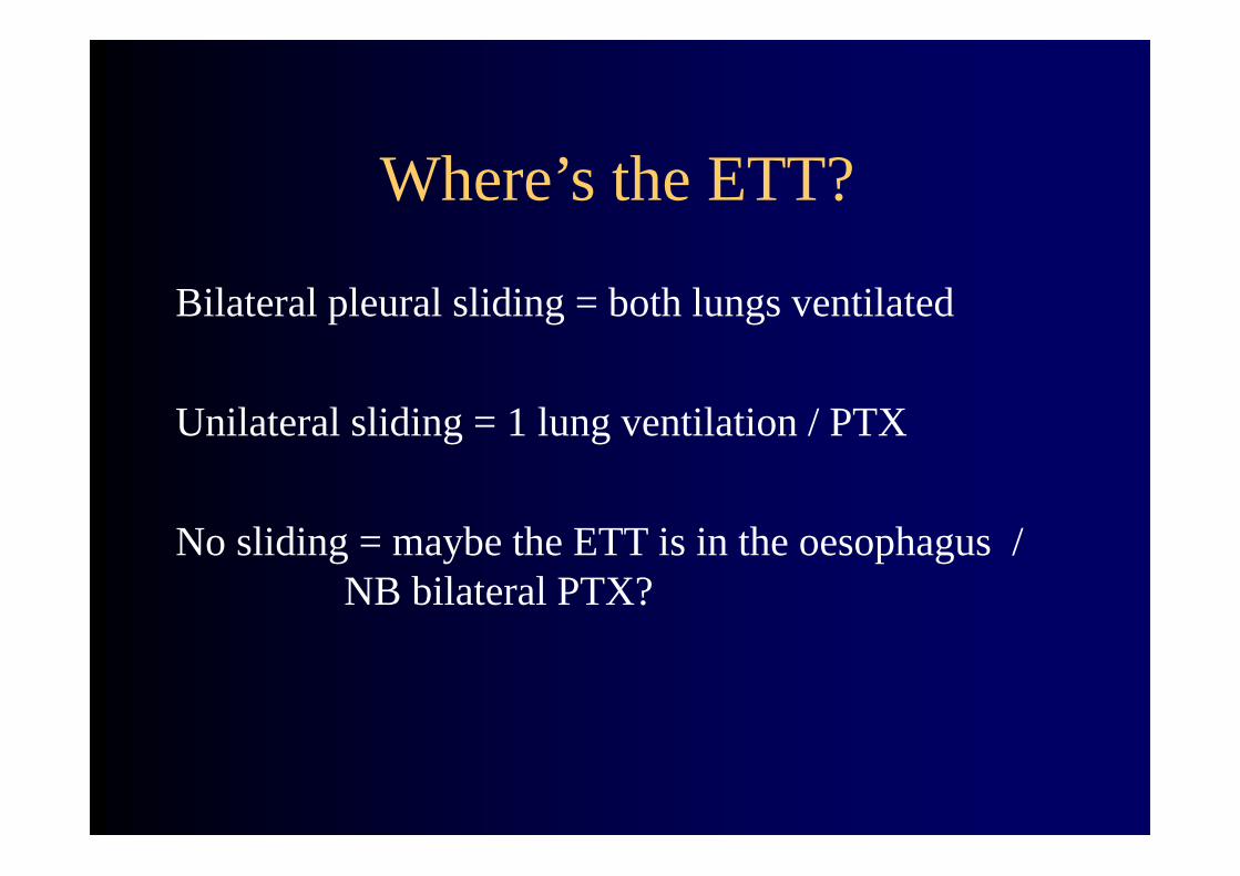
Where’s the ETT?
Bilateral pleural sliding = both lungs ventilated
Unilateral sliding = 1 lung ventilation / PTX
No sliding = maybe the ETT is in the oesophagus / NB bilateral PTX?

The arrest screen
13
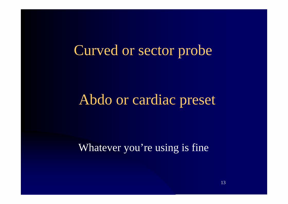
Curved or sector probe
Whatever you’re using is fine
Abdo or cardiac preset

14
Lungs
IVC
Heart
Other?
15
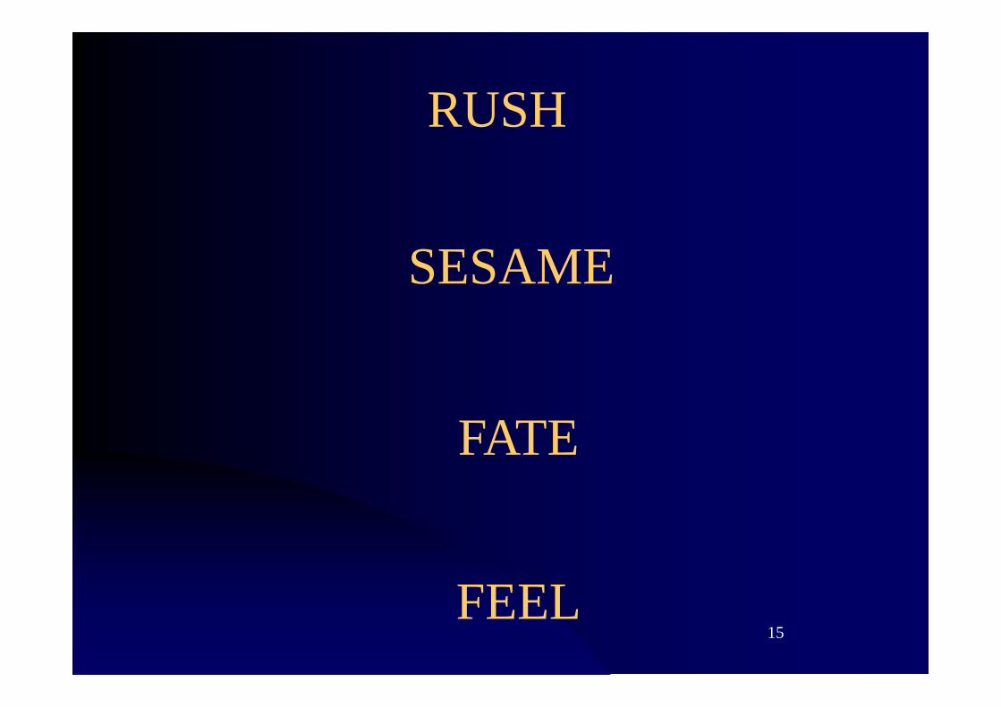
RUSH
SESAME
FATE
FEEL

16
JB’s version
17
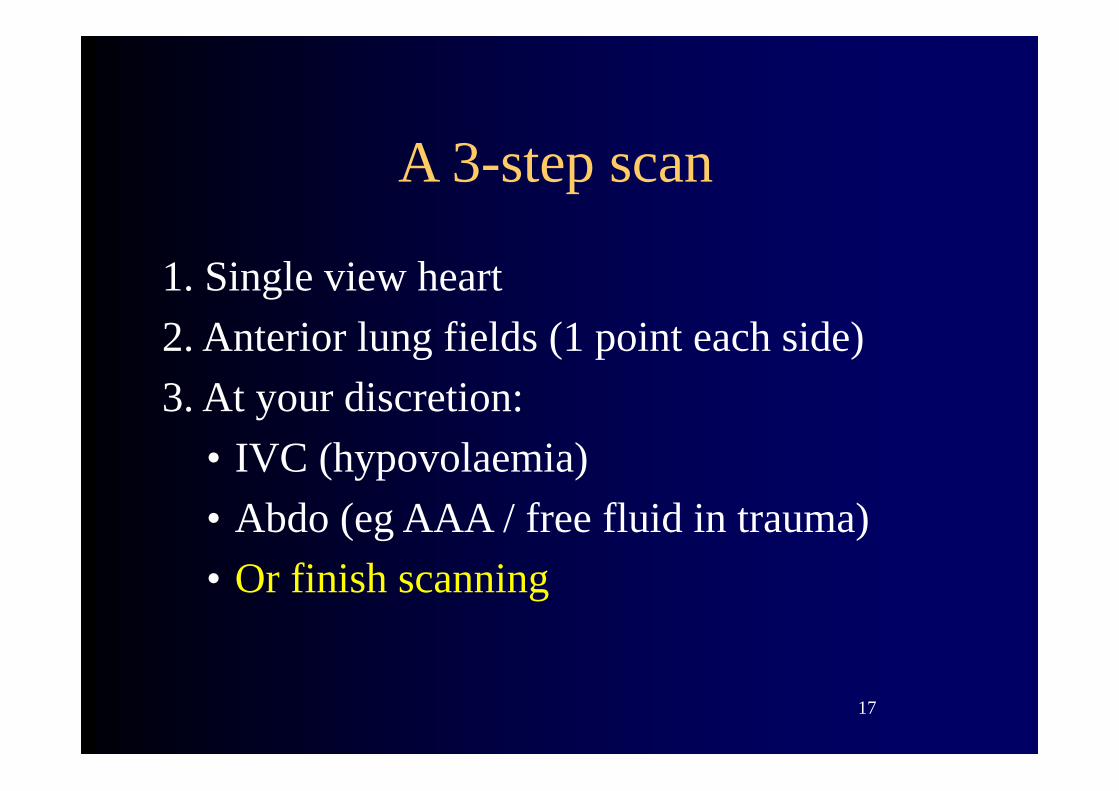
A 3-step scan
1. Single view heart2. Anterior lung fields (1 point each side)3. At your discretion:
• IVC (hypovolaemia)• Abdo (eg AAA / free fluid in trauma)• Or finish scanning

18
Don’t get in the way of CPR
You need to scan during the rhythm checkYou have ten seconds!
CPRScan/save heart imageCPR & discuss imagesScan/ save lung imagesCPR & discuss images

19

Step 1
Single view of heart

What am I looking for?
Is there a heartbeat?Pericardial effusion?
RV > LV?

22
Step 1: single view heart
Subcostal window is easiestBut any window will do
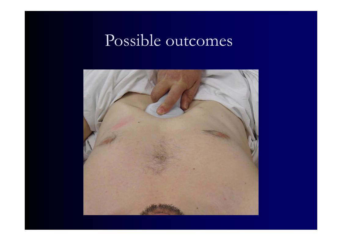
Possible outcomes

24

25
Big RV
squashing LVIt's a PE
Caveats:• Is it chronic? Thickened RV wall
• Is it dilated? Intra arrest
Action: consider thrombolysisIf in doubt, consider 3-point DVT scan

26
27
Pericardial fluid
It’s a tamponade
Caveat: it might be an incidental findingWhat if you’re wrong?
What have you got to lose?
Action: pericardiocentesis /ED thoracotomy /
NB OT?

28
29
Active heart, small LV = PEA
Hypovolaemia?
Action: replace volumeContinue CPR
Find & treat cause

30
31
Cardiac standstill
Exclude other reversible causes (Hs, Ts)
Action: cease CPR

32
Inadequate view
Options:Try another window
Try cardiac probeTry step 2Get help

Step 2
Anterior chest

NB not always necessary

35
Step 2: anterior chest
• Probe sagittal, 5cm depth• Just 1 spot on each side• Ideally the most elevated portion of chest
36
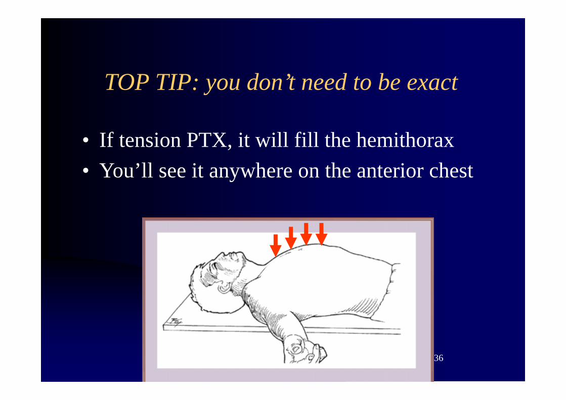
TOP TIP: you don’t need to be exact
• If tension PTX, it will fill the hemithorax• You’ll see it anywhere on the anterior chest


Step 2 findings
Neither lung is sliding?
One lung is not sliding?
Both lungs sliding

Step 2 findings
Neither lung is sliding?
One lung is not sliding?
Both lungs ventilating
PTX or1 lung ventilation
Not ventilating!(NB bilateral PTX?)
No PTXLungs are ventilating

Step 2 findings
Neither lung is sliding?
One lung is not sliding?
Both lungs sliding
PTX or1 lung ventilation
Not ventilating! No PTXETT in trachea
Check the airway Check the ETTDrain PTX?
Go to step 3

41
Step 3
Hypovolaemia What’s the cause?At your discretion:
• Review clinical picture• Scan the IVC ( confirm hypovolaemia)
• Scan the abdomen (eg AAA / free fluid in trauma)

Recap: the arrest screen

43
A 3-step scan
1. Single view heart2. Anterior lung fields3. At your discretion:
• IVC (hypovolaemia)• Abdo (eg AAA / free fluid in trauma)• Or finish scanning

44
Thanks to
Daniel LichtensteinJames DentKari SmallYogi Tikare