Embed Size (px)
Citation preview
1092 Emerging Infectious Diseases • www.cdc.gov/eid • Vol. 23, No. 7, July 2017
Clonal Clusters and Virulence Factors of Group C and G Streptococcus Causing
Severe Infections, Manitoba, Canada, 2012–2014
Sylvain A. Lother, Walter Demczuk, Irene Martin, Michael Mulvey, Brenden Dufault, Philippe Lagacé-Wiens, Yoav Keynan
In support of improving patient care, this activity has been planned and
implemented by Medscape, LLC and Emerging Infectious Diseases. Medscape,
LLC is jointly accredited by the Accreditation Council for Continuing Medical
Education (ACCME), the Accreditation Council for Pharmacy Education
(ACPE), and the American Nurses Credentialing Center (ANCC), to provide
continuing education for the healthcare team.
Medscape, LLC designates this Journal-based CME activity for a maximum
of 1.00 AMA PRA Category 1 Credit(s)™. Physicians should claim only the credit commensurate with
the extent of their participation in the activity.
All other clinicians completing this activity will be issued a certificate of participation. To participate in
this journal CME activity: (1) review the learning objectives and author disclosures; (2) study the
education content; (3) take the post-test with a 75% minimum passing score and complete the evaluation
at http://www.medscape.org/journal/eid; and (4) view/print certificate. For CME questions, see page 1232. Release date: June 15, 2017; Expiration date: June 15, 2018
Learning Objectives
Upon completion of this activity, participants will be able to:
• Identify clinical features of patients with group C and G Streptococcus (GCGS)
bacteremia, based on a retrospective study in Manitoba, Canada.
• Assess outcomes in patients with GCGS bacteremia.
• Determine genetic determinants of GCGS bacteremia.
CME Editor Claudia Chesley, Technical Writer/Editor, Emerging Infectious Diseases. Disclosure: Claudia Chesley has disclosed no relevant financial relationships.
CME Author Laurie Barclay, MD, freelance writer and reviewer, Medscape, LLC. Disclosure: Laurie Barclay, MD, has disclosed the following relevant financial relationships: owns stock, stock options, or bonds from Alnylam; Biogen; Pfizer.
Authors Disclosures: Sylvain A. Lother, MD; Walter Demczuk, BSc; Irene Martin, BSc; Michael Mulvey, PhD; Brenden Dufault, MSc (Biostatistics); and Yoav Keynan, MD, PhD, have disclosed no relevant financial relationships. Philippe Lagacé-Wiens, MD, has disclosed the following relevant financial relationships: served as an advisor or consultant for Merck, Cubist; served as a speaker or a member of a speakers bureau for Merck.
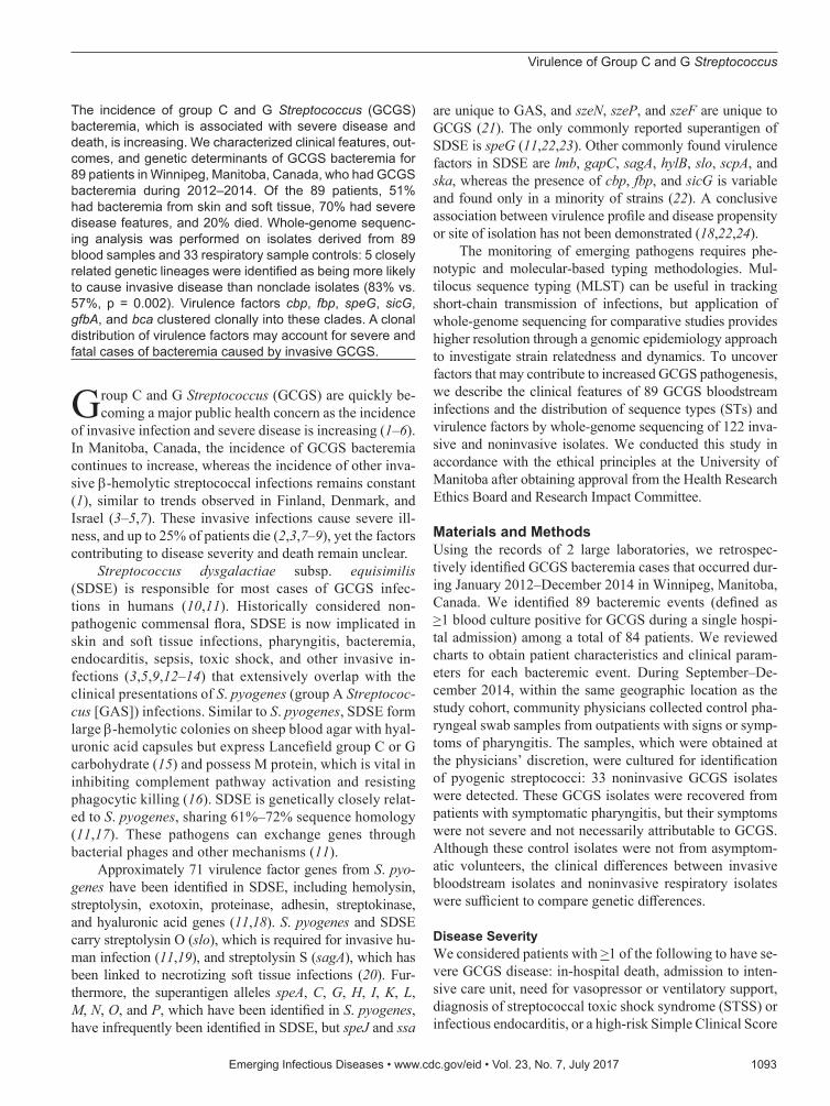
Author affiliations: University of Manitoba, Winnipeg, Manitoba, Canada (S.A. Lother, B. Dufault, P. Lagacé-Wiens, Y. Keynan);
National Microbiology Laboratory, Winnipeg (W. Demczuk,
I. Martin, M. Mulvey); Diagnostic Service Manitoba, Winnipeg
(P. Lagacé-Wiens)
DOI: https://dx.doi.org/10.3201/eid2307.161259
Emerging Infectious Diseases • www.cdc.gov/eid • Vol. 23, No. 7, July 2017 1093
The incidence of group C and G Streptococcus (GCGS) bacteremia, which is associated with severe disease and death, is increasing. We characterized clinical features, out-comes, and genetic determinants of GCGS bacteremia for 89 patients in Winnipeg, Manitoba, Canada, who had GCGS bacteremia during 2012–2014. Of the 89 patients, 51% had bacteremia from skin and soft tissue, 70% had severe disease features, and 20% died. Whole-genome sequenc-ing analysis was performed on isolates derived from 89 blood samples and 33 respiratory sample controls: 5 closely related genetic lineages were identified as being more likely to cause invasive disease than nonclade isolates (83% vs. 57%, p = 0.002). Virulence factors cbp, fbp, speG, sicG, gfbA, and bca clustered clonally into these clades. A clonal distribution of virulence factors may account for severe and fatal cases of bacteremia caused by invasive GCGS.
Group C and G Streptococcus (GCGS) are quickly be-coming a major public health concern as the incidence
of invasive infection and severe disease is increasing (1–6). In Manitoba, Canada, the incidence of GCGS bacteremia continues to increase, whereas the incidence of other inva-sive β-hemolytic streptococcal infections remains constant (1), similar to trends observed in Finland, Denmark, and Israel (3–5,7). These invasive infections cause severe ill-ness, and up to 25% of patients die (2,3,7–9), yet the factors contributing to disease severity and death remain unclear.
Streptococcus dysgalactiae subsp. equisimilis (SDSE) is responsible for most cases of GCGS infec-tions in humans (10,11). Historically considered non-pathogenic commensal flora, SDSE is now implicated in skin and soft tissue infections, pharyngitis, bacteremia, endocarditis, sepsis, toxic shock, and other invasive in-fections (3,5,9,12–14) that extensively overlap with the clinical presentations of S. pyogenes (group A Streptococ-cus [GAS]) infections. Similar to S. pyogenes, SDSE form large β-hemolytic colonies on sheep blood agar with hyal-uronic acid capsules but express Lancefield group C or G carbohydrate (15) and possess M protein, which is vital in inhibiting complement pathway activation and resisting phagocytic killing (16). SDSE is genetically closely relat-ed to S. pyogenes, sharing 61%–72% sequence homology (11,17). These pathogens can exchange genes through bacterial phages and other mechanisms (11).
Approximately 71 virulence factor genes from S. pyo-genes have been identified in SDSE, including hemolysin, streptolysin, exotoxin, proteinase, adhesin, streptokinase, and hyaluronic acid genes (11,18). S. pyogenes and SDSE carry streptolysin O (slo), which is required for invasive hu-man infection (11,19), and streptolysin S (sagA), which has been linked to necrotizing soft tissue infections (20). Fur-thermore, the superantigen alleles speA, C, G, H, I, K, L, M, N, O, and P, which have been identified in S. pyogenes, have infrequently been identified in SDSE, but speJ and ssa
are unique to GAS, and szeN, szeP, and szeF are unique to GCGS (21). The only commonly reported superantigen of SDSE is speG (11,22,23). Other commonly found virulence factors in SDSE are lmb, gapC, sagA, hylB, slo, scpA, and ska, whereas the presence of cbp, fbp, and sicG is variable and found only in a minority of strains (22). A conclusive association between virulence profile and disease propensity or site of isolation has not been demonstrated (18,22,24).
The monitoring of emerging pathogens requires phe-notypic and molecular-based typing methodologies. Mul-tilocus sequence typing (MLST) can be useful in tracking short-chain transmission of infections, but application of whole-genome sequencing for comparative studies provides higher resolution through a genomic epidemiology approach to investigate strain relatedness and dynamics. To uncover factors that may contribute to increased GCGS pathogenesis, we describe the clinical features of 89 GCGS bloodstream infections and the distribution of sequence types (STs) and virulence factors by whole-genome sequencing of 122 inva-sive and noninvasive isolates. We conducted this study in accordance with the ethical principles at the University of Manitoba after obtaining approval from the Health Research Ethics Board and Research Impact Committee.
Materials and Methods
Using the records of 2 large laboratories, we retrospec-tively identified GCGS bacteremia cases that occurred dur-ing January 2012–December 2014 in Winnipeg, Manitoba, Canada. We identified 89 bacteremic events (defined as >1 blood culture positive for GCGS during a single hospi-tal admission) among a total of 84 patients. We reviewed charts to obtain patient characteristics and clinical param-eters for each bacteremic event. During September–De-cember 2014, within the same geographic location as the study cohort, community physicians collected control pha-ryngeal swab samples from outpatients with signs or symp-toms of pharyngitis. The samples, which were obtained at the physicians’ discretion, were cultured for identification of pyogenic streptococci: 33 noninvasive GCGS isolates were detected. These GCGS isolates were recovered from patients with symptomatic pharyngitis, but their symptoms were not severe and not necessarily attributable to GCGS. Although these control isolates were not from asymptom-atic volunteers, the clinical differences between invasive bloodstream isolates and noninvasive respiratory isolates were sufficient to compare genetic differences.
Disease Severity
We considered patients with >1 of the following to have se-vere GCGS disease: in-hospital death, admission to inten-sive care unit, need for vasopressor or ventilatory support, diagnosis of streptococcal toxic shock syndrome (STSS) or infectious endocarditis, or a high-risk Simple Clinical Score
Virulence of Group C and G Streptococcus
RESEARCH
1094 Emerging Infectious Diseases • www.cdc.gov/eid • Vol. 23, No. 7, July 2017
>8 or Rapid Emergency Medicine Score >10. We defined STSS according to guidelines of the Working Group on Se-vere Streptococcal Infections (25). We calculated Simple Clinical Scores and Rapid Emergency Medicine Scores primarily by using patient vital signs and other clinical fea-tures; high-risk scores are associated with a 9.0%–10.3% risk for death by 30 days after admission (26–28).
Collection and Identification of BacteriaAt the discretion of the healthcare provider, patient blood samples were collected at symptom onset into BacT/Alert bottles (bioMérieux, Saint-Laurent, QC, Canada) accord-ing to institutional protocol and incubated using the BacT/Alert blood culture instrument (bioMérieux). Isolates were stored in frozen stocks in skim milk at −70°C and later re-trieved by subculture for further analysis.
A total of 92 GCGS isolates were recorded during the study period; 90 were retrieved, 2 were lost in storage, and 1 was identified as S. equi subsp. zooepidemicus by 16S rRNA sequence similarity and excluded from the study. We plated the 89 remaining isolates onto sheep blood agar (Oxoid, Ne-pean, ON, Canada) and aerobically incubated them for 24 h at 37°C in the presence of 5% CO2. We confirmed isolate identification by using MALDI-TOF (matrix-assisted laser desorption/ionization time-of-flight) mass spectrometry with the MALDI BioTyper system (Bruker, Boston, MA, USA) according to the manufacturer’s protocol. To confirm isolates with ambiguous MALDI-TOF mass spectrometry identifica-tions, we used latex agglutination to Lancefield antigens C and G and the Vitek2 system (bioMérieux) for biochemical identification. All isolates were identified as S. dysgalactiae.
Whole-Genome Sequencing
We extracted DNA from cultures, created multiplexed librar-ies, assembled reads, and performed core nucleotide variation phylogenetic analyses (online Technical Appendix 1, https://wwwnc.cdc.gov/EID/article/23/7/16-1259-Techapp1.pdf). In brief, we generated paired-end, 300-bp indexed reads on the Illumina MiSeq platform (Illumina, San Diego, CA, USA); the average yield was 1,015,107 reads/genome, and the average genomic coverage was 145×. Read quality was assessed by using FastQC version 0.11.4 (http://www.bioin-formatics.babraham.ac.uk/projects/fastqc/), assembled with SPAdes version 3.6.2 (http://cab.spbu.ru/software/spades/), and annotated with Prokka version 1.11 (http://www.vicbio-informatics.com/software.prokka.shtml), yielding an aver-age contig length of 39,313 bp and an average N50 contig length of 82,867 bp (29–31). The high-quality reads were then mapped to the publicly available reference genome, S. dys-galactiae subsp. equisimilis AC-2713 (GenBank accession no. NC_019042.1), by using SMALT version 0.7.5 (http://www.sanger.ac.uk/science/tools/smalt-0). Single-nucleotide variations (SNVs) were called using FreeBayes version
0.9.20 (https://github.com/ekg/freebayes) and SAMtools mpileup (http://www.htslib.org/) (32). The percentage of bases in the core was 82.8%, and 21,746 sites were used to generate the phylogeny.
We constructed a maximum-likelihood phylogenetic tree of informative SNV positions by using PhyML ver-sion 3.0 (http://www.atgc-montpellier.fr/phyml/) (33) and visualized the tree by using FigTree version 1.4.1 (http://tree.bio.ed.ac.uk/software/figtree/) (34). We determined phylogenetic clades by cluster analysis on the full dataset of blood and respiratory isolates (n = 122) and on isolates from blood only (n = 89) by using ClusterPicker version 1.2.4 (http://hiv.bio.ed.ac.uk/software.html) with the fol-lowing settings: initial and main support thresholds = 0.9, genetic distance threshold = 4.5, and the large cluster threshold = 10 (34). We submitted whole-genome se-quencing read data to the NCBI Sequence Read Archive (https://www.ncbi.nlm.nih.gov/sra/) under BioProject ac-cession no. PRJNA325743.
Molecular Typing
We used the whole-genome sequencing data for in silico determination of MLST STs; virulence factors (lmb, gapC, cba, cbp, fbp, sagA, slo, hylB, spegg, sicG, fbsA, pavA, fnbA, fnbB, gfbA, scpA, scpB, bca, cylE, ska, skc and skg) (22,35); and superantigens (speA, speB, speC, speF, spegg, speH, speI, speJ, speL, mf-2, mf-3, and smeZ) (21,23). We determined Lancefield serogroups from sequences anno-tated with Prokka and confirmed them by serologic test-ing using commercial latex antisera (SSI Diagnostica, Hillerød, Denmark). We submitted MLST allelic profiles to the Streptococcus dysgalactiae MLST database (https://pubmlst.org/sdysgalactiae/). We used allelic profiles to compute a goeBURST (global optimal eBurst; http://www.phyloviz.net/goeburst/) full minimum spanning tree us-ing PHYLOViZ (http://www.phyloviz.net/) (36); groups were assigned by a single-locus variation from a founding ST. All strains were confirmed to belong to S. dysgalac-tiae subsp. equisimilis by BLASTn (37) alignment of 16S rRNA sequences to reference genomes of S. dysgalactiae subsp. dysgalactiae ATCC27957 and S. dysgalactiae sub-sp. equisimilis ATCC12394 (PubMed accession nos. NZ_CM001076.1 and NC_017567.1, respectively).
Statistical Methods
We used descriptive statistics, χ2 test, Kruskal-Wallis test, and Fisher exact test to compare demographics be-tween clusters of SDSE to determine whether they dif-fered with respect to key risk factors. We used Fisher exact test to compare risk of death and other disease severity markers between ST clusters and clades. No observations were censored, so survival analysis tech-niques were not necessary.
Emerging Infectious Diseases • www.cdc.gov/eid • Vol. 23, No. 7, July 2017 1095
Results
Patient Characteristics and Disease Severity
We investigated 89 GCGS bacteremic events in 84 patients in Winnipeg during 2012–2014. Most patients (63%) were male, and the mean age was 61 years (SD ± 18.4 years). Many patients had co-existing conditions, predominantly cardiovascular disease (47%) and diabetes mellitus (43%). The most common source of bacteremia was from skin and soft tissue infections (51%), and 37% of patients had pri-mary bacteremia. Infectious endocarditis was confirmed or suspected in 7% of patients. No patients had necrotizing fasciitis or pharyngitis (Table 1).
In 70% of the cases, bacteremia was associated with markers of severe disease, including admission to an inten-sive care unit (26%) and the need for vasopressor (19%) or ventilatory (17%) support. Seventeen percent of patients
had a diagnosis of STSS, and 35%–61% of patients had high-risk disease severity scores. Twenty percent of pa-tients with GCGS bacteremia died while in the hospital (Table 2).
SDSE Isolate Characteristics
SDSE isolates from blood represented 89 (73%) of 122 to-tal isolates; 33 (37%) of the 89 isolates were from female patients and 56 (63%) were from male patients. These iso-lates were classified as Lancefield groups G (63%) and C (37%). Respiratory isolates represented 27% (33/122) of the isolates; information regarding the number from female and male patients was not available. These isolates also were classified as Lancefield groups G (52%) and C (48%).
Core Single-Nucleotide Variation
Phylogenetic Analysis
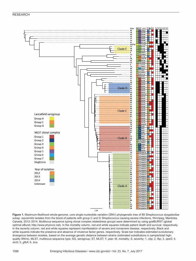
Phylogenetic analysis of all 122 isolates showed no as-sociation between infection type and patient sex, age, or disease severity (online Technical Appendix 1 Figure). Compared with the heterogeneous nonclade isolates, those that clustered into clades A–E represented a higher propor-tion of blood isolates (25/45 [57%] vs. 64/77 [83%], re-spectively; p = 0.002). In addition, compared with the other clades combined, clade A was represented by significantly fewer blood isolates (36/38 [95%] vs. 28/39 [72%], respec-tively; p = 0.017). In silico molecular determinants (MLST, Lancefield serogroups, and virulence factors) were clus-tered in a clonal distribution (online Technical Appendix 1 Figure). However, we found no significant associations when comparing blood and respiratory isolates.
Cluster analysis of the 89 blood isolates yielded 5 clades, A–E (n = 64); the other 25 heterogeneous isolates were outside these lineages. Clade A isolates were Lance-field serogroup C, clades B–E were serogroup G, and the heterogeneous nonclade isolates were serogroups C (n = 5) and G (n = 20) (Figure 1). Isolate numbers 35, 49, 26, 40, 47, 45, and 51 were most genetically distant from the other blood isolates, averaging 3,897–3,987 SNVs.
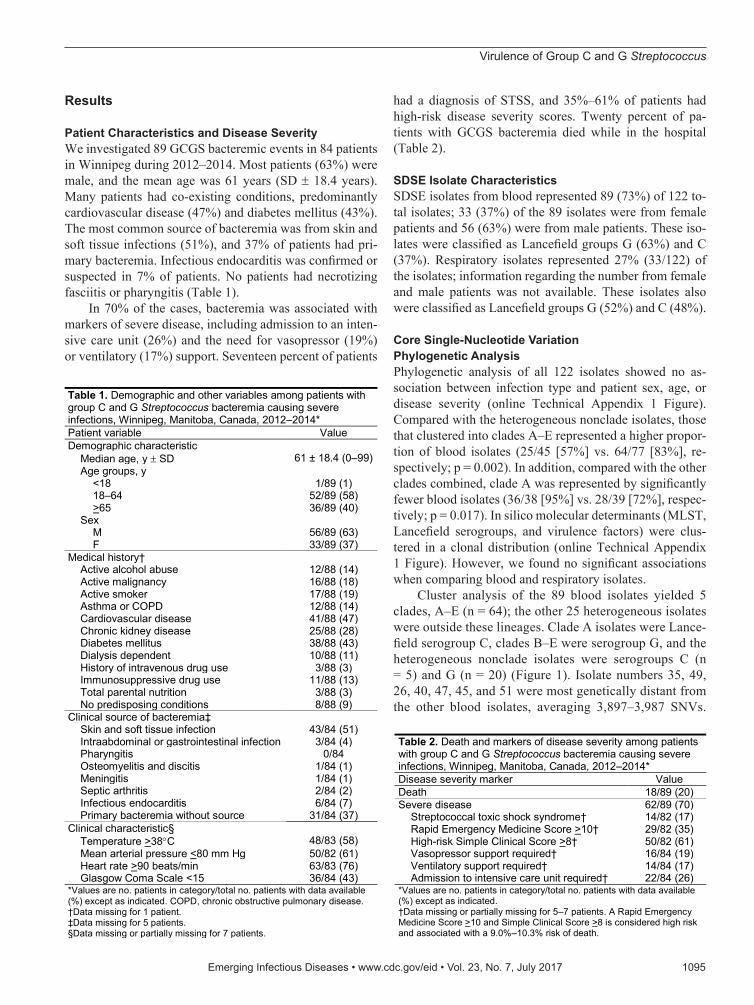
Table 1. Demographic and other variables among patients with group C and G Streptococcus bacteremia causing severe infections, Winnipeg, Manitoba, Canada, 2012–2014* Patient variable Value Demographic characteristic Median age, y r SD 61 ± 18.4 (0–99) Age groups, y <18 1/89 (1) 18–64 52/89 (58) >65 36/89 (40) Sex M 56/89 (63) F 33/89 (37) Medical history† Active alcohol abuse 12/88 (14) Active malignancy 16/88 (18) Active smoker 17/88 (19) Asthma or COPD 12/88 (14) Cardiovascular disease 41/88 (47) Chronic kidney disease 25/88 (28) Diabetes mellitus 38/88 (43) Dialysis dependent 10/88 (11) History of intravenous drug use 3/88 (3) Immunosuppressive drug use 11/88 (13) Total parental nutrition 3/88 (3) No predisposing conditions 8/88 (9) Clinical source of bacteremia‡ Skin and soft tissue infection 43/84 (51) Intraabdominal or gastrointestinal infection 3/84 (4) Pharyngitis 0/84 Osteomyelitis and discitis 1/84 (1) Meningitis 1/84 (1) Septic arthritis 2/84 (2) Infectious endocarditis 6/84 (7) Primary bacteremia without source 31/84 (37) Clinical characteristic§ Temperature >38qC 48/83 (58) Mean arterial pressure <80 mm Hg 50/82 (61) Heart rate >90 beats/min 63/83 (76) Glasgow Coma Scale <15 36/84 (43) *Values are no. patients in category/total no. patients with data available (%) except as indicated. COPD, chronic obstructive pulmonary disease. †Data missing for 1 patient. ‡Data missing for 5 patients. §Data missing or partially missing for 7 patients.
Table 2. Death and markers of disease severity among patients with group C and G Streptococcus bacteremia causing severe infections, Winnipeg, Manitoba, Canada, 2012–2014* Disease severity marker Value Death 18/89 (20) Severe disease 62/89 (70) Streptococcal toxic shock syndrome† 14/82 (17) Rapid Emergency Medicine Score >10† 29/82 (35) High-risk Simple Clinical Score >8† 50/82 (61) Vasopressor support required† 16/84 (19) Ventilatory support required† 14/84 (17) Admission to intensive care unit required† 22/84 (26) *Values are no. patients in category/total no. patients with data available (%) except as indicated. †Data missing or partially missing for 5–7 patients. A Rapid Emergency Medicine Score >10 and Simple Clinical Score >8 is considered high risk and associated with a 9.0%–10.3% risk of death.
Virulence of Group C and G Streptococcus
RESEARCH
1096 Emerging Infectious Diseases • www.cdc.gov/eid • Vol. 23, No. 7, July 2017
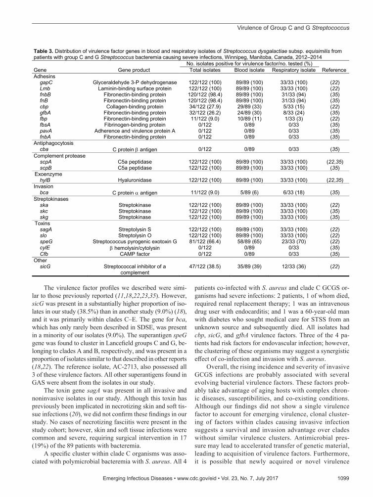
Figure 1. Maximum-likelihood whole-genome, core single-nucleotide variation (SNV) phylogenetic tree of 89 Streptococcus dysgalactiae subsp. equisimilis isolates from the blood of patients with group C and G Streptococcus causing severe infections, Winnipeg, Manitoba, Canada, 2012–2014. Multilocus sequence typing clonal complex relatedness groups were determined by using goeBURST (global optimal eBurst; http://www.phyloviz.net). In the mortality column, red and white squares indicate patient death and survival, respectively. In the severity column, red and white squares represent manifestation of severe and nonsevere disease, respectively. Black and white squares indicate the presence and absence of virulence factor genes, respectively. Scale bar indicates estimated evolutionary divergence between isolates, based on the average genetic distance between strains (estimated substitutions in sample/total high-quality SNVs). MLST, multilocus sequence type; SG, serogroup; ST, MLST; Y, year; M, mortality; S, severity; 1, cbp; 2, fbp; 3, speG; 4, sicG; 5, gfbA; 6, bca.
Emerging Infectious Diseases • www.cdc.gov/eid • Vol. 23, No. 7, July 2017 1097
The greatest difference was 5,110 SNVs between isolate numbers 30 and 51 (online Technical Appendix 2, https://wwwnc.cdc.gov/EID/article/23/7/16-1259-Techapp2.xlsx). Clade C was the most genetically homogenous, showing a maximum of 138 SNVs between isolates in the clade. Clade B was the most diverse, showing a maximum difference of 600 SNVs between isolates (online Techni-cal Appendix 1 Table 1).
MLST
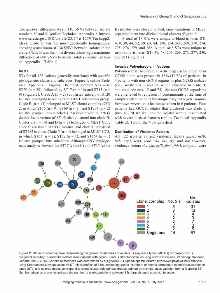
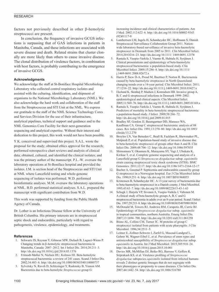
STs for all 122 isolates generally correlated with specific phylogenetic clades and subclades (Figure 1; online Tech-nical Appendix 1 Figure). The most common STs were ST20 (n = 28), followed by ST17 (n = 16) and ST15 (n = 9) (Figure 2). Clade A (n = 28) consisted entirely of ST20 isolates belonging to a singleton MLST relatedness group. Clade B (n = 13) belonged to MLST clonal complex (CC) 2, in which ST15 (n = 9), ST69 (n = 1), and ST274 (n = 2) isolates grouped into subclades. An isolate with ST276 (a double-locus variant of ST15) also clustered into clade B. Clades C (n = 14) and D (n = 5) belonged to MLST CC1; clade C consisted of ST17 isolates, and clade D consisted of ST282 isolates. Clade E (n = 4) belonged to MLST CC3, in which ST63 (n = 2), ST52 (n = 1), and ST164 (n = 1) isolates grouped into subclades. Although SNV phyloge-netic analysis showed that ST17 (clade C) and ST15 (clade
B) isolates were closely related, large variations in MLST separated them into distinct clonal clusters (Figure 2).
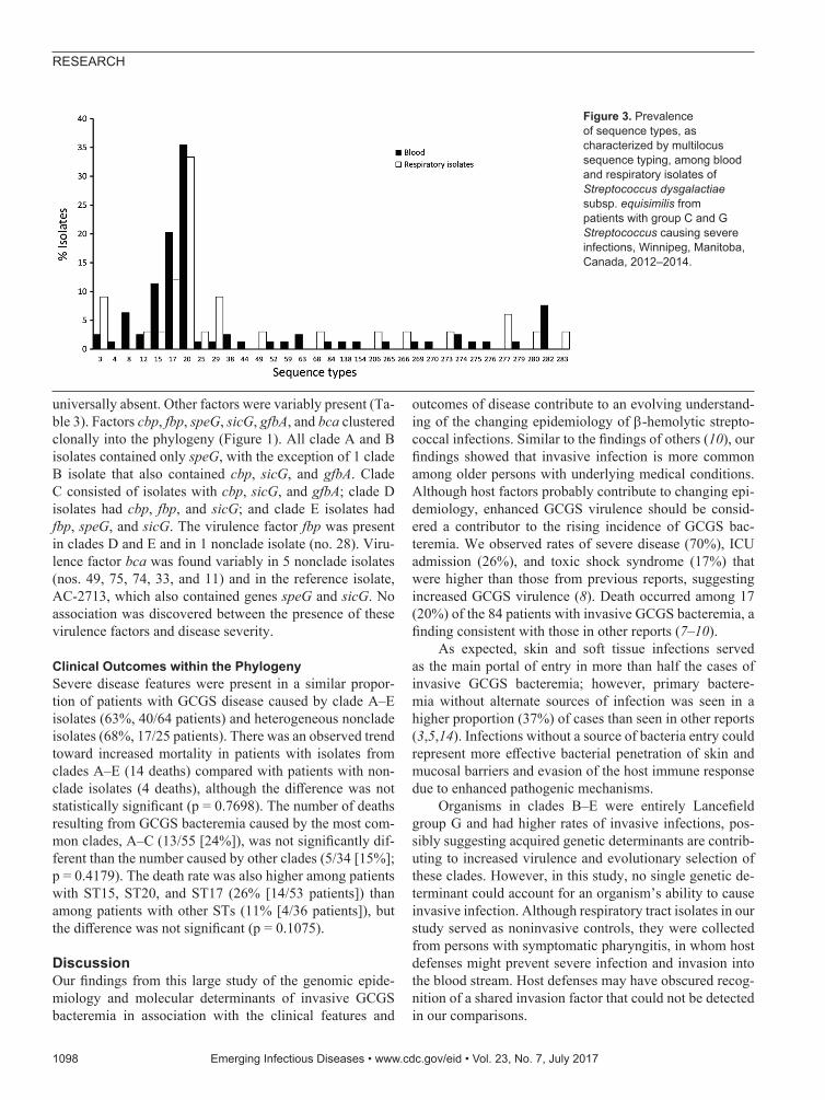
A total of 18 STs were unique to blood isolates: STs 4, 8, 38, 44, 52, 59, 63, 84, 138, 154, 265, 269, 270, 274, 275, 276, 279, and 282. A total of 8 STs were unique to respiratory isolates: STs 49, 68, 206, 266, 273, 277, 280, and 283 (Figure 3).
Invasive Polymicrobial Infections
Polymicrobial bacteremia with organisms other than GCGS alone was present in 18% (16/89) of patients. In 4 patients with non-GCGS organisms plus GCGS isolates (i.e., isolate nos. 3 and 57, which clustered in clade B; and nonclade nos. 12 and 74), the non-GCGS organisms were believed to represent 1) contaminants at the time of sample collection or 2) the nonprimary pathogen. Staphy-lococcus aureus co-infection was seen in 6 patients. Four patients had GCGS isolates that clustered into clade C (nos. 41, 70, 82, 85), and the isolates were all associated with severe disease features (online Technical Appendix Table 2). Two of the 4 patients died.
Distribution of Virulence Factors
All 122 isolates carried virulence factors gapC, hylB, lmb, sagA, scpA, scpB, ska, skc, skg, and slo; however, virulence factors cba, cfb, cylE, fbsA, fnbA, and pavA were
Figure 2. Minimum spanning tree representing the genetic relatedness of multilocus sequence types (MLSTs) of Streptococcus dysgalactiae subsp. equisimilis isolates from patients with group C and G Streptococcus causing severe infections, Winnipeg, Manitoba, Canada, 2012–2014. Genetic relatedness was determined by full goeBURST (global optimal eBurst; http://www.phyloviz.net) analysis using Streptococcus dysgalactiae MLST allelic profiles of 7 housekeeping genes. Numbers on nodes correspond to individual sequence types (STs) and colored nodes correspond to clonal cluster relatedness groups defined by a single-locus variation from a founding ST. Number labels on branches indicate the number of allelic variations between STs; branch lengths are not to scale.
Virulence of Group C and G Streptococcus
RESEARCH
1098 Emerging Infectious Diseases • www.cdc.gov/eid • Vol. 23, No. 7, July 2017
universally absent. Other factors were variably present (Ta-ble 3). Factors cbp, fbp, speG, sicG, gfbA, and bca clustered clonally into the phylogeny (Figure 1). All clade A and B isolates contained only speG, with the exception of 1 clade B isolate that also contained cbp, sicG, and gfbA. Clade C consisted of isolates with cbp, sicG, and gfbA; clade D isolates had cbp, fbp, and sicG; and clade E isolates had fbp, speG, and sicG. The virulence factor fbp was present in clades D and E and in 1 nonclade isolate (no. 28). Viru-lence factor bca was found variably in 5 nonclade isolates (nos. 49, 75, 74, 33, and 11) and in the reference isolate, AC-2713, which also contained genes speG and sicG. No association was discovered between the presence of these virulence factors and disease severity.
Clinical Outcomes within the Phylogeny
Severe disease features were present in a similar propor-tion of patients with GCGS disease caused by clade A–E isolates (63%, 40/64 patients) and heterogeneous nonclade isolates (68%, 17/25 patients). There was an observed trend toward increased mortality in patients with isolates from clades A–E (14 deaths) compared with patients with non-clade isolates (4 deaths), although the difference was not statistically significant (p = 0.7698). The number of deaths resulting from GCGS bacteremia caused by the most com-mon clades, A–C (13/55 [24%]), was not significantly dif-ferent than the number caused by other clades (5/34 [15%]; p = 0.4179). The death rate was also higher among patients with ST15, ST20, and ST17 (26% [14/53 patients]) than among patients with other STs (11% [4/36 patients]), but the difference was not significant (p = 0.1075).
Discussion
Our findings from this large study of the genomic epide-miology and molecular determinants of invasive GCGS bacteremia in association with the clinical features and
outcomes of disease contribute to an evolving understand-ing of the changing epidemiology of β-hemolytic strepto-coccal infections. Similar to the findings of others (10), our findings showed that invasive infection is more common among older persons with underlying medical conditions. Although host factors probably contribute to changing epi-demiology, enhanced GCGS virulence should be consid-ered a contributor to the rising incidence of GCGS bac-teremia. We observed rates of severe disease (70%), ICU admission (26%), and toxic shock syndrome (17%) that were higher than those from previous reports, suggesting increased GCGS virulence (8). Death occurred among 17 (20%) of the 84 patients with invasive GCGS bacteremia, a finding consistent with those in other reports (7–10).
As expected, skin and soft tissue infections served as the main portal of entry in more than half the cases of invasive GCGS bacteremia; however, primary bactere-mia without alternate sources of infection was seen in a higher proportion (37%) of cases than seen in other reports (3,5,14). Infections without a source of bacteria entry could represent more effective bacterial penetration of skin and mucosal barriers and evasion of the host immune response due to enhanced pathogenic mechanisms.
Organisms in clades B–E were entirely Lancefield group G and had higher rates of invasive infections, pos-sibly suggesting acquired genetic determinants are contrib-uting to increased virulence and evolutionary selection of these clades. However, in this study, no single genetic de-terminant could account for an organism’s ability to cause invasive infection. Although respiratory tract isolates in our study served as noninvasive controls, they were collected from persons with symptomatic pharyngitis, in whom host defenses might prevent severe infection and invasion into the blood stream. Host defenses may have obscured recog-nition of a shared invasion factor that could not be detected in our comparisons.
Figure 3. Prevalence of sequence types, as characterized by multilocus sequence typing, among blood and respiratory isolates of Streptococcus dysgalactiae subsp. equisimilis from patients with group C and G Streptococcus causing severe infections, Winnipeg, Manitoba, Canada, 2012–2014.
Emerging Infectious Diseases • www.cdc.gov/eid • Vol. 23, No. 7, July 2017 1099
The virulence factor profiles we described were simi-lar to those previously reported (11,18,22,23,35). However, sicG was present in a substantially higher proportion of iso-lates in our study (38.5%) than in another study (9.0%) (18), and it was primarily within clades C–E. The gene for bca, which has only rarely been described in SDSE, was present in a minority of our isolates (9.0%). The superantigen speG gene was found to cluster in Lancefield groups C and G, be-longing to clades A and B, respectively, and was present in a proportion of isolates similar to that described in other reports (18,22). The reference isolate, AC-2713, also possessed all 3 of these virulence factors. All other superantigens found in GAS were absent from the isolates in our study.
The toxin gene sagA was present in all invasive and noninvasive isolates in our study. Although this toxin has previously been implicated in necrotizing skin and soft tis-sue infections (20), we did not confirm these findings in our study. No cases of necrotizing fasciitis were present in the study cohort; however, skin and soft tissue infections were common and severe, requiring surgical intervention in 17 (19%) of the 89 patients with bacteremia.
A specific cluster within clade C organisms was asso-ciated with polymicrobial bacteremia with S. aureus. All 4
patients co-infected with S. aureus and clade C GCGS or-ganisms had severe infections: 2 patients, 1 of whom died, required renal replacement therapy; 1 was an intravenous drug user with endocarditis; and 1 was a 60-year-old man with diabetes who sought medical care for STSS from an unknown source and subsequently died. All isolates had cbp, sicG, and gfbA virulence factors. Three of the 4 pa-tients had risk factors for endovascular infection; however, the clustering of these organisms may suggest a synergistic effect of co-infection and invasion with S. aureus.
Overall, the rising incidence and severity of invasive GCGS infections are probably associated with several evolving bacterial virulence factors. These factors prob-ably take advantage of aging hosts with complex chron-ic diseases, susceptibilities, and co-existing conditions. Although our findings did not show a single virulence factor to account for emerging virulence, clonal cluster-ing of factors within clades causing invasive infection suggests a survival and invasion advantage over clades without similar virulence clusters. Antimicrobial pres-sure may lead to accelerated transfer of genetic material, leading to acquisition of virulence factors. Furthermore, it is possible that newly acquired or novel virulence
Table 3. Distribution of virulence factor genes in blood and respiratory isolates of Streptococcus dysgalactiae subsp. equisimilis from patients with group C and G Streptococcus bacteremia causing severe infections, Winnipeg, Manitoba, Canada, 2012–2014
Gene Gene product No. isolates positive for virulence factor/no. tested (%)
Reference Total isolates Blood isolate Respiratory isolate Adhesins gapC Glyceraldehyde 3-P dehydrogenase 122/122 (100) 89/89 (100) 33/33 (100) (22) Lmb Laminin-binding surface protein 122/122 (100) 89/89 (100) 33/33 (100) (22) fnbB Fibronectin-binding protein 120/122 (98.4) 89/89 (100) 31/33 (94) (35) fnB Fibronectin-binding protein 120/122 (98.4) 89/89 (100) 31/33 (94) (35) cbp Collagen-binding protein 34/122 (27.9) 29/89 (33) 5/33 (15) (22) gfbA Fibronectin-binding protein 32/122 (26.2) 24/89 (30) 8/33 (24) (35) fbp Fibronectin-binding protein 11/122 (9.0) 10/89 (11) 1/33 (3) (22) fbsA Fibrinogen-binding protein 0/122 0/89 0/33 (35) pavA Adherence and virulence protein A 0/122 0/89 0/33 (35) fnbA Fibronectin-binding protein 0/122 0/89 0/33 (35) Antiphagocytosis cba C protein E antigen 0/122 0/89 0/33 (35) Complement protease scpA C5a peptidase 122/122 (100) 89/89 (100) 33/33 (100) (22,35) scpB C5a peptidase 122/122 (100) 89/89 (100) 33/33 (100) (35) Exoenzyme hylB Hyaluronidase 122/122 (100) 89/89 (100) 33/33 (100) (22,35) Invasion bca C protein D antigen 11/122 (9.0) 5/89 (6) 6/33 (18) (35) Streptokinases ska Streptokinase 122/122 (100) 89/89 (100) 33/33 (100) (22) skc Streptokinase 122/122 (100) 89/89 (100) 33/33 (100) (35) skg Streptokinase 122/122 (100) 89/89 (100) 33/33 (100) (35) Toxins sagA Streptolysin S 122/122 (100) 89/89 (100) 33/33 (100) (22) slo Streptolysin O 122/122 (100) 89/89 (100) 33/33 (100) (22) speG Streptococcus pyrogenic exotoxin G 81/122 (66.4) 58/89 (65) 23/33 (70) (22) cylE E hemolysin/cytolysin 0/122 0/89 0/33 (35) Cfb CAMP factor 0/122 0/89 0/33 (35) Other sicG Streptococcal inhibitor of a
complement 47/122 (38.5) 35/89 (39) 12/33 (36) (22)
Virulence of Group C and G Streptococcus
RESEARCH
1100 Emerging Infectious Diseases • www.cdc.gov/eid • Vol. 23, No. 7, July 2017
factors not previously described in other β-hemolytic streptococci are present.
In conclusion, the frequency of invasive GCGS infec-tions is surpassing that of GAS infections in patients in Manitoba, Canada, and these infections are associated with severe disease and death. Related strains that cluster clon-ally are more likely than others to cause invasive disease. The clonal distribution of virulence factors, in combination with host factors, is probably contributing to the emergence of invasive GCGS.
Acknowledgments
We acknowledge the staff at St-Boniface Hospital Microbiology Laboratory who collected control respiratory isolates and assisted with the culturing, identification, and shipment of organisms to the National Microbiology Laboratory (NML). We also acknowledge the hard work and collaboration of the staff from the Streptococcus and STI Unit at the NML. We express our gratitude to the staff of the NML Science Technology Cores and Services Division for the use of their infrastructure, analytical pipelines, technical support and guidance and to the NML Genomics Core Facility for their next-generation sequencing and analytical expertise. Without their interest and dedication to this project, this work would not have been possible.
Y.K. conceived and supervised this project. S.A.L. wrote the proposal for the study; obtained ethics approval for the research; performed retrospective chart reviews and analysis of clinical data; obtained, cultured, and identified the clinical isolates; and was the primary author of the manuscript. P.L.-W. oversaw the laboratory operations at St-Boniface hospital and provided the isolates. I.M. is section head of the Streptococcus and STI Unit at NML where Lancefield testing and whole-genome sequencing of isolates was performed. W.D. performed bioinformatic analyses. M.M. oversaw the laboratory operations at NML. B.D. performed statistical analyses. S.A.L. prepared the manuscript with significant contribution from W.D.
This work was supported by funding from the Public Health Agency of Canada.
Dr. Lother is an Infectious Disease fellow at the University of British Columbia. His primary interests are in streptococcal septic shock and endocarditis, particularly with regard to pathogenesis, virulence, epidemiology, and treatments.
References
1. Schwartz IS, Keynan Y, Gilmour MW, Dufault B, Lagacé-Wiens P. Changing trends in β-hemolytic streptococcal bacteremia in Manitoba, Canada: 2007–2012. Int J Infect Dis. 2014;28:211–3. http://dx.doi.org/10.1016/j.ijid.2014.03.1376
2. Frimodt-Møller N, Nielsen HU, Kolmos HJ. Beta-hemolytic streptococcal bacteremia: a review of 241 cases. Scand J Infect Dis. 2002;34:483–6. http://dx.doi.org/10.1080/00365540110080737
3. Sylvetsky N, Raveh D, Schlesinger Y, Rudensky B, Yinnon AM. Bacteremia due to beta-hemolytic Streptococcus group G:
increasing incidence and clinical characteristics of patients. Am J Med. 2002;112:622–6. http://dx.doi.org/10.1016/S0002-9343 (02)01117-8
4. Lambertsen LM, Ingels H, Schønheyder HC, Hoffmann S; Danish Streptococcal Surveillance Collaboration Group 2011. Nation-wide laboratory-based surveillance of invasive beta-haemolytic streptococci in Denmark from 2005 to 2011. Clin Microbiol Infect. 2014;20:O216–23. http://dx.doi.org/10.1111/ 1469-0691.12378
5. Rantala S, Vuopio-Varkila J, Vuento R, Huhtala H, Syrjänen J. Clinical presentations and epidemiology of beta-haemolytic streptococcal bacteraemia: a population-based study. Clin Microbiol Infect. 2009;15:286–8. http://dx.doi.org/10.1111/ j.1469-0691.2008.02672.x
6. Harris P, Siew D-A, Proud M, Buettner P, Norton R. Bacteraemia caused by beta-haemolytic streptococci in North Queensland: changing trends over a 14-year period. Clin Microbiol Infect. 2011; 17:1216–22. http://dx.doi.org/10.1111/j.1469-0691.2010.03427.x
7. Ekelund K, Skinhøj P, Madsen J, Konradsen HB. Invasive group A, B, C and G streptococcal infections in Denmark 1999–2002: epidemiological and clinical aspects. Clin Microbiol Infect. 2005;11:569–76. http://dx.doi.org/10.1111/j.1469-0691.2005.01169.x
8. Rantala S, Vuopio-Varkila J, Vuento R, Huhtala H, Syrjänen J. Predictors of mortality in beta-hemolytic streptococcal bacteremia: a population-based study. J Infect. 2009;58:266–72. http://dx.doi.org/10.1016/j.jinf.2009.01.015
9. Bradley SF, Gordon JJ, Baumgartner DD, Marasco WA, Kauffman CA. Group C streptococcal bacteremia: analysis of 88 cases. Rev Infect Dis. 1991;13:270–80. http://dx.doi.org/10.1093/clinids/13.2.270
10. Broyles LN, Van Beneden C, Beall B, Facklam R, Shewmaker PL, Malpiedi P, et al. Population-based study of invasive disease due to beta-hemolytic streptococci of groups other than A and B. Clin Infect Dis. 2009;48:706–12. http://dx.doi.org/10.1086/597035
11. Shimomura Y, Okumura K, Murayama SY, Yagi J, Ubukata K, Kirikae T, et al. Complete genome sequencing and analysis of a Lancefield group G Streptococcus dysgalactiae subsp. equisimilis strain causing streptococcal toxic shock syndrome (STSS). BMC Genomics. 2011;12:17. http://dx.doi.org/10.1186/1471-2164-12-17
12. Bucher A, Gaustad P. Septicemia and endocarditis caused by group G streptococci in a Norwegian hospital. Eur J Clin Microbiol Infect Dis. 1990;9:251–6. http://dx.doi.org/10.1007/BF01968055
13. Kristensen B, Schønheyder HC. A 13-year survey of bacteraemia due to beta-haemolytic streptococci in a Danish county. J Med Microbiol. 1995;43:63–7. http://dx.doi.org/10.1099/00222615-43-1-63
14. Schugk J, Harjola VP, Sivonen A, Vuopio-Varkila J, Valtonen M. A clinical study of beta-haemolytic groups A, B, C and G streptococcal bacteremia in adults over an 8-year period. Scand J Infect Dis. 1997;29:233–8. http://dx.doi.org/10.3109/00365549709019034
15. McDonald M, Towers RJ, Andrews RM, Carapetis JR, Currie BJ. Epidemiology of Streptococcus dysgalactiae subsp. equisimilis in tropical communities, northern Australia. Emerg Infect Dis. 2007;13:1694–700. http://dx.doi.org/10.3201/eid1311.061258
16. Bisno AL, Collins CM, Turner JC. M proteins of group C streptococci isolated from patients with acute pharyngitis. J Clin Microbiol. 1996;34:2511–5.
17. Leitner E, Zollner-Schwetz I, Zarfel G, Masoud-Landgraf L, Gehrer M, Wagner-Eibel U, et al. Prevalence of emm types and antimicrobial susceptibility of Streptococcus dysgalactiae subsp. equisimilis in Austria. Int J Med Microbiol. 2015;305:918–24. http://dx.doi.org/10.1016/j.ijmm.2015.10.001
18. Davies MR, McMillan DJ, Beiko RG, Barroso V, Geffers R, Sriprakash KS, et al. Virulence profiling of Streptococcus dysgalactiae subspecies equisimilis isolated from infected humans reveals 2 distinct genetic lineages that do not segregate with their phenotypes or propensity to cause diseases. Clin Infect Dis. 2007;44:1442–54. http://dx.doi.org/10.1086/516780
Emerging Infectious Diseases • www.cdc.gov/eid • Vol. 23, No. 7, July 2017 1101
19. Nakagawa I, Amano A, Mizushima N, Yamamoto A, Yamaguchi H, Kamimoto T, et al. Autophagy defends cells against invading group A Streptococcus. Science. 2004;306:1037–40. http://dx.doi.org/10.1126/science.1103966
20. Humar D, Datta V, Bast DJ, Beall B, De Azavedo JCS, Nizet V. Streptolysin S and necrotising infections produced by group G streptococcus. Lancet. 2002;359:124–9. http://dx.doi.org/10.1016/S0140-6736(02)07371-3
21. Commons RJ, Smeesters PR, Proft T, Fraser JD, Robins-Browne R, Curtis N. Streptococcal superantigens: categorization and clinical associations. Trends Mol Med. 2014;20:48–62. http://dx.doi.org/ 10.1016/j.molmed.2013.10.004
22. Lo HH, Cheng WS. Distribution of virulence factors and association with emm polymorphism or isolation site among beta-hemolytic group G Streptococcus dysgalactiae subspecies equisimilis. APMIS. 2015;123:45–52. http://dx.doi.org/10.1111/apm.12305
23. Hashikawa S, Iinuma Y, Furushita M, Ohkura T, Nada T, Torii K, et al. Characterization of group C and G streptococcal strains that cause streptococcal toxic shock syndrome. J Clin Microbiol. 2004;42:186–92. http://dx.doi.org/10.1128/JCM.42.1.186-192.2004
24. Pinho MD, Melo-Cristino J, Ramirez M. Clonal relationships between invasive and noninvasive Lancefield group C and G streptococci and emm-specific differences in invasiveness. J Clin Microbiol. 2006;44:841–6. http://dx.doi.org/10.1128/JCM.44.3.841-846.2006
25. Breiman RF, Davis JP, Facklam RR, Gray BM, Hoge CW, Kaplan EL, et al.; The Working Group on Severe Streptococcal Infections. Defining the group A streptococcal toxic shock syndrome. Rationale and consensus definition. JAMA. 1993 ;269:390–1. http://dx.doi.org/10.1001/jama.1993.03500030088038
26. Kellett J, Deane B. The Simple Clinical Score predicts mortality for 30 days after admission to an acute medical unit. QJM. 2006;99:771–81. http://dx.doi.org/10.1093/qjmed/hcl112
27. Olsson T, Terent A, Lind L. Rapid Emergency Medicine score: a new prognostic tool for in-hospital mortality in nonsurgical emergency department patients. J Intern Med. 2004;255:579–87. http://dx.doi.org/10.1111/j.1365-2796.2004.01321.x
28. Ghanem-Zoubi NO, Vardi M, Laor A, Weber G, Bitterman H. Assessment of disease-severity scoring systems for patients with sepsis in general internal medicine departments. Crit Care. 2011;15:R95. http://dx.doi.org/10.1186/cc10102
29. Magoč T, Salzberg SL. FLASH: fast length adjustment of short reads to improve genome assemblies. Bioinformatics. 2011;27:2957–63. http://dx.doi.org/10.1093/bioinformatics/btr507
30. Bankevich A, Nurk S, Antipov D, Gurevich AA, Dvorkin M, Kulikov AS, et al. SPAdes: a new genome assembly algorithm and its applications to single-cell sequencing. J Comput Biol. 2012;19:455–77. http://dx.doi.org/10.1089/cmb.2012.0021
31. Seemann T. Prokka: rapid prokaryotic genome annotation. Bioinformatics. 2014;30:2068–9. http://dx.doi.org/10.1093/ bioinformatics/btu153
32. Li H, Handsaker B, Wysoker A, Fennell T, Ruan J, Homer N, et al.; 1000 Genome Project Data Processing Subgroup. The Sequence Alignment/Map format and SAMtools. Bioinformatics. 2009;25:2078–9. http://dx.doi.org/10.1093/bioinformatics/btp352
33. Guindon S, Dufayard J-F, Lefort V, Anisimova M, Hordijk W, Gascuel O. New algorithms and methods to estimate maximum-likelihood phylogenies: assessing the performance of PhyML 3.0. Syst Biol. 2010;59:307–21. http://dx.doi.org/10.1093/sysbio/syq010
34. Ragonnet-Cronin M, Hodcroft E, Hué S, Fearnhill E, Delpech V, Brown AJL, et al.; UK HIV Drug Resistance Database. Auto-mated analysis of phylogenetic clusters. BMC Bioinformatics. 2013;14:317. http://dx.doi.org/10.1186/1471-2105-14-317
35. Wang X, Zhang X, Zong Z. Genome sequence and virulence factors of a group G Streptococcus dysgalactiae subsp. equisimilis strain with a new element carrying erm(B). Sci Rep. 2016;6:20389. http://dx.doi.org/10.1038/srep20389
36. Francisco AP, Vaz C, Monteiro PT, Melo-Cristino J, Ramirez M, Carriço JA. PHYLOViZ: phylogenetic inference and data visualization for sequence based typing methods. BMC Bioinformatics. 2012;13:87. http://dx.doi.org/10.1186/ 1471-2105-13-87
37. Altschul SF, Madden TL, Schäffer AA, Zhang J, Zhang Z, Miller W, et al. Gapped BLAST and PSI-BLAST: a new generation of protein database search programs. Nucleic Acids Res. 1997;25:3389–402. http://dx.doi.org/10.1093/nar/25.17.3389
Address for correspondence: Sylvain A. Lother, University of Manitoba, BMSB 507-745 Bannatyne Ave, Winnipeg, Manitoba R3E 0J9, Canada; email: [email protected]
EID Adds Advanced Search Features for Articles Emerging Infectious Diseases now has an advanced search feature that makes it easier to find articles by using keywords, names of authors, and specified date ranges. You can sort and refine search results by manuscript number, volume or issue number, or article type. A quick start guide and expandable help section show you how to optimize your searches.
https://wwwnc.cdc.gov/eid/AdvancedSearch
EID’s new mapping feature allows you to search for articles from specific countries by using a map or table to locate countries. You can refine search results by article type, volume and issue, and date, and bookmark your search results.
https://wwwnc.cdc.gov/eid/ArticleMap
Virulence of Group C and G Streptococcus