Embed Size (px)
Citation preview

Name.....................................................................
Trinity AcademyPsychology – Year
Two
BiopsychologyAQA Specification from 2015
1
BiopsychologyAQA A level (Yr13) Component 2
Paper 2Candidates should be able to: -
GREEN AMBER RED
3.2.2
Biopsychology
1. Divisions of the nervous system; central and peripheral (somatic & autonomic)
2. Structure and function of sensory, relay and motor neurons. Synaptic transmission, neurotransmitters, excitation and inhibition
3. Function of the endocrine system: glands and hormones4. Fight or flight response including the role of adrenaline5. Localisation of function in the brain and hemispheric lateralisation; motor, somatosensory, visual, auditory and language centres; Broca's and Wernicke's areas, split brain research. Plasticity and functional recovery of the brain after trauma.
6. Ways of studying the brain; Scanning - PET, fMRI, EEG and event-related potentials (ERP). Post-mortem
7. Biological rhythms, circadian, infradian and ultradian. Endogenous and exogenous zeitgebers.
5. Localisation of function in the Brain
2

This refers to the principle that specific functions (language, memory, hearing etc.) have specific locations within the brain. The early 19th century witnessed a growth of interest in the localisations of functions in the brain. Franz Gall’s theory of phrenology (the study of the structure of the skull to determine a person’s character and capacity) was undoubtedly influential but was quickly discredited. Shortly after, using animal experimentation, Pierre Flourens was able to demonstrate that the main divisions of the brain were responsible for different functions. Since Gall and Flourens’ day, the techniques have grown considerably more sophisticated, as has our understanding of the functional organization of the human brain.
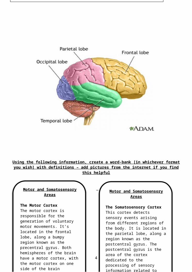
Using the following information, create a word-bank [in whichever format you wish] with definitions – add pictures from the internet if you find this
helpful
Motor and Somatosensory Areas
The Motor CortexThe motor cortex is responsible for the generation of voluntary motor movements. It’s located in the frontal lobe, along a bumpy region known as the precentral gyrus. Both hemispheres of the brain have
Motor and Somatosensory Areas
The Somatosensory CortexThis cortex detects sensory events arising from different regions of the body. It is located in the parietal lobe, along a region known as the postcentral gyrus. The postcentral
Label the diagram with the areas of the brain using the following key terms: Auditory Area Broca’s Area Motor Sensory Area Somatosensory Area Visual Area Wernicke’s Area Cerebral Cortex
3
Motor and Somatosensory Areas
The Motor CortexThe motor cortex is responsible for the generation of voluntary motor movements. It’s located in the frontal lobe, along a bumpy region known as the precentral gyrus. Both hemispheres of the brain have
Motor and Somatosensory Areas
The Somatosensory CortexThis cortex detects sensory events arising from different regions of the body. It is located in the parietal lobe, along a region known as the postcentral gyrus. The postcentral
Visual and Auditory Centres
Visual CentresThe primary visual centre in the brain is located in the visual cortex, in the occipital lobe of the brain. However, visual processing actually begins in the retina, at the back of the eye, where light enters and strikes the photoreceptors (rods and cones). Nerve impulses from the retina are then transmitted to the brain via the optic nerve. Some nerve impulses from the retina travel to areas of the brain involved in the coordination of circadian rhythms, but the majority terminate in an area of the brain called the thalamus, which acts as a relay station, passing this info on to the visual cortex.
The visual cortex spans both hemispheres, with the right hemisphere receiving its input from the left-hand side of the visual field, while the visual cortex in the left hemisphere receives its input from the right-hand side of the visual field. The visual cortex contains several different areas, with each of these areas processing different types of visual information, such as colour, shape or movement.
Auditory CentresThe auditory centre in the brain is concerned with hearing. Most of this area lies within the temporal lobes on both sides of the brain, where we find the auditory cortex. The auditory pathways begin in the cochlea in the inner ear, where sound waves are converted to nerve impulses, which travel via the auditory nerve to the auditory cortex.
On the journey from the cochlea to the brain, the first stop is the brain stem. Within the brain stem a basic decoding takes place e.g. the duration and intensity of a sound. The next stop is the thalamus where it carries out further processing of the auditory stimulus.
The last stop in the journey is the auditory cortex. Although the sound has been largely decoded by this stage, in the auditory cortex it’s recognized and may result in an appropriate response.
Language Centres
Broca’s areaThis area is named after Paul Broca, the French neurosurgeon who treated a patient he referred to as ‘Tan’ because that was the only syllable he could express. Tan had an unusual disorder. Although he had been able to understand spoken language, he was unable to speak, nor express his thoughts in writing.
Subsequently, Broca studied 8 other patients, all of whom had similar language deficits, along with lesions in their left frontal hemisphere. Patients with damage in these areas in the right hemisphere did not have the same language problems. This led
4
Evaluate
Challenges to Localisation: equipotentialityNot all researchers agree with the view that cognitive functions are localized in the brain. An influential, conflicting, view is the equipotentiality theory (Lashley, 1930). Supporters of this theory believed that the basic motor and sensory functions were localised, but that higher mental functions were not. Lashley claimed that intact areas of the cortex could take over responsibility for specific cognitive functions following injury to the area normally responsible for that function. According to this point of view, the effects of damage to the brain would be determined by the extent rather than the location of the damage. This view received some support from the discovery that humans were able to regain some of their cognitive abilities following damage to specific areas of the brain.
Communication may be more important than localisationResearch suggests that what might be more important is how the brain areas communicate with each other, rather than which specific brain regions control a particular cognitive process. Wernicke claimed that although different areas of the brain specialised in different functions, they are interdependent in the sense that in order to work they must interact with each other.
E.g. in 1892 a French neurologist, Joseph Dejerine, described a case in which the loss of an ability to read resulted in resulted in damage to the connection between the visual cortex and Wernicke’s area. This suggests that complex behaviours such as language, reading and movement are built up gradually as a stimulus enters the brain, then moves through different structures before a response is produced.
Language Centres
Broca’s areaThis area is named after Paul Broca, the French neurosurgeon who treated a patient he referred to as ‘Tan’ because that was the only syllable he could express. Tan had an unusual disorder. Although he had been able to understand spoken language, he was unable to speak, nor express his thoughts in writing.
Subsequently, Broca studied 8 other patients, all of whom had similar language deficits, along with lesions in their left frontal hemisphere. Patients with damage in these areas in the right hemisphere did not have the same language problems. This led
5
Damage to the connection between any two points in this process results in impairments that resemble damage to the localised brain region associated with that specific function.
Support for language centres from aphasia studiesEvidence for the different functions of Broca’s and Wernicke’s areas in language production and understanding comes from the discovery that damage to these different areas results in different types of aphasia. Aphasia refers to an ability (or impaired ability) to understand or produce speech as a result of brain damage. Expressive aphasia (also known as Broca’s aphasia) is an impaired ability to produce language. In most cases this is caused by brain damage in Broca’s area, demonstrating the important role played by this brain region in the production of language. Receptive aphasia (aka Wernicke’s aphasia) is an impaired ability to understand language, an inability to extract meaning from spoken or written words. This form of aphasia is usually the result of damage (e.g. from a stroke) in Wernicke’s area, demonstrating the important role played by this brain region in the comprehension of language.
There are individual differences in language areasThe pattern of activation observed in response to various language activities can vary from individual to individual. E.g. in a study of silent reading, Bavelier et al (1997) found a large variability in individual patterns of activation across different individuals. They observed activity on the right temporal lobe as well as in the left frontal, temporal and occipital lobes. Other studies have found significant gender differences in the size of the brain areas associated with language. E.g. Harasty et al (1997) found that women have proportionally larger Broca’s and Wernicke’s areas than men, the result of women’s greater use of language.
Language production may not be confined to Broca’s area aloneDronkers et al (2007) re-examined the preserved brains of 2 of Broca’s patients, Louis Leborgne (Tan) and Lazare Lelong. The purpose of this study was to identify the extent of any lesions in more detail by using modern high-resolution brain MRI imaging. The MRI findings revealed that other areas besides Broca’s area could have also contributed to the patient’s reduced speech abilities. This finding is significant because although legions to Broca’s area alone can cause temporary speech disruption, they do not usually result in severe disruption of spoken language. This study suggests that language and cognition are far more complicated than once thought and involve networks of brain regions rather than being localised to specific areas.
6
Lateralisation and Split Brain Research
The idea that different areas of the brain specialise in different tasks can be traced back to Marc Dax, a French doctor. In the early 1800s, Dax treated a significant number of patients who had lost the power of _____________ as a result of brain damage. He observed that in every case there had been damage in the left hemisphere but no damage in the right__________________. This suggested to Dax that language was located in the left hemisphere (this would be researched later in split-brain research). The split brain patients had received surgery to isolate the 2 hemispheres from each other, something that enabled psychologists to study each hemisphere __________________.
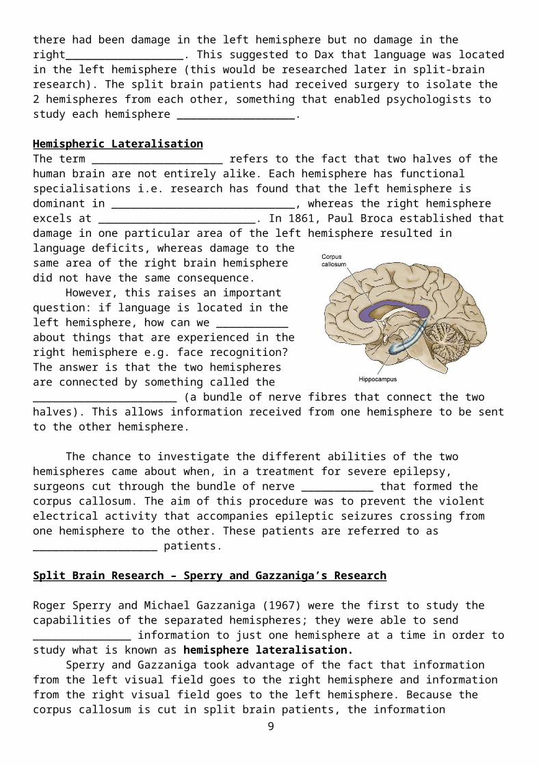
Hemispheric LateralisationThe term ____________________ refers to the fact that two halves of the human brain are not entirely alike. Each hemisphere has functional specialisations i.e. research has found that the left hemisphere is dominant in ____________________________, whereas the right hemisphere excels at ________________________. In 1861, Paul Broca established that damage in one particular area of the left hemisphere resulted in language deficits, whereas damage to the same area of the right brain hemisphere did not have the same consequence.
However, this raises an important question: if language is located in the left hemisphere, how can we ___________ about things that are experienced in the right hemisphere e.g. face recognition? The answer is that the two hemispheres are connected by something called the ______________________ (a bundle of nerve fibres that connect the two halves). This allows information received from one hemisphere to be sent to the other hemisphere.
The chance to investigate the different abilities of the two hemispheres came about when, in a treatment for severe epilepsy, surgeons cut through the bundle of nerve ___________ that formed the corpus callosum. The aim of this procedure was to prevent the violent electrical activity that accompanies epileptic seizures crossing from one hemisphere to the other. These patients are referred to as ___________________ patients.
7

Split Brain Research – Sperry and Gazzaniga’s Research
Roger Sperry and Michael Gazzaniga (1967) were the first to study the capabilities of the separated hemispheres; they were able to send _______________ information to just one hemisphere at a time in order to study what is known as hemisphere lateralisation.
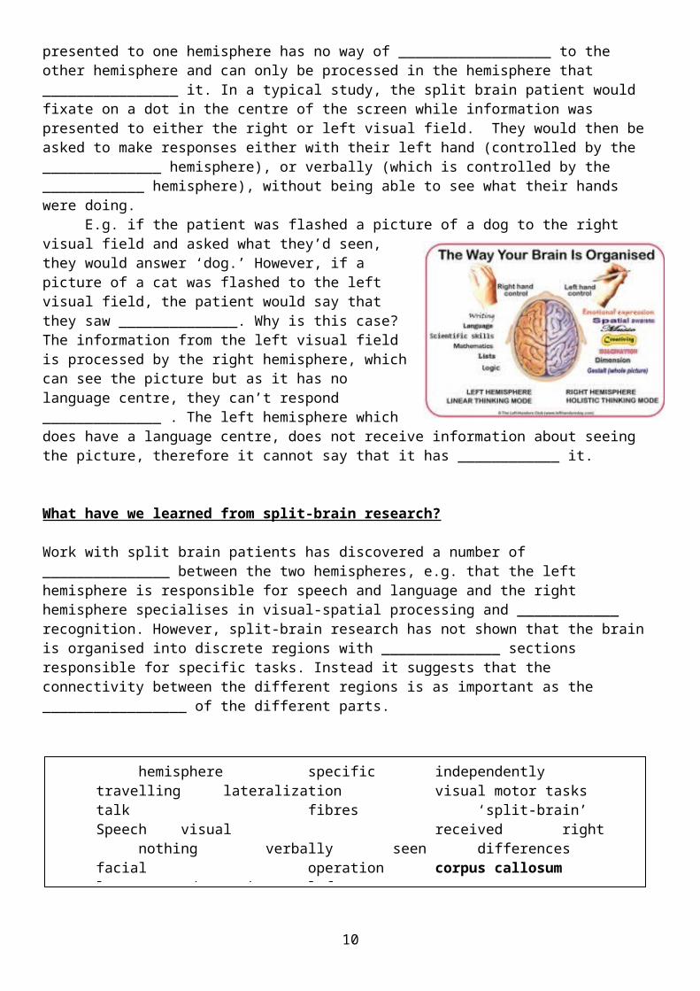
Sperry and Gazzaniga took advantage of the fact that information from the left visual field goes to the right hemisphere and information from the right visual field goes to the left hemisphere. Because the corpus callosum is cut in split brain patients, the information presented to one hemisphere has no way of __________________ to the other hemisphere and can only be processed in the hemisphere that ________________ it. In a typical study, the split brain patient would fixate on a dot in the centre of the screen while information was presented to either the right or left visual field. They would then be asked to make responses either with their left hand (controlled by the ______________ hemisphere), or verbally (which is controlled by the ____________ hemisphere), without being able to see what their hands were doing. E.g. if the patient was flashed a picture of a dog to the right visual field and asked what they’d seen, they would answer ‘dog.’ However, if a picture of a cat was flashed to the left visual field, the patient would say that they saw ______________. Why is this case? The information from the left visual field is processed by the right hemisphere, which can see the picture but as it has no language centre, they can’t respond ______________ . The left hemisphere which does have a language centre, does not receive information about seeing the picture, therefore it cannot say that it has ____________ it.
What have we learned from split-brain research?
Work with split brain patients has discovered a number of _______________ between the two hemispheres, e.g. that the left hemisphere is responsible for speech and language and the right hemisphere specialises in visual-spatial processing and ____________ recognition. However, split-brain research has not shown that the brain is organised into discrete regions with ______________ sections responsible for specific tasks. Instead it suggests that the connectivity between the different regions is as important as the _________________ of the different parts.
TASK: Produce a summary table of the following evaluation points…[P.T.O.]
hemisphere specific independently travellinglateralization visual motor tasks talk
fibres ‘split-brain’ Speech visualreceived right nothing verbally
seen differences facial operationcorpus callosum language and speech left
8
Evaluation of Lateralisation
Evaluation of split-brain research
Advantages of hemisphere lateralisation
It’s generally assumed that the main advantage of brain lateralisation is that it increases neural processing capacity. By using only one hemisphere to engage in a particular task (e.g. language or mathematical ability), this would leave the other hemisphere free to engage in another function. However, despite this assumption, very little empirical evidence has been provided to show that lateralisation confers any advantage to the functioning of the brain, Rogers et al (2004) found that, in the domestic chicken, brain lateralisation is associated with an enhanced ability to perform two tasks simultaneously – finding food and being vigilant for predators. This finding does provide some evidence that brain lateralisation enhances brain efficiency in cognitive tasks that demand the simultaneous but different use of both hemispheres.
Lateralisation and immune system functioning
There are a number of advantages and disadvantages associated with hemispheric lateralisation. E.g., architects and the mathematically gifted tend to have superior right-hemispheric skills but are also much more likely to be left-handed and to suffer higher rates of allergies and problems with the immune system. E.g. Tonnesson et al (1993) found a small but significant relationship between handedness and immune disorders. This suggests that the same genetic processes that lead to lateralisation may also affect the development of the immune system. Likewise, Morfit and Weekes (2001) lent support to this suggestion, finding that left handers had a higher incidence of immune disorders in their immediate families than did right handers.
Lateralisation changes with age
Lateralisation of function appears not to stay exactly the same throughout an individual’s lifetime, but changes with normal ageing. Across many types of tasks and many brain areas, lateralised patterns found in younger individuals tend to switch to bilateral patterns in healthy older adults. Szaflarski et al (2006) found that language became more lateralised to the left hemisphere with increasing age in children and adolescents, but after the age of 25, lateralisation decreased with each decade of life. It is difficult to know why this is the case. One possibility is that using the extra processing resources of the other hemisphere may in some way compensate for age-related declines in function.
Language may not be restricted to the left hemisphere
Gazzaniga (1998) suggests that some of the early discoveries from split-brain research have been disconfirmed by more recent discoveries. E.g. split brain research had suggested that the right hemisphere was unable to handle even the most rudimentary language. Damage to the left hemisphere was found to be far more detrimental to language function than was damage to the right. However, case studies have demonstrated that this was not necessarily the case. One patient, known as J.W., developed the capacity to speak out of the right hemisphere, with the result that J.W. can now speak about information presented to the left or to the right brain (Turk et al 2002).
9
Plasticity and functional recovery of the brain
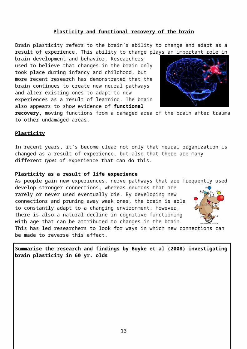
Brain plasticity refers to the brain’s ability to change and adapt as a result of experience. This ability to change plays an important role in brain development and behavior. Researchers used to believe that changes in the brain only took place during infancy and childhood, but more recent research has demonstrated that the brain continues to create new neural pathways and alter existing ones to adapt to new experiences as a result of learning. The brain also appears to show evidence of functional recovery, moving functions from a damaged area of the brain after trauma to other undamaged areas.
Plasticity
Language may not be restricted to the left hemisphere
Gazzaniga (1998) suggests that some of the early discoveries from split-brain research have been disconfirmed by more recent discoveries. E.g. split brain research had suggested that the right hemisphere was unable to handle even the most rudimentary language. Damage to the left hemisphere was found to be far more detrimental to language function than was damage to the right. However, case studies have demonstrated that this was not necessarily the case. One patient, known as J.W., developed the capacity to speak out of the right hemisphere, with the result that J.W. can now speak about information presented to the left or to the right brain (Turk et al 2002).
Limitations of split-brain research
The split-brain procedure is rarely carried out these days. Therefore patients who have had this procedure are rarely encountered in sufficient numbers to be useful for research. Andrewes (2001) argues that many studies have only 3 (sometimes even just 1 ppt). Therefore he claims conclusions have been drawn from individuals who have either a confounding physical disorder that made the procedure necessary or who had a less complete sectioning of the 2 hemispheres than was originally believed. Andrewes claims that these ‘rogue’ cases are often only identified when the results of a study have failed to be replicated.
10

In recent years, it’s become clear not only that neural organization is changed as a result of experience, but also that there are many different types of experience that can do this.
Plasticity as a result of life experienceAs people gain new experiences, nerve pathways that are frequently used develop stronger connections, whereas neurons that are rarely or never used eventually die. By developing new connections and pruning away weak ones, the brain is able to constantly adapt to a changing environment. However, there is also a natural decline in cognitive functioning with age that can be attributed to changes in the brain. This has led researchers to look for ways in which new connections can be made to reverse this effect.
Summarise the research and findings by Boyke et al (2008) investigating brain plasticity in 60 yr. olds
Playing video gamesPlaying video games makes many different complex cognitive and motor demands. Kuhn et al (2014) compared a control group with a video game training group that was trained for 2 months for at least 30 mins per day on the game Super Mario. They found a significant increase in grey matter in various brain areas including the cortex, hippocampus and cerebellum. This increase was not evident in the control group. The researchers concluded that video game training had resulted in new synaptic connections in brain areas involved in spatial navigation, strategic planning and working memory and motor performance - skills that were important in playing the game successfully.
What is grey matter?
MeditationResearchers working with Tibetan monks have been able to demonstrate that meditation can change the inner workings of the brain.
11
Summarise the research and findings by Davidson et al (2004) comparing 8 practitioners of Tibetan meditation with 10 student volunteers with no previous meditation experience.
Functional Recovery after trauma
In the 1960s, researchers studied cases in which stroke victims were able to regain functioning. They discovered that when brain cells are damaged or destroyed, as they are during a stroke, the brain re-wires itself over time so that some level of function can be regained. Although parts of the brain may be damaged or even destroyed as a result of trauma, other parts appear to take over the functions that were lost. Neurons next to damaged brain areas can form new circuits that resume some of the lost function.
Mechanisms for recoveryRegenerative developments in brain function arise from the brain’s plasticity, its ability to change structurally and functionally following trauma. Two ways in which the brain is able to do this are neuronal unmasking and stem cells.
Neuronal UnmaskingWall (1977) first identified what he called ‘dormant synapses’ in the brain. These are:
Increasing the rate of input to these synapses:
Stem cellsStem cells are:
12
There are a number of views on how stem cells might work to provide treatments for brain damage caused by injury or neurodegenerative disorders.
- The first view is that stem cells implanted into the brain would:
- A second possibility is that transplanted stem cells secrete:
- A third possibility is that transplanted cells from a neural network, which:
Evaluation of plasticity
Evaluation of functional recovery after trauma
Research support from animal studies
Evidence for the brain’s ability to change as a result of experience comes from animal studies. Kempermann et al (1998) investigated whether an enriched environment can alter the number of neurons in the brain. They found evidence of an increased number of new neurons in the brains of rats housed in complex environments compared to rats housed in lab cages. In particular, the rats housed in complex environments showed an increase in neurons in the hippocampus, a part of the brain associated with the formation of new memories, and the ability to navigate form one location to another.
Research support from human studies
Research has shown that rats are not the only animals to demonstrate plasticity after exposure to an enriched environment. Maguire et al (2000) studied London taxi drivers to discover whether changes in the brain could be detected as a result of their extensive navigational experience. Using an MRI scanner, the researchers calculated the amount of grey matter in the brains of taxi drivers and a set of control ppts. The posterior hippocampi of taxi drivers were significantly larger relative to those of control ppts and posterior hippocampal volume was positively correlated with the amount of time they’ spent taxi driving.
Research support from animal studies
Tajiri et al (2013) provided evidence for the role of stem cells in recovery from brain injury. They randomly assigned rats with traumatic brain injury to one of 2 groups. One group received transplants of stem cells into the region of the brain affected by traumatic injury. The control group received a solution infused into the brain containing no stem cells. Three months after the brain injury, the brains of stem cell rats showed clear development of neuron-like cells in the area of injury. This was accompanied by a solid stream of stem cells migrating to the brain’s site of injury. This was not the case with the control group.
But what is the problem with animal research?
13
Ways of Studying the Brain
The brain is the main focus of neuroscience. Studying the brain gives us important insights into the underlying
Age differences in functional recovery
It’s a commonly accepted view that functional plasticity reduces with age (Huttenlocher, 2002). According to this view, the only option following traumatic brain injury beyond childhood is to develop compensatory behavioural strategies to work around the deficit (such as seeking social support or to develop strategies to deal with cognitive deficits). However, studies have suggested that even abilities commonly thought to be fixed in childhood can still be modified in adults with intense retraining. Despite these indications of adult plasticity, Elbert et al (2001) conclude that the capacity for neural reorganization is much greater in children than in adults, as demonstrated by the extended practice that adults require in order to produce changes.
Educational attainment and functional recovery
Schneider et al (2014) found that patients with the equivalent of a college education are seven times more likely than those who didn’t finish high school to be disability-free one year after a moderate to severe traumatic brain injury. They carried out a retrospective study based on data from the US Traumatic Brain Injury Systems Database. Of the 769 patients studied, 214 had achieved disability-free recovery (DFR) after one year. Of these, 39.2% of the patients with 16 or more years of education had achieved DFR, as had 30.8% of those with 12-15 years of education, and just 9.7% of those with less than 12 years of education achieved DFR after just one year. The researchers concluded that ‘cognitive reserve’ (associated with greater educational attainment) could be a factor in neural adaptation during recovery from traumatic brain injury.
14

foundations of our behavior and mental processes. A variety of methods are used by research scientists in order to study the functions of different areas of the brain. Some involved scanning the living brain, looking for patterns of electrical activity associated with the performance of particular tasks. Other methods involve studying sections of a deceased brain to investigate anatomical reasons for behavior observed while the patient was alive.
Post-Mortem Examinations
1. Post-mortem examinations are used to establish the underlying neurobiology of a particular behaviour. For example, researchers may study a person who displays behaviour while they’re alive that suggests possible underlying brain damage. Subsequently, when the person dies, the researchers can examine their brains to look for abnormalities that might explain that behavior and which are not found in control individuals.
Below, provide explanations of two famous case studies: 1. Tan 2. HM
The use of post-mortem studies has also been used to establish a link between psychiatric disorders, such as schizophrenia and depression, and underlying brain abnormalities. E.g. post-mortem studies have found evidence of reduced numbers of glial cells in the frontal cortex of patients with depression (Cotter et al 2001).
Scanning Techniques: fMRI, EEG and ERPs
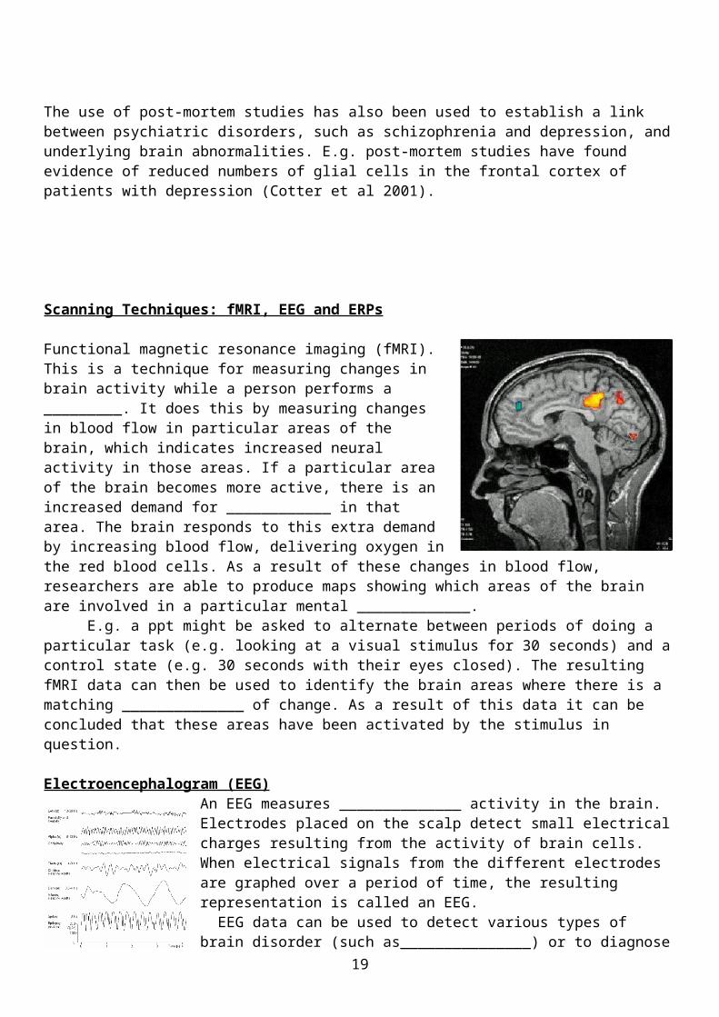
Functional magnetic resonance imaging (fMRI).This is a technique for measuring changes in brain activity while a person performs a _________. It does this by measuring changes in blood flow in particular areas of the brain, which indicates increased neural activity in those areas. If a particular area of the brain becomes more active, there is an increased demand for ____________ in that area. The brain responds to this extra demand by increasing blood flow, delivering oxygen in the red blood cells. As a result of these changes in blood flow, researchers are able to produce
15

maps showing which areas of the brain are involved in a particular mental _____________.
E.g. a ppt might be asked to alternate between periods of doing a particular task (e.g. looking at a visual stimulus for 30 seconds) and a control state (e.g. 30 seconds with their eyes closed). The resulting fMRI data can then be used to identify the brain areas where there is a matching ______________ of change. As a result of this data it can be concluded that these areas have been activated by the stimulus in question.
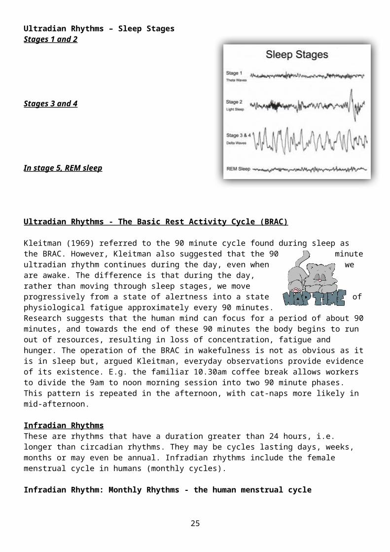
Electroencephalogram (EEG)An EEG measures ______________ activity in the brain. Electrodes placed on the scalp detect small electrical charges resulting from the activity of brain cells. When electrical signals from the different electrodes are graphed over a period of time, the resulting representation is called an EEG.
EEG data can be used to detect various types of brain disorder (such as_______________) or to diagnose other disorders that influence brain activity such as Alzheimer’s disease. E.g. readings of patients with epilepsy show spikes of electrical activity. EEG patterns in patients with brain disease and brain
injury show overall ____________ of electrical activity. The four basic patterns are ___________ waves, ____________ waves, ___________
waves and ___________ waves. When a person is awake but relaxed rhythmical alpha waves are recorded. When the person is physiologically aroused, their EEG pattern shows low amplitude and fast frequency beta waves. Beta waves are also found in _________ sleep when the eyes move rapidly back and forth. Delta and theta waves occur during sleep. As the person moves from light to deep sleep the occurrence of alpha waves decreases and are replaced first by lower frequency theta waves and then by delta waves.
Event-Related Potentials (ERPs)ERPs are very small voltage changes in the brain that are triggered by specific events or stimuli. ERPs are difficult to pick out from all the other electrical activity being generated within the brain at a given time. To establish a specific response to a target ____________ requires many presentations of the stimulus and these responses are then averaged together. Any extraneous neural activity that is not related to the specific stimulus will not occur consistently, whereas activity linked to the stimulus will. This has the effect of ______________ out the background neural ‘noise’, making the specific response to the stimulus in question stand out more clearly.
ERPs can be divided into two categories. Waves occurring within the first 100 milliseconds after presentation of the stimulus are termed ‘____________’ ERPs as they reflect an initial response to the physical characteristics of the stimulus. ERPs after the first 100 milliseconds reflect the manner in which the subject evaluates the stimulus and are termed ‘____________’ ERPs as they demonstrate information processing.
Evaluation: Ways of Studying the BrainStrengths Limitations
sensory task alpha oxygen cognitive activity stimulus pattern delta electricalcancelling theta epilepsy slowing beta REM
16
fMRI
EEG
ERP
Post-Mortem Examina
tions
7. Biological Rhythms: Circadian Rhythms
Biological rhythms are cyclical changes in the way that biological systems have evolved because the environment in which organisms live has cyclic changes, day/night, summer/winter and so on. The most important of these rhythms are the circadian rhythms – any cycle that lasts about 24 hours. In fact, the word circadian comes from the Latin ‘circa’ (about) plus ‘dies’ (a day). Nearly all organisms possess a biological representation of the 24 hour day. These circadian rhythms (often referred to as the body clock) optimize an organism’s physiology and behavior to best meet the varying demands of the day/night cycle.
TASK: Research the missing key terms…
The Nature of Circadian Rhythms
Our circadian rhythms are driven by our body clocks, found in all of the cells of the body, and synchronised by the master ______________ pacemaker, the suprachiasmatic nuclei (SCN), found in the _________________. This pacemaker must constantly be reset so that our bodies are in synchrony with the outside
17
world. ____________ provides the primary input to this system, setting the body clock to the correct time in a process termed _________________________. In mammals, light-sensitive cells within the eye act as brightness detectors, sending messages about environmental light levels direct to the SCN. The SCN then uses this information to coordinate the activity of the entire circadian ________________. The most familiar of the circadian rhythms subject to this entrainment process is the _________________ cycle.
The Sleep-Wake cycle
The circadian rhythm not only dictates when we should be sleeping, but also when we should be ________________. Light and darkness are the external signals that determine when we feel the need to sleep and when we need to wake up. The circadian rhythm also dips and rises at certain times of the _______, so our strongest sleep drive usually occurs in two dips (between ____ - ____am and between _____-_____pm, the ‘post lunch dip’). The sleepiness we feel during these circadian dips is less intense if we had sufficient sleep, and more intense when we’re sleep _____________.
Sleep and wakefulness are not determined by the circadian rhythm alone, but are also under homeostatic control. When we have been awake for a long period of time, ________________ tells us that the need for sleep is increasing because of the amount of energy used up during wakefulness. This homeostatic drive for sleep increases gradually throughout the day, reaching its maximum in the late evening when most people fall asleep.
Therefore, the circadian system keeps us awake as long as there is _________________, prompting us to sleep as it becomes dark. The homeostatic system tends to make us sleepier as time goes on throughout the waking period, regardless of whether it’s day or night. The internal circadian ‘__________’ is described as ‘free running’ i.e. it will maintain a cycle of about 24-25 hours, even in the absence of external cues. The circadian system is, however, intolerant of any major alterations in sleep and wake schedules (e.g. through jet travel, shift work) because this causes the biological clock (and the internal
physiological systems that are dependent on this) to become completely out of _____________.
Other circadian rhythms
Core Body temperatureCore body temperature is one of the best indicators of the
circadian rhythm. It is at its lowest (about 36 degrees Celsius) at about __________am and at its highest (about 38 degrees Celsius) at about ______pm. During the normal circadian rhythm, sleep occurs when the core temperature begins to drop, and the body temperature starts to rise during the last hours of sleep, promoting a feeling of alertness in the morning. A small drop in body temperature also occurs in most people between 2pm and 4pm, which may explain why many people feel sleepy in the afternoon.
Hormone ProductionHormone release follows a circadian rhythm. For example, the production and release of melatonin from the __________ gland in the brain follows this rhythm, with peak levels
occurring during the hours of darkness. By activating chemical receptors in the brain, ______________ encourages feelings of sleep. When it’s dark, more melatonin is produced, and when it’s light again, the production of melatonin drops and the person ____________.
A Case Study – Michel Siffre
Evidence for a ‘free running’ circadian rhythm comes from a series of studies conducted by the French cave explorer, Michel Siffre. On several occasions Siffre has subjected himself to long periods of time living
18
Evaluation
A Case Study – Michel Siffre
Evidence for a ‘free running’ circadian rhythm comes from a series of studies conducted by the French cave explorer, Michel Siffre. On several occasions Siffre has subjected himself to long periods of time living
Individual Differences
There are two important types of individual differences in circadian rhythms. One is the cycle length; research has found that cycle length can vary from 13-65 hours (Czeisler et al 1999). The other type of individual difference relates to cycle onset; individuals appear to be innately different in terms of when their circadian rhythms reach their peak. E.g. Duffy et al (2001) found that morning people tend to rise early and go to bed early (about 6am and 10pm), whereas evening people prefer to wake and go to bed later (10am and 1am).
Research Methodology
Early research studies of circadian rhythms suffered from an important flaw when estimating the ‘free running’ cycle of the human circadian rhythm. In most studies, ppts are isolated form variables that might affect their circadian rhythms e.g. clocks, radios, daylight. However, they were not isolated from artificial light because it was generally thought that dim artificial light, in contrast to daylight, wouldn’t affect circadian rhythms. But research suggests this might not be true. Czeisler (1999) altered ppts circadian rhythms down to 22 hours & up to 28 hours by dim lighting.
Chronotherapeutics
One real-world application of circadian rhythms is chronotherapeutics – the study of how timing affects drug treatments. The specific time a patient takes their medication is very important as it can have a significant impact on treatment success. It is essential that the right concentration of a drug is released in the target area of the body at the time that the drug is most needed. For example, the greatest risk of heart attack is during the early morning hours after awakening. Therefore, chronotherapeutic medications have been developed with a novel drug delivery system. These medication scan be administered before the person goes to sleep at 10pm, but the actual drug is not released before the
19

Ultradian and Infradian Rhythms
The circadian rhythm isn’t the only biological rhythm. There are two other important rhythms that determine human behavior. Ultradian rhythms span a period of less than one day. Infradian rhythms span a much longer period than one day. An example is the monthly menstrual cycle in women, although in humans there is even evidence for an annual infradian rhythm in some behaviours.
Ultradian RhythmsIn humans, daily cycles of wakefulness and sleep follow a circadian rhythm. However, within the sleep portion of this cycle another type of rhythm exists – an ultradian rhythm. A biological rhythm is referred to as ultradian if its period is shorter than 24 hours. In humans, a classic example of such a rhythm would be the five stages that make up a typical night’s sleep.
Ultradian Rhythms – Sleep StagesStages 1 and 2
Stages 3 and 4
In stage 5, REM sleep
Ultradian Rhythms - The Basic Rest Activity Cycle (BRAC)
Kleitman (1969) referred to the 90 minute cycle found during sleep as the BRAC. However, Kleitman also suggested that the 90 minute ultradian rhythm continues during the day, even when we are awake. The difference is that during the day, rather than moving through sleep stages, we move progressively from a state of alertness into a state of physiological fatigue approximately every 90 minutes. Research suggests that the human mind can focus for a period of about 90 minutes, and towards the end of these 90 minutes the body begins to run out of resources, resulting in loss of concentration, fatigue and hunger. The operation of the BRAC in wakefulness is not as obvious as it is in sleep but, argued Kleitman, everyday observations provide evidence of its existence. E.g. the familiar 10.30am coffee break allows workers to divide the 9am to noon morning
Chronotherapeutics
One real-world application of circadian rhythms is chronotherapeutics – the study of how timing affects drug treatments. The specific time a patient takes their medication is very important as it can have a significant impact on treatment success. It is essential that the right concentration of a drug is released in the target area of the body at the time that the drug is most needed. For example, the greatest risk of heart attack is during the early morning hours after awakening. Therefore, chronotherapeutic medications have been developed with a novel drug delivery system. These medication scan be administered before the person goes to sleep at 10pm, but the actual drug is not released before the
20

session into two 90 minute phases. This pattern is repeated in the afternoon, with cat-naps more likely in mid-afternoon.
Infradian RhythmsThese are rhythms that have a duration greater than 24 hours, i.e. longer than circadian rhythms. They may be cycles lasting days, weeks, months or may even be annual. Infradian rhythms include the female menstrual cycle in humans (monthly cycles).
Infradian Rhythm: Monthly Rhythms - the human menstrual cycleA woman’s reproductive cycle is known as a menstrual cycle because it lasts about one month (__________ is Latin meaning month). There are considerable variations in the length of this cycle with some women experiencing a relatively short 23 day cycle whereas others have a cycle as long as 36 days (Refinetti 2006). The average appears to be around _______ days. The menstrual cycle is regulated by __________------, which either promote ovulation or stimulate the uterus for fertilisation. Ovulation occurs roughly through the menstrual cycle, when _______________ levels peak, and usually lasts for 16 to 32 hours. After the ovulatory phase, _____________________ levels increase in preparation for the possible implantation of an embryo into the uterus.
Infradian Rhythm: Annual RhythmsIn most animals, annual rhythms are related to the seasons (e.g. migration as a response to lower temperatures and decreased food sources in the winter), but in humans the calendar year appears to influence behavior regardless of changes in temperature. Research suggests a seasonal variation in mood in humans, especially in women (Magnusson, 2000), with some people becoming severely depressed during the winter months (seasonal affective disorder). The winter is also associated with an increase in heart attacks, which varies seasonally and peaks in winter. In fact, there is
a robust annual rhythm in human deaths, with most deaths occurring in January (Trudeau, 1997).
Evaluation
Individual Differences in sleep stages
Differences in the sleep patterns of individuals are usually attributed to differences in non-biological factors e.g. room temperature, sleep hygiene. However, a study by Tucker et al (2007) suggests that these differences are in large part biologically determined and may even be genetic in origin. Ppts were studied over 11 consecutive days and nights in a strictly controlled lab environment. The researchers assessed sleep duration, time to fall asleep and the amount of time in each
21

Individual Differences in sleep stages
Differences in the sleep patterns of individuals are usually attributed to differences in non-biological factors e.g. room temperature, sleep hygiene. However, a study by Tucker et al (2007) suggests that these differences are in large part biologically determined and may even be genetic in origin. Ppts were studied over 11 consecutive days and nights in a strictly controlled lab environment. The researchers assessed sleep duration, time to fall asleep and the amount of time in each
The menstrual cycle – the role of exogenous cues
The menstrual cycle is normally governed by an endogenous system – the release of hormones by the pituitary gland. However, it can also be controlled by exogenous cues. When several women of childbearing age live together and do not take oral contraceptives, their menstrual cycles tend to synchronise. In one study, daily samples of sweat were collected from one group of women and rubbed onto the upper lips of women in a second group. The groups were kept separate yet their menstrual cycles became synchronized with their ‘odour donor’ (Russell et al 1980). This suggests that the synchronisation of menstrual cycles can be affected by pheromones. Pheromones act in a similar way to hormones, but have an effect on the bodies of people close by rather than the body of the person producing them
The menstrual cycle influences mate choice
Research by Penton-Voak et al (1999) suggests that human mate choice varies across the menstrual cycle, with different preferences at different stages of the cycle. In their research, they found that women generally expressed a preference for ‘slightly feminised’ male faces when picking a partner for a long term relationship. However, when in the ovulatory phase, women showed a preference for more masculine faces. This preference is believed to represent a preference for kindness and cooperation in parental care in long term mates, but a preference for males with good genes for short term liaisons so that these genes might be passed to their offspring.
Belief in lunar rhythms
Despite empirical evidence to the contrary, the belief in an infradian rhythm based on the phases of the moon remain strong. For example, many midwives still believe that more babies are born during a full moon than during a new moon, but statistics show that this is a purely subjective association (Arliss et al 2005). Likewise, surveys of workers in the mental health profession have shown a persistent belief that the full moon can alter behavior (Vance, 1995). Yet study after study has failed to show any consistent association between the moon and human psychopathology. Occasional studies have found correlations between the phase of the moon and various aspects of human behavior, but there is no evidence of a causal relationship (Foster and Roenneberg, 2008).
22

Endogenous Pacemakers and Exogenous Zeitgebers
Biological rhythms like the ones we have studied must be constantly fine-tuned in order to stay in tune with the external world. In order to achieve this we have endogenous pacemakers, sometimes referred to as biological clocks, and exogenous (or external) zeitgebers, which reset this clock every day to maintain its coordination with the external world. So how do these two processes work?
Endogenous Pacemakers
The term endogenous refers to anything whose origins are within the organism i.e. within our bodies. These pacemakers are most probably the products of inherited genetic mechanisms and allow us to keep pace with changing cycles in the environment. The most important pacemaker in human beings is the ____________________ nucleus.
The Suprachiasmatic NucleusIn mammals, the main endogenous pacemaker is a tiny cluster of ___________ cells called the suprachiasmatic nucleus (SCN), which lies in the __________________. The SCN plays an important role in generating the body’s circadian rhythms. It acts as the ‘master clock’, which links to other brain regions that control sleep and arousal, and has control over other biological clocks throughout the ____________. Neurons within the SCN spontaneously
Research methods – Criticising research:
If they had asked the midwives and workers in mental health about their beliefs about lunar activity and behavior with a questionnaire or interview; what could have affected their answers (which in turn would decrease the validity of their answers)?
23
synchronise with each other, so that their target neurons in sites elsewhere in the body receive correctly ____________________ time-coordinated signals. These peripheral clocks can maintain a circadian rhythm, but not for very long, which is why they must be controlled by the SCN.
This is possible because of the SCN’s built-in circadian rhythm, which only needs resetting when external light levels ___________. The SCN receives information about light levels via the _______________________. This happens even when our eyes are shut, because light penetrates the eyelids. If our biological clock is running slow (e.g. the sun rises earlier than on the previous day), then morning light ___________________ adjusts the clock, putting its rhythm in step with the world outside. The SCN also regulates the manufacture and secretion of melatonin in the pineal gland via an interconnecting neural pathway.
The pineal glandThe SCN sends signals to the pineal gland, directing it to increase production and secretion of the hormone ______________ at night and to decrease it as light levels increase in the morning. Melatonin induces sleep by inhibiting the brain mechanisms that promote wakefulness. The pineal gland and the SCN function jointly as ______________ pacemakers in the brain. The sensitivity of the pineal gland and the SCN to light, and the role of melatonin in controlling sleep and activity, mean that, despite the endogenous nature of these clocks, their activity must be ________________ with the light-dark rhythm of the world outside.
Exogenous Zeitgebers
The term ‘exogenous’ refers to anything whose origins are outside the organism. The term ‘zeitgeber’ comes from the German words Zeit and Geber, meaning ‘_________________.’ Exogenous zeitgebers are environmental events that are responsible for entraining the biological clock of an organism. The most important zeitgeber for most animals is ________.
LightReceptors in the SCN are sensitive to changes in light levels during the day and use this information to synchronise the activity of the body’s ___________________________. Light resets the internal biological clock each day, keeping it on a 24 hour cycle. Rods and cones in the _____________ of the eye detect light to form visual images. However, there is a third type of light detecting cell in the retina that gauges overall brightness to help reset the internal biological clock. A protein called ________________, which is sensitive to natural light, is critical in this system. A small number of retinal cells contain melanopsin and carry signals to the SCN to set the daily body cycle.
Social CuesSocial stimuli, such as mealtimes and social activities, may also have a role as zeitgebers. ________________ (1971) showed that individuals are able to compensate for the absence of zeitgebers such as natural light by responding to social zeitgebers instead.
One of the earliest studies on jet lag (Klein and Wegmann, 1974) found that the circadian rhythms of air travelers adjusted more quickly if they went ______________ more at their destination. This was thought to be because they were exposed to the social cues of their new time zone, which acted as a zeitgeber.
24
Likewise, the circadian rhythms of blind people were thought to be no different to sighted people as both groups were exposed to the same social cues. We now know that both examples can be better explained in terms of light exposure acting as a zeitgeber. The sleep-wake cycle of most blind people is still influenced by light during the day, even though they have no visual perception. This is because connections exist between the eye and the SCN that do not involve those parts of the visual system on which the perception of light depends.
Evaluation of Endogenous Pacemakers
The role of the SCN
The importance of the SCN as endogenous pacemaker had been demonstrated in animal studies. Morgan (1995) bred a strain of hamsters so that they had abnormal circadian rhythms of 20 hours rather than 24. SCN neurons from these abnormal hamsters were then transplanted into the brains of normal hamsters. These normal hamsters then displayed the same abnormal circadian rhythm of 20 hours, showing that the transplanted SCN had imposed its pattern onto the hamster. Further confirmation of the importance of the SCN came in the reverse experiment, planting SCN neurons from normal hamsters into the brains of abnormal hamsters. Rather than maintaining their abnormal circadian rhythm, the recipient hamsters changed to a circadian pattern of 24 hours.
Separate rhythms
Under normal conditions the ‘master clock’ (the SCN) coordinates all other bodily rhythms. However, in some circumstances these rhythms can become out of step with each other. Folkard (1996) studied a university student, Kate Aldcroft, who volunteered to spend 25 days in the controlled environment of a laboratory. During her time in the lab she had no access to daylight or other zeitgebers that might have reset the SCN, at the end of the 25 days her core temperature rhythm was still at 24 hours. However, her sleep-wake cycle had extended to 30 hours, with periods of sleep as long as 16 hours.
synchronised organs and glands Suprachiasmatic Ascoff et al light nerve change melatonin
hypothalamus optic nerve outside body time giver endogenous melanopsin retina time-coordinated automatically
25

Evaluation of Exogenous Zeitgebers
Exam Questions (A Level)
1) Read the item and then answer the questions that follow.
The role of the SCN
Support for the role of melanopsin in setting the circadian rhythm comes from studies of blind people. Some blind people are still able to reliably entrain their circadian rhythm in response to light despite a total lack of image forming visual perception (i.e. non-functioning rods and cones). Skene and Arendt (2007) estimate that the vast majority of blind people who still have some light perception have normally entrained circadian rhythms. This suggests that the pathway from retinal cells containing melanopsin to the SCN is still intact. As further evidence for the importance of this pathway in setting the biological clock, people without light perception show abnormal circadian entrainment.
Using light Exposure to Avoid Jet Lag
Burgess et al (2003) found that exposure to bright light prior to an east-west flight decreased the time needed to readjust to local time on arrival. Volunteers participated in one of three treatments (continuous bright light, intermittent bright light, and dim light), each of which shifted their sleep-wake cycle back by one hour a day over three days. Ppts exposed to continuous bright light shifted their circadian rhythm by 2.1 hours over the course of the study. Those exposed to intermittent bright light shifted their rhythm by 1.5 hours, and a third group exposed to dim light shifted theirs by 0.6 hours. As a result, ppts in the first treatment group felt sleepier 2 hours earlier in the evening and woke 2 hours earlier in the morning i.e. closer to the local times conditions they would find after an east-west flight.
The role of artificial light as a zeitgeber
Vetter et al (2011) investigated the importance of light in the regulation of the sleep-wake and activity-rest patterns of two groups of volunteer ppts over a 5 week study period. One group remained in normal ‘warm’ artificial light while the other group experienced artificial ‘blue enriched’ light that had a spectral composition close to daylight. All ppts kept a daily sleep log and wore devices that measured their movement over each 24 hour period. Ppts working under the warmer light synchronised their circadian rhythms each day with the natural light of dawn. Over the course of the study, sunrise advanced by 42 minutes. The ppts who were exposed to blue enriched light did not show the same 42 minute adjustment and instead synchronized their rhythms to office hours. The results confirm that light is the dominant zeitgeber for the SCN and that its effectiveness depends on its spectral composition.
26
Figure 1 shows the left hemisphere of the human brain. Six areas of cortical specialisation are labelled A, B, C, D, E and F.
Figure 1: Left hemisphere of the human brain
Using your knowledge of localisation of function in the brain, identify the area of cortical specialisation. Shade one box only for each area.
. Broca’s area
. Somatosensory cortex
. Visual cortex
. Wernicke’s area
. Motor cortex
[1 mark] [1 mark] [1 mark] [1 mark] [1 mark]
2) The electroencephalogram (EEG) and event-related potentials (ERPs) both involve recording the electrical activity of the brain. Outline one difference between the EEG and ERPs.
[2 marks]
3) Read the item and then answer the question that follows.
Sam is a police officer. She has just started working the night shift and after a week, she finds that she has difficulty sleeping during the day and is becoming tense and irritable. Sam is also worried that she is less alert during the night shift itself.
Using your knowledge of endogenous pacemakers and exogenous zeitgebers, explain Sam’s experiences.
[4 marks]
4) The human female menstrual cycle is an example of one type of biological rhythm; it is called a:
27
A circadian rhythm
B infradian rhythm
C ultradian rhythm
[1 mark]
5) Outline the structures and processes involved in synaptic transmission.
[6 marks]
6) Split brain patients show unusual behaviour when tested in experiments. Briefly explain how unusual behaviour in split brain patients could be tested in an experiment.
[2 marks]
7) Briefly evaluate research using split brain patients to investigate hemispheric lateralisation of function.
[4 marks]
24 mark total
28