Embed Size (px)
Citation preview

Dr Colin Walker
Melbourne Bird Veterinary Clinic, 1 George Street, Scoresby, Victoria 3179, AustraliaEmail: [email protected]
Rota Virus in Pigeons. The Australian experience
Introduction
The key word that describes the entire Rota virus outbreak in Australia is “speed”. A highly
infectious, high mortality disease rapidly spread throughout the country in a period of months, killing
tens of thousands of pigeons and causing considerable disruption to the entire pigeon sport.
The veterinary response needed to match the severity of the problem. It was on 12 th December 2016
that the first case was seen on the eastern seaboard of Australia and yet, by the beginning of July
2017, in only 7 months, the condition had been diagnosed, available vaccines both in Australia and
overseas had been investigated for potential cross immunity, the need to make a new vaccine had been
identified, a trial vaccine had, in fact, been made, vaccine trials had been completed and the vaccine
was available to go to commercial production. At the same time, the nature of the disease had been
investigated so that an understanding of the clinical disease caused by Rota virus had been developed.
In addition, the length of time of the carrier state and the persistence of immunity had been
investigated.
We quickly realised that we were dealing with a new disease in the world. All of this work had been
done in Victoria, Australia. It was a good example of the Department of the Environment and
Primary Industry (DEPI), Agribio ( the diagnostic branch of DEPI ), Latrobe University, The
University of Melbourne, Tredlia Biovet ( a vaccine manufacturing company ) , the Australian
National Racing Pigeon Board (ANRPB) and the Victorian pigeon industry all working together in a
collaborative way . Government and education organisations had combined with the pigeon industry
to deliver much clinical information and a trial vaccine that could then be adapted to commercial
production. It was a great result for all concerned. I think all involved learnt a lot. Many of us had
made new acquaintances, some of whom became friends.
The Beginning – the first cases in Western Australia
An outbreak of disease occurred in racing pigeons in Perth, Western Australia (WA) in May 2016.
Sickness and death appeared within 3 to 4 days of race birds returning from the first Pigeon Racing
Federation of Western Australia (PRF) race of 2016. Affected birds developed vomiting and a green
mucoid diarrhoea. Mortality rates in various lofts ranged from 10 to 50 % with an average of 25
%.The birds had been exposed in the race transporter to pigeons that had experienced similar
symptoms a week or so earlier. The origin of the infection appears to be linked to illness and deaths
1

that were not investigated in pigeons from lofts that shared a training trailer several weeks prior to the
first race. Approximately 100 members competed in the race and every competitor’s loft became
infected. Only the three members who did not compete remained free of the disease. At the time, the
disease was diagnosed by the attending veterinarian as a combined infection of pigeon Adeno virus
Type 1 and Type 2. The significance of this misdiagnosis was that the importance of quarantine was
not realised. Adeno virus is not uncommon in Australia. Failure to identify the cause as a new and
exotic virus meant that bird movement continued.
After several weeks the surviving WA birds regained their health and racing resumed. A normal
racing season was completed in October 2016. In that same month, however, in one of the final races,
the PRF transporter collected some pigeons from a country club based in Busselton, 200 km south of
Perth, and transported these birds with their own to the race release point. These birds developed the
disease after return.
The Department of Agriculture and Food, Western Australia (DAFWA) released a diagnostic
summary 8 months later in January 2017, stating the likely cause was a Reo virus.
Perth is a city of 2 million people located on the west coast of Australia and has approximately 150
racing pigeon fanciers. It is 3000km from the west coast to the eastern seaboard of Australia, which is
where most Australians live. The west coast is separated from the east coast by a huge desert, called
the Nullabor Plain, which forms a natural barrier between the two areas. As the end of 2016
approached although the disease had not been fully diagnosed, it appeared to have been confined to
WA and to have “died out”. Interest in it and diagnostic momentum were fading.
Figure 1. The size of Australia compared to Europe and Poland.
2

The disease spreads
Melbourne is located on the east coast of Australia, 3000km from Perth. On Monday 12 December
2016, veterinarians at the Melbourne Bird Veterinary Clinic ( MBVC ) were presented throughout the
day with racing pigeons by three separate fanciers . The birds in all three lofts had vomiting and
diarrhoea and about a quarter of the birds had died. All fanciers described how they had been to a
pigeon sale at Kyabram ( a country town 150 km north of Melbourne ) the previous weekend on
Sunday 4 December. No Western Australian pigeons were at the sale, however, it subsequently
transpired that birds were offered for sale from lofts where birds were unwell with similar symptoms
and mortality rates to that seen in WA. It is strongly suspected that WA birds had been introduced into
these lofts in the few weeks prior to the sale. These fanciers, however, denied introducing birds from
WA but did believe that their birds were affected by the same problem as that seen in WA despite not
having their birds’ health problem investigated. Birds were placed from these lofts into the sale at
Kyabram.
Birds from the Kyabram sale travelled to South Australia, southern New South Wales (NSW), rural
Victoria and Melbourne and they took the virus with them. By the end of the year the disease had
been diagnosed in Port Augusta (South Australia), Finley ( NSW ), Kyabram ( country Victoria ) and
several Melbourne lofts. Throughout January 2017 an unfortunate trend developed in Melbourne,
where fanciers deliberately exposed their birds to the virus, which still at this time had yet to be
diagnosed. They had received veterinary advice ( from the same vet who was the attending vet in
Perth ) that there was no long term way of protecting the birds through vaccination, that once birds
had had the disease they could not catch it again and that it was only a matter of time until any one
loft became infected. He advised that it was better to get the infection over and done with and
proposed a mass infection day on 3 February 2017. A significant proportion of fanciers followed this
advice, particularly those in the north-western suburbs of Melbourne.
By 11 January, 11 cases had been diagnosed in Melbourne. One of these was in Dandenong ( an outer
eastern suburb ). The rest were in Melbourne’s north- west. Three were in non-racing lofts.
Throughout January, the number of cases actually diagnosed in Melbourne remained low. There were,
however, a significant number of lofts reporting birds with typical symptoms and mortality rates but
failing to present birds for diagnosis. Social media reported that there were 100 lofts infected in
Melbourne and the disease was in “plague proportions”.
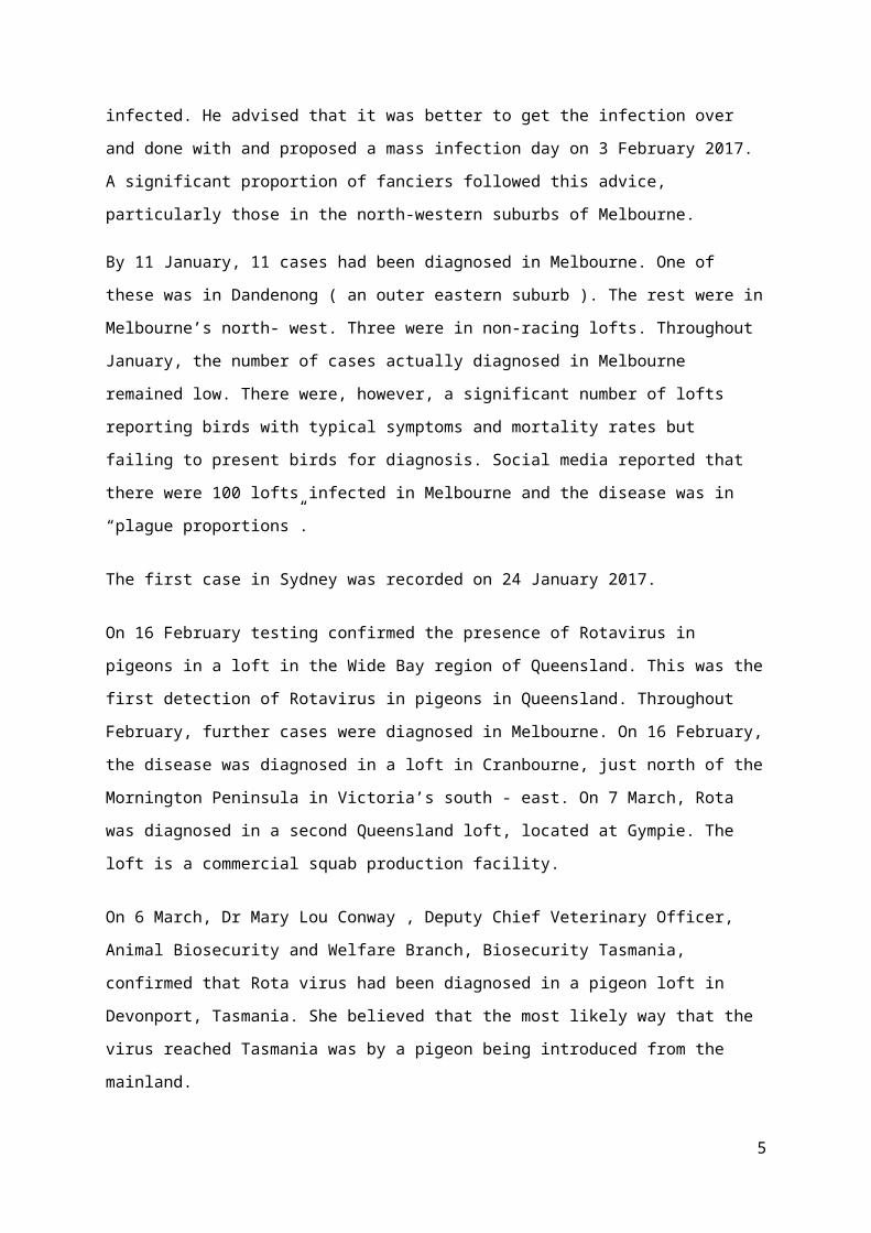
The first case in Sydney was recorded on 24 January 2017.
On 16 February testing confirmed the presence of Rotavirus in pigeons in a loft in the Wide Bay
region of Queensland. This was the first detection of Rotavirus in pigeons in Queensland. Throughout
February, further cases were diagnosed in Melbourne. On 16 February, the disease was diagnosed in a
3

loft in Cranbourne, just north of the Mornington Peninsula in Victoria’s south - east. On 7 March,
Rota was diagnosed in a second Queensland loft, located at Gympie. The loft is a commercial squab
production facility.
On 6 March, Dr Mary Lou Conway , Deputy Chief Veterinary Officer, Animal Biosecurity and
Welfare Branch, Biosecurity Tasmania, confirmed that Rota virus had been diagnosed in a pigeon loft
in Devonport, Tasmania. She believed that the most likely way that the virus reached Tasmania was
by a pigeon being introduced from the mainland.
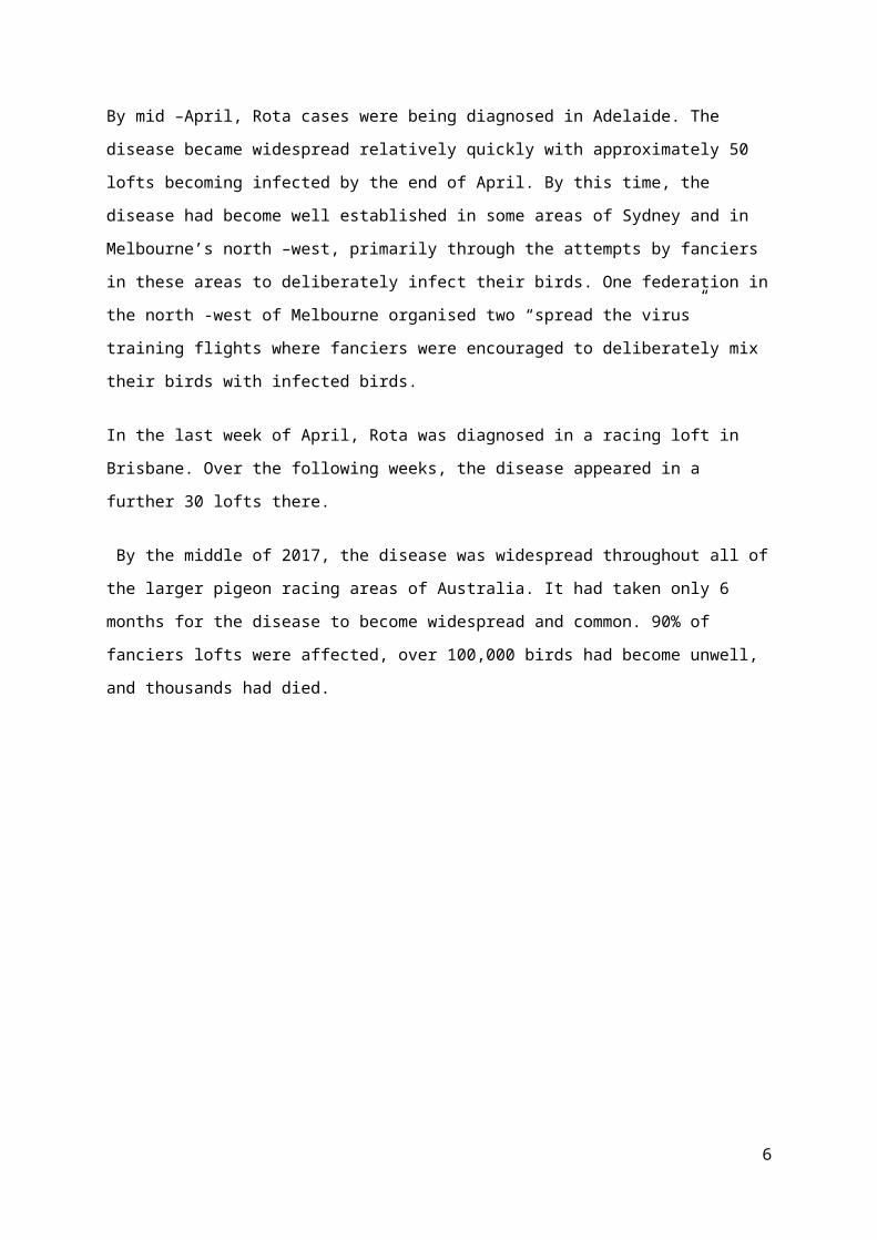
By mid –April, Rota cases were being diagnosed in Adelaide. The disease became widespread
relatively quickly with approximately 50 lofts becoming infected by the end of April. By this time, the
disease had become well established in some areas of Sydney and in Melbourne’s north –west,
primarily through the attempts by fanciers in these areas to deliberately infect their birds. One
federation in the north -west of Melbourne organised two “spread the virus” training flights where
fanciers were encouraged to deliberately mix their birds with infected birds.
In the last week of April, Rota was diagnosed in a racing loft in Brisbane. Over the following weeks,
the disease appeared in a further 30 lofts there.
By the middle of 2017, the disease was widespread throughout all of the larger pigeon racing areas of
Australia. It had taken only 6 months for the disease to become widespread and common. 90% of
fanciers lofts were affected, over 100,000 birds had become unwell, and thousands had died.
Figure 2. The spread of pigeon Rota virus throughout Australia.
4
Origin, where did this disease come from?
Where did this virus come from? It probably did not come from anywhere. The virus, or at least its
parts, has probably been in Australia for a long time but in a slightly different form.
The sequenced virus has significant similarities to Rota viruses isolated previously from the Red Fox
(Vulpes vulpes) and also from the Spotted neck dove ( Streptopelia sp ). Red foxes are a common and
widespread feral pest in Australia. Active extermination programs are in place due to the damage they
inflict on native Australian mammals and birds but their populations appear remarkably resilient.
Foxes are found in all Australian environments and are surprisingly common not only in rural areas
but also residential suburbs and indeed large city centres. Three species of Streptopelia dove were
introduced into Australia towards the end of the 19th century from south-east Asia. All have
established large feral populations. Streptopelia chinensis and Streptopelia tigrina are found on the
eastern seaboard while Streptopelia senegalis is located on the west coast. The eastern and western
populations are discreet, being geographically separated not only by distance but by the huge central
desert areas of Australia
So where did this disease come from? The suggestion is that an antigenic shift in a Streptopelia Rota
virus has occurred or alternatively a viral recombination has occurred that has altered the virus
sufficiently that both the pattern of disease it causes and the species it affects have changed.
Reaching a diagnosis
Throughout December 2016 and January 2017, extensive diagnostic work involving autopsy,
bacteriology, histopathology, electron microscopy, virus culture and genetic sequencing was done in
Victoria. When the initial unwell birds were presented to veterinarians at the MBVC on 12 December
a fairly standard diagnostic path was followed. Following clinical examination and standard screening
tests including microscopic examination of the faeces and crop flushes several birds were selected for
autopsy. Full sets of tissue samples were collected for histopathology, swabs for microscopic
examination, bacterial culture and antibiotic sensitivity (M C and S) were taken from the liver and
bowel and swabs were collected for PPMV (Pigeon Paramyxovirus) and Circo virus PCR
( Polymerase Chain Reaction) tests. The PCRs returned negative results while the bacterial cultures
grew normal bacteria.
Gross autopsy was unremarkable, however, on histology the lesion looked viral, according to the
pathologists at AgriBio, so electron microscopy was performed (at the Australian Animal Health
Laboratory (AAHL) ) and abundant viral particles were seen. Back at AgriBio, viral cultures in
embryonated eggs were commenced. Isolation proved tricky, but identification of a Rotavirus on next
5
generation sequencing allowed some modifications, which led to isolation, but not amplification.
(Staff at AgriBio, including Dr Christina McCowan, plan to do more work on this.)
Next generation sequencing and Sanger PCR confirmed the virus to be an A group rotavirus, sub type
G18P,of previously undescribed genotype.
Further testing in Victoria on samples collected in Western Australia in May 2016, showed the
disease in Western Australia to be caused by the same virus.
Figure 3. Transmission electron micrograph of virus particles in a preparation from the liver of an affected pigeon. (Photo courtesy DAFWA)
Diagnostic Features
Autopsy and histology changes
As mentioned above gross autopsy is unremarkable with no reliable gross abnormalities. Some birds
have enlarged spleens, many do not. Sometimes the liver is slightly pale or perhaps a bit friable.
Essentially however autopsy is not diagnostic.
Histologically the principle lesion is hepatic necrosis seen as dissociation, rounding up and
hypereosinophlia of hepatocytes with shrinking and distortion of nuclei, often with little or no
inflammation. A moderate to marked macrophage response, some with prominent cytoplasmic
vacuolation and intravacuolar cellular debris and yellow/brown granular pigment is seen.
Inflammatory infiltrates (lymphocytic or lymphohistiocytic) were often, but not always seen in the
spleen. Histiocytic infiltrates (associated with resolving the necrosis) and sometimes a lymphocytic
6

infiltrate are seen in the liver but are not reliable features. Gut lesions are inconsistent with most birds
not having them at all. Other organs are not reliably involved.
Professor Amir Noormohammadi at the University of Melbourne accidentally found Rota virus in the
kidneys of some birds on electron microscopy (EM). However there is no significant renal lesion,
apart from urate nephrosis in some birds which is thought to be probably secondary to dehydration
(which is not surprising with vomiting and diarrhoea). The indication is that the virus is not causing
any pathology in the kidney even if it is there in some birds.
Clinical details
1/ The virus has an incubation period of 3 – 7 days. Birds start to become unwell 3 – 7 days after they
have been exposed to the virus.
2/ Initially, birds develop a hunched posture, fluff their feathers and become reluctant to move. They
then develop a green mucoid diarrhoea and start to vomit.
3/ Deaths start 12 – 24 hours after the first birds become unwell. Deaths peak on day 4 and stop
usually by day 7. In lofts in Melbourne, mortality rates have ranged from 6 to 36 % with an average of
22%. However, in some of the worst affected lofts in Sydney, mortality rates have been up to 50%.
Approximately seven days after the onset of symptoms most birds in the loft appear to regain their
health and appear clinically normal, however, the occasional death can be expected to occur for
several weeks after the outbreak due to complications associated with recovery in individual birds,
( such as adhesions to adjacent organs and secondary bacterial infections ).
4/ During the outbreak, every bird in the loft is thought to become infected, however, more extensive
testing of larger numbers of birds needs to be done to confirm this. Not all birds become unwell.
Some birds continue to behave normally. Others display symptoms that may be mild to severe, and
then recover. However, in many lofts, a significant proportion of birds that develop symptoms die.
Terminally, these birds develop a hunched posture and become dyspneic. Their mucous membranes
are congested and cyanotic. The glottis is rounded on inspiration. Many terminal birds become
sternally recumbent. During the outbreak food intake is reduced within the loft by approximately
50%.
5/ During infection, the virus penetrates the bowel wall and presumably is carried in the bloodstream
to the liver and occasionally the kidneys. In the liver, the virus causes massive necrosis of
hepatocytes. The primary cause of death is viral hepatic necrosis. Secondary causes of death include
dehydration and hypothermia.
7

6/ Surviving birds from infected lofts become carriers of the virus. Testing ( by cloacal Rota PCR )
has shown that surviving birds from infected lofts shed the virus in their faeces for 9 to 12 weeks and
are infectious to previously non-infected birds during this time. In testing done at the MBVC, 13 of 14
birds were positive on cloacal swab 9 weeks post infection while 10 of 10 were negative on cloacal
swab at 13 weeks. Two birds tested by liver Rota PCR at 9 weeks post infection were positive while
two tested at 15 weeks were both negative. These results suggest that the carrier state persists for 9-12
weeks, however, they are at odds with the spread of the virus from WA to Victoria via the auction at
Kyabram about 5-6 months after the initial outbreak in WA had died down. It may be that, where
most birds stop shedding by 9-12 weeks, as detected by cloacal PCR, some birds carry the virus for
much longer. Under natural conditions evidence suggests shedding may last up to 6 months in at least
some birds. It has been suggested that the auction was like a natural experiment, which measured the
duration of infection in a different and in some respects more realistic way, perhaps with the stress of
transport contributing to the renewal of shedding. As larger numbers of birds are tested the duration of
the carrier state and nature of shedding will become clearer
8/ It is thought that wild birds, in particular Streptopelia spp. (lace neck ) doves, and also flies, may
transmit the virus. This, however, needs to be validated. To date, the only wild, free-flying birds that
the virus has been detected in are feral pigeons. However, very few birds other than pigeons have
been presented for testing. One of our biggest missing pieces of information is how the virus spreads
locally from loft to loft in nearby suburbs.
9/ Anecdotally, it appears that the virus is not particularly robust in the environment and does not
survive under ambient conditions for more than 2- 3weeks. Deliberate attempts to infect birds with the
faeces of recovered birds failed if more than 3 weeks had passed since collection.
Figure 4. Typical diarrhoea and regurgitated grain from infected pigeons on the loft floor.
8
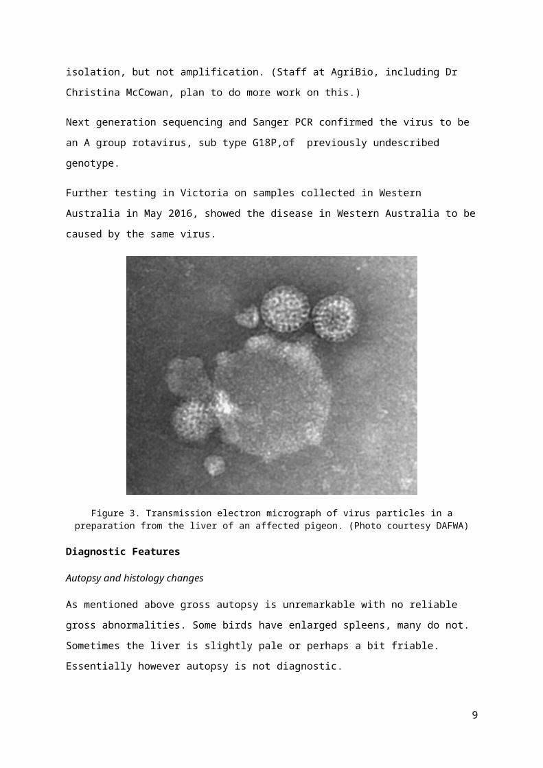
Figure 5. Typical appearance of infected pigeons. Horizontal posture (suggestive of abdominal pain), fluffed feathers over rump (also suggestive of abdominal pain), empty crop (reduced appetite, vomiting), “cocky
comb” – fluffed feathers at the base of the cere over the frontal sinus (indicative of general malaise), dropped wings, spread tail, partially closed eyes (all due to general weakness). This bird died 18 hours after the photo
was taken.
Figure 6. Terminally infected pigeons are often sternally recumbent and dyspnoeic.
I
9

Figure 7. Vomited grain on perches. Droppings indicate anorexia and polyuria.
Figure 8. Vomited grain on perches, mucoid diarrhoea.
Figure 9. Polyuria.
10

Figure 10. Two images of mucoid diarrhoea.
Figure 11. Polyuria, decreased faecal volume and biliverdinuria indicating impaired kidney function, decreased food intake or delayed crop emptying and impaired liver function, respectively.
11

Figure 12. Vomit containing grit, suggestive of either recent ingestion of a large amount of grit (in this situation likely associated with crop or stomach pain or an attempt to replace lost
minerals/electrolytes) or deep vomiting from the ventriculus.
Figure 13. General appearance of loft floor (cleaned 24 hrs earlier).
12
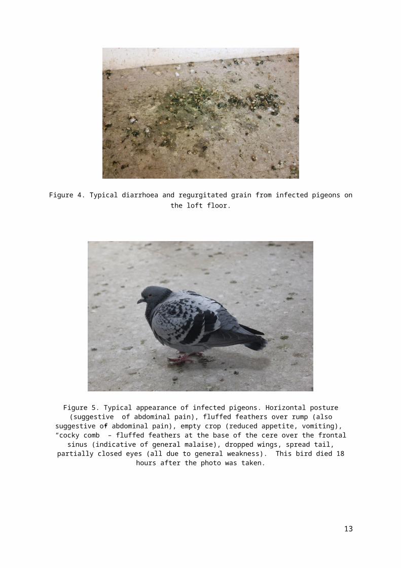
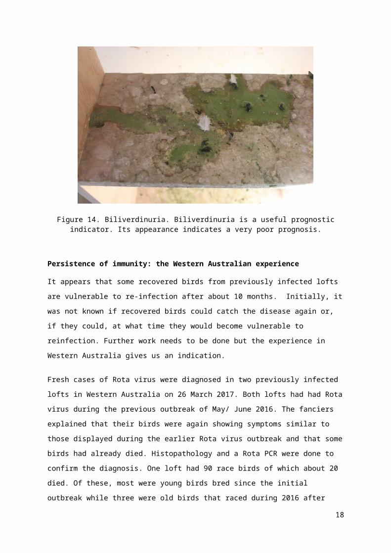
Figure 14. Biliverdinuria. Biliverdinuria is a useful prognostic indicator. Its appearance indicates a very poor prognosis.
Persistence of immunity: the Western Australian experience
It appears that some recovered birds from previously infected lofts are vulnerable to re-infection after
about 10 months. Initially, it was not known if recovered birds could catch the disease again or, if
they could, at what time they would become vulnerable to reinfection. Further work needs to be done
but the experience in Western Australia gives us an indication.
Fresh cases of Rota virus were diagnosed in two previously infected lofts in Western Australia on 26
March 2017. Both lofts had had Rota virus during the previous outbreak of May/ June 2016. The
fanciers explained that their birds were again showing symptoms similar to those displayed during the
earlier Rota virus outbreak and that some birds had already died. Histopathology and a Rota PCR
were done to confirm the diagnosis. One loft had 90 race birds of which about 20 died. Of these, most
were young birds bred since the initial outbreak while three were old birds that raced during 2016
after surviving the outbreak. In the second loft two of 100 died with about 10 % of birds showing mild
depression, diarrhoea and vomiting.
The PRF of Western Australia commenced their 2017 racing season in June. Within 4 weeks of
starting the season, changes to the race season had to be made in order to accommodate the effect of
Rota virus, ( racing continued but championship points were not allocated). Approximately 40% of
competitors lofts had become infected with Rota virus. It appeared that the young birds, when mixing
in the race baskets with birds from other lofts for the first time, had been exposed to Rota virus. The
13

most likely source of the exposure was older birds that had survived the previous year’s outbreak and
were carriers of the virus. As all fanciers with unwell birds had previously had Rota in their lofts in
2016, it seems that, although it is possible for some immunity to be passed from parents to their
youngsters during breeding, it is insufficient to prevent disease reliably. Compared to the previous
year only small numbers (less than 10 %) of birds in each loft became obviously unwell and even
lower numbers died. The majority of birds that died were youngsters. Presumably some immunity
passes from recovered birds, which are subsequently bred from, to their youngsters. As these young
ones mature this passively acquired immunity gradually fades making them increasingly vulnerable to
clinical disease. Partial immunity can modify the severity of symptoms. As in the year before, after
about 4 weeks most birds in most lofts had recovered and racing was resumed and the season
completed.
Identifying the need to make a vaccine
Initial diagnostic work in 2016 identified a Reo virus as a possible cause of the disease outbreak. In
January and February 2017 the possibility of using an existing vaccine to immunise Australian
pigeons was investigated. The outbreak was dynamic and urgent solutions were being sought.
Although there are no Reo virus vaccines in Australia there are chicken Reo virus vaccines available
in other parts of the world. These are used as an aid in protecting chickens against diseases caused by
a Reo virus, such as Infectious Bursal Disease. It was hoped that, once the new pigeon virus was
sequenced , sufficient antigenic overlap would be identified between it and a virus that already had a
vaccine that some cross- immunity may occur. One of these vaccines could then be used to protect the
Australian flock. Although there are considerable regulatory hurdles to importing vaccines into
Australia, it was felt that these could be addressed. Sequencing was completed on 24 January 2017.
This identified the virus, in fact, as a Rota virus ( a member of the Reoviridae ). The good thing about
this information was that there are Rota virus vaccines already available in Australia. They are used in
cattle. The investigation moved away from evaluating chicken Reo vaccines for potential cross
immunity to evaluating cattle Rota vaccines. The companies that manufacture these vaccines made
these sequences available to us and experts at AgriBio and Deakin and Latrobe University’s compared
these with the sequence of pigeon Rota virus. The disappointing conclusion was that there was
unlikely to be any cross- immunity. We had therefore exhausted our options of using an available
vaccine. Further investigations revealed that no chicken, turkey , pigeon or indeed any avian Rota
virus vaccines had ever been made anywhere in the world at any time. We therefore came to the
disconcerting realisation that in order to protect our birds we would need to make one.
Production of the vaccine
Background
14

Rotaviruses have been identified as one of the main aetiological agents of diarrhoea and enteritis in
mammals, including humans, and in avian species. Rotaviruses are non-enveloped viruses in the
family Reoviridae, genus Rotavirus. Virions are triple-layered and contain a viral genome that
consists of 11 segments of double-stranded RNA (dsRNA). These dsRNAs encode six structural
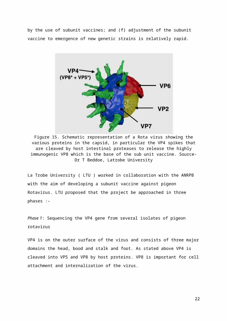
proteins (VPs) and six non-structural proteins (NSPs). The capsid is composed of an inner VP2 layer,
VP6 forming the intermediate capsid, and an outer layer made of VP7. VP4 forms 60 spikes on the
outer surface of the virus. The VP7 layer is essential for the virion to be able to infect a cell.
The spike protein VP4 gets cleaved by proteases located in the host intestinal lumen into VP5 and
VP8. VP8 is highly immunogenic and has been proven to elicit high levels of homotypic and
heterotypic neutralising antibodies when injected into mice.
A subunit vaccine against rotavirus based on V8 protein has been proposed and has been shown to
provide protection. Subunit vaccines offer a range of benefits in protection against rotavirus, which
include the following: (a) the recombinant subunit vaccine is produced in cells and there is no risk of
disease caused by incomplete inactivation or reversal to virulence of inactivated or attenuated virus,
respectively; (b) there is no need to infect live embryos (as in the production of some viral vaccines),
impacting animal welfare; (c) the procedure is controlled and repeatable; (d) the vaccinated individual
is exposed mainly to the relevant epitopes, eliminating induction of an immune response to a whole
virus, where most epitopes are irrelevant for the induction of a protective response; (e) in some cases,
suppressive elements are produced by viruses and the immune response against the vaccine, as well as
the general immune response, are reduced. This is eliminated by the use of subunit vaccines; and (f)
adjustment of the subunit vaccine to emergence of new genetic strains is relatively rapid.
Figure 15. Schematic representation of a Rota virus showing the various proteins in the capsid, in particular the VP4 spikes that are cleaved by host intestinal proteases to release the highly
15

immunogenic VP8 which is the base of the sub unit vaccine. Source- Dr T Beddoe, Latrobe University
La Trobe University ( LTU ) worked in collaboration with the ANRPB with the aim of developing a
subunit vaccine against pigeon Rotavirus. LTU proposed that the project be approached in three
phases :-
Phase 1: Sequencing the VP4 gene from several isolates of pigeon rotavirus
VP4 is on the outer surface of the virus and consists of three major domains the head, bood and stalk
and foot. As stated above VP4 is cleaved into VP5 and VP8 by host proteins. VP8 is important for
cell attachment and internalization of the virus.
Three different isolates of pigeon Rota virus had their genomes sequenced and the deduced VP4
amino acid sequence was identical in all three isolates. Therefore, it was expected that a single subunit
vaccine against VP8 would be effective and sufficient. The amino acid sequence of VP8 was used in
phase 2 for recombinant protein production.
Phase 2: Production of VP8 protein in E.coli for use as vaccine antigen
It was hoped that two antigens could be designed, expressed and purified representing different
lengths of VP8. The reason for this was that the antigenic properties of VP8 vary with the length of
the protein. However this proved problematic and so one construct was created. The construct was
VP8 26-224 representing residues 26-224. The genes encoding VP8 were chemically synthesized by
Bioneer (a biotechnology & bioscience company in Melbourne) and cloned into a modified pET-28 E.
coli expression vector which produces VP8 with a N-terminal HexHis tag and a 3CV protease site.
The overexpressed VP8 variant was purified by a combination of Ni-NTA and size exclusion
chromatography. By the end of this phase, an expression system had been developed that could be
used to produce large amounts of antigen.
Phase 3: Vaccination trial of recombinant VP8 protein
The purified antigen produced in Phase 2 was used for injection into racing pigeons (seven groups of
birds; five birds per group) to measure the immune response. Because only one antigen was produced,
this was injected at two different strengths in the presence of three different adjuvants. Two
inoculations were given four weeks apart. After boosting, blood was drawn and serum antibodies to
VP8 constructs were measured by ELISA. The outcome of this experiment showed which VP8
strength and adjuvant were most suitable for vaccine production.
16

The laboratory scale production developed by LTU was then transferred to Treidlia Biovet with the
aim being to adapt this production to a commercial setting and produce sufficient vaccine to
immunise the Australian pigeon flock.
Figure 16. The team at Treidlia Biovet. From left to right: Dr Mark White BVSc PhD, Director,Treidlia Biovet ; Mr Pieter Swanepoel BSc (Hons) PG Cert IP Law, Production Manager; Dr Dale Swanepoel BSc PhD, Regulatory and R&D Officer; Dr Colin Walker (visiting from Melbourne);
Dr Travis Beddoe , BVSc, PhD, Senior Lecturer, La Trobe, in charge of research and development (on Skype from Melbourne ); Dr Marcia dos Santos BVSc PhD, Vaccinologist/Virologist; Dr Dinuka
Warnakulasooriya BVSc, Vet Officer.
Summary
Put simply, a number of Rota isolates were sequenced and the VP4 amino acid sequence in each was
found to be the same. The part of the Rota genome that codes for the highly antigenic VP8 protein
was identified and inserted into an E. coli to produce a genetically modified E.coli. Although the virus
proved hard to grow, this E.coli can be more easily grown, in the process producing large amounts of
VP8 protein which could then be used as the antigen in a sub unit vaccine. A trial was conducted in
which the antigen at two different strengths in three different adjuvants was used to identify the
combination that stimulated the strongest immune response. The lab-scale production techniques that
had been developed by LTU to produce this experimental vaccine were then passed to Treidlia Biovet
to be adapted to produce a commercial vaccine.
Vaccine Trial Results
Everything happened very quickly with the production of the vaccine. The initial trial results came out
on 3 July, only 7 months after the first birds started to die on the eastern seaboard. The six trial
vaccines were initially all given to the trial pigeons on 1 May 2017. Blood was drawn for the first
time on 17 May 2017, 16 days later and showed low levels of immunity that were unlikely to protect
17
the birds. A second batch of the six trial vaccines was prepared and this was given on 30 May 2017,
29 days after the first vaccination. Dr Travis Beddoe suggested drawing blood for the second time 4
weeks after this second vaccination. This was duly done on 3 July 2017, 63 days after the first
vaccination. It was pleasing, and with some relief, that levels of immunity likely to be protective were
identified.
VP8 stimulates the production of an antibody called IgY. The level of immunity was estimated by
measuring IgY levels with an Elisa test. The trial vaccine that gave the highest value contained
20microg of antigen in M206 adjuvant. In vaccinated birds four weeks after the second inoculation
IgY levels ranged from 0.3 to 0.8 with an average of 0.57.
Figure 17. Vaccine trial results 4 weeks after the second inoculation. The amount of antigen (20 and 50 µg) together with each adjuvant (M206, Al and Emulsion ) are on the horizontal axis. OD450
levels (estimating IgY) are on the vertical axis. Source- Dr T Beddoe, Latrobe University.
It is still unclear, however, what level of antibody is required to block the effects of the virus and
therefore protect the birds from disease. LTU were scheduling virus neutralisation tests but this has
not, as yet, been done. As a guide, however, we can make comparisons with antibody titres in
recovered naturally infected birds. IgY levels had previously been measured in recovered naturally
infected birds five weeks after recovery. Anecdotally, it appears that recovered naturally infected
birds are refractory to reinfection for at least 10 months. In naturally infected recovered birds the level
of IgY, five weeks post-recovery ranged from 0.35 to 0.8 with an average of 0.48. (There was no
difference between the “Brisbane variant’ (see below) and other isolates)
Extrapolating this information to the higher levels of IgY produced by the vaccine, it seems likely that
the vaccine would be protective and probably for at least10 months. These results must be considered,
however, in the light of the small number of birds in the trial and the variation in results.
18

The Brisbane “variant”
Fanciers in Melbourne, Sydney and Adelaide reported variation in the severity of symptoms and
mortality rates in their birds when infected. Mortality rates ranged from 5% to 50 % and were on
average about 22% .In some lofts only a proportion, perhaps 10 %, of birds developed quite mild
vomiting and diarrhoea while in other lofts virtually every bird would be severely sick.
In Brisbane, however, fanciers reported a different pattern of disease. Their birds displayed mild
symptoms of vomiting and diarrhoea .These symptoms persisted for only 3 – 4 days and very few of
the birds died. Full histopathology and a Rota virus PCR test on the liver had been done at the time.
There was no significant pathology and, although the pattern of disease was very different, the Rota
PCR had returned a positive result showing that a Rota virus was involved. It was arranged that
samples from one of the affected lofts be sent to Melbourne from Brisbane for testing.
The samples were:-
1/ 10 cloacal swabs from 10 birds selected at random from the loft.
Two birds were culled. From these birds were collected:-
2/ two full sets of tissue samples for histopathology;
3/ a cloacal and liver swab from each bird for Rota PCR;
4/ a swab from the upper bowel of each bird for bacterial culture.
A feral pigeon that was hanging around the affected loft was trapped and euthanised and the following
samples collected from it:-
5/ a full set of tissue samples for histopathology;
6/ a cloacal and liver swab for Rota PCR;
7/ a swab from the upper bowel for bacterial culture.
The results of the testing and how they compare to the pattern of disease seen in Melbourne associated
with identified G18P Rota virus are below.
1/ Cloacal PCR swabs – Of the 10 cloacal swabs collected from birds in the loft, two were positive for
Rota. The two cloacal swabs from the autopsied birds were both negative. This means that two of 12
swabs or 16.5% of birds were shedding Rota virus in their droppings 3 ½ weeks post- infection. With
19

the Rota virus studies conducted in Melbourne , 100% of birds were still shedding 4 weeks post-
infection and 15 of 22 or 68% were, in fact, still shedding at 8 weeks post-infection.
2/ Liver PCR swabs – The liver PCR swabs from the two euthanised birds were both negative (3 ½
weeks post-infection). With the Melbourne birds, Rota virus persists in the liver for longer than
cloacal shedding occurs. We would therefore expect with Melbourne-infected birds to detect Rota in
the liver for at least 10 weeks.
3/ Histopathology – Histopathology done both at the time of infection and in the tissue samples sent
to us 3 ½ weeks later revealed that there was no significant pathology. In particular, there was no
hepatocellular necrosis. This was surprising because the Rota virus PCR done at the time of infection
was positive, which showed that the virus was there (presumably in the blood) but was not causing
liver damage – the primary cause of death in Melbourne birds.
4/ The bacterial cultures of the bowel identified normal bacteria.
5/ The feral pigeon –The feral pigeon results were particularly interesting. Its cloacal and liver PCRs
were both positive and yet it was not unwell, had no hepatocellular pathology and had normal bowel
bacteria. These results are consistent with the above results. They suggest that the bird had been
infected with Rota, had recovered and was now in the shedding stage only.
From these results, it appears that the Brisbane birds were infected with a Rota virus but the disease
did not involve the liver, was less severe, of shorter duration, had a lower mortality rate and the birds
recovered and cleared the virus more quickly. This can be explained in two ways. Either we were
1/ dealing with a less pathogenic Rota virus that may be a variant of the Rota virus found in
Melbourne or a Rota virus that has always been in Queensland but was only now being detected or
2/ there are some modifying factors in south-eastern Queensland that were affecting the way the
highly pathogenic Melbourne virus that killed large numbers of pigeons in Victoria was behaving.
In order to investigate whether the pattern of disease was being modified by local factors in
Queensland, a less sophisticated (and potentially controversial) investigative procedure was to
introduce the virus into a single loft in Melbourne. This was done in early June 2017. Droppings from
the original Brisbane loft were sent to Melbourne and placed in a single Victorian loft. Nothing
happened for seven days. Some birds started vomiting and developed diarrhoea on the seventh day.
Over the next five days the virus rolled through the loft with about 10% of birds having diarrhoea or
vomiting on any given day but essentially the birds remained well. The birds’ appetite remained about
normal and each day the majority trained normally and flew for about an hour. On the sixth day after
20
the first symptoms, there was no vomiting and the droppings appeared normal. No birds died. A Rota
PCR was done on droppings from this loft which was positive. The trial was then extended to eleven
lofts, two in NSW, two in SA, one in the ACT and six in Melbourne. The observed pattern of disease
in each of these was the same. In total, in the eleven lofts, 4000 birds were infected, none died.
Genetic sequencing will help in our understanding of these results.
Treatment of Rota virus
General treatment principles
There is no specific treatment for Rota virus infections in people or other animal groups. In
particular antibiotics and antiviral drugs are not thought to be of benefit. Preventing
dehydration is the biggest concern. This also seems to be the case with Rota virus infection in
pigeons. Treatment is supportive. Supportive treatment aims to keep sick birds alive while the
virus runs its course. This is achieved by keeping birds hydrated and in a positive energy
balance so that normal body functions can continue. In our experience, the treatments that
seem most effective are oral rehydration preparations and probiotics.
Oral rehydration preparations
Diarrhoea and vomiting that come on severely and suddenly, as occurs with Rota virus,
where death can occur within 12 -24 hours of the onset of symptoms, can cause a quick loss
of fluid and electrolytes. Using a specifically formulated veterinary rehydration preparation
containing a balance of sugars and electrolytes would be expected to reduce mortalities.
When managing a Rota virus outbreak it is recommended that a rehydration fluid be given as
soon as symptoms are noticed and continued until signs have abated (about 7 days).
Probiotics
Probiotics are thought to protect the bowel from disease in a number of ways - by producing
protective slime layers, maintaining a weakly acidic environment, setting up a physical barrier of
bacteria, preferentially occupying receptor sites etc etc. It is therefore reasonable to presume that
probiotics would be of benefit in treating birds with a Rota virus infection. Their use may lead to
reduced penetration of the bowel by the virus and aid in reducing mortalities.
Antibiotics
Bacterial infections were identified in 10% - 30 % of cases (depending on the area), and almost all
were heavy pure E.coli infections in the liver and kidney. In these cases, the E. coli did not cause
21
death. Birds die of overwhelming Rota viral hepatocellular necrosis. It does however seem reasonable
that by treating a concurrent E. coli infection, that some birds are more likely to recover. Essentially
we still don’t know if this is the case. Some of the lofts that experienced high mortalities gave
antibiotics while some lofts with low mortalities did not give antibiotics. I guess all we can say at the
moment is that there is no correlation, when looking at cases confirmed by the larger pathology
services, with antibiotic use and the mortality rate. Antibiotic use should perhaps be reserved to treat
individual birds assessed on an individual basis.
Other treatment considerations
Interferon stimulation
Giving a live vaccine such as the ND4 for PPMV can stimulate the release of an immune
mediator called interferon. This may give some protection against other viruses for a period
of time. Vaccinating birds at risk, for example birds housed near an infected loft, before they
start to show symptoms may be of benefit.
Natural substances
Anecdotally fennel tea has been shown to help stimulate crop emptying and maintain normal bowel
function. Fennel tea can be purchased from health food stores. Simply make a cup of fennel tea as you
make a cup of normal tea for yourself. This can then be added to the drinker. There is no strict dose
rate. It smells like licorice but, unless mixed too strongly, the birds drink it readily. Chlorophyll, the
natural substance that makes plants green, also available from health food stores, has also been shown
to stimulate the crop to empty. The green powder can be added to the drinker until the water turns
pale green.
Current treatment recommendations at Melbourne Bird Veterinary Clinic
Flock recommendations
Mildly affected birds can be managed as a flock. For the flock the most important things are to
maintain hydration and electrolyte balance and protect the normal population of bowel bacteria
1/ Consider using probiotics on the food or in the drinker. I prefer to place probiotics on the food as
they are fragile and can deteriorate in the drinker. Seed can be pre-moistened with a seed oil before
the probiotic is applied
2/At the first sign of symptoms, start a rehydration solution and Fennel tea in the drinker.
22

3/ Grit, pink minerals and bentonite clay-based mineral blocks should be readily available.
4/ Do not give antibiotics on a flock basis.
Recommendations to treat individual unwell birds
The more severely affected birds often stop eating and drinking. With Rota virus, this can lead to a
rapid decline, with these birds becoming dehydrated, hypoglycaemic and hypothermic. Unless these
processes are corrected, they quickly progress to a fatal end. With correct management, these trends
can be reversed in some birds. What a fancier notices in the loft is a bird that becomes fluffed up,
reluctant to move, has vomiting and diarrhoea and often goes to a corner. Because of the rapidly
progressive nature of this disease, prompt action is imperative
Once birds stop eating, they are provided with an easy-to-digest, high-energy vitamin and mineral
rich, liquid food that is given via crop tube. These foods are usually offered frequently in smaller
amounts. Debilitated birds often have delayed crop emptying. If the crop is distended by eating a large
meal in a bird that is already nauseated this seems to trigger a vomiting reflex.
1/ Most avian vets find it easiest to crop-feed debilitated birds with commercially available hand
rearing formulas made for seed-eating birds such as parrots. Being formulated for an immature
growing bird, they are easy to digest and contain higher levels of vitamins, amino acids and minerals
than those found in adult rations. These formulas are ideal to treat pigeons debilitated with diseases
such as Rota. These preparations are available as powders. Warm water is added until a smooth
creamy preparation is produced. This is fed via crop tube.
Formulas are made more dilute if the crop is slow. Also if the crop is slow, smaller amounts are given
more frequently.
2/ If the crop is emptying, rehydration solutions can be given via crop tube. If crop stasis has
occurred, injectable solutions can be given using standard veterinary techniques and volumes.
3/ Give antibiotics – if the birds are vomiting or the crop is slow, this will need to be given by
injection.
4/ Pigeons with slow crops often develop elevated trichomonad levels. Including a nitro imidazole in
the treatment protocol may be of benefit.
5/ Place into a veterinary hospital cage set at 20 -23C with supplemental oxygen.
Other points to consider
23

1. Loft based factors
A number of loft-based factors may also affect mortality rates and the number of birds that become unwell. Overcrowding, poor hygiene and a poor diet, particularly one low in protein, may exacerbate the disease. Lofts in which birds become unwell in hot weather and lofts that are poorly ventilated also seem to experience higher mortalities. Because of the loss of body fluid associated with the vomiting and diarrhoea during the disease, a common secondary cause of death is dehydration. Hot, stuffy conditions increase fluid demand dramatically in pigeons, which, in turn, makes it even harder for affected birds to remain hydrated and survive.
2. Other disease in the loft
The number of birds that die and the number of birds that become sick appear to be affected by other diseases that the birds may be carrying. It was noticed that lofts where the birds were concurrently infected with other diseases sometimes experienced higher mortalities and larger numbers of birds became unwell. Furthermore, it was observed that concurrent disease also seemed to modify the symptoms displayed by the birds as well as the typical pattern of disease, and possibly extended disease over a longer period. It appears that a good way, therefore, of keeping Rota deaths to a minimum is through good control of disease generally in the loft. In one loft tested at the MBVC, the birds had Rota virus, PMV, coccidia and wet canker!
Any stress would be expected to make the birds an easier target for Rota. Fanciers should ensure that the loft is kept clean, is not overcrowded, that the birds are fed a nutritious diet and there is good control of the parasitic diseases. Keeping a healthy loft would be expected to minimise the severity of Rota virus if a fancier was unlucky enough for his birds to become infected.
24

Figure 18. Images from a loft infected with Rota virus. This loft had 66 birds of 200 die in 3 days.
“Swollen eyelids” – related to Rota virus?
In August 2017, fanciers in one of Melbourne’s four federations started reporting that significant
numbers of their race birds were developing “ swollen eyelids” .The condition quickly spread to other
clubs and federations as birds mixed in training and race baskets. The birds had lower eyelid
blepharospasm and hyperaemic and swollen conjunctivae. Usually the condition was unilateral. At the
MBVC, a standard diagnostic path was followed:
1/ Blood was drawn for complete biochemistry and haematology. All biochemistry tests were
normal. The haematology was normal except for a lymphopoenia, most likely indicating either
chronic stress or a long-term infection that had depleted lymphocyte numbers.
2/ Chlamydia Immunocomb test – measuring IgG. Positive
3/ Chlamydial PCR. A swab was taken from the underside of the eyelids and tested. Negative.
4/ Bacterial culture. A swab was also collected from the underside of the eyelid for M.C. and S. No
bacteria of any significance were found.
5/ Mycoplasma PCR. Another swab from the eyelid was taken and used to test for Mycoplasma
DNA. This test was positive.
6/ Conjunctival biopsy. Conjunctival biopsies were collected for histopathology. This showed a
lymphocytic infiltrate consistent with a Mycoplasma infection.
7/ Mycoplasma sequencing failed due to too small a sample.
25

A diagnosis of Mycoplasmal conjunctivitis was made, based on positive Mycoplasmal PCR and the
pattern of inflammation on histopathology being consistent with a Mycoplasmal infection. The tested
birds had had an earlier Chlamydial infection as indicated by the positive Immunocomb but negative
PCR.
The condition responded well to treatment with doxycycline and also doxycycline/tylosin
combinations, however, pigeons that were not treated also recovered uneventfully but in a longer
period of time.
Mycoplasmosis is not an unusual diagnosis in pigeons. It is one of the common causes of respiratory
infection and ‘eye colds’ in young pigeons, particularly in the post-weaning time. It is unusual,
however, for Mycoplasma to cause this type of disease in large numbers of race age birds that are
being looked after well. So what we had was a common disease behaving in an uncommon way. The
question that was invariably asked was “Is this related to Rota virus?” Basically, we just don’t know.
It may be coincidental that this outbreak of readily infectious Mycoplasma disease occurred in many
Melbourne lofts in different federations in the same year as the Rota virus outbreak. It was thought,
however, that the problem could be indirectly related to Rota virus. Many of the pigeons that raced in
2017 were infected with Rota virus when they were young. The resultant disease may have caused a
check in their development and interfered with their ability to develop normal levels of immunity as
they grew. This is known to occur with other problems that interfere with health during growth such
as severe parasitism and poor nutrition. Since this initial outbreak in Melbourne other outbreaks have
occurred around Australia. Australian fanciers are starting to regard this problem as being ancillary to
Rota virus, at least anecdotally anyway. As more cases are examined, the situation will become
clearer.
Does having had a Rota virus infection compromise subsequent racing in recovered birds?
This was a common and understandable question from fanciers quite early in the course of
the disease outbreak. Before racing started, it seemed reasonable that some birds at least
would have their subsequent racing careers compromised. Rota virus damages the liver and
possibly the kidneys in some birds. Some fanciers had reported a persistent thirst in their
birds after clinical recovery from Rota infection. Some birds developed polyuria during the
infection and in some this persisted for weeks and even months after otherwise clinical
recovery. These symptoms raised the possibility of kidney damage. Although the liver has a
good capacity to repair, it seemed reasonable that some birds, particularly those that were
more severely affected or carried the virus for a longer time, would risk the possibility of
subsequent performance compromise . As it turned, out the 2017 racing season was a
26

particularly hard one with many head-wind, low-velocity races. All competing birds can be
assumed to have earlier been infected with Rota virus. Some fanciers had experienced severe
disease in their lofts with significant losses while others had deliberately exposed their birds
to the “Brisbane variant”. These fanciers’ birds had experienced milder disease and generally
losses of less than 1%. Many federations around the country experienced very poor returns
throughout the season. This was particularly evident when conditions were warm. Could an
earlier Rota virus infection have compromised these birds ability to race competitively and
return? At the end of the season, some fanciers had won more prizes but more importantly
they experienced good returns, including from the long races under difficult conditions. Some
previously successful fanciers, however, experienced late clocking and extremely poor
returns. We were given the opportunity to investigate several of these lofts thoroughly.
An Investigation into the effect of Rota virus on racing in 2017
In the first loft investigated, the following tests were done.
1/ Clinical examination. Birds that had returned late on Saturday and on Sunday from the weekend’s race were presented at the MBVC on Monday for examination. The birds had the typical appearance of birds that had recently raced. There were no outstanding clinical features apart from the fact that some birds had yellow plaques at the beak margins, which looked like pigeon pox vesicles.
2/ Microscopic examination of the droppings. Low levels of coccidia eggs were detected
3/ Microscopic examination of a crop flush. Large numbers of trichomonads were detected, ie the birds had wet canker.
4/ Blood was collected for a Chlamydia Immunocomb test. The test was negative.
5/ Blood was drawn for complete biochemistry and haematology. This blood was drawn on Tuesday morning from a bird that had returned late on Sunday. We were particularly interested in the liver and kidney parameters. The results are below.
Haematology
Test Result UnitsPCV 0.56 l/lThrombocytes Clumped and adequateWBC 8.0 x 10⁹/lHeterophils 58 %Heterophils 4.6 x 10⁹/lLymphocytes 41 %Lymphocytes 3.3 x 10⁹/lMonocytes 1 %Monocytes 0.1 x 10⁹/l
27

Eosinophils 0 %Eosinohils 0.0 x 10⁹/lBasophils 0 %Basophils 0.0 x 10⁹/lBlood smear examination Red cell and white cell
morphology normal
Biochemistry
Test Results UnitGlucose, serum 19.0 mmol/lUrea 1.5 mmol/lCalcium 2.0 mmol/lProtein, total 26 g/lAlbumin 9 g/lGlobulin 17 g/lA:G ratio 0.5AST 329 iu/lCK 634 iu/lCholesterol 5.5 mmol/lAmylase 1500 iu/lGLDH 8 iu/lUric acid 0.56 mmol/lBile acids fasting 32 umol/lSample appearance Mild haemolysis
The principal changes were:
a/ Elevated GLDH :– GLDH is a hepatocellular leakage enzyme. When liver cells are damaged, this
enzyme leaks from inside liver cells into the bloodstream. An elevation in the blood is a direct
indicator of liver damage. In health, the reading should be less than 1. Anything over 3 is regarded as
significant at the MBVC. This bird had a value of 8.
b/ Elevated urea :– urea is a body waste . It is excreted from the body by healthy kidneys. When the
kidneys are working normally, the urea level should be between 0.4 and 0.7. This bird’s value was
1.5. Body wastes that should have been excreted in the urine were accumulating in the blood. It is
worth noting that uric acid levels were still within normal range (0.15 -0.77 ). Uric acid is passively
excreted and only becomes elevated with severe kidney disease or dehydration.
c/ Elevated PCV : -- PCV stands for Packed Cell Volume. This is the concentration of red blood
cells in the circulation. A number of factors affect this but the most common cause of an elevation is
dehydration. Normal PCR is about 0.42. This bird’s value was 0.56.The interesting thing is that this
bird had been home for 48 hours with free access to water before the blood was drawn and yet it was
still dehydrated. The kidneys have two main jobs : to excrete body wastes and to maintain a normal
level of hydration. When kidneys are not working properly , not only do wastes accumulate in the
28

blood but the kidneys lose the ability to concentrate urine and therefore conserve body fluid and
maintain a normal level of hydration Because of this, unless the pigeon can drink regularly,
dehydration occurs.
d/ The total number of white blood cells was low, and the actual type of white blood cell that was
low was a lymphocyte. This suggested that an inflammatory process was going on in the body that
was lymphocyte based.
These results were all regarded as significant. The blood results indicated the presence of liver
inflammation and impaired kidney function. The results also told us that the bird was dehydrated and
that there was a focus of inflammation somewhere in the body that was involving lymphocytes.
Racing pigeons are not like parrots sitting in an aviary. They are athletic birds that are usually not just
healthy but also fit. It takes quite a bit to cause changes in blood values such as these
6/ Autopsy and histopathology :– Without histopathology we would not know what was the actual
cause of the liver and kidney problems. A different bird that had also been late from the race was
selected. It was euthanized and a full set of tissue samples collected and forwarded to the specialist
pathologists at AgriBio for microscopic examination. The pathologists confirmed the presence of
coccidia and determined the ulcers in the mouth to be pigeon pox. The pattern of inflammation in the
liver was described as lymphocytic and heterophilic. What this means is that it is these two types of
white blood cell that were primarily involved in the inflammatory response. In the report, the
pathologist states, “This type of inflammatory response is not consistent with clinical Rota virus
infection (at least not the fatal infection) as there is no necrosis or phagocytic macrophages present. I
would presume that the hepatitis is associated with Rota virus though”. The final diagnosis by the
pathologist is a presumed sub fatal Rota virus infection.
7/ A Rota virus PCR test that detects DNA was done on the liver and this was positive. This loft
had been infected with Rota virus in mid- June. Now in late October, the pigeon was still carrying
Rota virus .
At this stage in our understanding of Rota virus, the diagnosis can only be presumed. No other cause
of the hepatitis was detected and Rota virus was present. We just don’t know however if this is the
typical pattern of inflammation and liver damage that is seen in some lofts under some circumstances
in some recovered birds or those with chronic Rota virus infections.
Similar investigations were carried out in other lofts. Some had similar but less severe changes while
investigations in some lofts failed to reveal any abnormalities.
29

It does appear, however, that at least some apparently recovered pigeons that have been previously
infected with Rota virus do suffer long term damage. This damage involves active liver inflammation
and impaired kidney function. Furthermore, it appears that being infected with Rota virus does
subsequently interfere with some pigeons’ ability to race. Because of the possibility of impaired
kidney function and resultant reduced ability to excrete body wastes and prevent dehydration, it is
logical that this effect will be more marked in races where birds need to spend longer on the wing and
in races that are conducted during hot weather.The effect does, however, appear variable between
lofts and so it is possible that this affect is modified by as yet unknown other factors that may be loft
based . More investigative work needs to be done on more birds from different lofts.
Pigeon Rota Virus and the World.
Australia fulfilled its international obligations with its trade partners and notified these countries of
the identification of a new highly infectious, high mortality pigeon disease. Currently, however, there
are no barriers to exporting pigeons from Australia. It is up to importing countries to decide whether
they will allow these imports to occur. New Zealand no longer allows the import of Australian
pigeons. Australian pigeons were refused entry to the “Million Dollar race” in South Africa in 2017.
The potential for spread of this virus to the rest of the world is extremely high. Clinically normal
carriers of the virus could easily introduce the virus to remote pigeon-racing locations. The clothing
and, in particular, the shoes of international travelling Australian fanciers could also act as fomites of
the disease. In the same way that the virus spread 3000km across the Nullabor Plain from Western
Australia to the east coast of Australia, the virus could spread from Australia to the rest of the world.
If the virus gained entry to Europe or China it is logical to expect it to behave the same way and cause
the death of 10% or more of all the pigeons there. Currently the only test we have to detect carriers is
a PCR but experience tells us that not all carriers consistently shed the virus and so some infectious
birds would register negative on this test. Further testing on the vaccine, once it is made, will be
required to determine if the level of immunity produced is enough, to not only prevent clinical disease
but also to prevent birds becoming infected with the virus and also act as carriers.
International veterinarians have a significant role to play. This involves monitoring for the disease at a
clinical level, having a management plan in place should the disease appear, and also providing expert
advice to the government decision makers and policy creators.
Achnowledgements
Understanding the Rota virus and developing the trial vaccine was a team effort. Key contributions were made by Dr Christina McCowan and Dr Travis Beddoe. Dr McCowan is a veterinary pathologist at AgriBio, Victoria, Australia, I thank her for her enthusiasm and willingness to share her insights and expertise in histopathology. Dr Beddoe is Director of the Centre for Livestock Interactions with Pathogens in the Department of Animal, Plant and Soil Sciences at AgriBio. The production of the
30

vaccine and trial protocol are his work. I thank him for his time , molecular biology expertise and patience in explaining to a clinician the intricacies of this project.
31