Embed Size (px)
Citation preview
WHITE MATTER OF THE CEREBRUMThe Association & Commissural Fibres
ByProf. Dr. Muhammad Imran Qureshi
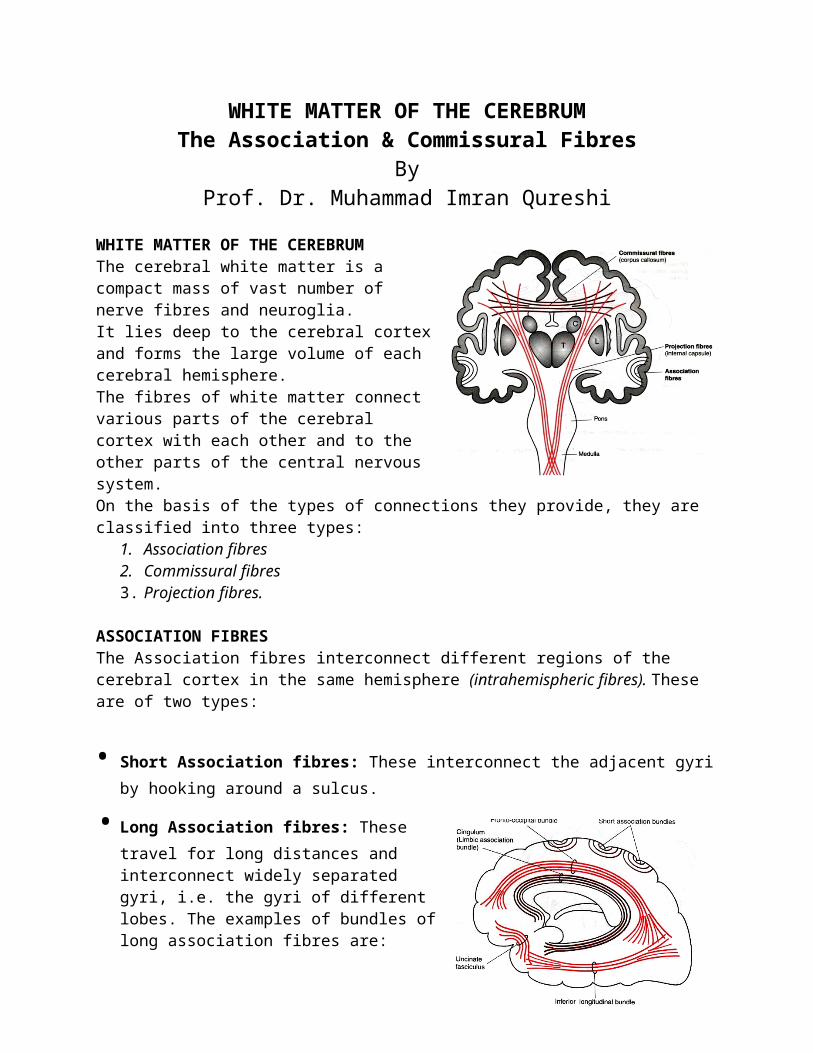
WHITE MATTER OF THE CEREBRUMThe cerebral white matter is a compact mass of vast number of nerve fibres and neuroglia.It lies deep to the cerebral cortex and forms the large volume of each cerebral hemisphere.The fibres of white matter connect various parts of the cerebral cortex with each other and to the other parts of the central nervous system.On the basis of the types of connections they provide, they are classified into three types:
1. Association fibres2. Commissural fibres3. Projection fibres.
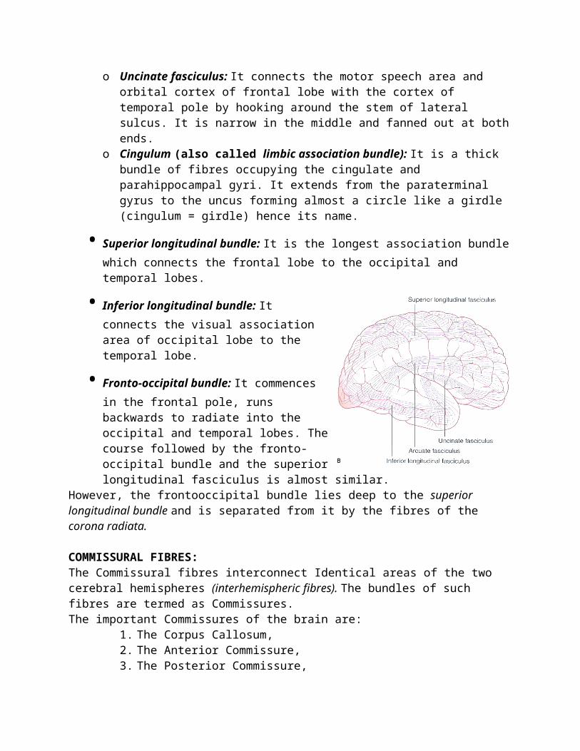
ASSOCIATION FIBRESThe Association fibres interconnect different regions of the cerebral cortex in the same hemisphere (intrahemispheric fibres). These are of two types:
• Short Association fibres: These interconnect the adjacent gyri by hooking around a sulcus.
• Long Association fibres: These travel for long distances and interconnect widely separated gyri, i.e. the gyri of different lobes. The examples of bundles of long association fibres are:
o Uncinate fasciculus: It connects the motor speech area and orbital cortex of frontal lobe with the cortex of temporal pole by hooking around the stem of lateral sulcus. It is narrow in the middle and fanned out at both ends.
o Cingulum (also called limbic association bundle): It is a thick bundle of fibres occupying the cingulate and parahippocampal gyri. It extends from the paraterminal gyrus to the uncus forming almost a circle like a girdle (cingulum = girdle) hence its name.
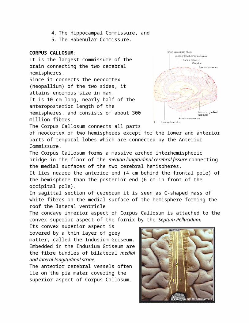
• Superior longitudinal bundle: It is the longest association bundle which connects the frontal lobe to the occipital and temporal lobes.
• Inferior longitudinal bundle: It connects the visual association area of occipital lobe to the temporal lobe.
• Fronto-occipital bundle: It commences in the frontal pole, runs backwards to radiate into the occipital and temporal lobes. The course followed by the frontooccipital bundle and the superior longitudinal fasciculus is almost similar.
However, the frontooccipital bundle lies deep to the superior longitudinal bundle and is separated from it by the fibres of the corona radiata.
COMMISSURAL FIBRES:The Commissural fibres interconnect Identical areas of the two cerebral hemispheres (interhemispheric fibres). The bundles of such fibres are termed as Commissures.The important Commissures of the brain are:
1. The Corpus Callosum,2. The Anterior Commissure,3. The Posterior Commissure,4. The Hippocampal Commissure, and5. The Habenular Commissure.
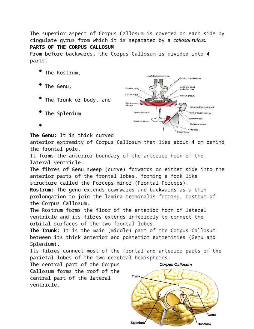
CORPUS CALLOSUM: It is the largest commissure of the brain connecting the two cerebral hemispheres.Since it connects the neocortex (neopallium) of the two sides, it attains enormous size in man.It is 10 cm long, nearly half of the anteroposterior length of the hemispheres, and consists of about 300 million fibres.The Corpus Callosum connects all parts of neocortex of two hemispheres except for the lower and anterior parts of temporal lobes which are connected by the Anterior Commissure.The Corpus Callosum forms a massive arched interhemispheric bridge in the floor of the median longitudinal cerebral fissure connecting the medial surfaces of the two cerebral hemispheres.It lies nearer the anterior end (4 cm behind the frontal pole) of the hemisphere than the posterior end (6 cm in front of the occipital pole).In sagittal section of cerebrum it is seen as C-shaped mass of white fibres on the medial surface of the hemisphere forming the roof the lateral ventricle The concave inferior aspect of Corpus Callosum is attached to the convex superior aspect of the fornix by the Septum Pellucidum.
Its convex superior aspect is covered by a thin layer of grey matter, called the Indusium Griseum. Embedded in the Indusium Griseum are the fibre bundles of bilateral medial and lateral longitudinal striae.The anterior cerebral vessels often lie on the pia mater covering the superior aspect of Corpus Callosum.The superior aspect of Corpus Callosum is covered on each side by cingulate gyrus from which it is separated by a callosal sulcus.PARTS OF THE CORPUS CALLOSUMFrom before backwards, the Corpus Callosum is divided into 4 parts:
• The Rostrum,
• The Genu,
• The Trunk or body, and
• The Splenium
•The Genu: It is thick curved anterior extremity of Corpus Callosum that lies about 4 cm behind the frontal pole.It forms the anterior boundary of the anterior horn of the lateral ventricle.The fibres of Genu sweep (curve) forwards on either side into the anterior parts of the frontal lobes, forming a fork like structure called the Forceps minor (Frontal Forceps).Rostrum: The genu extends downwards and backwards as a thin prolongation to join the lamina terminalis forming, rostrum of the Corpus Callosum.The Rostrum forms the floor of the anterior horn of lateral ventricle and its fibres extends inferiorly to connect the orbital surfaces of the two frontal lobes.The Trunk: It is the main (middle) part of the Corpus Callosum between its thick anterior and posterior extremities (Genu and Splenium).Its fibres connect most of the frontal and anterior parts of the parietal lobes of the two cerebral hemispheres.The central part of the Corpus Callosum forms the roof of the central part of the lateral ventricle.
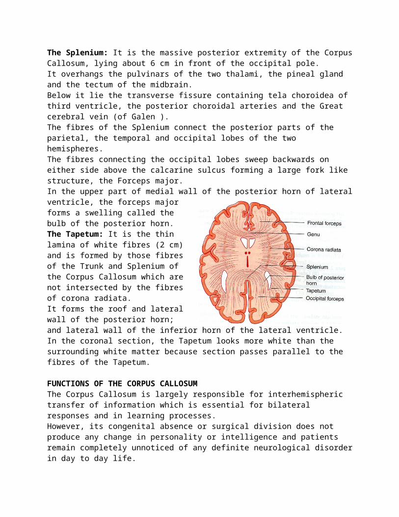
The Splenium: It is the massive posterior extremity of the Corpus Callosum, lying about 6 cm in front of the occipital pole.It overhangs the pulvinars of the two thalami, the pineal gland and the tectum of the midbrain.Below it lie the transverse fissure containing tela choroidea of third ventricle, the posterior choroidal arteries and the Great cerebral vein (of Galen ).The fibres of the Splenium connect the posterior parts of the parietal, the temporal and occipital lobes of the two hemispheres.The fibres connecting the occipital lobes sweep backwards on either side above the calcarine sulcus forming a large fork like structure, the Forceps major.In the upper part of medial wall of the posterior horn of lateral ventricle, the forceps major forms a swelling called the bulb of the posterior horn. The Tapetum: It is the thin lamina of white fibres (2 cm) and is formed by those fibres of the Trunk and Splenium of the Corpus Callosum which are not intersected by the fibres of corona radiata.It forms the roof and lateral wall of the posterior horn; and lateral wall of the inferior horn of the lateral ventricle.In the coronal section, the Tapetum looks more white than the surrounding white matter because section passes parallel to the fibres of the Tapetum.
FUNCTIONS OF THE CORPUS CALLOSUMThe Corpus Callosum is largely responsible for interhemispheric transfer of information which is essential for bilateral responses and in learning processes.However, its congenital absence or surgical division does not produce any change in personality or intelligence and patients remain completely unnoticed of any definite neurological disorder in day to day life.Only special tests of tactile and visual systems will reveal any abnormality.
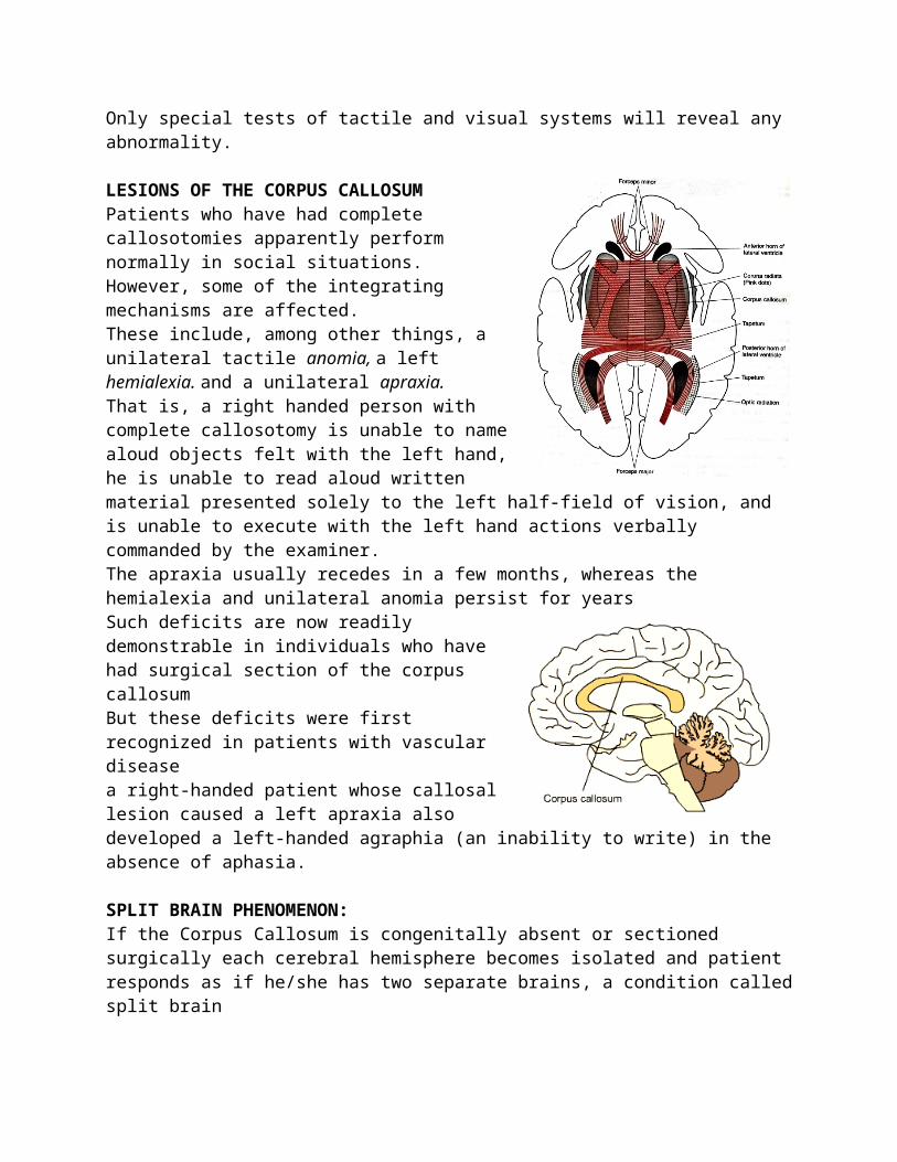
LESIONS OF THE CORPUS CALLOSUMPatients who have had complete callosotomies apparently perform normally in social situations.However, some of the integrating mechanisms are affected.These include, among other things, a unilateral tactile anomia, a left hemialexia. and a unilateral apraxia. That is, a right handed person with complete callosotomy is unable to name aloud objects felt with the left hand, he is unable to read aloud written material presented solely
to the left half-field of vision, and is unable to execute with the left hand actions verbally commanded by the examiner.The apraxia usually recedes in a few months, whereas the hemialexia and unilateral anomia persist for yearsSuch deficits are now readily demonstrable in individuals who have had surgical section of the corpus callosumBut these deficits were first recognized in patients with vascular diseasea right-handed patient whose callosal lesion caused a left apraxia also developed a left-handed agraphia (an inability to write) in the absence of aphasia.
SPLIT BRAIN PHENOMENON:If the Corpus Callosum is congenitally absent or sectioned surgically each cerebral hemisphere becomes isolated and patient responds as if he/she has two separate brains, a condition called split brainThe severance of Corpus Callosum in young monkeys produces split brain effect.If they are trained to perform a task with one hand, they are unable to repeat the same act with the other hand.
Anterior CommissureThe Anterior Commissure is a small round bundle of white fibres which crosses the midline in the upper part of the Lamina Terminalis.It is located immediately in front of the anterior column of the fornix and the interventricular foramen.The Anterior Commissure consists of two components:
• A larger Posterior Neocortical component, which interconnects the lower and anterior parts of the temporal lobes.
• A smaller Anterior Paleocortical component, which interconnects the olfactory regions (olfactory bulbs, olfactory tubercles, etc.) of the two hemispheres.
Habenular CommissureThe Habenular Commissure is a slender bundle of white fibres which crosses the midline through the superior lamina of the stalk of pineal gland. It interconnects the Habenular nuclei of the two sides.
Posterior CommissureThe Posterior Commissure is a slender bundle of white fibres which crosses the midline through the inferior lamina of the stalk of pineal gland. It interconnects the superior colliculi, pretectal and interstitial nuclei of two sides.
Hippocampal Commissure (Commissure of the Fornix)The Hippocampal Commissure interconnects the crura of fornix and thus the Hippocampal formations of the two sides.


![Chapter 1: Architectural Overview and Building a Simple ... · Building a Simple App in Angular. Graphic Bundle [ 2 ] Graphic Bundle [ 3 ] Graphic Bundle [ 4 ] Graphic Bundle [ 5](https://img.pdfslide.us/doc/110x75/5ee01311ad6a402d666b53e7/chapter-1-architectural-overview-and-building-a-simple-building-a-simple-app.jpg)

![Chapter 1: Getting Up and Running with Cassandra...Chapter 11: Cassandra Multi-Node Cluster Graphics Bundle [ 55 ] Graphics Bundle [ 56 ] Graphics Bundle [ 57 ] Graphics Bundle [ 58](https://img.pdfslide.us/doc/110x75/5f4a5ed088ed921a2d1ef796/chapter-1-getting-up-and-running-with-cassandra-chapter-11-cassandra-multi-node.jpg)