Embed Size (px)
Citation preview

Longitudinal differences in white matter integrity in youth at high
familial risk for bipolar disorder
Rossana Ganzola1, Thomas Nickson2, Mark E Bastin3, Stephen Giles2, Alix Macdonald2, Jessika Sussmann2,
Andrew M McIntosh2, Heather C. Whalley2, Simon Duchesne1,4
1Institut universitaire en santé mentale de Québec, Québec, Canada
2Division of Psychiatry, University of Edinburgh, Edinburgh, United Kingdom
3Centre for Clinical Brain Sciences, Western General Hospital, University of Edinburgh, Edinburgh, UK
4Départment de Radiologie, Faculté de Médecine, Université Laval, Québec, Canada
Corresponding author: Rossana Ganzola
e-mail address: [email protected]
Work address: Institut universitaire en santé mentale de Québec
2601 Chemin de la Canardière, Quebec City, Quebec
CANADA G1J 2G3
Work telephone: +1-418-6635000, ext.: 6714
Running head: WM changes in high-risk subjects for BD
Word count: 6,268
1
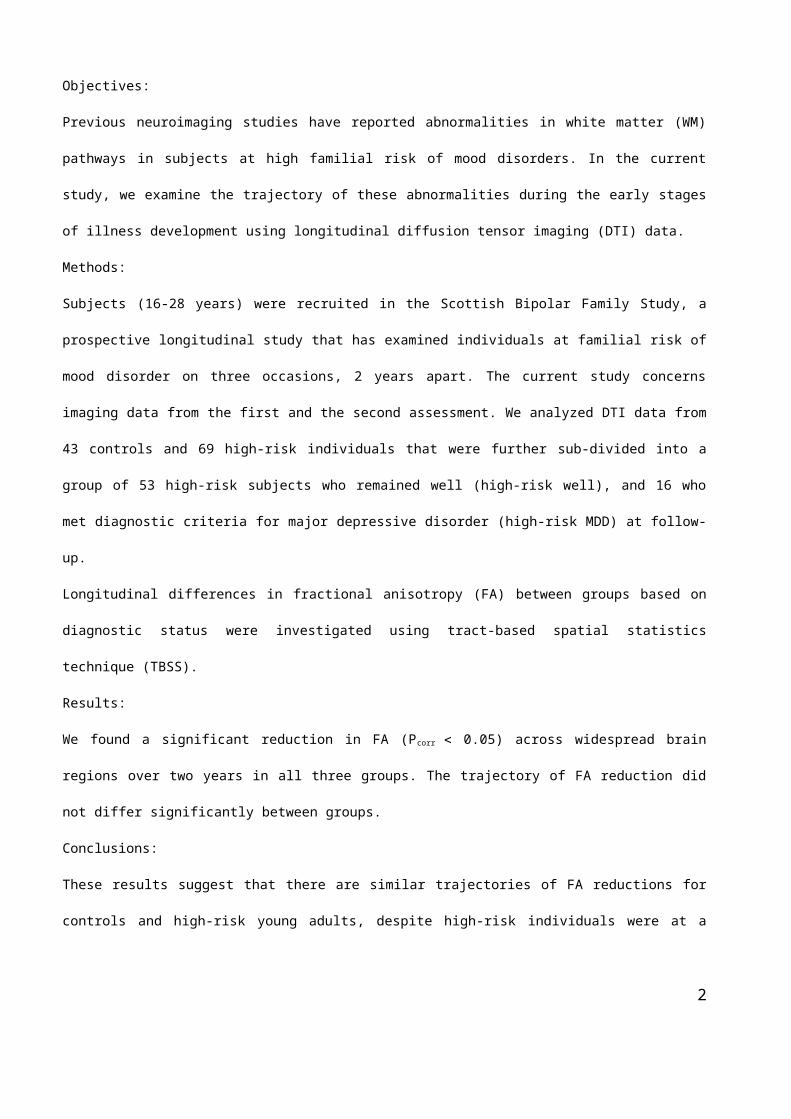
Objectives:
Previous neuroimaging studies have reported abnormalities in white matter (WM) pathways in subjects at
high familial risk of mood disorders. In the current study, we examine the trajectory of these abnormalities
during the early stages of illness development using longitudinal diffusion tensor imaging (DTI) data.
Methods:
Subjects (16-28 years) were recruited in the Scottish Bipolar Family Study, a prospective longitudinal study
that has examined individuals at familial risk of mood disorder on three occasions, 2 years apart. The current
study concerns imaging data from the first and the second assessment. We analyzed DTI data from 43
controls and 69 high-risk individuals that were further sub-divided into a group of 53 high-risk subjects who
remained well (high-risk well), and 16 who met diagnostic criteria for major depressive disorder (high-risk
MDD) at follow-up.
Longitudinal differences in fractional anisotropy (FA) between groups based on diagnostic status were
investigated using tract-based spatial statistics technique (TBSS).
Results:
We found a significant reduction in FA (Pcorr 0.05) across widespread brain regions over two years in all
three groups. The trajectory of FA reduction did not differ significantly between groups.
Conclusions:
These results suggest that there are similar trajectories of FA reductions for controls and high-risk young
adults, despite high-risk individuals were at a disadvantaged starting point considering their reduced WM
integrity detected at the baseline in previous studies.
Difference in WM integrity between high-risk and controls could therefore occur in earlier childhood and be a
necessary but not sufficient condition to develop future mood disorders.
Key words: bipolar disorder, major depressive disorder, high familial risk, longitudinal study, white matter
integrity, fractional anisotropy.
2

Introduction
Bipolar disorder (BD) and major depressive disorder (MDD) are highly heritable mood disorders sharing
overlapping symptomatology, neural basis, and genetic architecture. Whereas sporadic elevation of mood is
only present in BD, episodic depression is observed in both. The disorders seemingly share common
neurobiological traits; notably, it has been reported that a disconnection in cortical-limbic pathways are
significantly associated with both disorders (1).
To explain these findings and propose an aetiological model, the importance of development in MDD and BD
has been recently recognized and neurodevelopmental models have supplanted the classic hypothesis of
progressive degeneration (2, 3). In this perspective, offspring and relatives of BD patients are a population
rich in potential for revealing important aspects in the development of mood disorders before the onset of the
disease, considering that familial studies showed an increased frequency of both BD and MDD in first-degree
relatives of bipolar patients (4).
Diffusion tensor imaging (DTI) - a magnetic resonance technique able to quantify microstructural integrity
using indices of preferential diffusion such as fractional anisotropy (FA) - has been used in studies comparing
white matter (WM) microstructure in relatives of BD patients with those of subjects without a family history of
psychiatric disorders (controls). Recent evidence indicates that early WM abnormalities may have a
significant part in the pathophysiology of both BD and MDD, and could therefore represent an early marker of
mood disorders (5-8).
In a previous DTI study on this sample at baseline, we reported widespread FA reductions in unaffected
young relatives of BD compared to subjects without family history of psychiatric disorders. These subjects
were recruited in the Scottish Bipolar Family Study (BFS). Although the effects of familial risk were diffuse,
the associations with cyclothymia were more localized to fronto-temporal and prefrontal-thalamic connections
(9). These findings confirm that WM integrity is an endophenotype for mood disorder with important
behavioural association linked to the etiology of the condition. Moreover, it could be stated that abnormal
connectivity between regions may contribute to developmental alterations of key neural structures in BD.
However, there is a paucity of studies in the literature studying the longitudinal development of WM in young
adults at high-risk for mood disorders, and hence whether these initial differences remain or if their trajectory
changes through time. Only Versace and colleagues (10) have reported group by age interactions in FA
values in a cross-sectional design comparing healthy offspring of BD patients with individuals without familiar
3

history of psychiatric disorders. The results showed a linear increase in FA in controls in the left corpus
callosum and right inferior longitudinal fasciculus, whereas in BD offspring there was a linear decrease in FA
with age in the left corpus callosum, and no relationship between FA and age in the right inferior longitudinal
fasciculus.
To our knowledge, there have been no other longitudinal studies investigating changes in WM during the
young adulthood development of individuals at high-risk for mood disorders, and none with sufficient follow-
up to include subjects that went on to develop MDD. To fill this gap in knowledge, we propose in the current
study an analysis of findings from scans acquired at baseline and at two-year follow-up of individuals
recruited in the BFS..
We selected the subjects having the diffusion data available at the baseline and at the second follow-up in
the same cohort where differences in FA between subjects with familial risk and controls were already
detected at the baseline (9).
Individuals in the study, by virtue of the shared genetic architect of MDD/BD, are at risk for both mood
disorders. Over the course of the BFS, a number of individuals have developed one or the other, with MDD
being the predominant outcome (defined as meeting formal diagnostic criteria, Diagnostic and Statistical
Manual of Mental Disorder, 4th edition, DSM-IV) and in sufficient numbers as to make a longitudinal
comparison of well v. affected individuals (11). Our analysis focused on individuals at high family risk who
remained well at the time of the second assessment (the high-risk well) compared those who met the
diagnostic criteria for a MDD between baseline and follow-up.
We hypothesized, based on our previous results that WM integrity changes according to different trajectories
in controls than high-risk individuals that become subsequently ill and in those that remain unaffected.
In particular, in according to the literature, we expected to find a significant FA decrease with the
development in the corpus callosum of the high-risk subjects compared with controls as already reported in
the study of Versace et al. (10), and a further loss of white matter integrity in the high-risk individuals who
developed a major depressive disorder located in the fronto-limbic connections mainly involved in the mood
disorders (Shared white matter alterations across emotional disorders: A voxel-based meta-analysis of
fractional anisotropy.Jenkins LM1, Barba A1, Campbell M1, Lamar M1, Shankman SA1, Leow AD1, Ajilore
O1, Langenecker SA1).
4

Materials and Methods
Initial baseline recruitment
Baseline recruitment has been described in full previously (9, 12). Briefly, at the beginning of the study (time
1, T1), individuals with a diagnosis of bipolar I disorder were identified by psychiatrists across Scotland.
Diagnosis was confirmed with the OPCRIT symptom checklist (13) using data from clinical notes and the
Structured Clinical Interview for DSM-IV (SCID; (14). Each affected subject was asked to identify members of
close family aged 16–28 years. Following informed consent, unaffected individuals with at least one first-
degree or two second-degree relatives with BD I were invited to participate. Unaffected, unrelated
comparison subjects with no personal or family history of BD were identified from the social groups of the
high-risk subjects and matched for age, gender and intelligence quotient (IQ) to the high-risk group.
Comparison subjects were also screened using the SCID (14). Exclusion criteria for both groups at initial
recruitment included a personal history of major depression, mania or hypomania, psychosis, or any major
neurological or psychiatric disorder, a history of substance dependence, learning disability, or any history of
head injury that included loss of consciousness and any contraindications to magnetic resonance imaging
(MRI). No group differences in lifetime substance misuse were observed previously in this sample (12).
Written informed consent was obtained. The study was approved by the Multi-Centre Research Ethics
Committee for Scotland.
Current study population
Participants were recruited for the current study as part of the BFS as described above (9, 12).
Only the first two occasions [T1, time 2 (T2)] contained imaging assessments, the third assessment (T3)
being primarily a follow-up clinical assessment. All participants, controls and high risk, were interviewed by
one of two experienced psychiatrists (A.M.M., J.E.S.) using the SCID (15) to confirm the lifetime absence of
any Axis I disorders at T1, and at to determine the presence of any mood disorder meeting diagnostic criteria
over the intervening period. At T3 diagnostic status was determined either by face-to-face assessment, or
through accessing clinical records at the National Health Service (NHS) as to whether a clinical diagnosis had
been made or not (16). For a number of individuals (n = 19), it was not possible to determine the clinical
status at T3, either the general practitioner (GP) did not provide details or the GP address was unknown. In
the absence of further clinical information indicating that they had become unwell, and since they had
remained well over the previous two assessments, these individuals were presumed to have remained well.
5

If other disorders were present along with depressive features these individuals were excluded from the
current analysis. Moreover, we excluded MR images of subjects that did not pass quality control during the
different analysis steps.
Manic and depressive symptoms were rated using the Young Mania Rating Scale (YMRS; (17)) and the
Hamilton Rating Scale for Depression (HAM-D; (18)).
Estimates of trait-liability to mood disorder (cyclothymia, neuroticism and extraversion) were measured using
the TEMPS-A and NEO-FFI (19, 20).
The current study concerns structural brain changes between T1 and T2 in relation to the development of
MDD at the second time (T2) throughout the study.
Groups
The final sample consisted of individuals categorized firstly into two groups: (i) healthy controls who remained
well (n=43); and (ii) familial high-risk participants (n=69). Controls meeting criteria for psychiatric disorders at
the follow-up and high-risk patients developing conditions other than MDD were removed from the study
because numbers did not allow robust statistical testing. Moreover, controls and high-risk subjects whose
images did not pass quality check during the registration to the standard space were also excluded.
Appendix 1 details the subjects’ selection process.
The high-risk group was then split in two sub-groups, based on diagnosis at the time of the second MRI scan:
subjects who remained well between the baseline (T1) and the second assessment (T2) (high-risk well, n =
53); and familial high-risk participants who had developed MDD by the second assessment (high-risk MDD, n
=16).
Scan Acquisition and Preprocessing
The current study reports findings from imaging data collected at T1 and T2. MRI data were collected on a
1.5-T GE Signa Horizon HDX (General Electric, Milwaukee, Wisconsin) clinical scanner equipped with a self-
shielding gradient set (22 mT/m maximum gradient strength) and manufacturer-supplied “birdcage”
quadrature head coil. Whole brain DTI data were acquired for each subject with a single-shot pulsed gradient
spin-echo echo-planar imaging sequence with diffusion gradients (b = 1000 sec/mm2) applied in 64 non-
collinear directions and seven T2-weighted echo-planar imaging baseline scans. Fifty-three 2.5-mm
contiguous axial slices were acquired with a field-of-view of 240 x 240 mm, acquisition matrix of 96 x 96
(zero-filled to 128 x 128), giving an isotropic acquisition voxel dimension of 2.5 mm. In addition, a T1-
6

weighted volume was acquired with time of inversion = 500 msec, echo time = 4 msec, flip angle = 8°, and
voxel-size = 1.25 x 1.25 x 1.20mm (192 x 192voxels, 180 slices).
The DTI data were converted to 4D NIfTI volumes and preprocessed with standard tools available from the
Fmrib Software Library (FSL; http://www.fmrib.ox.ac.uk/fsl). This included the following processes: correction
for eddy current induced distortions and bulk subject motion in the scanner by registering the diffusion
weighed volumes to the first T2-weighted volume within each subject; brain extraction; and calculation of
diffusion tensor characteristics, including principal eigenvectors and FA values with DTIFIT.
Tract-Based Spatial Statistics
We used tract-based spatial statistics (TBSS) to study longitudinal WM changes across time and between
groups. TBSS was carried out according to standard FSL procedures (21) (http://www.fmrib.ox.ac.uk/fsl).
First, FA volumes of all subjects were nonlinearly registered to a standard template. Secondly, a mean of all
registered FA volumes was calculated, and a white matter “skeleton” created. This was achieved by
searching for the maximum FA values in directions perpendicular to the local tract direction in the mean FA
map. A threshold of FA > 0.25 was applied to the FA skeleton to exclude predominantly non-white matter
voxels. Thirdly, the maximum voxel FA value perpendicular to the local direction was projected onto the
skeleton at each point in all subjects. This resulted in one FA skeleton map per subject, assumed to contain
the anatomically corresponding centroids of the WM structure.
With the obtained TBSS skeletons we performed three statistical analyses. Firstly, to test for between-group
difference in FA changes over time, repeated meausures ANOVA were applied to the skeletons by using the
“randomize” function in FSL, with four regressors in the design matrix, one for each group and one for each
time of acquisition.
Secondly, we used TBSS to study the difference over time between high-risk Well and high-risk MDD groups.
In this case, we used six regressors in the design matrix, one for each group (controls/high-risk well/high-risk
MDD) and one for each time of acquisition (T1 andT2)
Thirdly, we substracted the FA volume obtained for each subject at the second scan to the FA volume
obtained at baseline. We used this FA difference to perform a correlation with age using age as covariate in
the GLM model to investigate the relationship between FA changes and development in each group.
Threshold-free cluster enhancement (TFCE) was applied to obtain cluster-wise statistics corrected for
multiple comparisons. Briefly, this method transforms local T-statistics into TFCE statistics that reflect both
7

the size of the local effect (or “height”) and the cluster extent (22). The major advantages are that no
predefined T-threshold is required, and that TFCE is sensitive to detect both large clusters of modest effects
and single voxels of large effects, at the same time.
With the obtained TFCE maps, “randomize” then calculates a p-value (p-corrected) for each voxel, corrected
for whole-brain family wise error (FWE) rate via permutation testing (5,000 permutations). These TFCE
corrected p-maps were thresholded at pFWE .05. We report the sizes of contiguous clusters of
suprathreshold voxels. Significant results were localized to white matter tracts/structures with the Johns
Hopkins University DTI-based white matter atlas and the Johns Hopkins University white matter tractography
atlas (23) digitally available in FSL.
Statistical analysis of demographic and clinical data was conducted using SPSS software, version 23.0
(http://www-01.ibm.com/software/analytics/spss/). Differences between groups were tested using one-way
ANOVA, t- chi-squared tests or using non-parametric tests as appropriate.
8

Results
Demographic and clinical features
We did not find any significant differences between groups with respect to gender, age, IQ, or scan interval
(Table 1, Table 2). Scores for the HAM-D, cyclothmic, depressive, and irritability traits as measured by the
TEMPS-A scale, as well as for neuroticism and extraversion factors as measured using the Five Factor
Inventory differed significantly between groups at baseline (Table 3). Post-hoc analysis revealed that High-
risk MDD individuals differed significantly for the HAM-D, cyclothymia, and extraversion scores from
Controls, and for depressive trait and neuroticism from both Controls and High-risk Well. These effects
were in the directions predicted in accordance with other studies, namely High-risk MDD > High-risk Well >
Controls for neuroticism, cyclothymia, depressive and HAM-D scores, and High-risk MDD < High-risk Well
< Controls for extraversion.
TBSS analysis
Results (a) were obtained by analysis of FA longitudinal changes in each group and comparisons of FA
changes over time between Controls and all High-risk subjects.
Results (b) were obtained by analysis of FA longitudinal changes in the two sub-high-risk groups and
comparisons of FA changes over time between the 53 High-risk Well and 16 High-risk MDD.
Results (c) describe TBSS results from the correlation between FA changes and age in each of the three
groups (Controls/High-risk Well /High-risk MDD).
a) Controls vs High-risk
a.1. FA changes over time in Control group
There was no significant increase in FA in the control group along the two-year time interval between
assessments, whereas a widespread decrease of FA was detected. TBSS followed by permutation testing
resulted in a large diffuse cluster (K=35,921, pcorr 0.05) extending over most of the white matter skeleton,
indicating decreases over time in the body and the splenium of the corpus callosum, internal and external
capsule (including thalamic radiation), inferior and superior longitudinal fasciculi, inferior fronto-occipital
fasciculi, corona radiata, parts of the cortical spinal tract and the right uncinate fasciculus (Figure 1).
a.2. FA changes over time in High-risk group
Like Controls, increase in FA over time in the high-risk group was not detected. An FA decrease surviving the
permutation test was observed in a larger cluster (K=57,451, pcorr 0.05), comprising the same regions as the
Controls, . However, there were further WM areas with FA loss for High-risk subjects includingthe left
9

uncinate fasciculum, genu of the corpus callosum, and the anterior portion of the thalamic radiation, corona
radiata, cingulum, and inferior fronto-occipital fasciculus (Figure 1).
a.3. Group x Time interactions
Despite the different pattern of FA loss found separately in the Control and High-risk group, there was no
significant differences surviving permutation test when FA changes between groups where directly compared.
b) Controls, High-risk Well, and High-risk MDD
b.1. FA changes over time in High-risk MDD and High-risk Well individuals
There was a significant widespread decrease in FA along the two-year time course in both high-risk groups.
Hovewer, High-risk Well individuals had relatively spared bilateral posterior thalamic radiation, body of the
corpus callosum, left inferior and fronto-occipital fasciculi, and left anterior cingulum and corona radiata in the
medial frontal lobe. Moreover, in that same group an increase of FA was detected in a small cluster (K= 133,
p 0.05) located in the right posterior superior longitudinal and fronto-occipital fasciculi (Figure 2).
b.2. Groups (Controls/High-risk Well/High-risk MDD) x Time interactions
There were no significant differences surviving the permutation test in any comparison of FA between groups
over time.
c) Correlation with age
Results of the TBSS correlation with age show a negative correlation between FA and age surviving the
permutation tests in the right inferior longitudinal and fronto-occipital fasciculi, and anterior thalamic radiation in
the Control group (Figure 3).
10

Discussion
We investigated longitudinal WM changes in youth at high familial risk for mood disorders compared to
unrelated controls. We further stratified the analysis by looking at two sub-groups, the first composed of those
high-risk subjects that met the criteria for MDD at two-year follow-up, compared to the second group of
subjects that remained healthy.
We found a significant decrease of FA over two years in all three groups, albeit diffuse over the whole brain.
No significant increase in FA over time was detected, except for a small cluster including posterior
connections in the High-risk Well group.
a) Lifetime trajectory of FA
A diffuse increase in FA from newborn to adolescence and a rapid widespread decrease from old age
onwards is a consistent finding in studies tracking WM changes across the lifespan. However, WM changes
during adulthood are not as well defined. Indeed, studies on WM variations in young and mid-adults have
reported conflicting results (24). An explanation for this discrepancy could be that the peak of FA for different
WM areas may be reached at varying ages during adulthood. Indeed, there is evidence for myelination
occurring into fronto-temporal cortical areas well into the late-adulthood (25), which will inevitably impact FA.
More specifically, FA of the corpus callosum and long association fibers such as the inferior longitudinal and
inferior fronto-occipital fasciculus peak in the early to mid-20s, while it has been shown that projection fibers
such as those in the anterior limb of the internal capsule as well as corticospinal tracts peak in the early to
mid-30s. Of note, the long association tracts connected with the limbic system have differing developmental
trajectories; the fornix and cingulum bundles reach their FA peak before 20 and after 40 years of age,
respectively (26). Overall, it can be said that the details of the WM maturation process for different brain
structures as well as within each structure over time are still not entirely understood. This is further supported
by the fact that our understanding of lifespan brain WM integrity is based on mostly cross-sectional studies of
different age periods, as demonstrated in a recent review on this subject (24). Cross-sectional results must
be considered as models of longitudinal outcome with caution. Longitudinal designs will provide better
evidence to elucidate the normal developmental course of WM changes with age, and path to illness.
Given this background, we can assess our results in Controls, where we found a negative correlation
between age and FA values in these subjects, aged between 16 and 28, therefore late adolescents to young
adults. After permutation testing, clusters of voxels were still significant in the WM fibers of the right medial
11

temporal lobe for the Control group. This result suggests that the decrease in FA over time found in Controls
could be partially explained by maturation processes related to normal development of these early adult
individuals.
b) FA trajectory in high-risk population
In this study, we found FA reductions in adolescents and young adults with and without familial risk for mood
disorders over a time course of two years, and in contrast to our initial hypothesis we did not find significant
differences in the amount of FA decrease between the groups surviving the permutation test.
We expected a significant reduction of FA with the development in high-risk subjects compared with controls,
in particular in the corpus callosum and in front-limbic connections but we did not find differences.
However, we need to consider that the only study that analyzed the FA changes related to age in the high-
risk subjects for bipolar disorder was a cross-sectional study that considered youngest high-risk individuals
(Versace et al).
All other studies that have detected a loss WM integrity in high-risk individuals when compared with controls
were also cross-sectional studies (REF).
To the best of our knowledge, the present study is the first longitudinal analysis on WM integrity changes over
time in adolescents and young adults with high familial risk of mood disorders.
Therefore, our results suggest that WM abnormalities found previously between high-risk individuals and
controls (9) belonging the same cohort acquired using identical scans and parameters, and studied with a
common standard procedure are stable by the late teens/early twenties.
Most of studies of WM in relatives of BD patients have focused on adult individuals. Studies assessing
macrostructural WM alterations revealed reduced WM volumes (27-29), and more WM hyperintensities (30).
DTI analysis comparing first-degree relatives of BD patients with controls detected significantly reduced FA in
the right anterior limb of the internal capsule and right uncinate fasciculus (31). FA reductions were also
observed using TBSS in unaffected siblings of BD probands, mainly restricted to the corpus callosum,
posterior thalamic radiations, and left superior longitudinal fasciculus (32). Using tractography analysis,
reduced FA were not detected in the siblings compared to the controls, except for a trend in the corpus
callosum (33). Presence of WM abnormalities in adult relatives of patients with BD was therefore a consistent
finding in MRI investigations.
As stated before, only two studies analyzed FA in younger subjects with familial risk for BD. Frazier et al. (34)
found that children aged between four and 12 years having a first-degree relative with the disorder showed
12

reduced FA relative to controls in bilateral superior longitudinal fasciculi. Versace et al. (10) observed that
asymptomatic youth with familial risk for BD aged between eight and 17 years had a linear decrease between
age and FA in the left corpus callosum, whereas healthy controls showed a linear increase in the same
regions. WM alterations were therefore already present in children and in adolescents with familial risk for
BD.
In the current study, we did not detect differences in FA changes comparing both high-risk young adults who
developed and who did not develop MDD with controls, despite reduced FA was detected in the same cohort
of high-risk subjects when compared with controls at the baseline.
Together these findings could suggest that differences between cases and controls probably don’t emerge
proximal to the onset of illness but may therefore have existed from birth or emerged earlier in childhood and
adolescence.
Longitudinal studies in children and early adolescents with familial risk for bipolar disorders are needed to
determine whether a loss of WM integrity occurs during childhood and adolescence and may precede the
onset of BD or other psychiatric disorder in youth at risk for BD.
Despite not finding significant differences surviving the permutation test comparing longitudinal FA changes
between groups, considering the progressive changes of WM integrity in each group, we detected a decrease
of FA in both controls and high-risk subjects with further FA loss in the High-risk group mainly located in
anterior regions, such as the uncinate fasciculum, and the anterior portion of corpus callosum, thalamic
radiation, corona radiata, cingulum, and inferior fronto-occipital fasciculus.
Convergent evidence supports a central role for altered development of frontotemporal neural systems in
BD. Frontotemporal WM abnormalities have been reported in many adult DTI studies related to BD (31, 35-
44). Studies of children and adolescents with BD have also shown findings of reduced structural integrity in
frontal WM regions, compared to healthy children and adolescents (5, 6, 34, 45-49). Moreover,
frontotemporal structural integrity abnormalities have been reported in DTI studies of adults with BD and their
high-risk BD adult relatives, both showing decreased FA values in the right uncinate fasciculus (31).
Taken together, these reports indicate that intra- and inter-hemispheric frontotemporal WM abnormalities are
already present in childhood, continue in adolescence and remain throughout adulthood, and therefore could
represent some of the earliest markers of the disorder.
In the current study, the sample size of the groups could be too modest in order to detect what could be
biologically meaningful small effects. Further samples are therefore needed to understand if a loss of WM
13

integrity is present during development in familial high-risk subjects for mood disorders when compared to
individuals without family history of psychiatric disorders.
c) Conclusion and future perspectives
In conclusion, we found FA progressive reductions in our sample of adolescents and young adults with and
without familial risk for mood disorders.
In contrast with our expectations, we did not find significant different changes between the groups.
We can explain these negative finding considering that:
- reduced WM integrity previously detected between cases and controls in the same cohort are stable by the
late teens/early twenties and probably don’t occur proximal to the onset of illness but may therefore have
existed from birth or emerged earlier in childhood and adolescence.
Longitudinal studies in younger high-risk individuals as children and early adolescents could clarify if these
period would be more vulnerable for the white matter integrity;
- this analysis may be underpowered due to the relatively small sample size within the high-risk MDD group.
The difficulties in conducting prospective longitudinal high-risk studies on young individuals with a family
history of mental illness particularly relating to cumulative attrition over the course of the study should not be
underestimated. Clinical longitudinal follow-up of these high-risk cohort and larger samples could clarify
relationships between WM integrity and the onset of mood symptoms.
Moreover, a standard longitudinal TBSS method has been recently developed and tested (Longitudinal
reliability of tract-based spatial statistics in diffusion tensor imaging, Madhyastha T 2014). Therefore, future
studies will be improved through a increased test’-retest reliability.
In summary, research on WM in BD supports an important role for frontotemporal WM. Neurodevelopmental
abnormalities that affect trajectories of WM development during childhood and adolescence could be
implicated before the onset of mood disorder symptoms in subjects with familial risk. Longitudinal studies of
high-risk children and adolescents and increased sample sizes might clarify these abnormalities in brain
changes to target for early identification, intervention and prevention strategies.
14

Acknowledgements
We would like to thank all of the participants who took part in the study and the radiographers who acquired
the MRI scans. This study was conducted at the Brain Research Imaging Centre (http://www.bric.ed.ac.uk)
which is supported by SINAPSE (Scottish Imaging Network, a Platform for Scientific Excellence,
http://www.sinapse.ac.uk).
The research leading to these results has received funding from the European Community's Seventh
Framework Programme (FP7/2007-2013) under grant agreement n° 602450. This paper reflects only the
author’s views and the European Union is not liable for any use that may be made of the information
contained therein. This work was also supported by a Wellcome Trust Strategic Award 104036/Z/14/Z, and
the IMAGEMEND grant. TN is supported by the Dr Mortimer and Theresa Sackler Foundation. HCW is
supported by a College Fellowship from the University of Edinburgh and a JMAS SIM fellowship from the
Royal College of Physicians of Edinburgh. JES is supported by a Clinical Research Training Fellowship from
the Wellcome Trust. AMM was supported by the Health Foundation through a Clinician Scientist Fellowship
(Ref: 2268/4295), by the Brain and Behaviour Research Foundation through a NARSAD Independent
Investigator Award and by a Scottish Funding Council Senior Clinical Fellowship. The investigators also
acknowledge the financial support of National Health Service (NHS) Research Scotland, through the Scottish
Mental Health Research Network (www.smhrn.org.uk) who provided assistance with subject recruitment and
cognitive assessments. All imaging aspects also received financial support from the Dr Mortimer and Theresa
Sackler Foundation.
We gratefully acknowledge financial support from the Alzheimer’s Society of Canada (#13-32), and the
Quebec Bioimaging Network (IUSMQ #257). S.D. is a Research Scholar from the Fonds de recherche du
Québec – Santé (#30801).
15

Financial disclosures
HCW, and AMM have received financial support from Pfizer (formerly Wyeth) in relation to imaging studies of
people with schizophrenia and bipolar disorder. AMM has also previously received grant support from Lilly
and Janssen. None of these studies are connected to the current investigation.
S.D. is officer and shareholder of True Positive Medical Devices inc..
Other authors RG, TN, MEB, SG, AM, and JS have no competing interests to declare.
16

References
1. McMahon FJ, Akula N, Schulze TG, Muglia P, Tozzi F, Detera-Wadleigh SD, et al. (2010): Meta-
analysis of genome-wide association data identifies a risk locus for major mood disorders on 3p21.1. Nature
genetics. 42:128-131.
2. Hagan CC, Graham JM, Wilkinson PO, Midgley N, Suckling J, Sahakian BJ, et al. (2015):
Neurodevelopment and ages of onset in depressive disorders. The lancet Psychiatry. 2:1112-1116.
3. Ansorge MS, Hen R, Gingrich JA (2007): Neurodevelopmental origins of depressive disorders.
Current opinion in pharmacology. 7:8-17.
4. McGuffin P, Rijsdijk F, Andrew M, Sham P, Katz R, Cardno A (2003): The heritability of bipolar
affective disorder and the genetic relationship to unipolar depression. Archives of general psychiatry. 60:497-
502.
5. Barnea-Goraly N, Chang KD, Karchemskiy A, Howe ME, Reiss AL (2009): Limbic and corpus
callosum aberrations in adolescents with bipolar disorder: a tract-based spatial statistics analysis. Biological
psychiatry. 66:238-244.
6. Adler CM, Adams J, DelBello MP, Holland SK, Schmithorst V, Levine A, et al. (2006): Evidence of
white matter pathology in bipolar disorder adolescents experiencing their first episode of mania: a diffusion
tensor imaging study. The American journal of psychiatry. 163:322-324.
7. Chen HH, Nicoletti MA, Hatch JP, Sassi RB, Axelson D, Brambilla P, et al. (2004): Abnormal left
superior temporal gyrus volumes in children and adolescents with bipolar disorder: a magnetic resonance
imaging study. Neuroscience letters. 363:65-68.
8. Bessette KL, Nave AM, Caprihan A, Stevens MC (2014): White matter abnormalities in adolescents
with major depressive disorder. Brain imaging and behavior. 8:531-541.
9. Sprooten E, Sussmann JE, Clugston A, Peel A, McKirdy J, Moorhead TW, et al. (2011): White matter
integrity in individuals at high genetic risk of bipolar disorder. Biological psychiatry. 70:350-356.
10. Versace A, Ladouceur CD, Romero S, Birmaher B, Axelson DA, Kupfer DJ, et al. (2010): Altered
development of white matter in youth at high familial risk for bipolar disorder: a diffusion tensor imaging study.
Journal of the American Academy of Child and Adolescent Psychiatry. 49:1249-1259, 1259 e1241.
11. Nickson T, Chan SW, Papmeyer M, Romaniuk L, Macdonald A, Stewart T, et al. (2016): Prospective
longitudinal voxel-based morphometry study of major depressive disorder in young individuals at high familial
risk. Psychological medicine.1-11.
17

12. Whalley HC, Sussmann JE, Chakirova G, Mukerjee P, Peel A, McKirdy J, et al. (2011): The neural
basis of familial risk and temperamental variation in individuals at high risk of bipolar disorder. Biological
psychiatry. 70:343-349.
13. McGuffin P, Farmer A, Harvey I (1991): A polydiagnostic application of operational criteria in studies
of psychotic illness. Development and reliability of the OPCRIT system. Archives of general psychiatry.
48:764-770.
14. First MBS, R.L.; Gibbon, M.; Williams, J.B. (2002): Structured Clinical Interview for DSM-IV-TR Axis
I Disorders, Research Version, Patient Edition with Psychotic Screen.
15. First MB, Pincus HA (2002): The DSM-IV Text Revision: rationale and potential impact on clinical
practice. Psychiatric services. 53:288-292.
16. Whalley HC, Sussmann JE, Romaniuk L, Stewart T, Kielty S, Lawrie SM, et al. (2015): Dysfunction of
emotional brain systems in individuals at high risk of mood disorder with depression and predictive features
prior to illness. Psychological medicine. 45:1207-1218.
17. Young RC, Biggs JT, Ziegler VE, Meyer DA (1978): A rating scale for mania: reliability, validity and
sensitivity. The British journal of psychiatry : the journal of mental science. 133:429-435.
18. Hamilton M (1960): A rating scale for depression. Journal of neurology, neurosurgery, and psychiatry.
23:56-62.
19. Akiskal HS, Mendlowicz MV, Jean-Louis G, Rapaport MH, Kelsoe JR, Gillin JC, et al. (2005):
TEMPS-A: validation of a short version of a self-rated instrument designed to measure variations in
temperament. Journal of affective disorders. 85:45-52.
20. McCrae RR, John OP (1992): An introduction to the five-factor model and its applications. Journal of
personality. 60:175-215.
21. Smith SM, Jenkinson M, Johansen-Berg H, Rueckert D, Nichols TE, Mackay CE, et al. (2006): Tract-
based spatial statistics: voxelwise analysis of multi-subject diffusion data. NeuroImage. 31:1487-1505.
22. Smith SM, Nichols TE (2009): Threshold-free cluster enhancement: addressing problems of
smoothing, threshold dependence and localisation in cluster inference. NeuroImage. 44:83-98.
23. van Zijl PCM (2005): MRI atlas of human white matter. Amsterdam: Elsevier.
24. Yap QJ, Teh I, Fusar-Poli P, Sum MY, Kuswanto C, Sim K (2013): Tracking cerebral white matter
changes across the lifespan: insights from diffusion tensor imaging studies. Journal of neural transmission.
120:1369-1395.
18

25. Bartzokis G (2011): Alzheimer's disease as homeostatic responses to age-related myelin breakdown.
Neurobiology of aging. 32:1341-1371.
26. Lebel C, Gee M, Camicioli R, Wieler M, Martin W, Beaulieu C (2012): Diffusion tensor imaging of
white matter tract evolution over the lifespan. NeuroImage. 60:340-352.
27. Bearden CE, van Erp TG, Dutton RA, Boyle C, Madsen S, Luders E, et al. (2011): Mapping corpus
callosum morphology in twin pairs discordant for bipolar disorder. Cerebral cortex. 21:2415-2424.
28. van der Schot AC, Vonk R, Brans RG, van Haren NE, Koolschijn PC, Nuboer V, et al. (2009):
Influence of genes and environment on brain volumes in twin pairs concordant and discordant for bipolar
disorder. Archives of general psychiatry. 66:142-151.
29. Walterfang M, Wood AG, Barton S, Velakoulis D, Chen J, Reutens DC, et al. (2009): Corpus
callosum size and shape alterations in individuals with bipolar disorder and their first-degree relatives.
Progress in neuro-psychopharmacology & biological psychiatry. 33:1050-1057.
30. Gulseren S, Gurcan M, Gulseren L, Gelal F, Erol A (2006): T2 hyperintensities in bipolar patients and
their healthy siblings. Archives of medical research. 37:79-85.
31. Linke J, King AV, Poupon C, Hennerici MG, Gass A, Wessa M (2013): Impaired anatomical
connectivity and related executive functions: differentiating vulnerability and disease marker in bipolar
disorder. Biological psychiatry. 74:908-916.
32. Sprooten E, Brumbaugh MS, Knowles EE, McKay DR, Lewis J, Barrett J, et al. (2013): Reduced
white matter integrity in sibling pairs discordant for bipolar disorder. The American journal of psychiatry.
170:1317-1325.
33. Sprooten E, Barrett J, McKay DR, Knowles EE, Mathias SR, Winkler AM, et al. (2016): A
comprehensive tractography study of patients with bipolar disorder and their unaffected siblings. Human brain
mapping.
34. Frazier JA, Breeze JL, Papadimitriou G, Kennedy DN, Hodge SM, Moore CM, et al. (2007): White
matter abnormalities in children with and at risk for bipolar disorder. Bipolar disorders. 9:799-809.
35. Beyer JL, Taylor WD, MacFall JR, Kuchibhatla M, Payne ME, Provenzale JM, et al. (2005): Cortical
white matter microstructural abnormalities in bipolar disorder. Neuropsychopharmacology : official publication
of the American College of Neuropsychopharmacology. 30:2225-2229.
19

36. Versace A, Almeida JR, Hassel S, Walsh ND, Novelli M, Klein CR, et al. (2008): Elevated left and
reduced right orbitomedial prefrontal fractional anisotropy in adults with bipolar disorder revealed by tract-
based spatial statistics. Archives of general psychiatry. 65:1041-1052.
37. McIntosh AM, Munoz Maniega S, Lymer GK, McKirdy J, Hall J, Sussmann JE, et al. (2008): White
matter tractography in bipolar disorder and schizophrenia. Biological psychiatry. 64:1088-1092.
38. Wang F, Kalmar JH, He Y, Jackowski M, Chepenik LG, Edmiston EE, et al. (2009): Functional and
structural connectivity between the perigenual anterior cingulate and amygdala in bipolar disorder. Biological
psychiatry. 66:516-521.
39. Sussmann JE, Lymer GK, McKirdy J, Moorhead TW, Munoz Maniega S, Job D, et al. (2009): White
matter abnormalities in bipolar disorder and schizophrenia detected using diffusion tensor magnetic
resonance imaging. Bipolar disorders. 11:11-18.
40. Versace A, Almeida JR, Quevedo K, Thompson WK, Terwilliger RA, Hassel S, et al. (2010): Right
orbitofrontal corticolimbic and left corticocortical white matter connectivity differentiate bipolar and unipolar
depression. Biological psychiatry. 68:560-567.
41. Lin F, Weng S, Xie B, Wu G, Lei H (2011): Abnormal frontal cortex white matter connections in
bipolar disorder: a DTI tractography study. Journal of affective disorders. 131:299-306.
42. Sui J, Pearlson G, Caprihan A, Adali T, Kiehl KA, Liu J, et al. (2011): Discriminating schizophrenia
and bipolar disorder by fusing fMRI and DTI in a multimodal CCA+ joint ICA model. NeuroImage. 57:839-855.
43. Benedetti F, Absinta M, Rocca MA, Radaelli D, Poletti S, Bernasconi A, et al. (2011): Tract-specific
white matter structural disruption in patients with bipolar disorder. Bipolar disorders. 13:414-424.
44. Versace A, Andreazza AC, Young LT, Fournier JC, Almeida JR, Stiffler RS, et al. (2014): Elevated
serum measures of lipid peroxidation and abnormal prefrontal white matter in euthymic bipolar adults: toward
peripheral biomarkers of bipolar disorder. Molecular psychiatry. 19:200-208.
45. Kafantaris V, Kingsley P, Ardekani B, Saito E, Lencz T, Lim K, et al. (2009): Lower orbital frontal
white matter integrity in adolescents with bipolar I disorder. Journal of the American Academy of Child and
Adolescent Psychiatry. 48:79-86.
46. Gao W, Jiao Q, Qi R, Zhong Y, Lu D, Xiao Q, et al. (2013): Combined analyses of gray matter voxel-
based morphometry and white matter tract-based spatial statistics in pediatric bipolar mania. Journal of
affective disorders. 150:70-76.
20

47. Gonenc A, Frazier JA, Crowley DJ, Moore CM (2010): Combined diffusion tensor imaging and
transverse relaxometry in early-onset bipolar disorder. Journal of the American Academy of Child and
Adolescent Psychiatry. 49:1260-1268.
48. Pavuluri MN, Yang S, Kamineni K, Passarotti AM, Srinivasan G, Harral EM, et al. (2009): Diffusion
tensor imaging study of white matter fiber tracts in pediatric bipolar disorder and attention-deficit/hyperactivity
disorder. Biological psychiatry. 65:586-593.
49. Lagopoulos J, Hermens DF, Hatton SN, Tobias-Webb J, Griffiths K, Naismith SL, et al. (2013):
Microstructural white matter changes in the corpus callosum of young people with Bipolar Disorder: a
diffusion tensor imaging study. PloS one. 8:e59108.
21

Table 1. Demographic measures comparing controls with all high-risk subjects group.
Controls (n=43) High-risk (n=69) SignificanceMean St dev Mean St dev P value
age baseline (yrs) 20.8 2.6 21.6 2.9 0.17age FU (yrs) 22.9 2.7 23.8 2.9 0.12Gender (M/F) 16/27 - 32/37 - 0.4 NART IQ 110 7.6 109* 9 0.49Scan interval (yrs) 2.1 0.3 2.2 0.3 0.16
P denotes significance on t- or Chi-squared-test
22

Table 2. Demographic measures comparing controls with high-risk subjects split in high-risk individuals who
remained well (high-risk well) and HR individuals who develop a major depression disorder (high-risk MDD)
at the first follow-up.
Controls (n=43) High-risk well (n=53) High-risk MDD (n=16) Significance
Median IQR Median IQR Median IQR P valueage baseline (yrs) 21.1 4.6 21.8 5.4 20.4 4.5 0.2age FU (yrs) 23.0 4.6 24.0 5.4 22.5 4.5 0.19Gender (M/F) 16/27 - 27/26 - 5 /11 - 0.2 NART IQ 110.0 8.0 110.0 11.0 107.0 8.0 0.27Scan interval (yrs) 2.1 0.1 2.0 0.4 2.2 0.5 0.1
P denotes significance on Kruskal-wallis. IQR = Interquartile range
23

Table 3. Clinical and temperamental features at the baseline
Variable Controls (n=36)
High-risk well (n=49)
High-risk MDD (n=14) P value
Clinical MeasuresYMRS 0 (0) 0 (0) 0 (1) 0.38
HAM-D 0 (1) 0 (2) 2 (6) 0.01a
Temperament and personality measuresCyclothymia 1 (6) 2 (3) 6.5 (8) 0.02a
Depressive 0 (1) 0 (1) 2.5 (4) 0.01b
Irritability 1 (2) 1 (2) 2 (4) 0.05
Hyperthymia 2 (3) 2 (2) 2 (3) 0.59
Anxious 1 (2) 0 (1) 1.5 (3) 0.09
NEO - Five Factor InventoryNeuroticism 19.5 (14) 20.5 (13) 32.5 (20) 0.01b
Extraversion 32 (7) 30 (8) 24.5 (13) 0.04a
Agreeableness 32.5 (6) 32 (11) 29.5 (14) 0.50
Coscientiousness 29.5 (10) 30 (12) 23.5 (14) 0.39
P denotes significance on Kruskal-wallis test.
Median (interquartile range) reported in the table
a: high-risk MDD controls at post-hoc analysis
b: high-risk MDD both controls and high-risk well at post-hoc analysis
24

Appendix 1. Flowchart for inclusion of subjects
25