Embed Size (px)
Citation preview
Surveillance for variant CJD in UK children
Surveillance for variant CJD – do UK children with undiagnosed neurodegenerative diseases
have autopsies?
Christopher Verity, Anne Marie Winstone, Robert Will, Alison Powell, Peter Baxter, Carlos de Sousa,
Paul Gissen, Manju Kurian, John Livingston, Robert McFarland, Suvankar Pal, Michael Pike, Richard
Robinson, Evangeline Wassmer, Sameer Zuberi.
Authors’ affiliations
Dr Christopher Verity FRCPCH, Anne Marie Winstone MSc, Dr Alison Powell PhD: PIND Research
Group, Addenbrooke’s Hospital, Cambridge CB2 0QQ. UK
Dr Peter Baxter MD: Sheffield Children’s NHS Foundation Trust, Sheffield S10 2TH, UK
Dr Carlos de Sousa MD: Great Ormond Street Hospital for Children NHS Foundation Trust, Great
Ormond Street, London, WC1N 3JH, UK
Professor Paul Gissen PhD: Great Ormond Street Hospital for Children NHS Foundation Trust, Great
Ormond Street, London, WC1N 3JH, UK
Professor Manju Kurian PhD: University College London Institute of Child Health and Department of
Neurology, Great Ormond Street Hospital for Children, London, WC1N 3JH, UK
Professor John Livingston MD: Department of Paediatric Neurology, Leeds General Infirmary, Leeds,
West Yorkshire, LS10 1HG, UK
Professor Robert McFarland PhD: Wellcome Trust Centre for Mitochondrial Research, Institute of
Neuroscience, The Medical School, Newcastle upon Tyne, NE2 4HH,UK
Dr Suvankar Pal MD: The National Creutzfeldt-Jakob Disease Research & Surveillance Unit, Bryan
Matthews Building, Western General Hospital, Crewe Road, Edinburgh, EH4 2XU, UK
Dr Michael Pike FRCPCH: Honorary Consultant Paediatric Neurologist, Oxford Children’s Hospital,
John Radcliffe Hospital site, Headley Way, Headington, Oxford OX3 9DU, UK
Page 1
Surveillance for variant CJD in UK children
Professor Emeritus Richard Robinson FRCPCH: Guy’s Hospital, St Thomas’ Street, London, SE1
9RT, UK
Dr Evangeline Wassmer MRCPCH: Birmingham Children's Hospital, Steelhouse Lane, Birmingham,
B4 6NH, UK
Professor Robert Will FRCP: The National Creutzfeldt-Jakob Disease Research & Surveillance Unit,
Bryan Matthews Building, Western General Hospital, Crewe Road, Edinburgh, EH4 2XU,UK
Professor Sameer Zuberi MD: Paediatric Neurosciences Research Group, Royal Hospital for Children
& School of Medicine, University of Glasgow, 1345 Govan Road, Glasgow G51 4TF, UK
Corresponding author
Dr CM Verity, PIND Research Group, Children’s Services, Box number 267, Addenbrooke’s Hospital,
Hills Road, Cambridge CB2 0QQ, UK
Tel: 01223-216299/217598 [email protected]
Word count
Abstract: 248 Text: 2,500
Page 2
Surveillance for variant CJD in UK children
Abstract
Objectives
To report investigations performed in children with progressive neurodegenerative diseases reported
to this United Kingdom (UK) study.
Design
Since 1997 paediatric surveillance for variant Creutzfeldt-Jakob disease (vCJD) has been performed
by identifying children aged less than 16 years with progressive intellectual and neurological
deterioration (PIND) and searching for vCJD among them.
Setting
The PIND Study obtains case details from paediatricians who notify via the British Paediatric
Surveillance Unit.
Participants
Between May 1997 and October 2017 2050 cases meeting PIND criteria had been notified and
investigated.
Results
Six children had vCJD. 1819 children had other diagnoses, made in 13 cases by ante-mortem brain
biopsy and in 14 by post-mortem investigations. 225 children were undiagnosed: only three had ante-
mortem brain biopsies and only 14 of the 108 who died were known to have had autopsies; post-
mortem neuropathological studies were carried out in just 10% (11/108) and only two had prion
protein staining of brain tissue. A relatively large proportion of PIND cases came from Asian British
families (29-43%).
Conclusions
Most of the notified children had a diagnosis other than vCJD to explain their neurological
deterioration. None of the undiagnosed cases had the clinical phenotype of vCJD but brain tissue was
Page 3
Surveillance for variant CJD in UK children
rarely studied to exclude vCJD. Clinical surveillance via the PIND Study remains the only practical
means of searching for vCJD in UK children.
Funding
Funded by the NIHR Policy Research Programme (PR-ST-1216-10001). The views expressed are not
necessarily those of the NHS, the NIHR or the Department of Health.
Page 4
Surveillance for variant CJD in UK children
What this paper adds
What is already known on this subject
Since 1997 the PIND Study has been performing United Kingdom-wide surveillance for
children with progressive intellectual and neurological deterioration (PIND) and has identified
six children with vCJD.
In 2017 variant Creutzfeldt Jakob Disease (vCJD) was reported in a man who was
methionine/valine heterozygous at PRNP codon 129: this novel finding reinforced the need for
continued vCJD surveillance in the United Kingdom.
What this study adds
Most of the Study children who fulfill the case definition for PIND have a diagnosis other than
vCJD to explain their deterioration but some of them remain undiagnosed even after death.
Of the 225 undiagnosed children with PIND only 3 had ante-mortem brain biopsies, only 10%
of those who died had post-mortem neuropathological studies and only two had prion protein
staining of brain tissue.
In the undiagnosed cases there was a particularly large proportion of children from Asian
British families and high levels of consanguinity.
The finding that only a minority of undiagnosed children with PIND had full autopsies
emphasises the need to obtain ante-mortem or post-mortem samples of blood and other
relevant tissues for later study.
Page 5
Surveillance for variant CJD in UK children
Introduction
The first cases of variant Creutzfeldt-Jakob disease (vCJD) in young adults were reported by the
National Creutzfeldt-Jakob Disease Research and Surveillance Unit (NCJDRSU) in 1996.[1] Until
2016 all the 177 definite and probable cases of vCJD[2] had occurred in patients who were
methionine homozygous (MM) at codon 129 of the prion protein gene (PRNP). However in January
2017 it was reported that vCJD had been confirmed in a man who was methionine/valine (MV)
heterozygous at PRNP codon 129.[3] In the general population of the United Kingdom (UK) about
44% have the MM genotype and 45% are MV,[4] which raises the possibility of a second wave of
vCJD cases in those with the MV genotype. In addition there are concerns about surgical[5] and
blood-borne transmission[6] so there is a need for continuing national surveillance for vCJD.
When vCJD was reported in adults it raised the possibility that it might occur in children and it was
therefore necessary to search systematically for cases of vCJD among the many neurodegenerative
disorders of childhood. In 1997 the PIND Study commenced via the British Paediatric Surveillance
Unit (BPSU). Paediatricians in the United Kingdom (UK) are asked to notify all patients aged less than
16 years old with “progressive intellectual and neurological deterioration” (PIND) because this group
of children is likely to include any cases of vCJD.
Most of the children notified to the PIND Study have an underlying diagnosis other than vCJD to
explain their deterioration. However vCJD could be hidden among undiagnosed cases. This paper
considers the public health issues raised by this undiagnosed group, including an analysis of any
differences according to patient ethnicity as there are relatively large numbers of PIND cases in
certain ethnic groups.[7]
Methods
The British Paediatric Surveillance Unit (BPSU) has studied rare childhood disorders since 1986.[8]
All UK consultant paediatricians are requested to notify children with conditions under surveillance
using a monthly on-line system. Currently 3580 paediatricians are involved and over 90% reply. The
Page 6
Surveillance for variant CJD in UK children
PIND study has used the BPSU system since 1997[9] asking for notifications of all children meeting
the case definition for PIND shown in Table 1 (see the protocol in the BPSU section of the Royal
College of Paediatrics and Child Health website[10]).
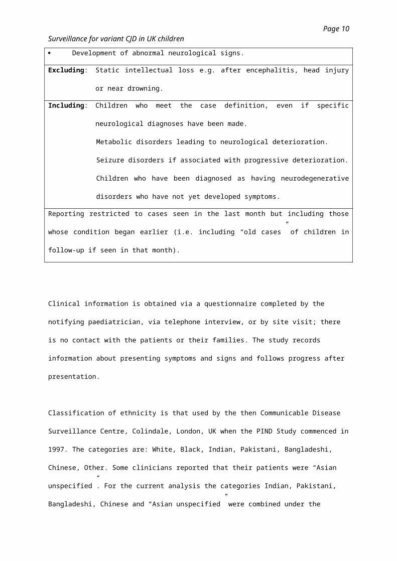
Table 1. Case definition – Progressive Intellectual and Neurological Deterioration.
Any child (under 16 years of age at onset of symptoms) who fulfils all of the following three criteria:
Progressive deterioration for more than three months
with
Loss of already attained intellectual or developmental abilities
and
Development of abnormal neurological signs.
Excluding: Static intellectual loss e.g. after encephalitis, head injury or near drowning.
Including: Children who meet the case definition, even if specific neurological diagnoses have
been made.
Metabolic disorders leading to neurological deterioration.
Seizure disorders if associated with progressive deterioration.
Children who have been diagnosed as having neurodegenerative disorders who have
not yet developed symptoms.
Reporting restricted to cases seen in the last month but including those whose condition began earlier
(i.e. including “old cases” of children in follow-up if seen in that month).
Clinical information is obtained via a questionnaire completed by the notifying paediatrician, via
telephone interview, or by site visit; there is no contact with the patients or their families. The study
records information about presenting symptoms and signs and follows progress after presentation.
Classification of ethnicity is that used by the then Communicable Disease Surveillance Centre,
Colindale, London, UK when the PIND Study commenced in 1997. The categories are: White, Black,
Indian, Pakistani, Bangladeshi, Chinese, Other. Some clinicians reported that their patients were
“Asian unspecified”. For the current analysis the categories Indian, Pakistani, Bangladeshi, Chinese
Page 7
Surveillance for variant CJD in UK children
and “Asian unspecified” were combined under the heading “Asian British” for comparison with the
2011 census of England and Wales where these groups were combined as “Asian/Asian British”.[11]
The PIND questionnaire asks if the parents of notified children are consanguineous, without defining
the term, but the details were obtained and only close blood relatives (i.e. cousins) were included in
this group.
The clinical data are anonymised by the PIND Study team and are then reviewed by an independent
PIND Expert Group to confirm and classify diagnoses. The Group consists of paediatric neurologists,
specialists in paediatric metabolic disease, and representatives from the NCJDRSU. If vCJD is
suspected it is suggested that the local paediatrician obtains consent to refer the child to the
NCJDRSU in Edinburgh. Otherwise the notified children are followed up by the PIND team via the
local clinician until a) a definite diagnosis is made, b) no further investigations are planned, or c) the
child dies. These children are often investigated by several national or international experts. Some
remain undiagnosed despite all investigations; they are not systematically followed up by the PIND
Study however the notifying paediatricians are asked to inform the Study if a diagnosis is
subsequently made.
For this paper an autopsy is defined as: “dissection and systematic examination of a body after death
in order to determine the cause of death or the presence of disease processes”. The term “post-
mortem pathological examination” is used to include full autopsies as well as more limited post
mortem examinations or biopsies of one or more tissues after death.
Consent for research was granted by Addenbrooke’s Cambridgeshire 2 Research Ethics Committee
(Ref: 97/010); Patient Information Advisory Group (PIAG/BPSU 2-10(c)(2005)), National Information
Governance Board (NIGB) and Confidentiality Advisory Group (CAG) under Section 251 of the NHS
Act 2006. Sponsored by Cambridge University Hospitals NHS Foundation Trust, R&D reference
A05001. Registered on the National Research Register; NRR ID is N0287023055. UKCRN ID 6636.
Role of the funding source. This report is based on independent research commissioned and
funded by the National Institute for Health Research (NIHR) Policy Research Programme. The views
Page 8
Surveillance for variant CJD in UK children
expressed in this publication are those of the author(s) and not necessarily those of the Department of
Health.
Results
Between May 1997 and October 2017 4244 children had been notified to the PIND Study. Of these
1933 were found not to meet the PIND criteria and 261 were either still under investigation or clinical
details were pending. The other 2050 cases met the PIND criteria and had been fully investigated; 6
had vCJD (identified between 1997 and 2000)[9,12] and 1819 (89% of the PIND cases) had a
diagnosis other than vCJD to explain their deterioration. In the remaining 225 children (11% of the
PIND cases) investigations had been negative or they had died undiagnosed.
The 1819 diagnosed cases. In the diagnosed group 977 were male, 842 were female; the ethnic
distribution is shown in Figure 1. The proportion of Asian British families in the diagnosed group was
29% (483/1683 for whom ethnicity was known). The rate of consanguinity in the diagnosed group was
32% (461/1440 for whom consanguinity was known). The 483 Asian British children comprised:
Pakistani 327, Indian 43, Bangladeshi 43, Chinese 5, Asian unspecified 65. The rate of consanguinity
in the families of Pakistani origin was 85% (279/327). In the whole diagnosed group there were more
than 190 different disorders; Figure 2 shows the ten disorders identified most frequently.
In 13 cases a brain biopsy had been performed during life and all helped to make a diagnosis. Of the
diagnosed children 241 were reported to have died; information about post-mortem examinations was
available for 109. 24/109 (22%) had a post-mortem examination (17 full autopsies, 1 coroner’s case
without neuropathology, 3 limited to brain and spinal cord pathology, 1 limited to biopsies of skin,
muscle and liver,1 liver biopsy only and 1 muscle biopsy only). 14 of the 24 post mortem studies
contributed to the diagnosis. In all the others the diagnosis was made without brain biopsies or post-
mortem examinations, that is in 1819 - (13+14) = 1792 cases (98%). The percentages known to have
had post mortems by ethnic group were: White 21/1104 = 2%, Pakistani 2/327 = 1%, Indian 1/43 =
2%. The last reported diagnostic post mortem was in 2007.
The 225 undiagnosed cases. In the undiagnosed group 118 were male, 107 were female; the ethnic
distribution is shown in Figure 1. There was a high rate of consanguinity - 52% of those for whom the
data were available (100/194). The proportion of Asian British families in the undiagnosed group was
Page 9
Surveillance for variant CJD in UK children
43% (94 of 217 for whom ethnicity was known). The 94 Asian British children comprised: Pakistani 72,
Indian 7, Bangladeshi 4, Asian unspecified 11. The rate of consanguinity in the families of Pakistani
origin was 89% (64/72), compared to 7% (8/108) in the White families.
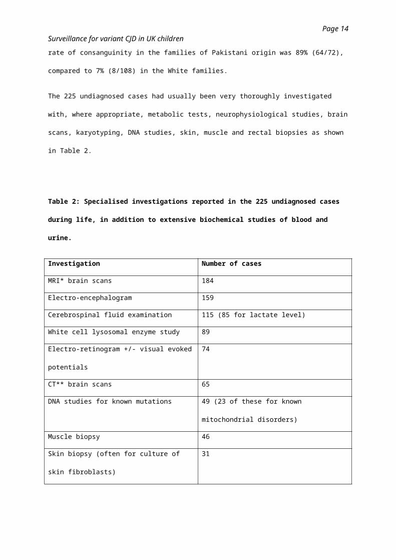
The 225 undiagnosed cases had usually been very thoroughly investigated with, where appropriate,
metabolic tests, neurophysiological studies, brain scans, karyotyping, DNA studies, skin, muscle and
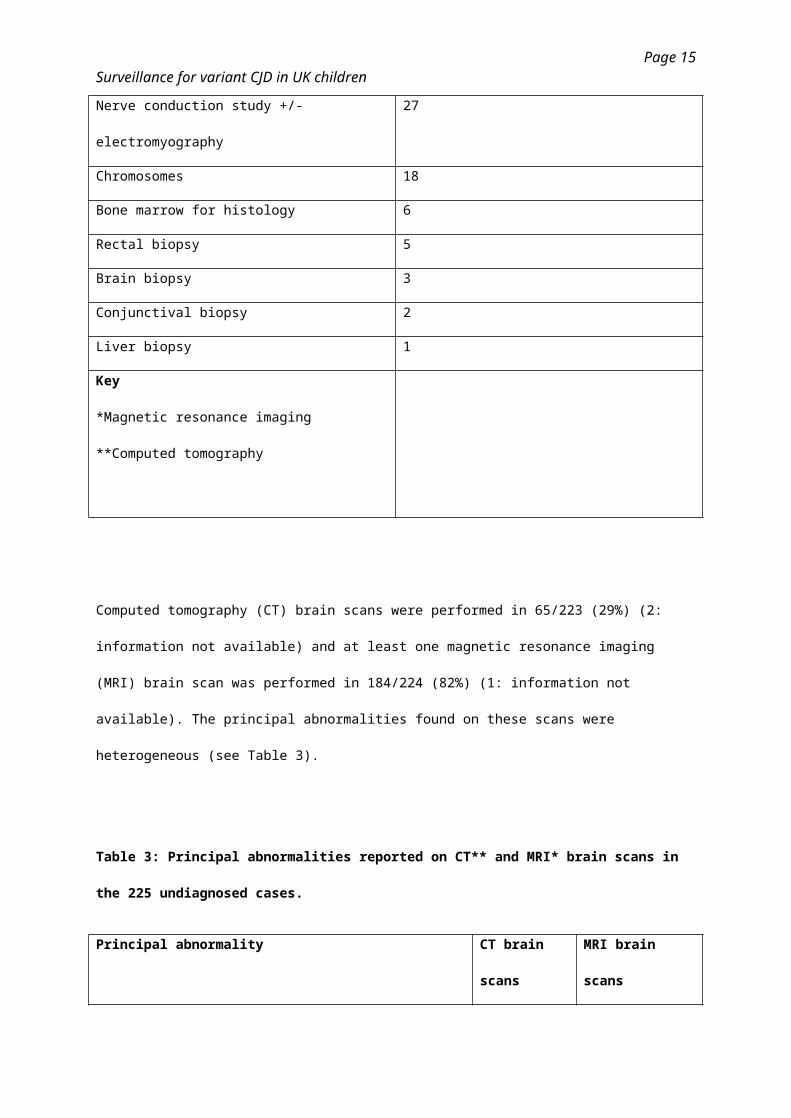
rectal biopsies as shown in Table 2.
Table 2: Specialised investigations reported in the 225 undiagnosed cases during life, in
addition to extensive biochemical studies of blood and urine.
Investigation Number of cases
MRI* brain scans 184
Electro-encephalogram 159
Cerebrospinal fluid examination 115 (85 for lactate level)
White cell lysosomal enzyme study 89
Electro-retinogram +/- visual evoked potentials 74
CT** brain scans 65
DNA studies for known mutations 49 (23 of these for known mitochondrial
disorders)
Muscle biopsy 46
Skin biopsy (often for culture of skin fibroblasts) 31
Nerve conduction study +/- electromyography 27
Chromosomes 18
Bone marrow for histology 6
Rectal biopsy 5
Brain biopsy 3
Conjunctival biopsy 2
Liver biopsy 1
Page 10
Surveillance for variant CJD in UK children
Key
*Magnetic resonance imaging
**Computed tomography
Computed tomography (CT) brain scans were performed in 65/223 (29%) (2: information not
available) and at least one magnetic resonance imaging (MRI) brain scan was performed in 184/224
(82%) (1: information not available). The principal abnormalities found on these scans were
heterogeneous (see Table 3).
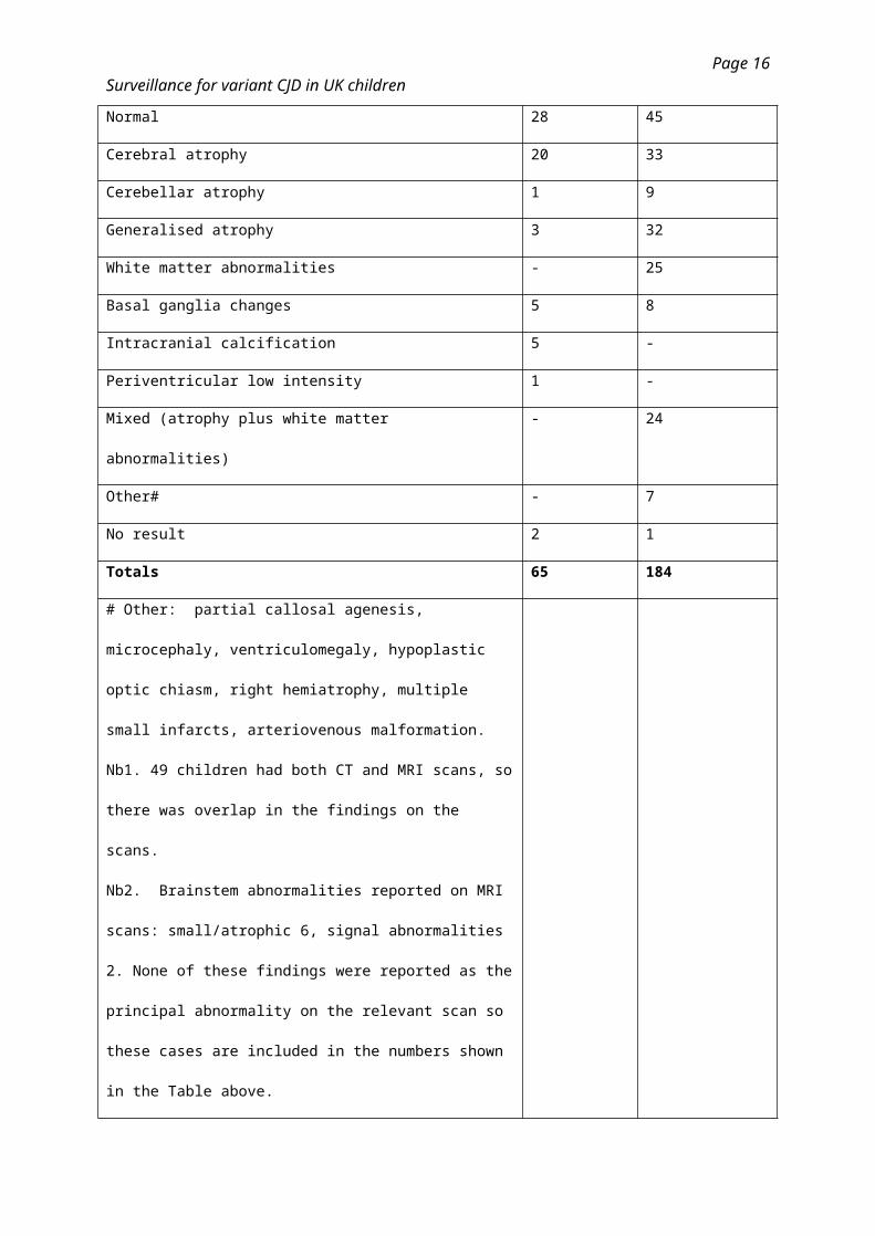
Table 3: Principal abnormalities reported on CT** and MRI* brain scans in the 225 undiagnosed
cases.
Principal abnormality CT brain
scans
MRI brain scans
Normal 28 45
Cerebral atrophy 20 33
Cerebellar atrophy 1 9
Generalised atrophy 3 32
White matter abnormalities - 25
Basal ganglia changes 5 8
Intracranial calcification 5 -
Periventricular low intensity 1 -
Mixed (atrophy plus white matter abnormalities) - 24
Other# - 7
No result 2 1
Totals 65 184
Page 11
Surveillance for variant CJD in UK children
# Other: partial callosal agenesis, microcephaly,
ventriculomegaly, hypoplastic optic chiasm, right hemiatrophy,
multiple small infarcts, arteriovenous malformation.
Nb1. 49 children had both CT and MRI scans, so there was
overlap in the findings on the scans.
Nb2. Brainstem abnormalities reported on MRI scans:
small/atrophic 6, signal abnormalities 2. None of these findings
were reported as the principal abnormality on the relevant
scan so these cases are included in the numbers shown in the
Table above.
Key
**Computed tomography
*Magnetic resonance imaging
It was reassuring that none of the MRI scans were reported to have the pulvinar sign associated with
vCJD.[13] MRI brain scans were performed in 73/94 (78%) of the Asian British children compared
with 92/108 (85%) of the White children, suggesting that Asian British children were being as
thoroughly investigated as white children because MRI scans are not easily performed in this age
group.
In three undiagnosed children a brain biopsy had been performed during life. In 123 cases the child’s
death had been reported. Post-mortem pathological examinations had been carried out in 14 (13%) of
the 108 cases for whom information was available: 9 had a full autopsy and 5 had partial
examinations (in 1 the brain was not examined, in 2 just the brain was examined, in 1 post mortem
liver and muscle biopsies were taken and 1 had a post-mortem liver biopsy). Post-mortem
neuropathological examination was carried out in 11 cases, in 2 of these brain tissue was sent to the
NCJDRSU where staining for abnormal prion protein was negative. In another case the
neuropathology was reviewed by the NCJDRSU and was not that of vCJD. In the ethnic groups the
percentages that had post mortems were: White 8/108 = 7%, Pakistani 4/72 = 6% and Indian 2/7 =
29%.
Page 12
Surveillance for variant CJD in UK children
Children were not routinely followed by the Study after being allocated to the undiagnosed group.
Therefore in October 2017 the National Health Service Spine Portal was used to discover how many
of the 225 undiagnosed cases had died by that time: the total number was 161, which is 38 cases
more than had been reported by the notifying paediatricians between May 1997 and October 2017.
Discussion
Principal findings
Post-mortems and diagnosis. The diagnosis was made without brain biopsy or post-mortem
neuropathology in 98% of the whole diagnosed group of children with PIND. Only 24 children in the
diagnosed group were known to have had post-mortem examinations. However these did help to
make the diagnosis in 14 cases. It is therefore a concern that only 14 of the 108 undiagnosed children
who died were known to have had post-mortem studies and in three of these the brain was not
examined.
Falling autopsy rates have been noted in adults and children.[14-16] Burton and Underwood[14]
reported that the overall autopsy rate in the UK in 1979 was 42.7%, falling to 15.3% in 2001. In 2015
Turnbull et al.[17] found that the mean autopsy rate in the UK was 0.69%: hospital autopsy rates in
children’s hospital NHS trusts ranged from 0% to 21%. A recent systematic review[18] of post-mortem
examinations in perinatal and paediatric settings stated that “...PM examinations result in clinically
significant findings in 22-76% of cases…”.
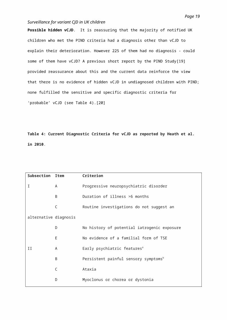
Possible hidden vCJD. It is reassuring that the majority of notified UK children who met the PIND
criteria had a diagnosis other than vCJD to explain their deterioration. However 225 of them had no
diagnosis - could some of them have vCJD? A previous short report by the PIND Study[19] provided
reassurance about this and the current data reinforce the view that there is no evidence of hidden
vCJD in undiagnosed children with PIND; none fulfilled the sensitive and specific diagnostic criteria for
‘probable’ vCJD (see Table 4).[20]
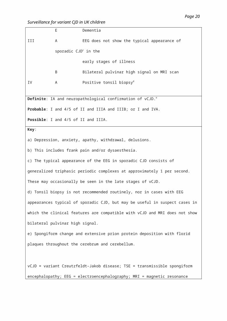
Table 4: Current Diagnostic Criteria for vCJD as reported by Heath et al. in 2010.
Page 13
Surveillance for variant CJD in UK children
Subsection Item Criterion
I A Progressive neuropsychiatric disorder
B Duration of illness >6 months
C Routine investigations do not suggest an alternative diagnosis
D No history of potential iatrogenic exposure
E No evidence of a familial form of TSE
II A Early psychiatric featuresa
B Persistent painful sensory symptomsb
C Ataxia
D Myoclonus or chorea or dystonia
E Dementia
III A EEG does not show the typical appearance of sporadic CJDc in the
early stages of illness
B Bilateral pulvinar high signal on MRI scan
IV A Positive tonsil biopsyd
Definite: IA and neuropathological confirmation of vCJD.e
Probable: I and 4/5 of II and IIIA and IIIB; or I and IVA.
Possible: I and 4/5 of II and IIIA.
Key:
a) Depression, anxiety, apathy, withdrawal, delusions.
b) This includes frank pain and/or dysaesthesia.
c) The typical appearance of the EEG in sporadic CJD consists of generalized triphasic periodic
complexes at approximately 1 per second. These may occasionally be seen in the late stages of
vCJD.
d) Tonsil biopsy is not recommended routinely, nor in cases with EEG appearances typical of
sporadic CJD, but may be useful in suspect cases in which the clinical features are compatible with
vCJD and MRI does not show bilateral pulvinar high signal.
e) Spongiform change and extensive prion protein deposition with florid plaques throughout the
Page 14
Surveillance for variant CJD in UK children
cerebrum and cerebellum.
vCJD = variant Creutzfeldt–Jakob disease; TSE = transmissible spongiform encephalopathy; EEG =
electroencephalography; MRI = magnetic resonance imaging.
However it is a concern that prion protein staining of brain tissue was carried out in just two of the
undiagnosed cases as this is the only way to completely exclude the diagnosis of vCJD.[21] Blood
and urine tests for diagnosing vCJD have been developed, but are not sufficiently validated for routine
clinical use.[22-24]
Ethnicity and consanguinity. The PIND study has reported that the greatest numbers of cases
come from areas where there are relatively large populations of Pakistani origin[7]. These new data
confirm that a large proportion of Study children are Asian British (29% in the diagnosed group and
43% in the undiagnosed group compared with 7.5% in the general population of England and Wales
in 2011[11]). There were high rates of consanguinity - in the families of undiagnosed children of
Pakistani origin with PIND it was 89%: possibly they have inherited neurodegenerative diseases that
have not yet been characterised. These diseases may become evident as new diagnostic
investigations are available.
Strengths and weaknesses of the PIND Study. The Study uses the well-established health
surveillance mechanism of the British Paediatric Surveillance Unit (BPSU).[8] The PIND Study Expert
Group includes representatives from the NCJDRSU and provides an independent opinion about the
diagnosis in every reported case. There is multisource ascertainment and the reporting is not
restricted to cases admitted to hospital. The Study has confirmed the diagnosis of more than 190
known neurological disorders and has published case series of the diagnosed groups.[25-28]
The BPSU relies on the voluntary support of consultant paediatricians: it does not provide complete
ascertainment of cases and it is not always possible to obtain all the relevant data. When PIND cases
are diagnosed they are not systematically followed up by the Study and follow up has to be
discontinued eventually in undiagnosed cases.
Page 15
Surveillance for variant CJD in UK children
UK surveillance for prion diseases is carried out by the NCJDRSU, the team that first identified this
new disease.[1] The NCJDRSU relies on a network of neurologists, psychiatrists, neuropathologists
and other clinical specialists who report adults with suspected vCJD. Because vCJD cases could be
hidden among the multitude of rare neurodegenerative diseases of childhood a system of direct
referral of suspected vCJD cases would not be reliable. The PIND Study therefore has to work via
local paediatricians and other specialists to screen all the children in the UK with PIND.
Policy implications and future research. There has been increasing use of minimally invasive post-
mortem investigations in infants and children. Post-mortem magnetic resonance imaging can provide
useful diagnostic information[29-31] and may identify significant neuropathology in infants and
children.[32] Storage of blood and other relevant tissues for metabolic and/or genetic studies is very
important; several PIND study children have been diagnosed via the UK and Ireland Deciphering
Developmental Disorders (DDD) Study.[33] Some children with undiagnosed PIND might be eligible
for inclusion in the 100,000 Genomes Project.[34] Because of the recent advances in genetics
undiagnosed children should be referred for an up-to-date opinion from a clinical geneticist.
The recent identification of the first patient with vCJD who was methionine/valine heterozygous at
PRNP codon 129 reinforces the need for continued vCJD surveillance ,[3] particularly as a study of
archived appendix samples from UK hospitals published in 2013 indicated that approximately 1 in
2000 of the UK population is carrying abnormal prion protein in the gastrointestinal tract.[35]
If all undiagnosed children with PIND underwent a full autopsy there would be no need for the PIND
Study. However the Study shows that undiagnosed children with neurological deterioration rarely
undergo ante- or post-mortem pathological examination of brain tissue. As there is currently no
validated vCJD screening test the PIND Study remains the only practical means of performing
systematic surveillance for vCJD in UK children.
Declaration of interest. The authors have no interests which might be perceived as posing a conflict
or bias.
Competing interest statement
Page 16
Surveillance for variant CJD in UK children
All authors have completed the ICMJE uniform disclosure form at
http://www.icmje.org/coi_disclosure.pdf and declare: no support from any organisation for the
submitted work [or describe if any]; no financial relationships with any organisations that might have
an interest in the submitted work in the previous three years [or describe if any], no other relationships
or activities that could appear to have influenced the submitted work [or describe if any.
Contributorship statement
All the authors made sufficient contributions to meet the recognised criteria for authorship of the
manuscript. All contributed to at least three of the following: literature search, figures, study design,
data collection, data analysis, data interpretation, writing.
Acknowledgements.
This report is based on independent research commissioned and funded by the National Institute for
Health Research (NIHR) Policy Research Programme (“To Undertake Prospective Multisource
Surveillance for all Cases of Progressive Intellectual and Neurological Deterioration Occurring in
Children in the UK” PR-ST-1216-10001). The views expressed in this publication are those of the
author(s) and not necessarily those of the NHS, the NIHR, the Department of Health, arm’s length
bodies or other government departments.
Many thanks to the paediatricians who notify cases, to Mr Richard Lynn and Mr Jacob Avis of the
BPSU and to the PIND Expert Group: Dr Peter Baxter, Dr Carlos de Sousa, Professor Paul Gissen,
Professor Manju Kurian, Professor John Livingston, Professor Robert McFarland, Dr Suvankar Pal, Dr
Michael Pike, Professor Richard Robinson, Dr Evangeline Wassmer, Professor Robert Will, Dr John
Wilson, and Professor Sameer Zuberi.
Page 17
Surveillance for variant CJD in UK children
References
1. Will RG, Ironside JW, Zeidler M et al. A new variant of Creutzfeldt-Jakob disease in the UK. Lancet
1996; 347: 921–5.
2. National Creutzfeldt-Jakob Disease Research and Surveillance website: latest NCJDSU CJD
monthly statistics: http://www.cjd.ed.ac.uk/sites/default/files/figs.pdf. Last accessed 31 November
2017.
3. Mok T, Jaunmuktane Z, Joiner S et al. Variant Creutzfeldt-Jakob disease in a patient with
heterozygosity at PRNP codon 129. N Engl J Med 2017; 376: 292–294. doi: 10.1056/NEJMc1610003.
4. Bishop MT, Pennington C, Heath CA et al. PRNP variation in UK sporadic and variant Creutzfeldt
Jakob disease highlights genetic risk factors and a novel non-synonymous polymorphism. BMC
Medical Genetics 2009; 10: 146. doi:10.1186/1471-2350-10-146.
5. Patient safety and reduction of risk of transmission of Creutzfeldt-Jakob disease (CJD) via
interventional procedures. Published 22 November 2006.
https://www.nice.org.uk/guidance/ipg196/resources/patient-safety-and-reduction-of-risk-of-
transmission-of-creutzfeldtjakob-disease-cjd-via-interventional-procedures-pdf-1899863525353669.
Last accessed 30 January 2018.
6. Seed CR, Hewitt PE, Dodd RY, Houston F, Cervenakova L. Creutzfeldt-Jakob disease and blood
transfusion safety. Vox Sanguinis 2018 DOI: 10.1111/vox.12631.
7. Devereux G, Stellitano L, Verity CM, Nicoll A, Rogers P. Variations in neurodegenerative disease
across the UK: findings from the national study of Progressive Intellectual and Neurological
Deterioration (PIND). Arch Dis Child 2004; 89: 8-12.
8. Verity C, Preece M. Surveillance for rare disorders by the BPSU. The British Paediatric
Surveillance Unit. Arch Dis Child 2002; 87: 269–271.
Page 18
Surveillance for variant CJD in UK children
9. Verity CM, Nicoll A, Will RG, Devereux G, Stellitano L. Variant Creutzfeldt-Jakob disease in UK
children: a national surveillance study. Lancet 2000; 356: 1224–27.
10. Royal College of Paediatrics and Child Health website: BPSU: Studies: Progressive Intellectual
and Neurological Deterioration. https://www.rcpch.ac.uk/pind. Last accessed 31 November 2017.
11. Office for National Statistics Date: 25 July 2013 Coverage: England and Wales Theme: Population
Comparison of mid-2010 Population Estimates by Ethnic Group against the 2011 Census.
file://ukbia04sfsrv001.a04.dt21.svcs.hp.com/users/a04/verityc/comparisonofmid2010peegsagainst201
1censusestimatesv2tcm77320363.pdf. Last accessed 18 August 2017.
12. Verity C, Winstone AM, Stellitano L et al. The epidemiology of progressive intellectual and
neurological deterioration in childhood. Arch Dis Child 2010; 95: 361-4. doi:
10.1136/adc.2009.173419. Epub 2009 Nov 29.
13. Collie DA, Summers DM, Sellar RJ et al. Diagnosing variant Creutzfeldt-Jakob disease with the
pulvinar sign: MR imaging findings in 86 neuropathologically confirmed cases. Am J Neuroradiol
2003; 24: 1560–69.
14. Burton JL, Underwood J. Clinical, educational, and educational value of autopsy. Lancet 2007;
369: 1471–80.
15. Shojania KG, Burton EC. The vanishing nonforensic autopsy. N Engl J Med 2008; 358: 873–5.
16. Turnbull A, Osborn M, Nicholas N. Hospital autopsy: endangered or extinct? J Clin Pathol 2015;
68: 601–4. doi:10.1136/jclinpath-2014-202700.
17. Turnbull A, Martin J, Osborn M. The death of autopsy? Lancet 2015; 386: 2141.
Page 19
Surveillance for variant CJD in UK children
18. Lewis C, Hill M, Arthurs OJ, Hutchinson C, Chitty LS, Sebire NJ. Factors affecting uptake of post-
mortem examinations in the prenatal, perinatal and paediatric setting. BJOG 2017;
DOI:10.1111/1471-0528.14600.
19.Verity CM, Winstone AM, Stellitano L, Nicoll A, Will RG. No clinical evidence of hidden vCJD in UK
children. Arch Dis Child 2006; 91: 608–609. doi: 10.1136/adc.2004.071266.
20. Heath CA, Cooper SA, Murray K et al. Validation of Diagnostic Criteria for Variant Creutzfeldt–
Jakob Disease. Ann Neurol 2010; 67: 761–770.
21. Ironside JW, Head MW, Bell JE, McCardle L, Will RG. Laboratory diagnosis of variant Creutzfeldt–
Jakob disease. Histopathology 2000; 37: 1–9. doi: 10.1046/j.1365-2559.2000.00946.x.
22. Moda F, Gambetti P, Notari S et al. Prions in the Urine of Patients with Variant Creutzfeldt–Jakob
Disease. N Engl J Med. 2014 August 7; 371(6): 530–539. doi:10.1056/NEJMoa1404401.
23. Concha-Marambio L, Pritzkow S, Moda F et al. Detection of prions in blood from patients with
variant Creutzfeldt-Jakob disease. Sci Transl Med 8, 370ra183 (2016) 21 December 2016.
24. Bougard D, Brandel J-P, Bélondrade M et al. Detection of prions in the plasma of presymptomatic
and symptomatic patients with variant Creutzfeldt-Jakob disease. Sci Transl Med 8, 370ra182 (2016)
21 December 2016.
25. Verity C, Winstone AM, Stellitano L, et al. The clinical presentation of mitochondrial diseases in
children with progressive intellectual and neurological deterioration: a national, prospective,
population-based study. Dev Med Child Neurol 2010; 52: 434–40.
26. Smith NJ, Winstone AM, Stellitano L, et al. GM2 gangliosidosis in a United Kingdom study of
children with progressive neurodegeneration: 73 cases reviewed. Dev Med Child Neurol 2012; 54:
176–82.
Page 20
Surveillance for variant CJD in UK children
27. Stellitano L, Winstone AM, van der Knaap MS, Verity CM. Leukodystrophies and genetic
leukoencephalopathies in childhood: a national epidemiological study. Dev Med Child Neurol 2016;
58: 680–9.
28. Winstone AM, Stellitano LA, Verity CM. Niemann-Pick type C as a cause of progressive
intellectual and neurological deterioration in childhood. Dev Med Child Neurol 2017; 59: 965–72. doi:
10.1111/dmcn.13476 Epub 2017 Jun 2.
29. Thayvil S, Chandrasekaran M, Chitty LS et al. Diagnostic accuracy of post-mortem magnetic
resonance imaging in fetuses, children and adults: a systematic review. Eur J Radiol 2010; 75: e142–
8. doi: 10.1016/j.ejrad.2009.10.007. Epub 2009 Nov 11.
30. Thayvil S, Sebire NJ, Chitty LS et al. Post-mortem MRI versus conventional autopsy in fetuses
and children: a prospective validation study. Lancet 2013; 382: 223–33.
31. Addison S, Arthurs OJ, Thayvil S. Post-mortem MRI as an alternative to non-forensic autopsy in
foetuses and children: from research into clinical practice. Br J Radiol 2014; 87: 20130621.
32. Arthurs OJ, Thayvil S, Pauliah SS et al. Diagnostic accuracy and limitations of post-mortem MRI
for neurological abnormalities in fetuses and children. Clin Radiol 2015; 70: 872–80.
33. Wright CF, Fitzgerald TW, Jones WD et al. Genetic diagnosis of developmental disorders in the
DDD study: a scalable analysis of genome-wide research data. Lancet 2015; 385: 1305–14.
34. The 100,000 Genomes Project. Genomics England. https://www.genomicsengland.co.uk/the-
100000-genomes-project/. Last accessed 22nd January 2018.
35. Gill ON, Spencer Y, Richard-Loendt A et al. Prevalent abnormal prion protein in human
appendixes after bovine spongiform encephalopathy epizootic: large scale survey. BMJ 2013; 347:
f5675. doi: 10.1136/bmj.f5675.
Page 21
Surveillance for variant CJD in UK children
Caption for Figure 1: Flow chart showing the ethnicity of the 2050 cases that met the PIND criteria
and had been fully investigated.
Caption for Figure 2: the 10 commonest diagnoses of the more than 190 different disorders in the
1819 diagnosed children with progressive intellectual and neurological deterioration identified
between May 1997 and October 2017.
Page 22