Embed Size (px)
Citation preview
Research Project Report 3
Investigating the role and localization of Tau V337M mutation in microtubules stabilization
Mihaela Veronica Foisor
201206342
Supervisor: Dr Natalia Sanchez Soriano
Internal Assessor: Dr John QuayleStrand: Medical Sciences
Word Count: 4120

2
Investigating the role and localization of Tau V337M mutation in microtubules stabilizationMihaela-Veronica FoisorInstitute of Translational Medicine, University of Liverpool, Crown St, Liverpool, L69 3BX, UK
Abstract
Frontotemporal dementia (FTD) represents one of the most common subtypes of dementia and are mostly classified as familial cases. Several mutations in the tau protein are known to be the main factor that drives FTD. Mutation V337M in has been proven to induce hyperphosphorylation of tau, resulting in alteration of tau function, microtubule destabilization and neurofibrillary tangles formation. In this study, we are trying to pinpoint the localization of the V337M tau proteins and to acquire a deeper understanding of the mechanisms behind V337M tau induced toxicity. Drosophila and chicken neuronal cultures expressing hTauWT and hTauV337M proteins have been generated and cell phenotypes compared after 1 week. Our results show that V337M mutation induces a strong phenotype in drosophila neurons, while in the chicken ones’ similar but weaker phenotype is present. Furthermore, we have also established that hTauV337M is actively located along the axons and dendrites in the chicken neurons. Although we do not have a complete image of how V337M mutation induces degeneration, our results are the first part of a series of experiments that will further work towards this goal.
Key words: Frontotemporal dementia, tau, V337M mutation, microtubules, drosophila neuronal cultures, chicken neuronal culturesIntroduction
Frontotemporal dementia (FTD) is represented by the degeneration of the frontotemporal lobar and is characterized by a progressive loss of neurons located in the frontal and/or temporal lobes, hence leading to a gradual deficiency in cognition, language ability and behavior [1]. Although it represents only a subtype of all the dementia cases, it accounts for approximately 20% of the young onset dementia cases, as most people affected are relatively young (55-65 years old)[2].
FTD occurs mostly in familial forms and frequently as sporadic events. Around 30% of diagnosed patents present hereditary forms of FTD, with up to 20% of these cases carrying different mutations in the MAPT (microtubule associated protein Tau) gene. Clinical cases of FTD due to mutations in the MAPT gene present a Parkinson like impairment - degeneration of subcortical area of the brain [3] and are categorized as a tauopathy type disease. Tauopathies are a class of neurodegenerative diseases characterized by
the existence in the human brains of tau protein deposits in various morphologies [4]. Currently, there are no effective treatments, preventative or curative, targeting tauopathies. Hence, acquiring a better understanding of tau function and roles in healthy pathology and disease models would allow identification of new possible targets for therapies.
Tau protein has a crucial role in neurons as it stabilizes microtubules, acts as a component of the neural cytoskeleton, defines neural polarity and facilitates axonal outgrowth [5]. As microtubules are the backbone of neuronal transport and synaptic plasticity, their regulation is directly linked to neurodegeneration [6]. Extensive evidence has demonstrated that dynamic microtubules growth is a decisive factor in the regulation of synaptic plasticity, and its turnover is linked to synaptic modifications and memory formation. Therefore, loss of microtubules dynamic and turnover due to failure of tau
3
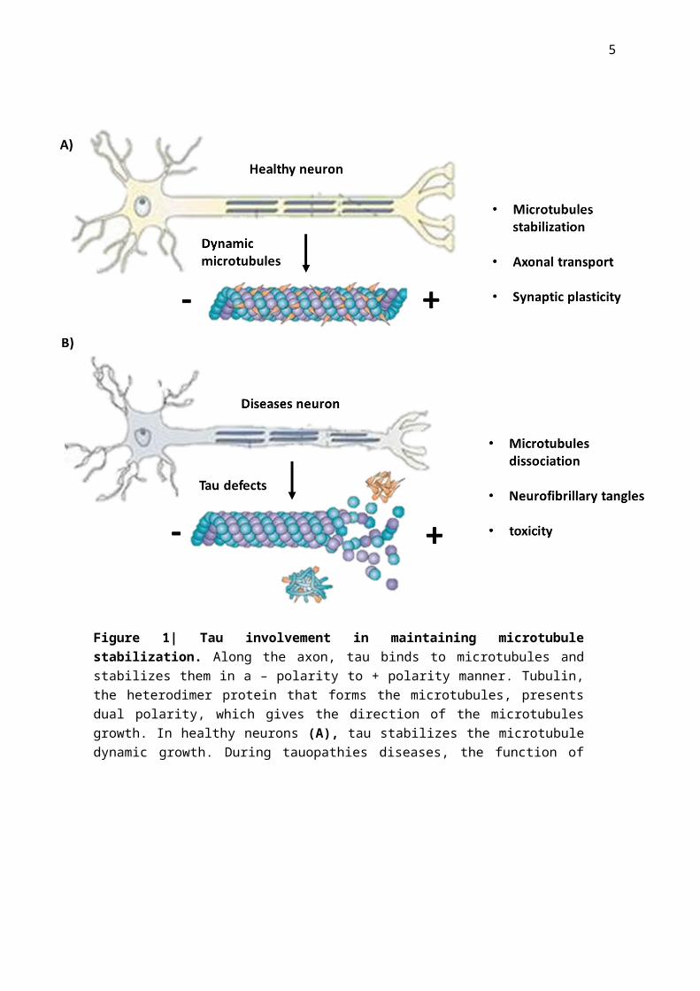
stabilization has been shown to be connected neurodegeneration (Figure 1) [7,8].
Figure 1| Tau involvement in maintaining microtubule stabilization. Along the axon, tau binds to microtubules and stabilizes them in a – polarity to + polarity manner. Tubulin, the heterodimer protein that forms the microtubules, presents dual polarity, which gives the direction of the microtubules growth. In healthy neurons (A), tau stabilizes the microtubule dynamic growth. During tauopathies diseases, the function of tau is compromised, resulting in less tau binding to microtubules and more neurofibrillary tangles formation, contributing to the neuropathology of the disease (adapted from [25]).
4
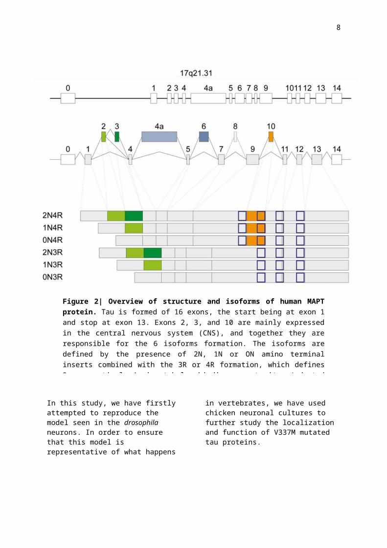
There are six different human Tau isoforms expressed in the adult brain; this being the result of alternative splicing of axons 2, 3 and 10 of the MAPT gene (Figure 2) [9]. In healthy brains, the ration between the different isoforms (4R and 3R) is balanced, however in FTD cases this ratio presents a significant shift towards the 4R isoforms, followed by 4R Tau protein deposits of neurofibrillary tangles being augmented [3]. One of the crucial questions that are currently being researched is the mechanisms behind the Tau tangles formations. Mutations of the Tau protein might represent part of the answer, as they may result in structurally and biochemically abnormal proteins and may induce an unbalance in the ratio between the 3R and 4R tau levels. In normal state, tau is classified as a soluble protein, however in FTD MAPT mutations pathologies, it is found in an insoluble form – it tends to become hyperphosphorylated and accumulate in filaments [10]. Consequently, hyperphosphorylation of the tau protein hallmarks the change in structure and plays a decisive role in generating FTD disease phenotypes. Tau contains a significant number of potential phosphorylation sites, around 85 sites per protein, which have a deciding influence on regulating its function. In heathy circumstances, tau has ~ 20 phosphate molecules attached, however as its phosphorylation increases, tau’s affinity for binding to microtubules changes, hindering its ability to regulate their outgrowth[11,12].
Site specific phosphorylation has been shown to induce changes in its function- phosphorylation of the proline-rich regions of tau can halt its microtubule regulation activity and stimulating an increase in its biochemical properties towards a more self-aggregating form[13,14]. Although none of the current known mutations of MAPT are directly influencing the phosphorylation status of the tau protein, several mutations have been
identified that can induce a heightened phosphorylation level [15]. One of these mutations that has been identified is V337M, which is a exonic mutation and has proven to affect all 6 tau human isoforms [16]. Moreover, V337M is been one of the first mutations in MAPT shown to be present in familial sporadic cases, and to have a substantial description at the phenotypic and genetic linkage levels [17,18]. Different studies have been associating the presence of the V337M mutation with significant increase of hyperphosphorylated tau in both 4R and 3R isoforms and an increase of neurofibrillary tangles present in the brain [19,20]. Current data infers a strong correlation between the presence of the V337M mutation and hyperphosphorylation state of tau; however, questions are still being raised as to how this mutation affects the localization of tau and how does this localization changes after hyperphosphorylation that leads to neurofibrillary tangles formation.
Experiments performed previously in our lab have demonstrated a strong phenotype of drosophila neurons exppressing the V337M mutation. In the first 6 hours to 2 days of cultures, the neurons presented a normal phenotype, however this was slowly changed and after 6 days we could see a significant difference between the control neurons and the V337M mutated ones. These results implying a degree of neurodegeneration over time that was significantly promoted by the induction of the V337M mutation.
As we have introduced the human 0N4R isoform into a drosophila model, the question of where does this human tau protein localize in the drosophila neurons. Furthermore, is the phenotype generated due to an imbalance of tau as we have an overexpression of the protein or due to the toxicity effect of V337M mutation?
5
In this study, we have firstly attempted to reproduce the model seen in the drosophila neurons. In order to ensure that this model is representative of what happens in
vertebrates, we have used chicken neuronal cultures to further study the localization and function of V337M mutated tau proteins.
Figure 2| Overview of structure and isoforms of human MAPT protein. Tau is formed of 16 exons, the start being at exon 1 and stop at exon 13. Exons 2, 3, and 10 are mainly expressed in the central nervous system (CNS), and together they are responsible for the 6 isoforms formation. The isoforms are defined by the presence of 2N, 1N or ON amino terminal inserts combined with the 3R or 4R formation, which defines 3, respectively 4 microtubules binding repeat sites (adapted from [26]).
6
Methods
Drosophila embryo selection and neural cultures. Drosophila crosses were performed over the weekend (Figure 3). The embryos were bleached (50% Bleach) for 90 seconds prior to selection. Stage 11 embryos were selected under the microscope; 30 embryos per genotype were used for each repeat performed. After selection, the embryos were transferred into 20µl of filtered Drosophila neuronal media containing 1:1000 Pen/Strep. After a 20 seconds’ wash with EtOH, the embryos were washed again with 100µl Drosophila neuronal media. After discarding the neuronal media, 100µl of Dispersion media was added and the embryos were homogenized a homogenization pestle. This was followed by a 3.5 minutes incubation period at 37°C. After the incubation, 200µl of Drosophila neuronal media was added and the solution was centrifugated for 5 minutes at 900g RT. The supernatant was removed and the cells were re-suspended in 90µl of Drosophila neuronal media. A 30µl drop was plated into a sterile chamber coated with acetone. The drop culture was sealed with Vaseline and a cover slip. From each genotype 3 distinct drop neuronal cultures were produced. After 3 hours, the culture was reversed so the neurons were attached to the cover slip. The cultures were kept in at 37°C in the dark until further processing.
Immunostaining of Drosophila neuronal cultures. After 6 days of incubation, the neuronal drop cultures were fixed in 4% PFA for 30 min at room temperature. This was followed by 2 washes with PBT, each for 10 min. Primary antibody staining (α-Tubulin 1:1000 dilution) was performed overnight at 4°C in the dark. The cultures were then washed two times with PBT and 2nd antibody was added for 1 hour. The HRP (Protein A) antibody was direct labelled, hence was
added as a 2nd antibody with a dilution of 1:20. This was followed by 1 hour PBT wash. The cover slips were then mounted using Vectashield (Vector Laboratories) and then sealed with nail polish. The figures were taken with Nikon Eclipse 90s Microscope and further processed using ImageJ programme.
Generating hTau plasmid with V337M mutation. Using the plasmid presented in Figure 4A, we have induced the V337M mutation in the Tau protein. The mutation was performed following the QuickChange Lightning Site-Directed Mutagenesis Kit (Agilent Technologies) protocol and using the following primers: forward 5’-CAGGAGGTGGCCAGATGGAAGTAAAATCTG-'3; reverse: 3’- CAGATTTTACTTCCATCTGGCCACCTCCTG-5’. The resulting plasmids were transformed into DH5α Bacterial cells as per protocol. The transformed cells were plated into Kanamycin plates (concentration of 50ng/µl) with 200µl and 400µl of transformed cells and incubated overnight at 37˚C. The successful colonies were transferred into a 4ml LB media at 37˚C overnight in a shaking incubator at 225rpm. DNA was extracted using the QIAprep Spin Miniprep Kit (Qiagen). The mutated clones were sequenced and the successful ones were further used for the chicken neuronal cultures transfection.
Chicken neurons cultures. Chicken eggs were incubated at 37°C for 7 days. On the 8th day the embryos were dissected and the 2 frontal lobes of the brain were extracted in 5 ml of Neurobasal media (containing 0.2% FBS, 0.4% Pen/Strep, 1% Glutamax, 1% Sodium Pyruvate; Gibco-Life Technologies). After all forebrains lobes were collected, 5 ml of Trypsin (Gibco-Life Technologies) were added for 30 min at 37°C.
7
The media was then discarded and the brain fragments were washed 3 times with 10ml PBS. After adding 1ml of neuronal media, 100µl of cells were added to each transfection reaction.
The transfection reaction was performed using Amaxa Chicken neuron Nucleofactor kit (Lonza) as per protocol. The transfected cells were then diluted in 10 ml of neuronal media, then plated in plates coated with poly-D-lysine (Sigma Aldrich; P6407). Every 7 days, half of media has changed for all plates. After 7 and 14 days pictures were taken with Marianas SDC 3i (Ametek) microscope. All
figures were further processed using ImageJ programme.
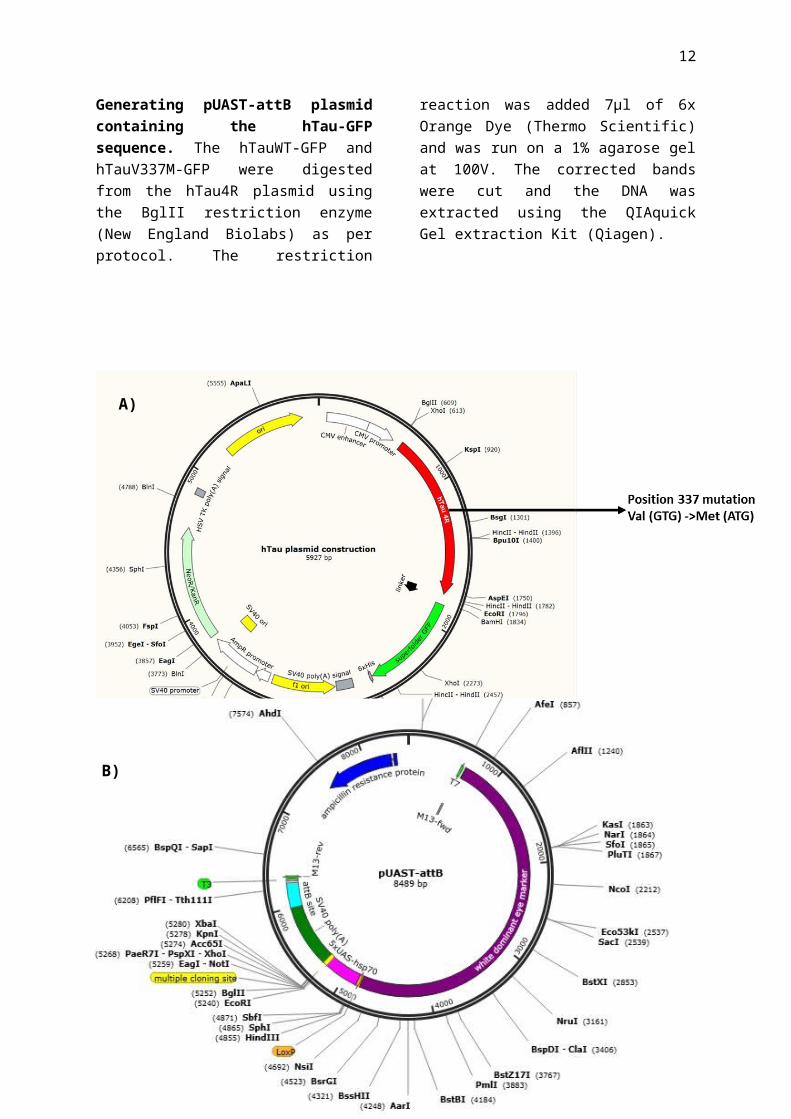
Generating pUAST-attB plasmid containing the hTau-GFP sequence. The hTauWT-GFP and hTauV337M-GFP were digested from the hTau4R plasmid using the BglII restriction enzyme (New England Biolabs) as per protocol. The restriction reaction was added 7µl of 6x Orange Dye (Thermo Scientific) and was run on a 1% agarose gel at 100V. The corrected bands were cut and the DNA was extracted using the QIAquick Gel extraction Kit (Qiagen).
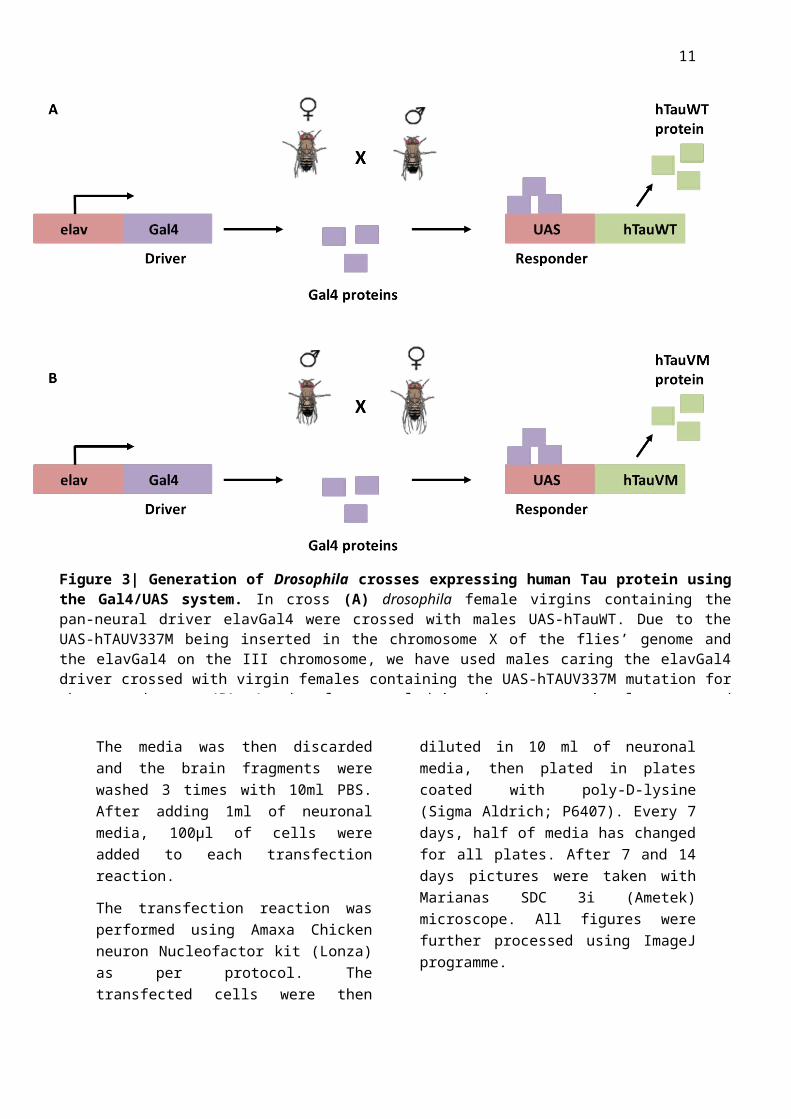
Figure 3| Generation of Drosophila crosses expressing human Tau protein using the Gal4/UAS system. In cross (A) drosophila female virgins containing the pan-neural driver elavGal4 were crossed with males UAS-hTauWT. Due to the UAS-hTAUV337M being inserted in the chromosome X of the flies’ genome and the elavGal4 on the III chromosome, we have used males caring the elavGal4 driver crossed with virgin females containing the UAS-hTAUV337M mutation for the second cross (B). As the elav neural driver become transiently expressed during development, GAL4 is transcribed and specifically binds to the UAS enhancer, activating gene transcription of hTauWT or hTauV337M.
8
A)
B)
Figure 4| A) hTau4R plasmid used for generating and expressing hTauV337M mutation in chicken neurons. B) pUAST-attB plasmid structure before the addition of the hTauWT/V337M sequence.

9
Before ligation, the pUAST-attB plasmid (Figure 4B) was dephosphorylated using Antarctic Phosphatase (New England Biolabs). The ligation of the hTauWT-GFP and hTauV337M-GFP to the pUAST-attB vector was performed with the T4 Ligase (New England Biolabs) as per protocol. DH5α bacterial cells were transformed using the pUAST-attB vector with the Tau protein. The resulting transformed cells were plated on Ampicillin plates using 400µl of cells and incubated overnight at 37˚C. When colonies were present, they were further cultured in 4ml Lb medium and incubated at 37˚C shaking incubator at 225rpm overnight. Following miniprep, digestion with the BamHI (Promega) restriction enzyme and run on an agarose gel (as described above), the samples were verified if they contained the insert in a correct orientation.
Results
Tau V337M mutation induces a specific neuronal phenotype in drosophila.
After performing the immunostaining on the 6 days Drosophila neuronal cultures, significant phenotypical changes were found between the controls and the V337M mutation neurons; throughout the experiment we have focused on individual neurons, as they allow a more precise quantification and analysis.
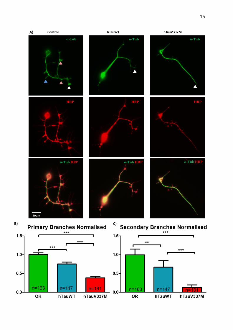
We have firstly looked at the number of primary and secondary branches. The morphology of hTauV337M neurons is less branched and more “stiff” than the hTauWT genotype. Performing the statistical analysis (Figure 5A-B) demonstrated that there is a significant difference between the two phenotypes. Hence, the hTauV337M neurons tend to partially lose the ability of branching and their microtubule growth is present mainly in the axons.
This deficiency in growth was further accentuated by the growth cone morphology of the different genotypes. Usually, as we can
see in the control neurons (Figure 5C), growth cones morphology is unbundled; sometimes presenting growing loops at the end of the growth cone. This phenotype allows an active growing and retracting of branches, permitting a more dynamic microtubule growth. While in the hTauWT phenotype there are less loops and more growth cones seems to be bundled, in hTauVM phenotype there is a significantly higher rate of bundling and loss of loops. This is to be expected, as it indicates that the growth cone is less dynamic and may explain why the number of branches is decreased. The statistical analysis performed on the number of loops and bundled/unbundled axon growth cones per neurones seen in the 3 genotypes are further illustrating our theory model (Figure 6 A-C).
Finally, we have performed analysis on the amount of disorganisation ratio per axon length. As expected, the hTauVM neurons showed significant less disorganisation; this results correlating with the model seen so far (Figure 6B). In all the features considered there is a significant statistical difference between the control phenotype and the addition of hTauWT proteins, which leads to the conclusion that the addition of the human tau itself presents a certain degree of toxicity to the neurons. However, we also show a significant difference between the addition of human tau and mutated tau in all cases, indicating that the V337M mutation itself adds to this toxicity and increases neuronal damage.
Taken together this results present a very strong phenotype in the hTauV337M genotype that is easily reproducible. As we have clearly shown that V337M mutation does affect microtubule growth, the question remains of where does this hTauV337M localize, the mechanisms behind the phenotype presented and if this model is valid in other animal models, especially vertebrates.
10
11
12
hTauV337M chicken neurons present a similar phenotype.
To further asses the reproductivity of the phenotypes seen in drosophila flies, we have performed similar experiments with chicken neurons. After the successful generation of the hTauV337M-GFP plasmid (Figure 4A), the plasmid was transfected into bacteria. Positive bacterial clones were sequenced and the presence of the GTG to ATG was verified (Figure 7A). The successful mutated plasmids were used for the transfection of the chicken neurons. After 7 days’ post transfection, the phenotypic differences between the controls and the V337M neurons are weak (7B). Although the V337M neurons presented longer axons and less branches, these characteristics were less significant than the ones present in drosophila neurons.
Analysation of GFP pattern denotes that both hTauWT and hTauV337M are indeed present throughout the cell body, axon and dendrites. As all of these places have microtubule formation activity, we can safely assume that the induced human tau protein does go along
the axon and is actively participating in regulating microtubules formation.
Generating the pUAST-attB plasmid with hTauWT/V337M sequences.
To ensure that the localization of the human tau proteins is the same in chicken neurons as in the drosophila ones, we planed on testing the hTauV337M-GFP on drosophila neural cultures. In order to achive this, we required to digest the hTauWt-GFP and hTauV337M-GFP gene sequences from the hTau4R plasmid and ligate them into the pUAST-attB plasmid, which contains the promoter specific for drosophila model. The rate of succesful bacterial transformations with the vectors was very low. To verify if the positive colonies have the insert in the corect orientation, we have cut the DNA with the BamHI restriction enzyme and calculated based on the sequence where it cuts what bands we would expect. For the insert to be in the right orientation, we would expect to see 4 bands- two around 1.5kb, one at 2kb and one at 8kb. As we can see in Figure 8, only one colony had the insert and it was in the reverse orientation. Further experiments are still necesarry for the corect insertion of the vector into the pUAST-attB plasmid.
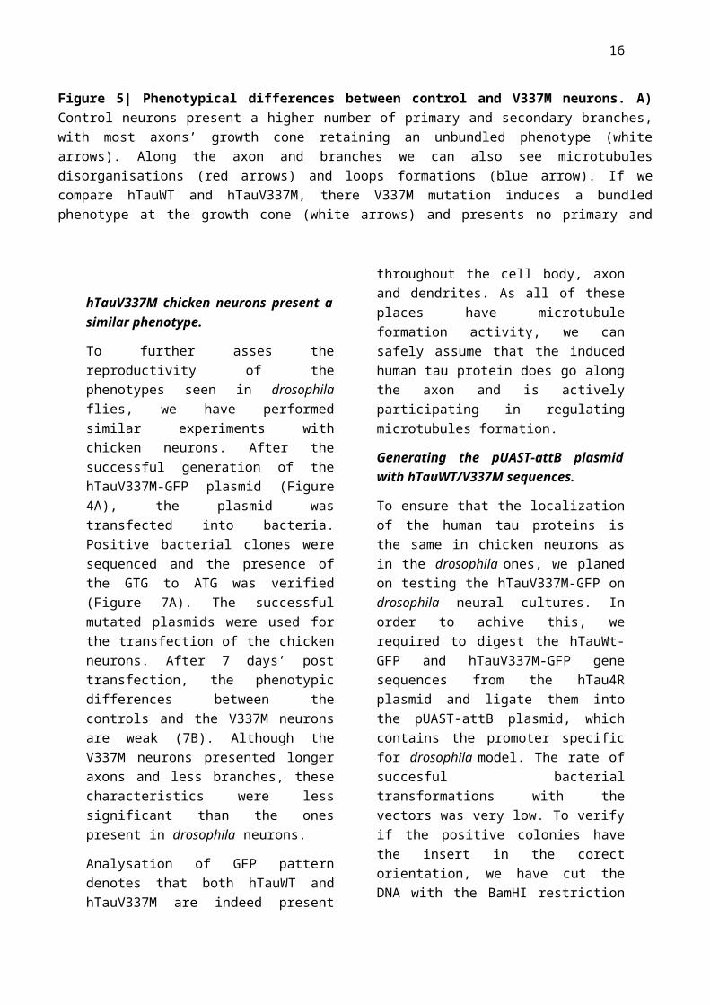
Figure 5| Phenotypical differences between control and V337M neurons. A) Control neurons present a higher number of primary and secondary branches, with most axons’ growth cone retaining an unbundled phenotype (white arrows). Along the axon and branches we can also see microtubules disorganisations (red arrows) and loops formations (blue arrow). If we compare hTauWT and hTauV337M, there V337M mutation induces a bundled phenotype at the growth cone (white arrows) and presents no primary and secondary branches. B) Statistical analysis of primary and secondary branches (Mann Whitney Test) demonstrate that there is significant loss of branching ability between the control (hTauWT) and the mutated neurons. The results shown are based on 2 independent repeat cultures.** p<0.001; *** p<0.0001; SEM; n-number of neurons analysed
13
Discussion
In this report, we have successfully reproduced V377M mutation in a drosophila neuronal model. Taken together, our results show that the induction of the human tau alone induces a certain amount of toxicity; the hTau cultures have shown constantly less branches, disorganisation and unbundle phenotype than the control neurons. These
results are consistent with the literature, as the addition of the extra tau brakes the balance of the endogenous expression tau levels. Overexpression of tau has been proven in different models to affect microtubule stability and increase cell toxicity [21,22]. We hypothesize that the extra human tau is either binding to microtubules and limiting their dynamics, or goes to the cell body and forms neurofibrillary tangles. Both theories may be proven correct as they are not mutually exclusive.
Figure 6| Statistical analysis results demonstrate a significant decrease in number of loops, unbundle phenotypes and disorganisation ratio between the V337M mutation and controls. For (A) and (B) Mann Whitney test was performed, while for (C) Fisher’s exact test. The results shown are based on 2 independent repeat cultures. ** p<0.001; *** p<0.0001; SEM; n-number of neurons analysed
14
After comparing the neurons expressing hTauWT versus hTauV337M, we can clearly distinguish a more elevated level of toxicity with the presence of the mutation. As we know that the V337M mutation affects the level of hyperphosphorylation of tau, we assume that this strong phenotype is partially due to the location of the tau protein following detachment from microtubes. This hypothesis was confirmed with the chicken neurons model, as we have clearly shown that the hTauV337M-GFP proteins added were
indeed localized along the axons, branches and dendrites. Further investigations into where do the hyperphosphorylated tau proteins localize could be performed using immunostaining techniques – staining for tau and tubulin would give us an estimation of the percent of tau binding to microtubules and the amount present in the soma. By comparing these data to the control neurons, we would be able to further confirm the localization of hTau proteins.
Figure 7| Transfection of chick neurons with hTauV337M-GFP and hTauWT-GFP plasmids. A) Sequencing results show the presence of mutation at base pair A (ATG codon) that transforms Valine into Methionine amino acid. B) Representation of a normal phenotype of control chick neurons (transfected with hTauWT). By comparison with V337M mutation neurons (C), the control phenotype presents a more branched phenotype. Moreover, small disorganisations within the axons (white arrows) are present.
15
Furthermore, performing a co-localization experiment via proximity labelling techniques would allow us to identify tau interactions with other proteins in its natural cellular environment. It would be interesting to perform the similar experiments without the endogenous expression of drosophila and chicken tau and with the hTauWT/V337M-GFP added and compare the results with our current experiments.
Mice models using V337M mutations in FTD have been performed [23,24], however none of the current research has been looking at the single neuronal phenotypical changes. Performing the experiments described above to a mouse neuronal cell culture would allow increase our confidence that the results shown to date are representative of other vertebrate models and may be translated to human pathology
Although both our experiments exhibit quite a distinctive phenotype, there are certain limitations to consider. Firstly, in the drosophila cultures, the genome integration of the UAS-hTauWT/V337M is random, which allows a different expression level between the two groups. This limitation is also present in the chicken experiments as each neuron
that was transfected could have uptake different amounts of the plasmid, which also would generate changes in expression of the human tau. This issue could be minimalized by performing a qPCR and/or Western Blotting experiments to verify approximately if such differences in expression are significant. Additionally, performing several more experiments using different crosses and normalize the results would attenuate further any differences between the levels of tau expression. Another factor to consider is that experiments performed were analysed after 6-7 days, however the effects on neurodegeneration require longer for chicken neurons than drosophila. Consequently, repeating the chicken neuronal cultures for a longer period of time would allow a better quantification of the level of degeneration seen. Finally, improvements could be made in the generation of the pUAST-attB plasmid containing the hTauWT/V337M sequence. To obtain a higher rate of successful integration of the insert into the plasmid, the ligation step could be left overnight. Changing the ration of the insert to vector in the ligation may increase the ratio of integration. Although we cannot impede the insert to ligate in both directions, by raising the ligation rate we also
Figure 8| Digestion of positive bacterial clones. 16 different colonies were digested, with only 2 containing the plasmid with an insert and both present in the wrong orientation. L-ladder; NI- no insert; WO- wrong direction
WONI
16
increase the probability of obtaining a positive clone in the right orientation.
Conclusion
In this report, we have tried to answer a serial of questions related to the function and localization of V337M tau proteins and the mechanisms behind how it promotes neurodegeneration. We have successfully demonstrated that the mutated tau protein does go along the axons and dendrites and is binding to microtubules, however several questions remain to be answered. Acquiring a better understanding of how this mutation induces cell toxicity and degeneration is the first step towards a future therapy design for familial cases of V337M FTD cases.
Acknowledgments
I would like to thank Dr Natalia Sanchez and Dr Pilar Okenve for their help and support throughout this project.
References
1 Boxer, A. L. and Miller, B. L. (2005) Clinical features of frontotemporal dementia. Alzheimer Dis. Assoc. Disord. 19 Suppl 1, S3–S6.
2 SNOWDEN, J. S. (2002) Frontotemporal dementia. Br. J. Psychiatry 180, 140–143.
3 Goedert, M. and Spillantini, M. G. (2011) Pathogenesis of the tauopathies. In Journal of Molecular Neuroscience, pp 425–431.
4 Gauthier-Kemper, A., Weissmann, C., Golovyashkina, N., Sebö-Lemke, Z., Drewes, G., Gerke, V., Heinisch, J. J. and Brandt, R. (2011) The frontotemporal dementia mutation R406W blocks tau’s interaction with the membrane in an annexin A2-dependent manner. J. Cell Biol. 192, 647–661.
5 Noble, W., Hanger, D. P., Miller, C. C. J. and Lovestone, S. (2013) The importance of tau phosphorylation for neurodegenerative diseases. Front.
Neurol.
6 Dixit, R., Ross, J. L., Goldman, Y. E. and Holzbaur, E. L. F. (2008) Differential Regulation of Dynein and Kinesin Motor Proteins by Tau. Science (80-. ). 319, 1086–1089.
7 Jaworski, J., Kapitein, L. C., Gouveia, S. M., Dortland, B. R., Wulf, P. S., Grigoriev, I., Camera, P., Spangler, S. A., Di Stefano, P., Demmers, J., et al. (2009) Dynamic Microtubules Regulate Dendritic Spine Morphology and Synaptic Plasticity. Neuron 61, 85–100.
8 Tackenberg, C. and Brandt, R. (2009) Divergent Pathways Mediate Spine Alterations and Cell Death Induced by Amyloid- , Wild-Type Tau, and R406W Tau. J. Neurosci. 29, 14439–14450.
9 Andreadis, A. (2012) Tau splicing and the intricacies of dementia. J. Cell. Physiol.
10 Goedert, M. and Spillantini, M. G. (2006) A Century of Alzheimer’s Disease. Science (80-. ). 314, 777–781.
11 Drewes, G., Trinczek, B., Illenberger, S., Biernat, J., Schmitt-Ulms, G., Meyer, H. E., Mandelkow, E. M. and Mandelkow, E. (1995) Microtubule-associated protein/microtubule affinity-regulating kinase (p110mark): A novel protein kinase that regulates tau-microtubule interactions and dynamic instability by phosphorylation at the Alzheimer-specific site serine 262. J. Biol. Chem. 270, 7679–7688.
12 Hanger, D. P., Anderton, B. H. and Noble, W. (2009) Tau phosphorylation: the therapeutic challenge for neurodegenerative disease. Trends Mol. Med.
13 Eidenmüller, J., Fath, T., Maas, T., Pool, M., Sontag, E. and Brandt, R. (2001) Phosphorylation-mimicking glutamate

17
clusters in the proline-rich region are sufficient to simulate the functional deficiencies of hyperphosphorylated tau protein. Biochem. J. 357, 759–67.
14 Liu, F., Li, B., Tung, E. J., Grundke-Iqbal, I., Iqbal, K. and Gong, C. X. (2007) Site-specific effects of tau phosphorylation on its microtubule assembly activity and self-aggregation. Eur. J. Neurosci. 26, 3429–3436.
15 Alonso, A. D. C., Mederlyova, A., Novak, M., Grundke-Iqbal, I. and Iqbal, K. (2004) Promotion of hyperphosphorylation by frontotemporal dementia tau mutations. J. Biol. Chem. 279, 34873–34881.
16 Goedert, M., Spillantini, M. G., Jakes, R., Rutherford, D. and Crowther, R. A. (1989) Multiple isoforms of human microtubule-associated protein tau: sequences and localization in neurofibrillary tangles of Alzheimer’s disease. Neuron 3, 519–526.
17 Sumi, S. M., Bird, T. D., Nochlin, D. and Raskind, M. a. (1992) Familial presenile dementia with psychosis associated with cortical neurofibrillary tangles and degeneration of the amygdala. Neurology 42, 120–127.
18 Bird, T. D., Wijsman, E. M., Nochlin, D., Leehey, M., Sumi, S. M., Payami, H., Poorkaj, P., Nemens, E., Rafkind, M. and Schellenberg, G. D. (1997) Chromosome 17 and hereditary dementia: linkage studies in three non-Alzheimer families and kindreds with late-onset FAD. Neurology 48, 949–954.
19 Spina, S., Schonhaut, D. R., Boeve, B. F., Seeley, W. W., Ossenkoppele, R., O’Neil, J. P., Lazaris, A., Rosen, H. J., Boxer, A. L., Perry, D. C., et al. (2017) Frontotemporal dementia with the V337M MAPT mutation. Neurology 88, 758–766.
20 Domoto-Reilly, K., Davis, M. Y., Keene, C. D. and Bird, T. D. (2017) Unusually long duration and delayed penetrance in a family with FTD and mutation in MAPT (V337M). Am. J. Med. Genet. Part B Neuropsychiatr. Genet. 174, 70–74.
21 Gorsky, M. K., Burnouf, S., Dols, J., Mandelkow, E. and Partridge, L. (2016) Acetylation mimic of lysine 280 exacerbates human Tau neurotoxicity in vivo. Sci. Rep. 6, 22685.
22 Tracy, T. E., Sohn, P. D., Minami, S. S., Wang, C., Min, S. W., Li, Y., Zhou, Y., Le, D., Lo, I., Ponnusamy, R., et al. (2016) Acetylated Tau Obstructs KIBRA-Mediated Signaling in Synaptic Plasticity and Promotes Tauopathy-Related Memory Loss. Neuron 90, 245–260.
23 Tanemura, K., Murayama, M., Akagi, T., Hashikawa, T., Tominaga, T., Ichikawa, M., Yamaguchi, H. and Takashima, A. (2002) Neurodegeneration with tau accumulation in a transgenic mouse expressing V337M human tau. J. Neurosci. 22, 133–141.
24 Tanemura, K., Akagi, T., Murayama, M., Kikuchi, N., Murayama, O., Hashikawa, T., Yoshiike, Y., Park, J. M., Matsuda, K., Nakao, S., et al. (2001) Formation of filamentous tau aggregations in transgenic mice expressing V337M human tau. Neurobiol. Dis. 8, 1036–1045.
25 Brunden, K. R., Trojanowski, J. Q. and Lee, V. M.-Y. (2009) Advances in tau-focused drug discovery for Alzheimer’s disease and related tauopathies. Nat. Rev. Drug Discov. 8, 783–793.
26 Arendt, T., Stieler, J. T. and Holzer, M. (2016) Tau and tauopathies 126, 238–292.