Embed Size (px)
Citation preview

Recognizing Different Sports Injuries and The Healing Process

Key Terminology
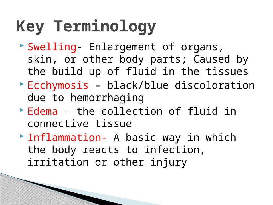
Swelling- Enlargement of organs, skin, or other body parts; Caused by the build up of fluid in the tissues
Ecchymosis – black/blue discoloration due to hemorrhaging
Edema – the collection of fluid in connective tissue
Inflammation- A basic way in which the body reacts to infection, irritation or other injury
Key Terminology

Modality - A therapeutic method or agent Analgesic – agent that relieves pain w/out
causing a complete loss of sensation Vasoconstriction – decrease in the diameter
of the blood vessel Vasodilation – Increase in the diameter of
the blood vessel Erythema – redness of the skin
Key Terminology

Everyone copes with pain differently. Pain is as much psychological as
physiological. Pain results from sensory input received
through the nervous system and indicates location of tissue damage.
Pain is not a useful indicator of injury severity.
Pain and Acute Injury

Cryotherapy ◦ Appropriate immediately following an injury (20min)◦ Direct application of cold causes vasoconstriction
in the affected tissues Constriction of the surrounding blood vessels
◦ Why?? Analgesic – decreases pain reduces muscle spasm (involuntary contraction of
muscle) Decreases blood flow, decreases temperature = a
decrease in secondary swelling ◦ Ice bags or packs, aerosol coolants, ice cups, ice
water immersion/whirlpool
Modalities

Thermotherapy◦ Appropriate 48-72 hours post injury◦ Considered an analgesic◦ Why should you not use heat for an acute injury?
Increases blood flow, Increases temperature = an Increase in swelling = longer recovery time
Your goal is to decrease swelling when dealing with an acute injury
◦ Hydrocollator packs, paraffin wax, warm whirlpool and ultrasound
Modalities

Non-Steroidal Anti-Inflammatory Drug
NSAIDs are very popular drugs. They work to decrease inflammation Common NSAIDs include Aspirin, Ibuprofen, Motrin,
Aleve, and Advil Rx (prescription) or OTC (over the counter)
NSAIDs

Acute◦ Result of sudden trauma; Isolated event◦ Examples???
Chronic◦ Caused by repetitive, overuse activities◦ Examples???
Acute or chronic in nature

Acute Traumatic Injuries
Fractures Result of extreme stress
and strain on bone – breaks bone
Bone Anatomy◦ Diaphysis -shaft
hollow and cylindrical
◦ Epiphysis - composed of cancellous bone and has hyaline cartilage covering
◦ Periosteum - dense, white fibrous covering contains blood vessels and
osteoblasts

Acute Fractures
Partial or complete Either closed or
open (through skin) Presents with
deformity, point tenderness, swelling and pain with movement

Load Characteristics
Bones can be stressed or loaded through: Tension Compression Bending twisting shearing
Greater force = more severe fracture
Some bones will require more force than others
Mechanical Forces of Injury
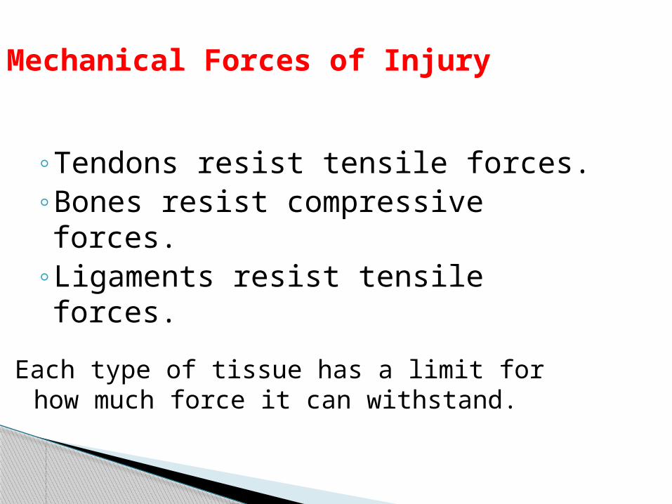
◦Tendons resist tensile forces.◦Bones resist compressive forces.◦Ligaments resist tensile forces.
Each type of tissue has a limit for how much force it can withstand.

Healing of a Fracture
◦ Generally requires immobilization (boot or cast) usually 6-8 wks
◦ Following cast removal, normal stresses and strains will aid in healing = Rehabilitation

Possible causes◦ Overload due to muscle contraction, ◦ Altered stress distribution due to muscle fatigue◦ Changes in surface◦ Too much activity too soon or in general
Begins with a dull ache becoming worse over time
Initially pain during activity and then progresses to pain following activity
Early detection is difficult, bone scan is useful X-ray is effective after several weeks If suspected, Stop activity and refer to
Orthopedic
Stress Fractures (fx)


• Dislocations– At least one bone in a joint is forced completely out of
the respected joint–Most common at the fingers, ankle, and shoulders
• Subluxations – Partial dislocation of the joint. Usually reducing itself
because the bone did not come completely out of the joint.
– Often occurs at the shoulder and patella• Signs and symptoms– Deformity – Swelling– Subluxation – pain and the sensation that it ‘came out’
of place
Dislocation’s and Subluxation’s

Signs and symptoms continued…
– Other factors associated with dislocations – 1) loss of limb function2) swelling and point tenderness
– Additional concerns• Avulsion fractures, ligament/muscular damage• “Once a dislocation, always a dislocation”
– Treatment (tx)• Reduction- generally done by a Physician• Dislocations (particularly first time) should always
be considered and treated as a fracture until ruled out
• X-ray is a MUST• Return to play often determined by extent of soft
tissue damage

Damage to a ligament (provides support to a joint)◦Connects a bone to another bone◦Extreme twist or rotation of the joint results
in the stretching or tearing of the ligament
Ligament Sprains

Grading System◦ Grade/degree 1 – Stretching of the Ligament
some pain, minimal loss of function, no abnormal motion, and mild point tenderness, slight swelling and joint stiffness
◦ Grade/degree II – Stretching and some tearing of ligament pain, moderate loss of function, swelling, and
instability, some tearing of ligament fibers ◦ Grade/degree III – Tearing/rupture of the
ligament Severe pain, significant loss of function, severe
instability and swelling, and may also represent subluxation


Ligament Sprains
Restoration of joint stability is difficult with grade I and II injuries◦ Must rely on other structures around the joint
Rely heavily on muscles surrounding joint◦ Ligament has been stretched/partially torn causing
development of inelastic scar Ligament will not regain original tension
Rehab = strengthening = improved joint stability

◦ Result of sudden blow to body, blunt force trauma◦ Deep or superficial◦ Hematoma = blood and lymph flow of tissue
bleeding results in discoloration of skin◦ Must be cautious of repeated blows to same area
Myositis Ossificans Calcium deposits form within the soft tissue Protect the area with padding Quadriceps and biceps are common sites
Contusions (Bruise)


MOI◦ Stretch, tear or rupture of muscle or connective tissue
Signs and Symptoms◦ Hear/feel a pop - Tightness◦ Deformity - feels like they have been ◦ Pain ‘shot’ ◦ Decreased ROM/flexibility
Rehabilitation◦Lengthy process regardless of severity◦ 6-8 weeks◦Return to activity too soon= re-injury
Muscle Strains

Grades/severity
– Grade/Deg I - some fibers have been stretched or actually torn resulting in tenderness and pain on active ROM; movement painful but full ROM present
–Grade/Deg II – more fibers have been torn and contraction is painful; usually a depression or divot is palpable; some swelling and discoloration are present
– Grade/Deg III- Complete rupture of muscle or musculotendinous junction; usual loss of function; initial great deal of pain that diminishes due to nerve damage


Muscle Cramps – Painful involuntary contraction– Attributed to dehydration/electrolyte imbalance
and fatigue
Muscle Soreness–Overexertion in strenuous exercise resulting in pain– Two types of soreness• Acute-onset muscle soreness - accompanies
fatigue, and is transient muscle pain experienced immediately after exercise
• Delayed-onset muscle soreness (DOMS) - pain that occurs 24-48 hours following activity that gradually subsides (pain free 3-4 days later)– Caused by slight microtrauma to muscle

MOI◦ Two main causes
compression and tension Acute or Chronic
◦ Causes pain and result in sensory responses pinch, burn, tingle, muscle weakness, radiating pain
Minor → Severe → Life Altering◦ Healing process is very slow and long term
Tx – ◦ Referral, rest, Anti-inflammatories◦ No sport related activity until asymptomatic
Nerve Injuries

Constant Inflammation ◦ Essential part of healing process
Must occur following tissue damage to initiate healing
◦ Signs and Symptoms Pain, Redness Swelling Loss of function Increase in Temperature
◦ If source of irritation is not removed then inflammatory process becomes chronic
Chronic Overuse Injuries

Most common overuse injury S/S
◦ swelling and pain◦ Decreased function and ROM◦ May also experience crepitus
Key treatment = rest◦ Maintain fitness through non-
weight bearing (NWB) or non impact activities Swimming, bike and elipitical
Tendinitis

Inflammation of synovial sheath◦ Area of the tendon that’s subject to ↑ of friction
Acute = sudden onset, crepitus, and diffuse swelling Chronic = thickening of tendon with pain and
crepitus Common in the long flexor tendons of
fingers Tx
◦ same as Tendinitis◦ NSAID’s
Tenosynovitis

– Bursa• Fluid filled sac found in areas of friction• Knees, shoulders, ankles, elbows, etc
– Acute bursitis• Sudden traumatic injury
– Chronic bursitis• Overuse and constant external trauma
– S/S– Swelling = increase in pressure due to limited
space around anatomical structures– Pain, and some loss of function
Bursitis

The Healing Process

Must understand the sequence and time frame of the various phases of healing
Interference with healing process will delay return to full activity
Need optimal healing environment◦ Little can be done to speed the process, while
much can be done to impede it
Importance of the Healing Process Following Injury

Inflammatory Fibroblastic (Repair) Maturation - Remodeling
3 Phases

Inflammatory Phase: 5 Cardinal signs of Inflammation◦ Swelling◦ Loss of function
◦ Pain
◦ Reddening of skin (erythema).
◦ Warmth – an increase in temperature of the affected area.

Severity of injury Swelling Muscle spasm Atrophy Infection Age Health/nutrition
Factors that interfere with Healing

Begins immediately following injury Without the inflammatory phase the other phases
will not occur◦ Vasoconstriction initially, followed by vasodilation◦ Damage to blood vessels results in blood flow into
interstitial spaces causing a hematoma.◦ Blood clot if formed to stop bleeding◦ Plasma proteins, platelets, and leukocytes move
out of capillaries and into damaged tissue. Leukocytes engage in phagocytosis
Phagocytosis occurs to clean the injured area ‘eats’ the debris and injured tissue
Inflammatory Phase

◦ Chemical mediators are released to facilitate healing
◦ Symptomatically presents with the 5 cardinal signs of inflammation
The acute inflammatory process results in a walling off of the damaged area from the rest of the body.
The process acts to clean up the debris and provide components for healing.
Inflammation phase lasts 2-4 days
Inflammatory Phase

Proliferative and regenerative activity occurs resulting in scar formation ◦ Special leukocytes migrate to the area which
break down debris and prepare the area for regeneration and repair.
◦ Unorganized development of scar tissue/collagen is laid down The collagen fibers are laid down randomly As the tissue continues to proliferate the area of injury
becomes stronger Therefore the tensile strength of wound ↑rapidly in
proportion to collagen synthesis This signals the beginning of the next phase
Fibroblastic Repair Phase

◦ Occurs within initial hours of injury and continues up to 4-6 weeks
◦ S&S of inflammatory phase subside Athlete will still experience some tenderness and
pain with motion With development of scar - complaints of pain and
tenderness will decrease Scar tissue can be 95% as strong as the original
tissue. Stress on the tissue is helpful for rehabilitation; exercises are critical to this process.
Fibroblastic Repair Phase

Long-term process◦ Re-alignment of scar tissue according to tensile
forces acting on tissue Re-align to position of maximum efficiency (parallel
to lines of tension)◦ Tissue gradually resumes normal appearance and
function◦ After 3 weeks
Firm, strong, contracted, nonvascular scar exists◦ Maturation may take several years to be totally
complete
Maturation-Remodeling Phase