Embed Size (px)
Citation preview

Bones and the SkeletonOsseus tissues

Function Support
› Allows us to stand erect. Protection
› Ribs protect the heart and lungs. Movement
› Serve as attachment for skeletal muscles Storage
› Mineral, including calcium Blood Cell Formation
› hematopoiesis

Tissue Types
Compact (dense) bone› Looks smooth and homogeneous
Spongy (cancellous) bone› Composed of small needlelike or flat pieces
called trabeculae.› Has a lot of open space.

Structure, microscopic Compact
bone:› Filled with
canals and passages for nerves and vessels.
› Structural unit is the osteon or Haversian system.

Structure, microscopic
Spongy Bone› Consists of
trabeculae.› No osteons are
present.

Structure

Structure, Cont.
Diaphysis- shaft of the long bone. Consists of a collar of compact bone surrounding a medullary cavity.
Epiphyses- ends Epiphyseal Line- remainder of the
growth plate. (cartilage) Endosteum- thin membrane lining the
medullary cavity. Contains osteoblasts and osteoclasts.

HEM
ATO
PO
IESIS
Click icon to add picture
Red marrow is found in long bones and flat bones. This is the area where blood cell production takes place.

Bone Marrow Donation
Some illnesses can only be cured with a bone marrow transplant.
What do you think you know about this process?

Bone Homeostasis
Remodeling› Coordinated by packets of osteoblasts and
osteoclasts called remodeling units.› In healthy adults, bone mass remains
constant.› Controled by 2 loops
1. mechanical and gravitational forces 2. negative feedback hormonal mechanism.
blood calcium release of PTH release of Ca from bone by osteoclasts.

Hom
eosta
sis
: rep
air
Types o
f fractu
res.
Phase
s of fra
cture
healin
g.
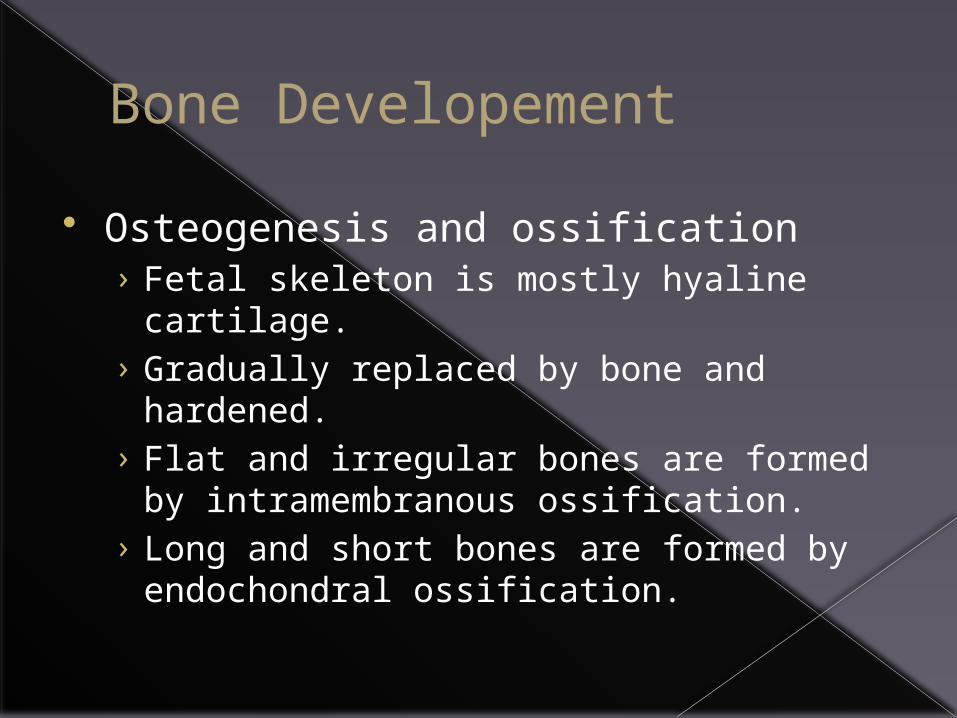
Bone Developement
Osteogenesis and ossification› Fetal skeleton is mostly hyaline cartilage.› Gradually replaced by bone and hardened.› Flat and irregular bones are formed by
intramembranous ossification.› Long and short bones are formed by
endochondral ossification.

Bone Growth
Longitudinal› Growth at the epiphyseal plate by mitosis.› Grow in length.
Appositional› Increases bone thickness.
*Growth hormone released during infancy and childhood

Homeostatic Imbalances Osteoporosis
› Reduced by mass due to increased bone reabsorption.
Osteomalacia/ Rickets› Bones do not have enough calcium,
making them soft.› Caused by Vit. D deficiency.
Paget’s Disease› Excessive and abnormal bone formation.

CLA
SS
IFICATIO
N O
F BO
NES
BY
SH
APE
:
•Flat
•long
•Short
•irregular

Bone Markings: every bump and grove has a name and purpose.
Two types of bone markings:› Projections (aka processes) that grow out
from the bone
› Depressions (cavities) that indent the bone

Joint Projections
Condyle: Rounded articular projection
Head: bony expansion on a narrow neck
Facet: smooth, nearly flat articular surface
head
Facet

Joint Projections
Ramus: Armlike bar of bone

Projections for ligament/ tendon attachment.Crest: Narrow ridge
of bone (Line: smaller than a crest)
Epicondyle: Raised area on or above a condyle

Tubercle: Small rounded projection
Tuberosity: large rounded or roughened projection
Trochanter: very large, blunt projection
(only on femur)

Spine: Sharp, pointed projectionEx: sharp points on the throacic vertebrae.

Depressions
Allow blood vessels or nerves to pass through.› Meatus: (me - A-
tus) Canal or tube› Sulcus, Groove or
Furrow: a shallow depression

Depressions
Sinus: Cavity within a bone; filled with air and lined with mucous membranes
Foramen: Round or oval opening
Foramen Magnum

Depressions
Fossa: shallow basin
Fissure: narrow, slit-like opening

Review of markings:
Projections1) Condyle2) Head3) Facet4) Ramus5) Crest6) Epicondyle7) Tubercle8) Tuberosity9) Trochanter10) Spine
Depressions1) Meatus2) Fossa3) Fissure4) Sinus5) Sulcus or Groove
or Furrow

The Skeleton Axial: skull,
vertebral column and bony thorax.
Appendicular: hang from 2 yokelike, bony girdles anchored to axial skeleton.

The Skull
· Two sets of bones
· Cranium
· Facial bones
· Bones are joined by sutures
· Only the mandible is attached by a freely movable joint

The Skull

Bones of the Skull
Figure 5.11

Human Skull, Superior View
Figure 5.8

Human Skull, Inferior View
Figure 5.9

Slide 5.27a
The Fetal Skull
Copyright © 2003 Pearson Education, Inc. publishing as Benjamin Cummings
· The fetal skull is large compared to the infants total body length
Figure 5.13

Slide 5.27b
The Fetal Skull
Copyright © 2003 Pearson Education, Inc. publishing as Benjamin Cummings
· Fontanelles – fibrous membranes connecting the cranial bones
· Allow the brain to grow
· Convert to bone within 24 months after birth
Figure 5.13

Slide 5.26
The Hyoid Bone
Copyright © 2003 Pearson Education, Inc. publishing as Benjamin Cummings
· The only bone that does not articulate with another bone
· Serves as a moveable base for the tongue
Figure 5.12

Slide 5.28
The Vertebral Column
Copyright © 2003 Pearson Education, Inc. publishing as Benjamin Cummings
· Vertebrae separated by intervertebral discs
· The spine has a normal curvature
· Each vertebrae is given a name according to its location
Figure 5.14

Slide 5.29
Structure of a Typical Vertebrae
Copyright © 2003 Pearson Education, Inc. publishing as Benjamin Cummings
Figure 5.16

The spinal column contains 33 bones termed vertebrae. The spine has five regions.
Region # of Vertebrae Body Area Abbreviation Cervical 7 Neck C1 - C7Thoracic 12 Chest T1 - T12Lumbar 5 or 6 Low Back L1 - L5Sacrum 5 (fused) Pelvis S1 - S5Coccyx 3 Tailbone None

TH
E A
TLA
S A
ND
TH
E A
XIS
Atlas (C1)› first cervical vertebra, C1. › supports the skull.› It looks different than other
vertebrae. The Atlas is a ring-shaped bone.
Axis (C2)› second cervical vertebra, C2.› It looks like a dull tooth and
sticks upward into the ring of the Atlas. The dens or tooth extends up from the C2 and joins with the inside of the C1 ring.
**The Atlas and Axis allow the head to turn from side to side.

Slide 5.31a
The Bony Thorax
Copyright © 2003 Pearson Education, Inc. publishing as Benjamin Cummings
· Forms a cage to protect major organs
Figure 5.19a

Slide 5.31b
The Bony Thorax
Copyright © 2003 Pearson Education, Inc. publishing as Benjamin Cummings
· Made-up of three parts
· Sternum
· Ribs
· Thoracic vertebrae
Figure 5.19a

Slide 5.32b
The Appendicular Skeleton
Copyright © 2003 Pearson Education, Inc. publishing as Benjamin Cummings
Figure 5.6c

Slide 5.33
The Pectoral (Shoulder) Girdle
Copyright © 2003 Pearson Education, Inc. publishing as Benjamin Cummings
· Composed of two bones
· Clavicle – collarbone
· Scapula – shoulder blade
· These bones allow the upper limb to have exceptionally free movement

Slide 5.34a
Bones of the Shoulder Girdle
Copyright © 2003 Pearson Education, Inc. publishing as Benjamin Cummings
Figure 5.20a, b

Slide 5.34b
Bones of the Shoulder Girdle
Copyright © 2003 Pearson Education, Inc. publishing as Benjamin Cummings
Figure 5.20c, d

Slide 5.35a
Bones of the Upper Limb
Copyright © 2003 Pearson Education, Inc. publishing as Benjamin Cummings
· The arm is formed by a single bone
· Humerus
Figure 5.21a, b

Slide 5.35b
Bones of the Upper Limb
Copyright © 2003 Pearson Education, Inc. publishing as Benjamin Cummings
• The forearm has two bones
• Ulna
• Radius
Figure 5.21c

Slide 5.36
Bones of the Upper Limb
Copyright © 2003 Pearson Education, Inc. publishing as Benjamin Cummings
· The hand
· Carpals – wrist
· Metacarpals – palm
· Phalanges – fingers
Figure 5.22

Slide 5.37
Bones of the Pelvic Girdle
Copyright © 2003 Pearson Education, Inc. publishing as Benjamin Cummings
· Hip bones· Composed of three pair of fused bones
· Ilium· Ischium· Pubic bone
· The total weight of the upper body rests on the pelvis
· Protects several organs· Reproductive organs· Urinary bladder· Part of the large intestine

Slide 5.38a
The Pelvis
Copyright © 2003 Pearson Education, Inc. publishing as Benjamin Cummings
Figure 5.23a

Slide 5.38b
The Pelvis
Copyright © 2003 Pearson Education, Inc. publishing as Benjamin Cummings
Figure 5.23b

Slide 5.39
Gender Differences of the Pelvis
Copyright © 2003 Pearson Education, Inc. publishing as Benjamin Cummings
Figure 5.23c

Slide 5.40a
Bones of the Lower Limbs
Copyright © 2003 Pearson Education, Inc. publishing as Benjamin Cummings
· The thigh has one bone
· Femur – thigh bone
Figure 5.35a, b

Slide 5.40b
Bones of the Lower Limbs
Copyright © 2003 Pearson Education, Inc. publishing as Benjamin Cummings
· The leg has two bones
· Tibia
· Fibula
Figure 5.35c

Slide 5.41
Bones of the Lower Limbs
Copyright © 2003 Pearson Education, Inc. publishing as Benjamin Cummings
· The foot
· Tarsus – ankle
· Metatarsals – sole
· Phalanges – toes
Figure 5.25

Slide 5.42
Arches of the Foot
Copyright © 2003 Pearson Education, Inc. publishing as Benjamin Cummings
· Bones of the foot are arranged to form three strong arches
· Two longitudinal
· One transverseFigure 5.26

Joints
Slide 5.43
Joints
Copyright © 2003 Pearson Education, Inc. publishing as Benjamin Cummings
· Functions
· Hold bones together
· Allow for mobility
· Ways joints are classified
· Functionally
· Structurally

Slide 5.44
Functional Classification of Joints
Copyright © 2003 Pearson Education, Inc. publishing as Benjamin Cummings
· Synarthroses – immovable joints
· Amphiarthroses – slightly moveable joints
· Diarthroses – freely moveable joints

Slide 5.45
Structural Classification of Joints
Copyright © 2003 Pearson Education, Inc. publishing as Benjamin Cummings
· Fibrous joints
· Generally immovable
· Cartilaginous joints
· Immovable or slightly moveable
· Synovial joints
· Freely moveable

Slide 5.46
Fibrous Joints
Copyright © 2003 Pearson Education, Inc. publishing as Benjamin Cummings
· Bones united by fibrous tissue
· Examples· Sutures
· Syndesmoses· Allows more
movement than sutures
· Example: distal end of tibia and fibula
Figure 5.27d, e

Slide 5.47
Cartilaginous Joints
Copyright © 2003 Pearson Education, Inc. publishing as Benjamin Cummings
· Bones connected by cartilage
· Examples
· Pubic symphysis
· Intervertebral joints
Figure 5.27b, c

Slide 5.48
Synovial Joints
Copyright © 2003 Pearson Education, Inc. publishing as Benjamin Cummings
· Articulating bones are separated by a joint cavity
· Synovial fluid is found in the joint cavity
Figure 5.27f–h

Slide 5.49
Features of Synovial Joints
Copyright © 2003 Pearson Education, Inc. publishing as Benjamin Cummings
· Articular cartilage (hyaline cartilage) covers the ends of bones
· Joint surfaces are enclosed by a fibrous articular capsule
· Have a joint cavity filled with synovial fluid
· Ligaments reinforce the joint

Slide 5.53
Inflammatory Conditions Associated with Joints
Copyright © 2003 Pearson Education, Inc. publishing as Benjamin Cummings
· Bursitis – inflammation of a bursa usually caused by a blow or friction
· Tendonitis – inflammation of tendon sheaths
· Arthritis – inflammatory or degenerative diseases of joints· Over 100 different types
· The most widespread crippling disease in the United States

Slide 5.54a
Clinical Forms of Arthritis
Copyright © 2003 Pearson Education, Inc. publishing as Benjamin Cummings
· Osteoarthritis· Most common chronic arthritis
· Probably related to normal aging processes
· Rheumatoid arthritis· An autoimmune disease – the immune system
attacks the joints
· Symptoms begin with bilateral inflammation of certain joints
· Often leads to deformities

Slide 5.54b
Clinical Forms of Arthritis
Copyright © 2003 Pearson Education, Inc. publishing as Benjamin Cummings
· Gouty Arthritis
· Inflammation of joints is caused by a deposition of urate crystals from the blood
· Can usually be controlled with diet