Embed Size (px)
Citation preview
1 23
Investigational New DrugsThe Journal of New Anticancer Agents ISSN 0167-6997Volume 34Number 4 Invest New Drugs (2016) 34:416-423DOI 10.1007/s10637-016-0354-7
Improved in vivo antitumor effect of adaunorubicin - GnRH-III bioconjugatemodified by apoptosis inducing agentbutyric acid on colorectal carcinomabearing miceBence Kapuvári, Rózsa Hegedüs, ÁkosSchulcz, Marilena Manea, József Tóvári,Alexandra Gacs, Borbála Vincze &Gábor Mező

1 23
Your article is published under the Creative
Commons Attribution license which allows
users to read, copy, distribute and make
derivative works, as long as the author of
the original work is cited. You may self-
archive this article on your own website, an
institutional repository or funder’s repository
and make it publicly available immediately.
PRECLINICAL STUDIES
Improved in vivo antitumor effect of a daunorubicin - GnRH-IIIbioconjugate modified by apoptosis inducing agent butyric acidon colorectal carcinoma bearing mice
BenceKapuvári1 &Rózsa Hegedüs2 &Ákos Schulcz1 &MarilenaManea3 & József Tóvári1 &
Alexandra Gacs1 & Borbála Vincze1 & Gábor Mező2
Received: 19 February 2016 /Accepted: 12 April 2016 /Published online: 5 May 2016# The Author(s) 2016. This article is published with open access at Springerlink.com
Summary Compared to classical chemotherapy, peptide-based drug targeting is a promising therapeutic approach forcancer, which can provide increased selectivity and decreasedside effects to anticancer drugs. Among various homing de-vices, gonadotropin-releasing hormone-III (GnRH-III) pep-tide represents a suitable targeting moiety, in particular in thetreatment of hormone independent tumors that highly expressGnRH receptors (e.g. colon carcinoma). We have previouslyshown that GnRH-III[4Lys(Ac),8Lys(Dau = Aoa)] bioconju-gate, in which daunorubicin was attached via oxime linkage tothe 8Lys of a GnRH-III derivative, exerted significant in vivoantitumor effect on subcutaneously developed HT-29 colontumor. In contrast, results of the study reported here indicatedthat this compound was not active on an orthotopically devel-oped tumor. However, if Lys in position 4 was acylated withbutyric acid instead of acetic acid, the resulting bioconjugateGnRH-III[4Lys(Bu),8Lys(Dau = Aoa)] had significant tumorgrowth inhibitory effect. Furthermore, it prevented tumor neo-vascularization, without detectable side effects. Nevertheless, thedevelopment of metastases could not be inhibited by the biocon-jugate; therefore, its application in combination with a metastasispreventive agentmight be necessary in order to achieve completetumor remission. In spite of this result, the treatment with GnRH-III[4Lys(Bu),8Lys(Dau = Aoa)] bioconjugate proved to have
significant benefits over the administration of free daunorubicin,which was used at the maximum tolerated dose.
Keywords Colon cancer . Targeted cancer therapy .
GnRH-III . Short chain fatty acids . Daunorubicin . In vivotumor growth inhibition
Introduction
Colorectal cancer is the third most common type of cancerworldwide, with nearly 1.4 million new cases diagnosed in2012. It is predicted that the number of cases will rise to 2.4million by 2035. About 54 % of colorectal cancer cases occurin more developed countries, with the highest incidence rate inEurope [1]. Thus, in addition to colon cancer prevention bychanging nutritional habits [2], the development of efficienttherapeutic strategies is of utmost importance. Peptide-basedtargeted tumor therapy, which has been investigated in the lastdecades, might be an effective targeted chemotherapeutic ap-proach to cure colon cancer as well [3]. Nevertheless, only afew research reports have been published in this field yet [4].
The principle of targeted tumor therapy relies on the struc-tural and/or functional differences between cancer cells andhealthy ones [5]. One of the possible targeted chemotherapeu-tic approaches is based on the attachment of an anticancerdrug to a targeting moiety, which recognizes tumor specificreceptors or cell surface structures that are highly expressed ontumor cells [6]. The homing device conjugated to the chemo-therapeutic agent enables the specific binding to the tumor cellsurface, without affecting the healthy tissues. Such targetingmoieties could be hormone peptides (e.g. gonadotropin releas-ing hormone (GnRH), somatostatin, bombesin), whose recep-tors are highly expressed on many types of tumors, while theirpresence on healthy tissues is limited [7].
Ákos Schulcz has passed away.
* Gábor Mező[email protected]
1 National Institute of Oncology, Budapest 1122, Hungary2 MTA-ELTE, Research Group of Peptide Chemistry, Pázmány P. stny.
1/A, Budapest 1117, Hungary3 Department of Chemistry and Zukunftskolleg, University of
Konstanz, 78457 Constance, Germany
Invest New Drugs (2016) 34:416–423DOI 10.1007/s10637-016-0354-7
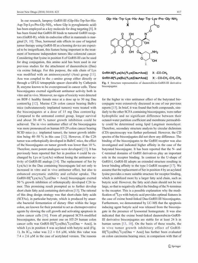
In our research, lamprey GnRH-III (Glp-His-Trp-Ser-His-Asp-Trp-Lys-Pro-Gly-NH2, where Glp is pyroglutamic acid)has been employed as a key molecule for drug targeting [8]. Ithas been found that GnRH-III binds to tumoral GnRH recep-tors (GnRH-R), while its endocrine effect in mammals is mar-ginal [9, 10]. Thus, hormonal side effects in case of targetedtumor therapy using GnRH-III as a homing device are expect-ed to be insignificant, this feature being important in the treat-ment of hormone independent tumors like colorectal cancer.Considering that lysine in position 8 of GnRH-III can be usedfor drug conjugation, this amino acid has been used in ourprevious studies for the attachment of daunorubicin (Dau)via oxime linkage. For this purpose, the side chain of Lyswas modified with an aminooxyacetyl (Aoa) group [11].Aoa was coupled to the ε-amino group either directly orthrough a GFLG tetrapeptide spacer cleavable by CathepsinB, enzyme known to be overexpressed in cancer cells. Thesebioconjugates exerted significant antitumor activity both invitro and in vivo. Moreover, no signs of toxicity were detectedon BDF-1 healthy female mice at a dose up to 30 mg Daucontent/kg [12]. Murine C26 colon cancer bearing Balb/cmice (subcutaneously implanted tumors) were treated withthe bioconjugates at a dose of 15 mg Dau content/kg.Compared to the untreated control group, longer survivaland about 30–40 % tumor growth inhibition could beachieved. The in vivo antitumor effect of the bioconjugateswas more pronounced on human HT-29 colon cancer bearingSCID mice (s.c. implanted tumor), the tumor growth inhibi-tion being 40–50 % in this case [12]. However, if the C26tumor was orthotopically developed in Balb/Cmice, the effectof the bioconjugates on tumor growth was lower than 10 %.Therefore, more potent analogues were developed [13]. It haspreviously been reported that Ser in position 4 could be ex-changed by Lys or Lys(Ac) without losing the antitumor ac-tivity of GnRH-III analogs [14]. The replacement of Ser byLys(Ac) in the Dau containing bioconjugate led not only toincreased in vitro and in vivo antitumor effect, but also toenhanced enzymatic stability and cellular uptake. TheGnRH-III[4Lys(Ac),8Lys(Dau = Aoa)] bioconjugate exerted50 % growth inhibition of orthotopically developed C26 tu-mor. This promising result prompted us to further developshort chain fatty acid containing derivatives [15]. The rationalof this drug design strategy was that short-chain fatty acids(SCFAs), in particular butyrate, which is produced by anaer-obe bacterial fermentation of dietary fiber within the largecolon, are known for their potential to act as chemopreventiveagents by slowing the cell growth and activating apoptosis incolon cancer cells [16]. From all prepared SCFA-modifiedbioconjugates, the most potent one on HT-29 human coloncancer cells was GnRH-III[4Lys(Bu),8Lys(Dau = Aoa)], inwhich Lys in position 4 was acylated with butyric acid (Fig.1). Its IC50 value was 2.2 ± 0.6 μM, while this value was7.4 ± 2.6 μM in the case of acetylated version. The reasons
for the higher in vitro antitumor effect of the butyrated bio-conjugate were extensively discussed in one of our previousreports [15]. In brief, it was found that both compounds, sim-ilarly to the other SCFA containing bioconjugates, were ratherhydrophilic and no significant difference between theiroctanol-water partition coefficient and membrane permeabili-ty could be determined using lipid Langmuir monolayer.Therefore, secondary structure analysis by circular dichroism(CD) spectroscopy was further performed. However, the CDspectra of the bioconjugates did not show any difference. Thebinding of the bioconjugates to the GnRH receptor was alsoinvestigated and indicated higher affinity in the case of thebutyrated bioconjugate. It has been reported that the N- andC-terminal domains of GnRH derivatives play an importantrole in the receptor binding. In contrast to the U-shape ofGnRH-I, GnRH-III adopts an extended structure resulting inlower binding affinity to the type I GnRH receptor [17]. Weassume that the replacement of Ser in position 4 by an acylatedlysine provides a more suitable structure for receptor binding,which is stabilized more by a larger fatty acid chain, such asbutyric acid. However, the fatty acid chain should not be toolarge, so that to negatively affect the binding of the N-terminusto the receptor. This is a possible explanation why the modi-fication of 4Lys with butyric acid provides optimal features inthe case of oxime bond-linked Dau-GnRH-III bioconjugates.Furthermore, we demonstrated by LC-MS that the apoptosisinducing agent butyric acid was released from the bioconju-gate in the presence of lysosomal homogenate. It was alsoindicated that the oxime bond-linked daunorubicin-GnRH-III derivative bioconjugates are stable for at least 24 h inhuman serum [13, 18]. On the basis of these results, thein vivo tumor growth inhibitory effect of GnRH-III[4Lys(Bu),8Lys(Dau = Aoa)] has further been evaluatedon colon carcinoma bearing mice, in comparison with that of
Fig. 1 Structure representation of daunorubicin-GnRH-III derivativebioconjugates
Invest New Drugs (2016) 34:416–423 417

GnRH-III[4Lys(Ac),8Lys(Dau = Aoa)] and the results are re-ported here.
Materials and methods
Daunorubicin-GnRH-III derivative bioconjugates
Daunorubicin-GnRH-III derivative bioconjugates were pre-pared by a combination of solid phase peptide synthesis andchemoselective ligation (e.g. oxime bond formation) as previ-ously described [13, 15]. The bioconjugates were purified byRP-HPLC using a mixture of 0.1 % TFA/water and 0.1 %TFA/MeCN-water (80:20, v/v). The freeze-dried bioconju-gates, without changing the tfa counter ions, were used inthe in vivo studies, in order to evaluate their tumor growthinhibitory effect on colon carcinoma bearing mice.
NSG mice
The immunodeficient NSG (Non Obese Diabetic SevereCombined ImmunoDeficient interleukine Gamma receptorchain knock-out) (NOD.Cg-Prkdcscid Il2rgtm1Wjl/SzJ) micewere originated from Jackson Laboratories. The mice wereheld in filter-top boxes in the experimental barrier roomsand every box-opening was done under a Class 100 laminar-flow hood by an operator dressed in sterilized surgical attire.The animal housing density was in accordance with the inter-national recommendations. The cage components, corn-cobbedding and food (VRF1 from Special Diet Services) weresteam-sterilized in autoclave (121 °C, 20 min). The distilledwater was acidified to pH 3 with hydrochloric acid.
The animals used in these studies were cared for accordingto the BGuiding Principles for the Care and Use of Animals^based upon the Helsinki declaration, and they were approvedby the local ethical committee. Our permission for breedingand performing experiments with laboratory animals is validuntil 2020/10/12 (registration numbers: 22.1/772/3/2010 andPEI/001/2574–6/2015).
Study of the chronic toxicity
NSG adult male mice, weighing 26–34 g, were used.Daunorubicin was dissolved in distilled water at a concentra-tion that allowed the dose to be given in a volume of 0.1 mL/10 g body weight. In this experiment, each group consisted of3 mice, which were treated by intraperitoneal (i.p.) adminis-tration on days 1, 4, 8, 12 and 16. The toxicity was assessed onthe basis of lifespan and body weight and the survival timewas followed for 20 days.
Development of the primary tumor
Female NSGmice, weighing 24–28 g, were used in this study.The xenografts were established by subcutaneous injection ofca. 106 HT-29 (GnRH receptor positive) human colon carci-noma cells, which were fromATCC and cultured according totheir standards. The mice with palpable tumors were killed bycervical-dislocation, disinfected with iodine, and the subcuta-neous tumor was dissected out aseptically. Tumor pieces of 2–3 mm3 were transplanted under aseptic conditions,orthotopically, into narcotized NSG mice.
Tumor transplantation
For direct implantation, mice were anesthetized (narcotic mix-ture: tiletamine, zolazepam, xylazine, butorphanol) and theabdomen was sterilized with iodine and alcohol swabs. Asmall midline incision (0.5 cm) was made and the colorectalpart of the intestine was exteriorized. Serosa of the site wherethe tumor pieces were to be implanted was removed. A pieceof 2 mm3 HT-29 human colon tumor was implanted on the topof the animal intestine; an 8/0 surgical (polypropylene) suturewas used to suture it on the wall of the intestine. The intestinewas returned to the abdominal cavity and the abdominal wallwas closed with 4/0 surgical (polyglycolic acid) sutures. Thewound was sterilized with iodine and alcohol swab again andthe animals were kept in a sterile environment. For pain re-lease and rehydration, mice were treated s.c. with 100 mg/kgalgopyrine in Ringer solution containing 5 % glucose. On thenext day, no signs of pain and stress of mice were observed.
Doses and treatments
Based on the results of the chronic toxicity study, we usedonly 1 mg (1.773 μmol)/kg free Dau, weekly, in the Dautreated group. Thirteen treatments were applied from the day5 after tumor transplantation until the day 50 in the groupstreated with the bioconjugates (every Monday and Thursday).The treatments were performed by i.v. (intravenous) or i.p.administration; the compounds were dissolved in distilled wa-ter (0.1 mL/10 g body weight).
In this experiment, 7 mice/group were used for the treatmentwith the bioconjugates (GnRH-III[4Lys(Ac),8Lys(Dau = Aoa)]and GnRH-III[4Lys(Bu),8Lys(Dau = Aoa)]) as well as in thecontrol group.
The bioconjugates were administered at different dosesduring the treatment period. In the first five treatments, theywere i.v. administered, at a dose of 28.435 μmol/kg bodyweight (15 mg Dau content in the bioconjugate/kg). Fromthe 6th to the 13th treatment, they were i.p. administered at adose of 14.217 μmol/kg body weight (7.5 mg Dau content inthe bioconjugate/kg). The first five doses represented the ini-tial round of treatments, which was followed by the
418 Invest New Drugs (2016) 34:416–423
maintenance therapy. In the maintenance treatments, 5 % glu-cose solution instead of distilled water was used, in order tominimize the harmful effects of tfa salts that were present inthe lyophilized bioconjugates.
The mice from all groups were sacrificed by deadly over-slept cocktail (0.3 mL/mouse concentrated nembutal(pentobarbitone) solution), on day 50 after tumortransplantation.
After that, the tumors were removed and the tumor weightwas determined in all cases.
Statistical analysis
The statistical analyses were performed by Medcalc® version12.1.3.0. (Broekstraat 52, B-9030 Mariakerke, Belgium)using the non-parametric Mann-Whitney (independent sam-ples) test. The experimental data were filtered by Gaussianstatistics g. P-values lower than 0.05 were considered statisti-cally significant.
Routine histological examination
The removed and fixed tissue samples were dehydrated in agraded series of ethanol, infiltrated with xylene and embeddedinto paraffin. Histological sections were stained with Harrishematoxylin and eosin (10:1, v/v, 1 % solutions) in acidified70 % ethanol and mounted. The histological samples wereexamined under a light microscope (Olympus CH30,Olympus Optical, Japan).
Determination of the proliferative indexand vascularization in orthotopic HT-29 tumor tissues
Before ending the experiment (30 min prior to sacrificing themice), the tumor bearing animals were injected i.p. with 5-bromo-2 V-deoxyuridine (BrdU, 200 mg/kg; Sigma-AldrichKft, Budapest, Hungary). After 6 h, 5–7 μm thick tissue sec-tions were excised from the newly frozen tumors and used forthe detection of BrdU positive cells using an anti-BrdUmono-clonal antibody (Becton Dickinson Hungary Kft., Budapest,Hungary), according to the manufacturer’s protocol. Positivecells were visualized with TRITC-conjugated anti-mouse IgG(1:100, Sigma). Endothelial cells were distinguished from tu-mor cells by treating the samples with rat anti-mouse CD31antibody and labelled with biotinylated anti-rat IgG andstreptavidin-FITC (Vector Laboratories, Burlingame, CA).The nuclei were stained with the Hoechst 33,342 dye(Molecular Probes, Eugene, OR). The number of labelledHT-29 tumor cells and the quantity of CD31-positive sampleswere determined in two different tumors by microscopic anal-ysis of 6 independent regions. The vascularization extent ofprimary tumors was determined by microscopic analysis oftwo different living tumors, where four independent areas of
living cells were counted and the percentage of separated en-dothelial cells covering a 10× magnification field of visionwas calculated [12].
Results
Chronic toxicity of daunorubicin on NSG healthy mice
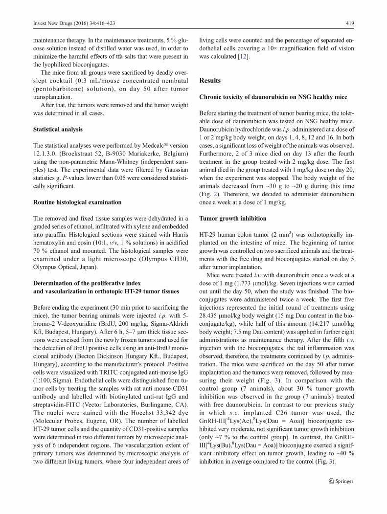
Before starting the treatment of tumor bearing mice, the toler-able dose of daunorubicin was tested on NSG healthy mice.Daunorubicin hydrochloride was i.p. administered at a dose of1 or 2 mg/kg body weight, on days 1, 4, 8, 12 and 16. In bothcases, a significant loss of weight of the animals was observed.Furthermore, 2 of 3 mice died on day 13 after the fourthtreatment in the group treated with 2 mg/kg dose. The firstanimal died in the group treated with 1 mg/kg dose on day 20,when the experiment was stopped. The body weight of theanimals decreased from ~30 g to ~20 g during this time(Fig. 2). Therefore, we decided to administer daunorubicinonce a week at a dose of 1 mg/kg.
Tumor growth inhibition
HT-29 human colon tumor (2 mm3) was orthotopically im-planted on the intestine of mice. The beginning of tumorgrowth was controlled on two sacrificed animals and the treat-ments with the free drug and bioconjugates started on day 5after tumor implantation.
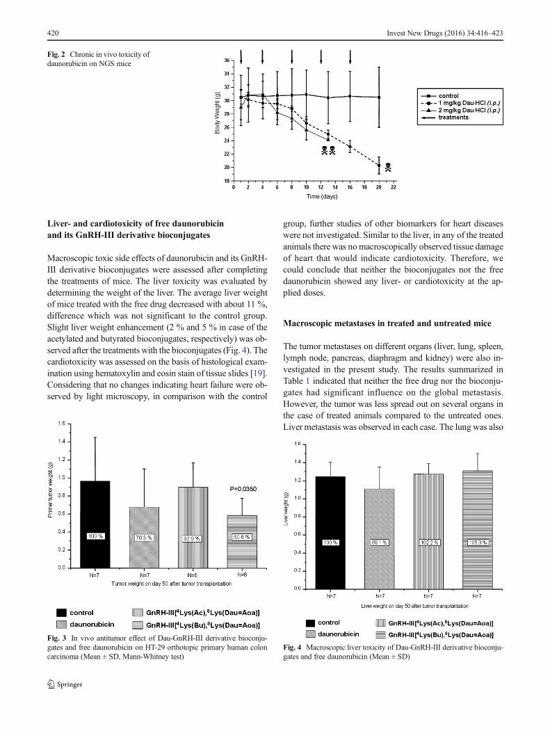
Mice were treated i.v. with daunorubicin once a week at adose of 1 mg (1.773 μmol)/kg. Seven injections were carriedout until the day 50, when the study was finished. The bio-conjugates were administered twice a week. The first fiveinjections represented the initial round of treatments using28.435 μmol/kg body weight (15 mg Dau content in the bio-conjugate/kg), while half of this amount (14.217 μmol/kgbody weight; 7.5 mg Dau content) was applied in further eightadministrations as maintenance therapy. After the fifth i.v.injection with the bioconjugates, the tail inflammation wasobserved; therefore, the treatments continued by i.p. adminis-tration. The mice were sacrificed on the day 50 after tumorimplantation and the tumors were removed, followed by mea-suring their weight (Fig. 3). In comparison with thecontrol group (7 animals), about 30 % tumor growthinhibition was observed in the group (7 animals) treatedwith free daunorubicin. In contrast to our previous studyin which s.c. implanted C26 tumor was used, theGnRH-III[4Lys(Ac),8Lys(Dau = Aoa)] bioconjugate ex-hibited very moderate, not significant tumor growth inhibition(only ~7 % to the control group). In contrast, the GnRH-III[4Lys(Bu),8Lys(Dau = Aoa)] bioconjugate exerted a signif-icant inhibitory effect on tumor growth, leading to ~40 %inhibition in average compared to the control (Fig. 3).
Invest New Drugs (2016) 34:416–423 419
Liver- and cardiotoxicity of free daunorubicinand its GnRH-III derivative bioconjugates
Macroscopic toxic side effects of daunorubicin and its GnRH-III derivative bioconjugates were assessed after completingthe treatments of mice. The liver toxicity was evaluated bydetermining the weight of the liver. The average liver weightof mice treated with the free drug decreased with about 11 %,difference which was not significant to the control group.Slight liver weight enhancement (2 % and 5 % in case of theacetylated and butyrated bioconjugates, respectively) was ob-served after the treatments with the bioconjugates (Fig. 4). Thecardiotoxicity was assessed on the basis of histological exam-ination using hematoxylin and eosin stain of tissue slides [19].Considering that no changes indicating heart failure were ob-served by light microscopy, in comparison with the control
group, further studies of other biomarkers for heart diseaseswere not investigated. Similar to the liver, in any of the treatedanimals there was nomacroscopically observed tissue damageof heart that would indicate cardiotoxicity. Therefore, wecould conclude that neither the bioconjugates nor the freedaunorubicin showed any liver- or cardiotoxicity at the ap-plied doses.
Macroscopic metastases in treated and untreated mice
The tumor metastases on different organs (liver, lung, spleen,lymph node, pancreas, diaphragm and kidney) were also in-vestigated in the present study. The results summarized inTable 1 indicated that neither the free drug nor the bioconju-gates had significant influence on the global metastasis.However, the tumor was less spread out on several organs inthe case of treated animals compared to the untreated ones.Liver metastasis was observed in each case. The lung was also
Fig. 2 Chronic in vivo toxicity ofdaunorubicin on NGS mice
Fig. 3 In vivo antitumor effect of Dau-GnRH-III derivative bioconju-gates and free daunorubicin on HT-29 orthotopic primary human coloncarcinoma (Mean ± SD, Mann-Whitney test)
Fig. 4 Macroscopic liver toxicity of Dau-GnRH-III derivative bioconju-gates and free daunorubicin (Mean ± SD)
420 Invest New Drugs (2016) 34:416–423
invaded by tumors almost in all tested animals. The observedmetastases on spleen were not significant in any group.Significant improvement in preventing metastasis was detect-ed on pancreas and lymph nodes in the treated groups com-pared to the control. The improvement on the latter one wasmore pronounced when the animals were treated with thebioconjugates. However, the diaphragm was more affectedby tumors , pa r t i cu l a r l y in the case o f GnRH-III[4Lys(Ac),8Lys(Dau = Aoa)] treated mice.
Tumor proliferation and vascularization
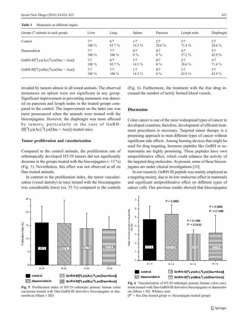
Compared to the control animals, the proliferation rate oforthotopically developed HT-29 tumors did not significantlydecrease in the groups treated with the bioconjugates (~15 %)(Fig. 5). Nevertheless, this effect was not observed at all onDau treated animals.
In contrast to the proliferation index, the tumor vasculari-zation (vessel density) in mice treated with the bioconjugateswas considerable lower (ca. 35 %) compared to the controls
(Fig. 6). Furthermore, the treatment with the free drug in-creased the number of newly formed blood vessels.
Discussion
Colon cancer is one of the most widespread types of cancer indeveloped countries; therefore, development of efficient treat-ment procedures is necessary. Targeted tumor therapy is apromising approach to treat different types of cancer withoutsignificant side effects. Among homing devices that might beused for drug targeting, hormone peptides like GnRH or so-matostatin are highly promising. These peptides have ownantiproliferative effect, which could enhance the activity ofthe targeted drug molecules. At present, some of these biocon-jugates are under clinical investigations [20].
In our research, GnRH-III peptide was mainly employed asa targeting moiety, due to its low endocrine effect in mammalsand significant antiproliferative effect on different types ofcancer cells. Our previous results showed that bioconjugates
Table 1 Metastasis on different organs
Groups (7 animals in each group) Liver Lung Spleen Pancreas Lymph node Diaphragm
Control 7/7100 %
6/785.7 %
1/714.3 %
2/728.6 %
5/771.4 %
2/728.6 %
Daunorubicin 7/7100 %
7/7100 %
0/70 %
0/70 %
4/757.2 %
3/742.9 %
GnRH-III[4Lys(Ac),8Lys(Dau = Aoa)] 7/7100 %
6/785.7 %
1/714.3 %
0/70 %
2/728.6 %
5/771.4 %
GnRH-III[4Lys(Bu),8Lys(Dau = Aoa)] 7/7100 %
7/7100 %
1/714.3 %
0/70 %
3/742.9 %
3/742.9 %
Fig. 5 Proliferation index of HT-29 orthotopic primary human coloncarcinoma treated with Dau-GnRH-III derivative bioconjugates or dau-norubicin (Mean ± SD)
Fig. 6 Vascularization of HT-29 orthotopic primary human colon carci-noma treated with Dau-GnRH-III derivative bioconjugates or daunorubi-cin (Mean ± SD, Whitney test)(Px = free Dau treated group vs. bioconjugate treated group)
Invest New Drugs (2016) 34:416–423 421
in which daunorubicin was attached to the side chain of Lys inposition 8 via oxime linkage exerted significant antitumoreffect both in vitro and in vivo. However, the efficacy of thebioconjugates was highly dependent on the tumor type andwhether this was subcutaneously or orthotopically developedin mice. Orthotopically developed tumors represent the nativeconditions much better than the subcutaneously implantedones. In this case, also the tumor metastases and the vascular-ization of primary tumors could occur easier than those ofisolated s.c. implanted tumors. Therefore, in the present study,HT-29 human colon tumor was implanted on the intestine ofimmunodeficient NSG mice. Two Dau-GnRH-III derivativebioconjugates that showed high antitumor activity in vitrowere selected for the treatment. These compounds containeda GnRH-III derivative in which Ser in position 4 was modifiedby Lys acylated with acetic or butyric acid (Fig. 1). Thesebioconjugates were not toxic on mice up to 30 mg daunoru-bicin content/kg body weight. However, the maximum toler-ated dose of free daunorubicin was 1–2 mg/kg/week in immu-nodeficient tumor bearing mice. According to the in vivochronic toxicity study, we used only 1 mg/kg free Dau, week-ly. In contrast to this, the bioconjugates were administered twotimes per week, at a dose of 15 mg Dau content in bioconju-gate/kg, five times as initial treatment and then 7.5 mg for themaintenance treatment. The results of this study indicated thatthe acetylated version of Dau-GnRH-III bioconjugate had noeffect on this type of tumor (Fig. 3). Interestingly, it has pre-viously shown significant antitumor effect (50 % tumorgrowth inhibition) on an orthotopically developed C26 mousecolon tumor [14]. A possible explanation of this result mightbe the difference in the doubling time of cancer cells. The C26tumor is an aggressive and fast proliferating tumor type, whileHT-29 grows much slower. In contrast to this, the butyric acidmodified bioconjugate exerted higher tumor growth inhibitionthan the free daunorubicin (40 % and 30 % to the control,respectively). The increased tumor growth inhibition ofGnRH-III[4Lys(Bu),8Lys(Dau = Aoa)] compared to GnRH-III[4Lys(Ac),8Lys(Dau = Aoa)] might partially be explainedby the higher receptor binding affinity and cellular uptake ofthe butyrated version, as determined in previous in vitrostudies [15].
The toxic side effects of daunorubicin and bioconjugateson different organs were also evaluated. Particular interest wasin determining the liver and heart toxicity, because of the wellknown cardiotoxity of anthracyclines. Except for a moderate(11 %) liver shrinkage in the case of daunorubicin treatment(Fig. 4), neither the bioconjugates nor the free daunorubicinshowed any toxicity at the applied doses.
The number of dividing cells in tumor tissues as well as thetissue vascularization are important factors of tumor growth.Therefore, the proliferative index was calculated and the new-ly formed blood vessels in the tumor were counted. In the caseof mice treated with the bioconjugates, the proliferative index
was lower than that of control animals and mice treated withthe free drug. However, the decrease was not significant.Interestingly, the number of blood vessels was considerablylower in the case of mice treated with the bioconjugates (64–67 % to the control), while a slightly increased number wasobserved in the case of daunorubicin treated animals (112 %).This might explain the higher tumor growth inhibition in-duced by the bioconjugate and could be an important aspectin its beneficial effect over the application of the free drug.
In conclusion, we clearly showed that GnRH-III[4Lys(Bu),8Lys(Dau = Aoa)] bioconjugate, in which dauno-rubicin was attached via a stable oxime linkage to 8Lys andSer in position 4 of the homing peptide was replaced byLys(Bu), provided benefits over the free drug in the treatmentof colon carcinoma. This bioconjugate exerted significantlyhigher tumor growth inhibitory effect on orthotopically devel-oped human HT-29 colon cancer bearing mice than the freeDau at the maximum tolerated dose. Furthermore, the biocon-jugate slightly decreased the tumor proliferation index andreduced the vascularization of tumor tissues at a high level.The bioconjugate did not show toxic side effects; however,similarly to the free drug it was not able to prevent metastaseson different organs.
Taken together these results, we can conclude that GnRH-III[4Lys(Bu),8Lys(Dau = Aoa)] bioconjugate is a promisingcandidate for targeted chemotherapy of colon cancer.However, its combination with a metastasis preventing agentmight be necessary and should be investigated in futurestudies.
Acknowledgments This work was supported by grants from theHungarian National Science Fund (OTKA 104045 and K116295) andUniversity of Konstanz (Zukunftskolleg, Project 634/12).
This project has received funding from the European Union’s
Horizon 2020 research and innovation program under the MarieSklodowska-Curie grant agreement No 642004.
Compliance with ethical standards
Conflict of interest There are no conflicts of interest and source offunding.
Open Access This article is distributed under the terms of theCreative Commons Attribution 4.0 International License (http://creativecommons.org/licenses/by/4.0/), which permits unrestricted use,distribution, and reproduction in any medium, provided you give appro-priate credit to the original author(s) and the source, provide a link to theCreative Commons license, and indicate if changes were made.
References
1. http://www.wcrf.org/cancer_statistics/data_specific_cancers/colorectal_cancer_statistics.php
2. Gonzales JF, Barnard ND, Jenkins DJ, Lanou AJ, Davis B, Saxe G,et al. (2014) Applying the precautionary principle to nutrition andcancer. J Am Coll Nutr 33:239–246
422 Invest New Drugs (2016) 34:416–423
3. Singh Y, Palombo M, Sinko JP (2008) Recent trends in targetedanticancer prodrug and conjugate designe. Curr Med Chem 15:1802–1826
4. Szepesházi K, Schally AV, Halmos G (2007) LH-RH receptors inhuman colorectal cancers: unexpected molecular targets for exper-imental therapy. Int J Oncol 30:1485–1492
5. Yim KL, Cunningham D (2011) Targeted drug therapies and can-cer. Recent Results Cancer Res 185:159–171
6. Schally AV, Nagy A (2004) Chemotherapy targeted to cancersthrough tumoral hormone receptors. Trends Endocrinol Metab 15:300–310
7. Mező G, Manea M (2010) Receptor-mediated tumor targetingbased on peptide hormones. Expert Opin Drug Deliv 7:79–96
8. Manea M, Mező G (2013) lGnRH-III - a promising candidate foranticancer drug development. Protein Pept Lett 20:439–449
9. Millar RP (2005) GnRHs and GnRH receptors. Anim Reprod Sci88:5–28
10. Sower SA, Chiang Y-C, Lovas S, Conlon JM (1993) Primary struc-ture and biological activity of a third gonadotropin-releasing hor-mone from lamprey brain. Endocrinology 132:1125–1131
11. Szabó I, Manea M, Orbán E, Csámpai A, Bosze S, Szabó R, et al.(2009) Development of an oxime bond containing daunorubicin-gonadotropin-releasing hormone-III conjugate as a potential anti-cancer drug. Bioconjug Chem 20:656–665
12. ManeaM, Tóvári J, TejedaM, Schulcz A, Kapuvári B, Vincze B, etal. (2012) In-vivo antitumour effect of daunorubicin-GnRH-III de-rivative conjugates on colon carcinoma-bearing mice. Anti-CancerDrugs 23:90–97
13. ManeaM, Leurs U, Orbán E, Baranyai Z, Öhlschläger P, MarquardtA, et al. (2011) Enhanced enzymatic stability and antitumor activityof daunorubicin-GnRH-III bioconjugates modified in position 4.Bioconjug Chem 22:1320–1329
14. Mező I, Lovas S, Pályi I, Vincze B, Kálnay A, Turi G, et al. (1997)Synthesis of gonadotropin-releasing hormone III analogs.Structure-antitumor activity relationships,. J Med Chem 40:3353–3358
15. Hegedüs R, Manea M, Orbán E, Szabó I, Kiss E, Sipos E, et al.(2012) Enhanced cellular uptake and in vitro antitumor activity ofshort-chain fatty acid acylated daunorubicin-GnRH-III bioconju-gates. Eur J Med Chem 56:155–165
16. Beyer-Sehlmeyer G, Glei M, Hartmann E, Hughes R, Persin C,Böhm V, et al. (2003) Butyrate is only one of several growth inhib-itors produced during gut flora-mediated fermentation of dietaryfibre sources. Br J Nutr 90:1057–1070
17. Pappa EV, Zompra AA, Diamantopoulou Z, Spyranti Z, Pairas G,Lamari FN, et al. (2012) Structure-activity studies of lGnRH-IIIthrough rational amino acid substitution and NMR conformationalstudies. Biopolymers 98:525–524
18. Orbán E, Mezo G, Schlage P, Csík G, Kulić Z, Ansorge P, et al.(2011) In vitro degradation and antitumor activity of oxime bond-linked daunorubicin-GnRH-III bioconjugates and DNA-bindingproperties of daunorubicin-amino acid metabolites. Amino Acids41:469–483
19. Sharkey LC, Radin MJ, Heller RLK, Tobias A, Matise I, et al.(2013) Differential cardiotoxicity in response to chronic doxorubi-cin treatment in male spontaneous hypertension-heart failure(SHHF), spontaneously hypertensive (SHR), and Wistar Kyoto(WKY) rats. Toxicol Appl Pharmacol 273:47–57
20. Emons G, Gorchev G, Harter P, Wimberger P, Stähle A, Hanker L,et al. (2014) Efficacy and safety of AEZS-108 (LHRH agonistlinked to doxorubicin) in women with advanced or recurrentendometrial cancer expressing LHRH receptors: a multicen-ter phase 2 trial (AGO-GYN5). Int J Gynecol Cancer 24:260–265
Invest New Drugs (2016) 34:416–423 423