Embed Size (px)
Citation preview
Skeletal System: Day ThreeBone Formation, Growth, and Remodeling, Bone Diseases and Disorders
2
Bone Development and Growth
Bones form by replacing connective tissues in the fetus.
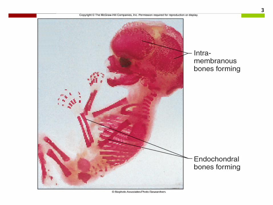
Some form within sheetlike layers of connective tissue (intramembranous bones), while others replace masses of cartilage (endochondral bones).
Ossification is another word for bone building.
CopyrightThe McGraw-Hill Companies, Inc. Permission required for reproduction or display.
3
4
Intramembranous Bones
The flat bones of the skull form as intramembranous bones that develop from layers of connective tissue.
Osteoblasts deposit bony tissue around themselves. Once osteoblasts deposit bone and are located in
lacunae, they are called osteocytes. Cells of the membranous connective tissue that lie
outside the developing bone give rise to the periosteum.
CopyrightThe McGraw-Hill Companies, Inc. Permission required for reproduction or display.
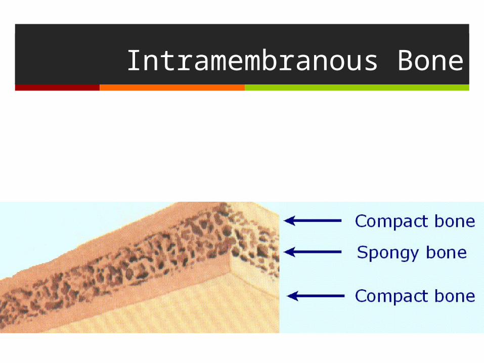
Intramembranous Bone
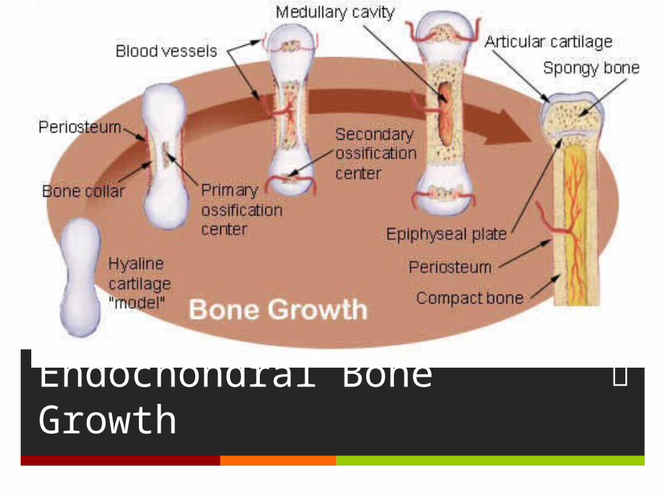
Endochondral Bone Growth
7
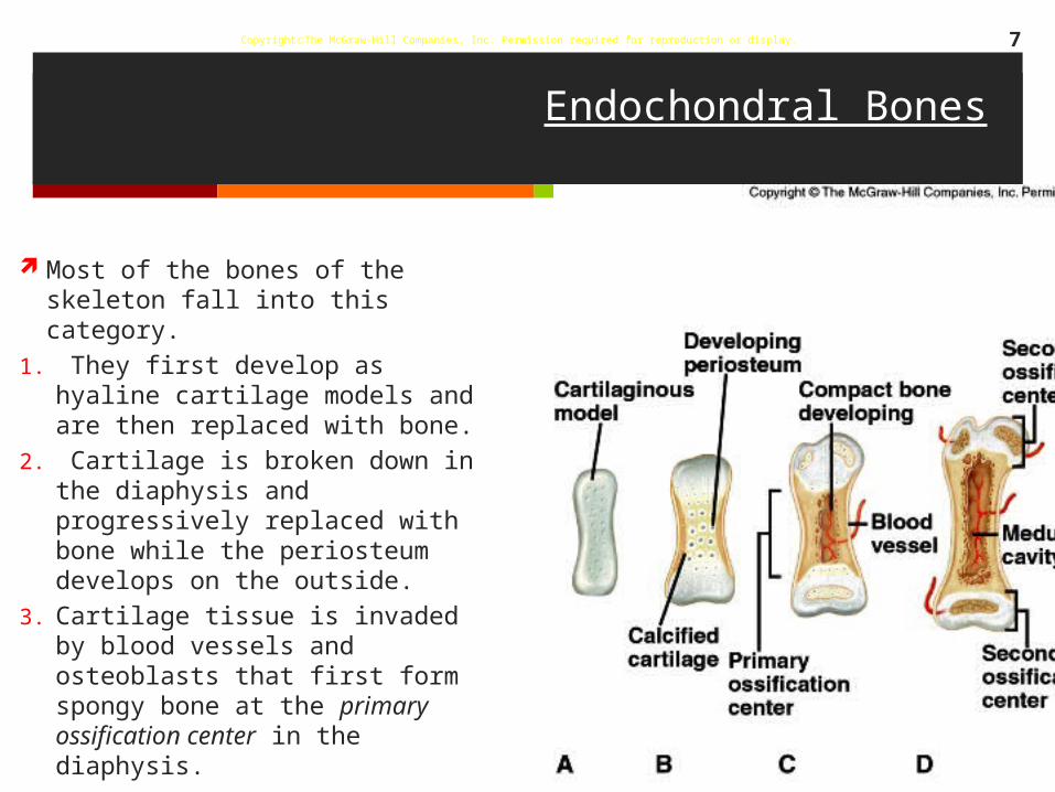
Endochondral Bones
Most of the bones of the skeleton fall into this category.
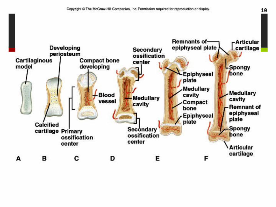
1. They first develop as hyaline cartilage models and are then replaced with bone.
2. Cartilage is broken down in the diaphysis and progressively replaced with bone while the periosteum develops on the outside.
3. Cartilage tissue is invaded by blood vessels and osteoblasts that first form spongy bone at the primary ossification center in the diaphysis.
CopyrightThe McGraw-Hill Companies, Inc. Permission required for reproduction or display.
8
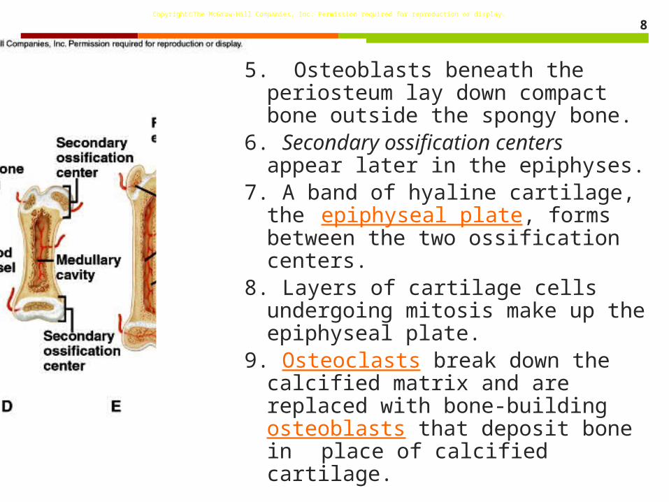
5. Osteoblasts beneath the periosteum lay down compact bone outside the spongy bone.
6. Secondary ossification centers appear later in the epiphyses.
7. A band of hyaline cartilage, the epiphyseal plate, forms between the two ossification centers.
8. Layers of cartilage cells undergoing mitosis make up the epiphyseal plate.
9. Osteoclasts break down the calcified matrix and are replaced with bone-building osteoblasts that deposit bone in place of calcified cartilage.
CopyrightThe McGraw-Hill Companies, Inc. Permission required for reproduction or display.
9
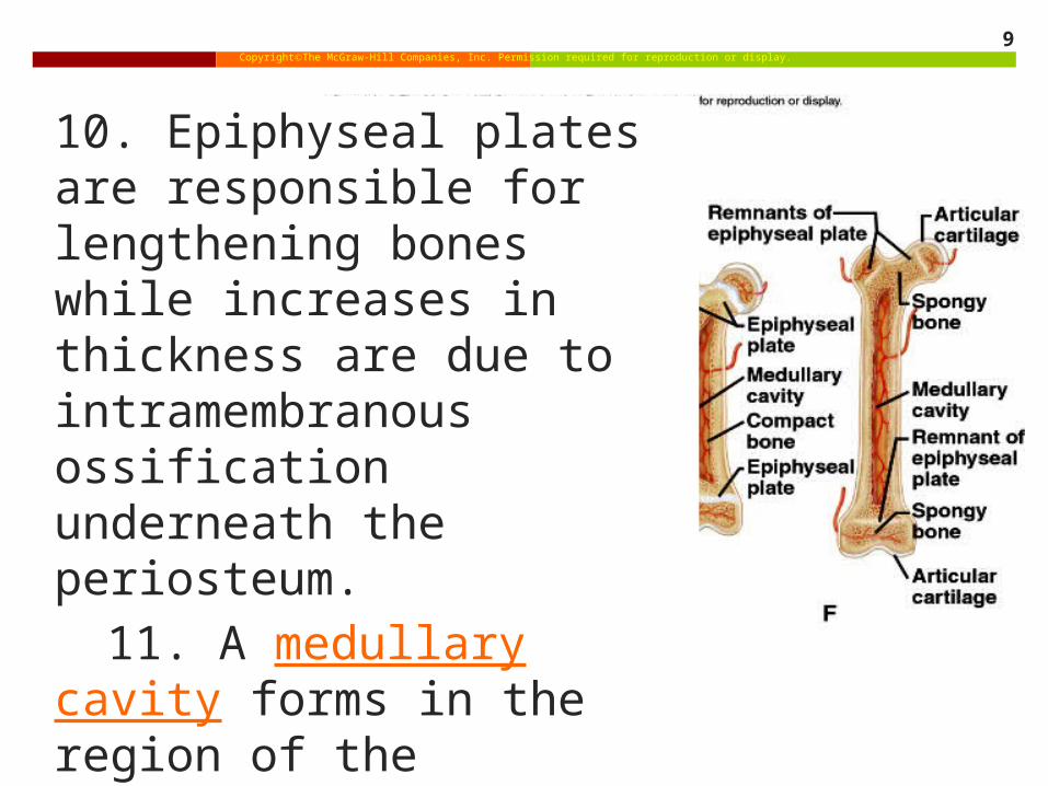
10. Epiphyseal plates are responsible for lengthening bones while increases in thickness are due to intramembranous ossification underneath the periosteum.
11. A medullary cavity forms in the region of the diaphysis due to the activity of osteoclasts.
CopyrightThe McGraw-Hill Companies, Inc. Permission required for reproduction or display.
10
Bone Formation: Ossification
By birth, most hyaline cartilage have been converted to bone except for two regions:
1. Articular cartilages• Cover long bone ends (epiphysis)• Persist for lifetime• Reduce friction at the joint surfaces
2. Epiphyseal Plate• Allows for longitudinal growth of long bones during
childhood• Will eventually disappear leaving only the epiphyseal
line
Bone Growth
New cartilage is continuously formed
Old cartilage is broken down and replaced by bony matrix
Process of long-bone growth controlled by hormones: growth hormone and sex
hormone Ends during adolescence when epiphyseal plate is converted
to bone
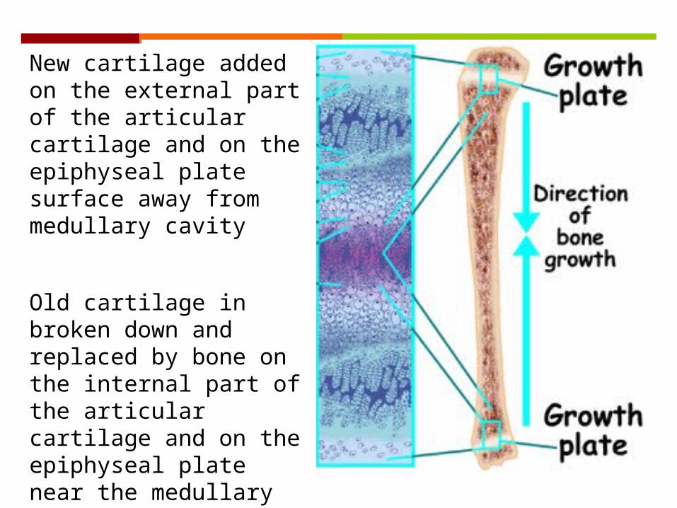
New cartilage added on the external part of the articular cartilage and on the epiphyseal plate surface away from medullary cavity
Old cartilage in broken down and replaced by bone on the internal part of the articular cartilage and on the epiphyseal plate near the medullary cavity
14
Homeostasis of Bone Tissue
Osteoclasts tear down and osteoblasts build bone throughout the lifespan with the processes of resorption and deposition, with an average of 3% to 5% of bone calcium exchanged annually.
CopyrightThe McGraw-Hill Companies, Inc. Permission required for reproduction or display.
Bone Remodeling
Bone is very active tissue
Bones are remodeled continuously in response to two factors:
1. Calcium levels in the blood2. Pull of gravity and muscles on the skeleton
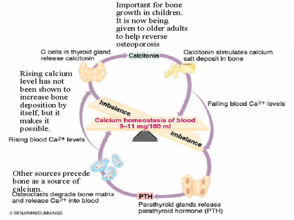
Bone Remodeling:Calcium levels in blood
Blood calcium low Parathyroid glands release parathyroid hormone (PTH) PTH activate osteoclasts (destroy bone cells) Osteoclasts break down bone and release calcium into the
blood
Blood calcium high Thyroid gland releases calcitonin Calcitonin stimulates Calcium to be deposited from the
blood into the bone
Bone Remodeling: Pull of gravity and muscles on the skeleton
Bones become thicker to increase strength where bulky muscles attached
Bones of physically inactive people tend to lose mass
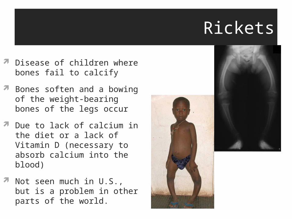
Rickets
Disease of children where bones fail to calcify
Bones soften and a bowing of the weight-bearing bones of the legs occur
Due to lack of calcium in the diet or a lack of Vitamin D (necessary to absorb calcium into the blood)
Not seen much in U.S., but is a problem in other parts of the world.
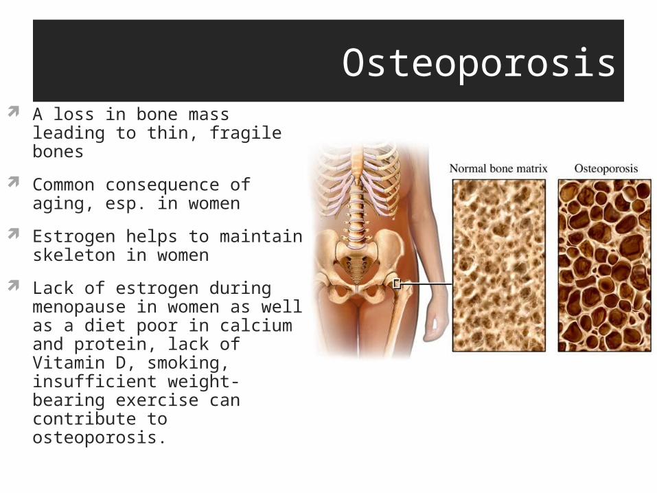
Osteoporosis A loss in bone mass leading
to thin, fragile bones
Common consequence of aging, esp. in women
Estrogen helps to maintain skeleton in women
Lack of estrogen during menopause in women as well as a diet poor in calcium and protein, lack of Vitamin D, smoking, insufficient weight-bearing exercise can contribute to osteoporosis.