Embed Size (px)
Citation preview
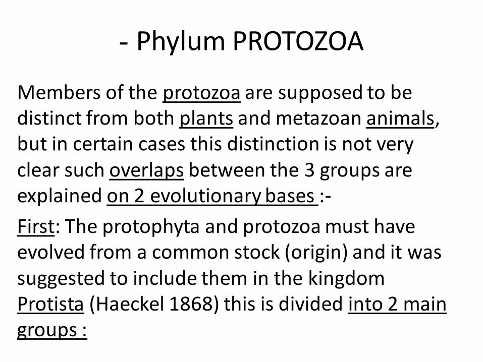
- Phylum PROTOZOA
Members of the protozoa are supposed to be distinct from both plants and metazoan animals, but in certain cases this distinction is not very clear such overlaps between the 3 groups are explained on 2 evolutionary bases :-
First: The protophyta and protozoa must have evolved from a common stock (origin) and it was suggested to include them in the kingdom Protista (Haeckel 1868) this is divided into 2 main groups :
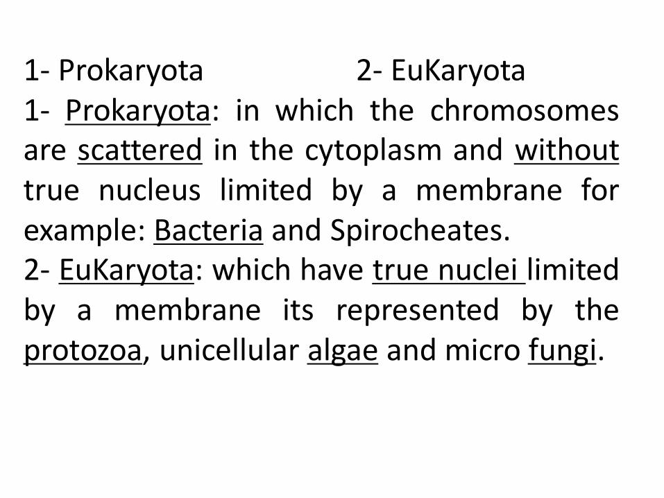
1- Prokaryota 2- EuKaryota
1- Prokaryota: in which the chromosomes are scattered in the cytoplasm and without true nucleus limited by a membrane for example: Bacteria and Spirocheates. 2- EuKaryota: which have true nuclei limited by a membrane its represented by the protozoa, unicellular algae and micro fungi.
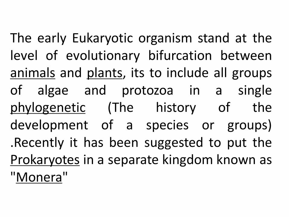
The early Eukaryotic organism stand at the level of evolutionary bifurcation between animals and plants, its to include all groups of algae and protozoa in a single phylogenetic (The history of the development of a species or groups) .Recently it has been suggested to put the Prokaryotes in a separate kingdom known as "Monera"
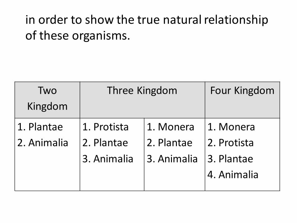
Two
Kingdom
Three Kingdom Four Kingdom
1. Plantae
2. Animalia
1. Protista
2. Plantae
3. Animalia
1. Monera
2. Plantae
3. Animalia
1. Monera
2. Protista
3. Plantae
4. Animalia
in order to show the true natural relationship of these organisms.
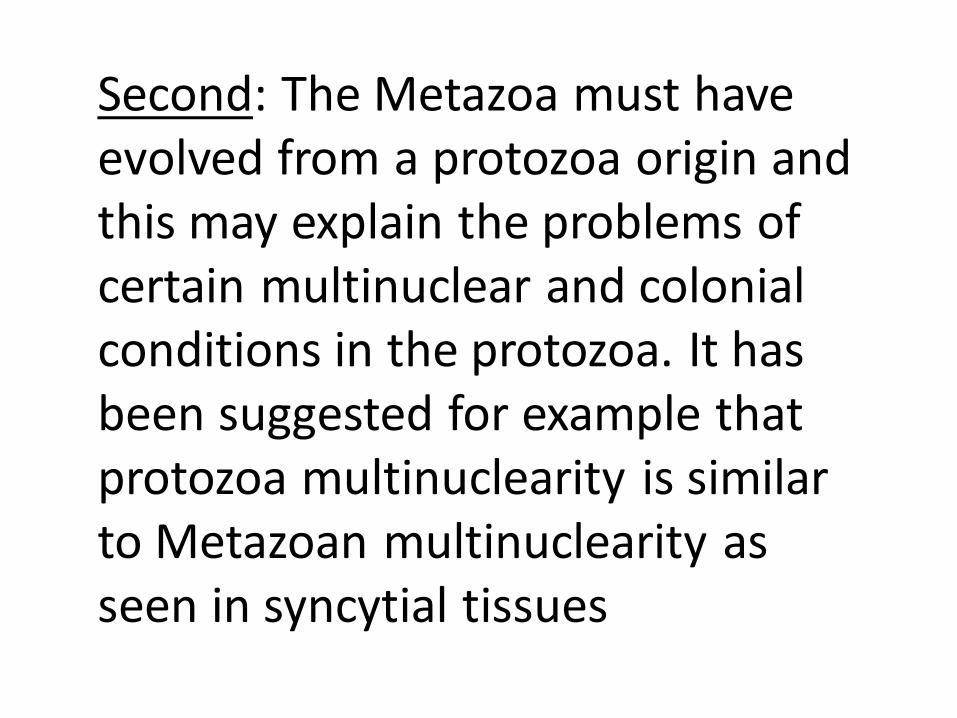
Second: The Metazoa must have evolved from a protozoa origin and this may explain the problems of certain multinuclear and colonial conditions in the protozoa. It has been suggested for example that protozoa multinuclearity is similar to Metazoan multinuclearity as seen in syncytial tissues
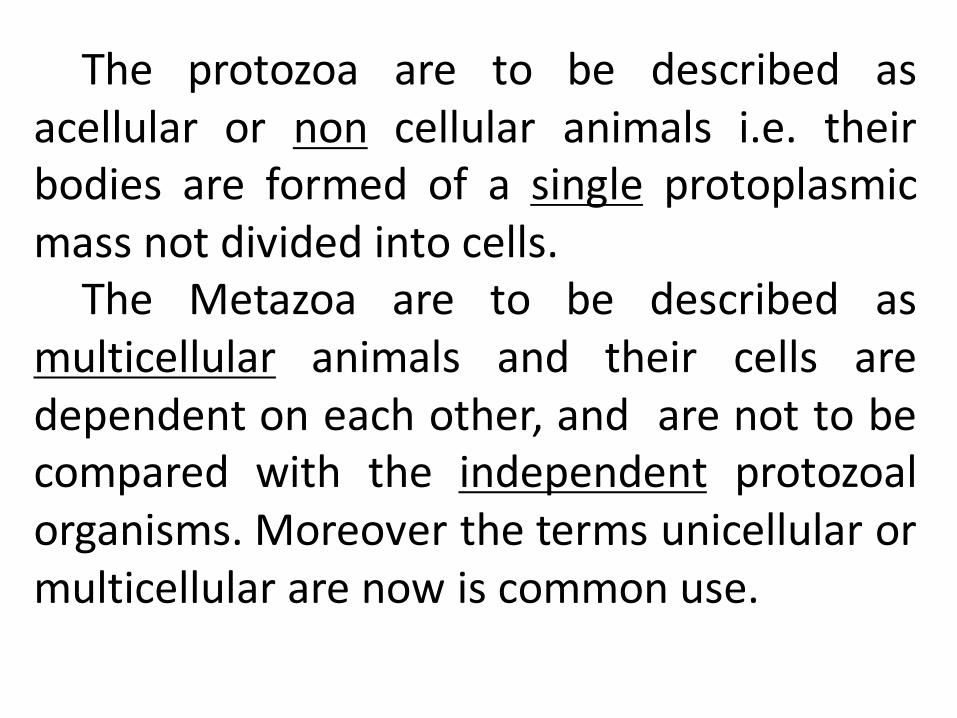
The protozoa are to be described as acellular or non cellular animals i.e. their bodies are formed of a single protoplasmic mass not divided into cells.
The Metazoa are to be described as multicellular animals and their cells are dependent on each other, and are not to be compared with the independent protozoal organisms. Moreover the terms unicellular or multicellular are now is common use.
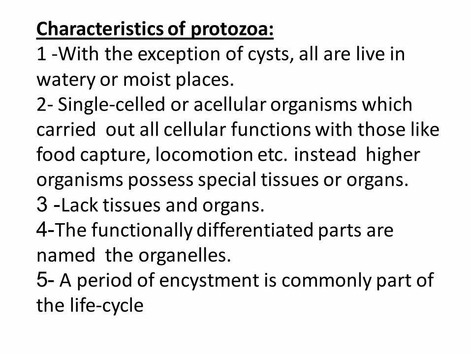
Characteristics of protozoa: 1 -With the exception of cysts, all are live in watery or moist places. 2- Single-celled or acellular organisms which carried out all cellular functions with those like food capture, locomotion etc. instead higher organisms possess special tissues or organs.
- 3 Lack tissues and organs. -4 The functionally differentiated parts are
named the organelles. -5 A period of encystment is commonly part of
the life-cycle
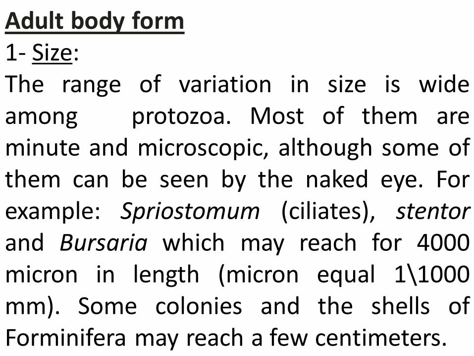
Adult body form
1- Size: The range of variation in size is wide among protozoa. Most of them are minute and microscopic, although some of them can be seen by the naked eye. For example: Spriostomum (ciliates), stentor and Bursaria which may reach for 4000 micron in length (micron equal 1\1000 mm). Some colonies and the shells of Forminifera may reach a few centimeters.
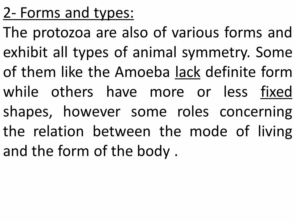
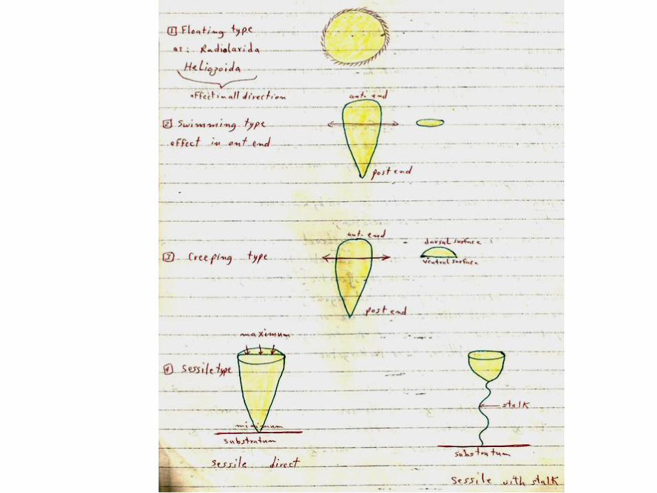
2- Forms and types: The protozoa are also of various forms and exhibit all types of animal symmetry. Some of them like the Amoeba lack definite form while others have more or less fixed shapes, however some roles concerning the relation between the mode of living and the form of the body .
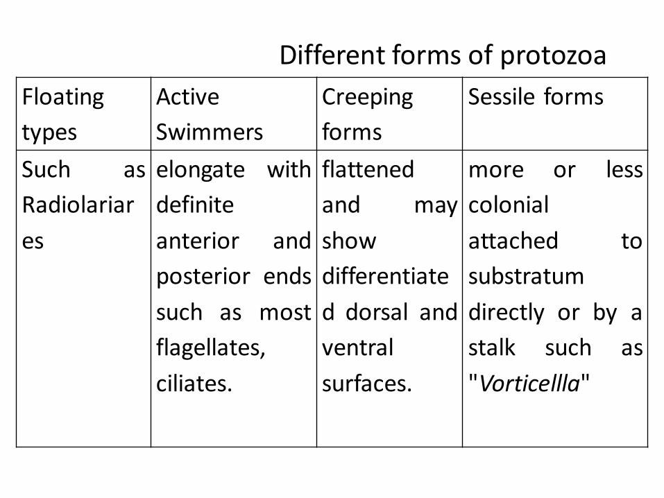
Floating
types
Active
Swimmers
Creeping
forms
Sessile forms
Such as
Radiolariar
es
elongate with
definite
anterior and
posterior ends
such as most
flagellates,
ciliates.
flattened
and may
show
differentiate
d dorsal and
ventral
surfaces.
more or less
colonial
attached to
substratum
directly or by a
stalk such as
"Vorticellla"
Different forms of protozoa
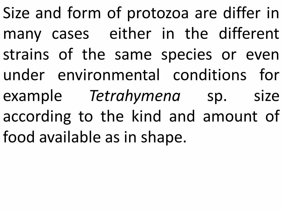
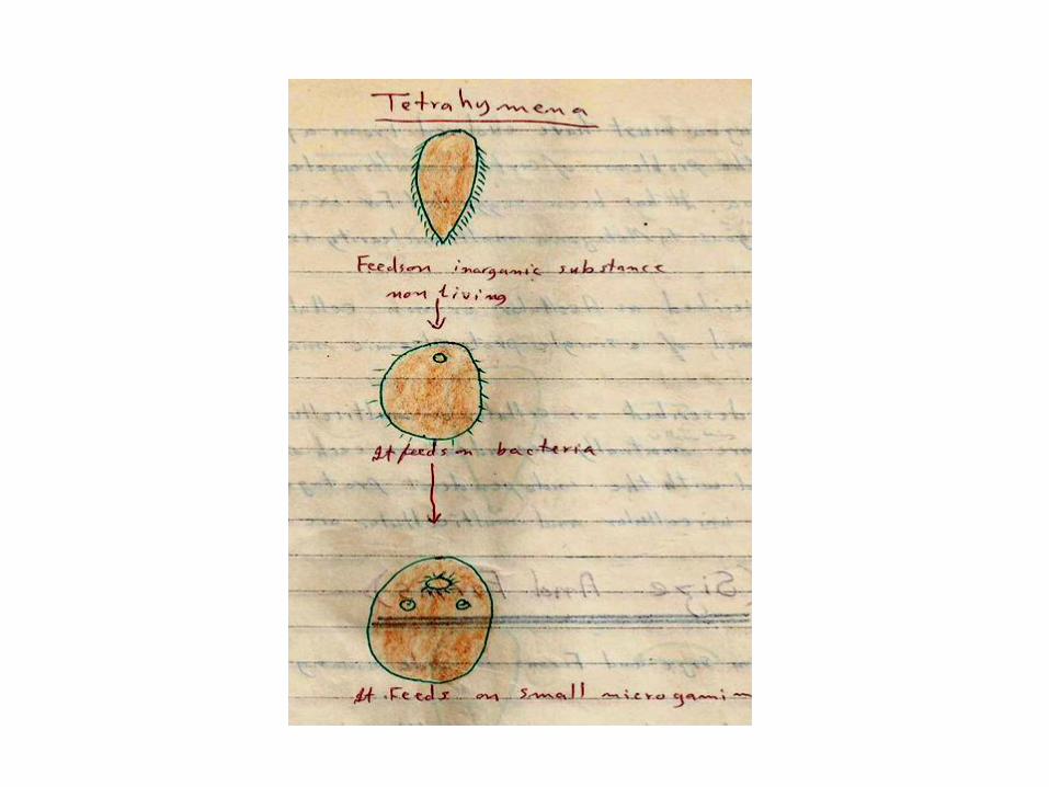
Size and form of protozoa are differ in many cases either in the different strains of the same species or even under environmental conditions for example Tetrahymena sp. size according to the kind and amount of food available as in shape.
Protozoa Aggregates: Most Protozoa are solitary but in some cases a number of individuals belonging to the same species become aggregated in a characteristic form, such aggregates may be includes the following :
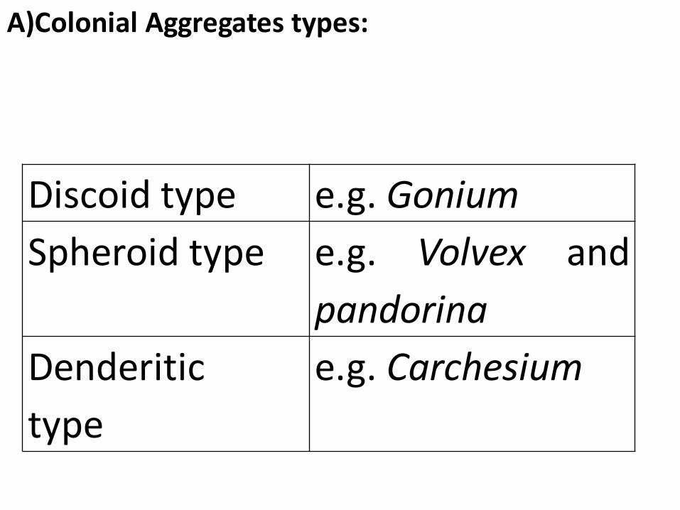
Discoid type e.g. Gonium
Spheroid type e.g. Volvex and
pandorina
Denderitic
type
e.g. Carchesium
A)Colonial Aggregates types:
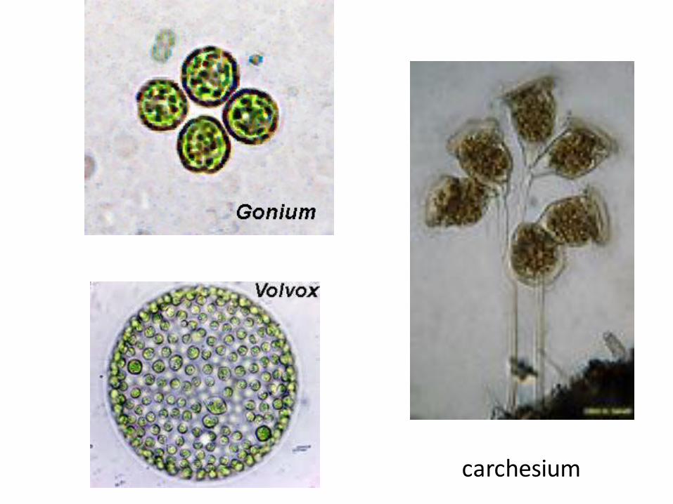
carchesium
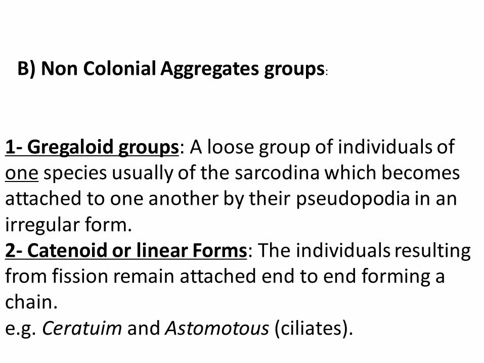
1- Gregaloid groups: A loose group of individuals of one species usually of the sarcodina which becomes attached to one another by their pseudopodia in an irregular form. 2- Catenoid or linear Forms: The individuals resulting from fission remain attached end to end forming a chain. e.g. Ceratuim and Astomotous (ciliates).
B) Non Colonial Aggregates groups:
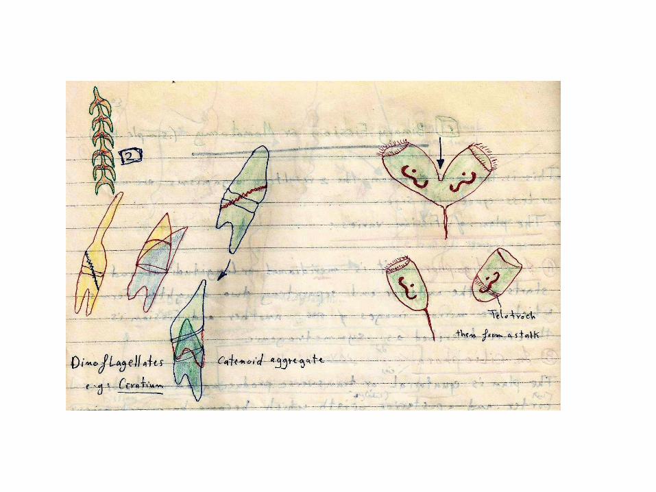
ceratuim
3- Plasmodial stages: This occurs mainly in the sarcodina the individuals unite and and their nuclei divide continuously forming a single multinucleate body. 4-Palmella Stages: This occurs mainly in the phytoflagellates. The individuals lose there flagella and undergo division with development of nucleus envelopes and form large bodies. This size of these bodies is not constant for each species.
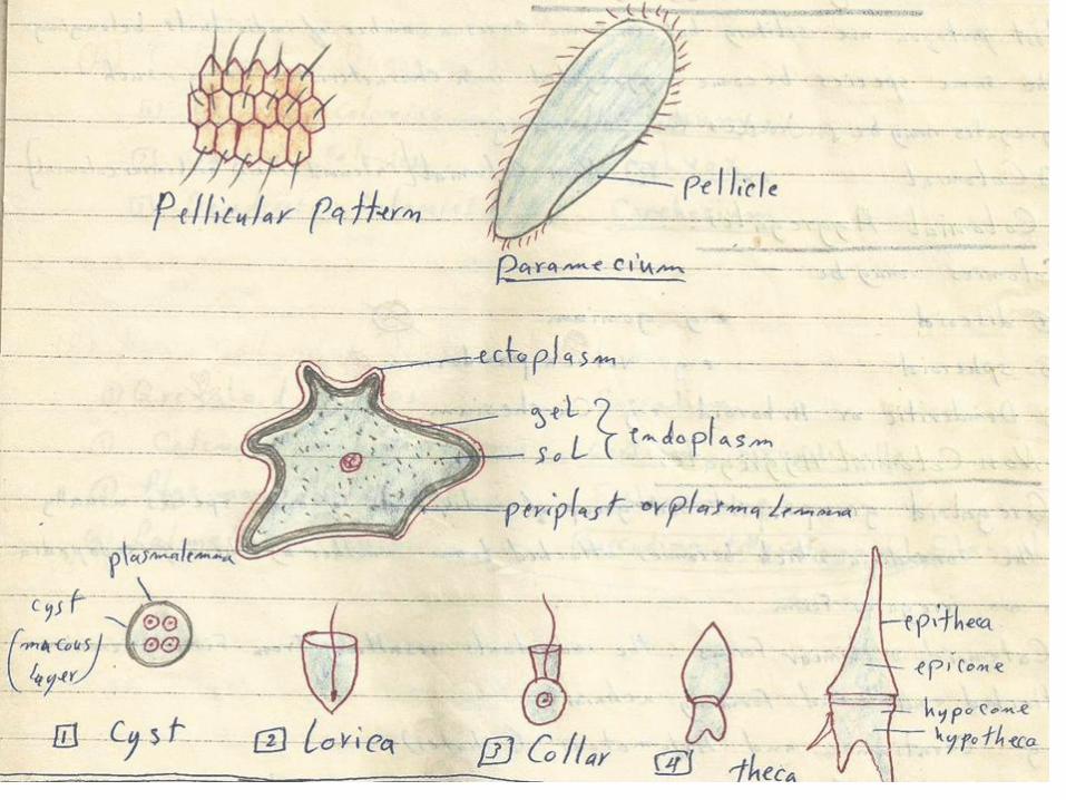
General Organization: The surface of ectoplasm may be differentiated into a thin layer known as a plasmalema or periblast or into a strong flexible layer known as a pellicle. The plasmalema allows for the formation of the pseudopodia and amoeboid movement. The pellicle may be sculptured as in paramecium and its flexibility allows for certain changes in the body form as a euglenoid and gregarine movement.
The ectoplasm is also capable of secreting various external covering structures: such as Cyst, Lorica, Collar, Theca and shell as in forminifera or internal structures such as Trichaetes, axostyle, pharyngeal basket, central capsule, siliceous skeleton in the Radiolaria.
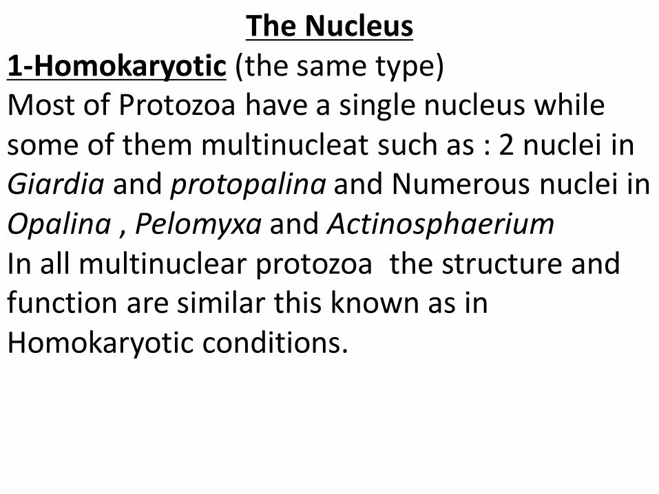
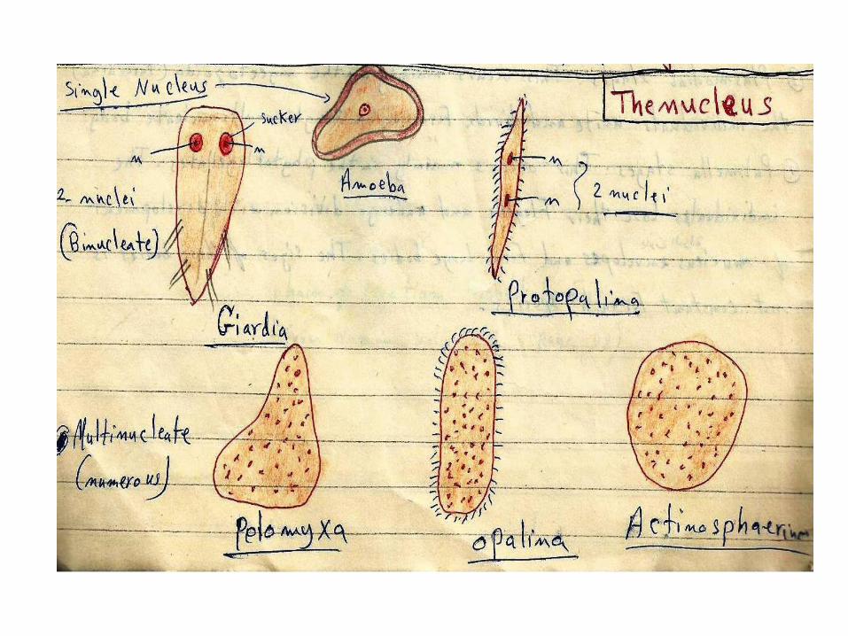
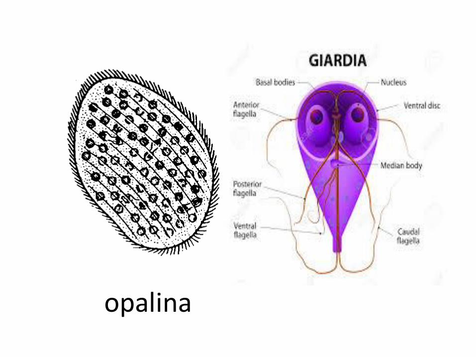
The Nucleus 1-Homokaryotic (the same type) Most of Protozoa have a single nucleus while some of them multinucleat such as : 2 nuclei in Giardia and protopalina and Numerous nuclei in Opalina , Pelomyxa and Actinosphaerium
In all multinuclear protozoa the structure and function are similar this known as in Homokaryotic conditions.
opalina
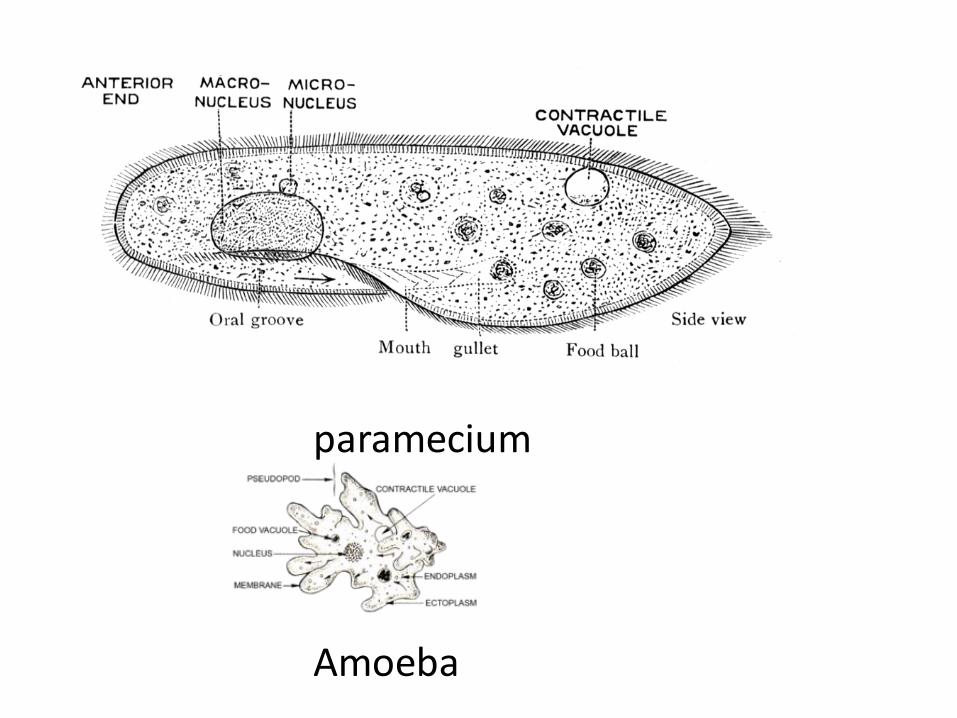
2-Heterokaryotic: (Nuclear Dimorphism): There are 2 kinds of nuclei different from each other in both structure and function . In ciliates there is a single macronucleus and one or more micronuclei and this known as a nuclear dimorphism (Hetero karyotic ) conditions such as Paramecium. The macronucleus: concerned with somatic or metabolic function and controls all metabolic activities and regeneration i.e. it determines the shape of the ciliate.
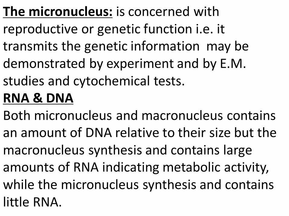
The micronucleus: is concerned with reproductive or genetic function i.e. it transmits the genetic information may be demonstrated by experiment and by E.M. studies and cytochemical tests. RNA & DNA Both micronucleus and macronucleus contains an amount of DNA relative to their size but the macronucleus synthesis and contains large amounts of RNA indicating metabolic activity, while the micronucleus synthesis and contains little RNA.
paramecium
Amoeba
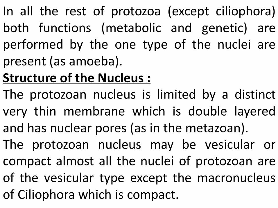
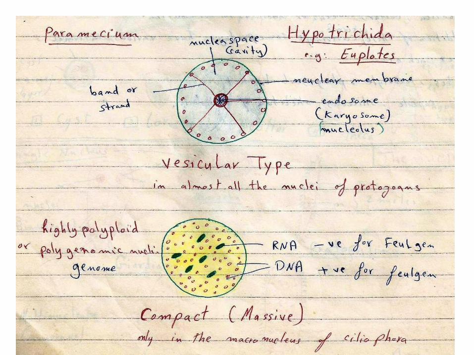
In all the rest of protozoa (except ciliophora) both functions (metabolic and genetic) are performed by the one type of the nuclei are present (as amoeba). Structure of the Nucleus : The protozoan nucleus is limited by a distinct very thin membrane which is double layered and has nuclear pores (as in the metazoan). The protozoan nucleus may be vesicular or compact almost all the nuclei of protozoan are of the vesicular type except the macronucleus of Ciliophora which is compact.
In the vesicular nucleus: Most of the nuclear space is free from chromatin and there may be one or more endosomes (Karyosomes) or (nucleolie) the nucleus is commonly spherical but may be oval or biconvex . In the compact or massive type: (i.e. the macronucleus of ciliates) the nucleus contain large amount of equal distributed chromatin granules. These consists of a number of feulgen –ve nucleoli . It containing few number of large granules RNA embedded among numerous smaller granules of DNA.
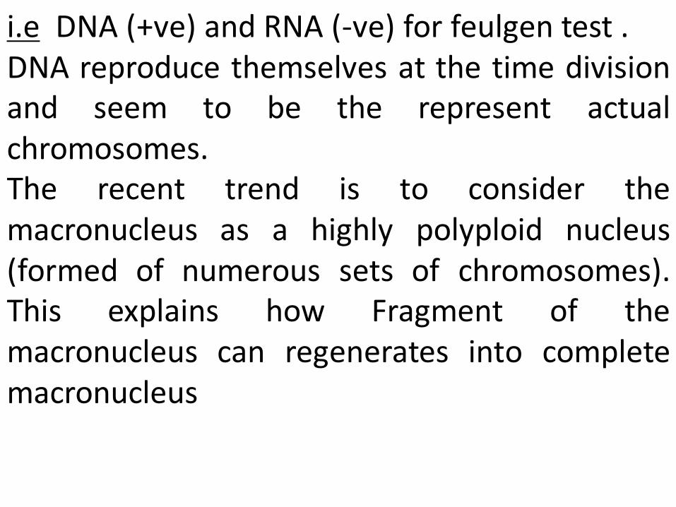
i.e DNA (+ve) and RNA (-ve) for feulgen test . DNA reproduce themselves at the time division and seem to be the represent actual chromosomes. The recent trend is to consider the macronucleus as a highly polyploid nucleus (formed of numerous sets of chromosomes). This explains how Fragment of the macronucleus can regenerates into complete macronucleus
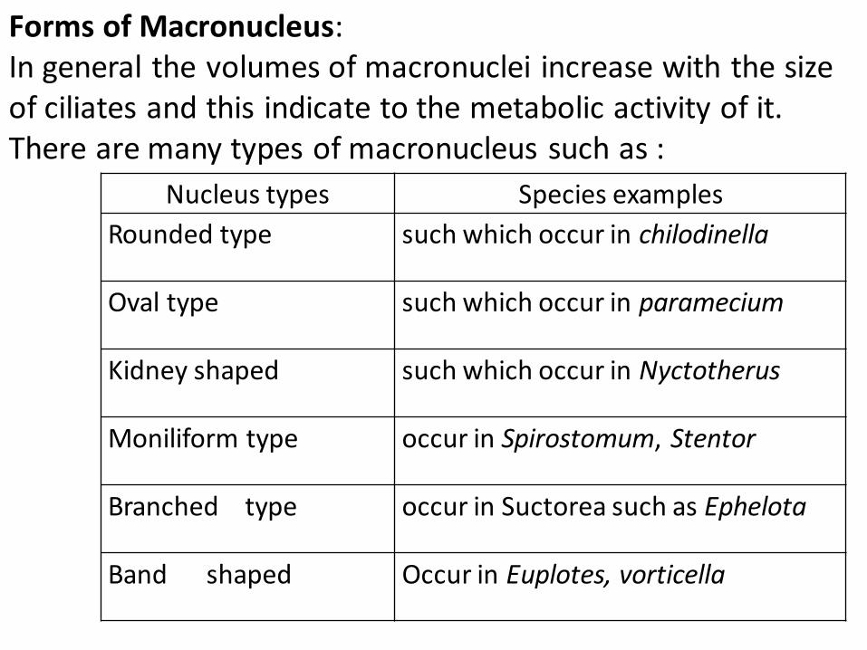
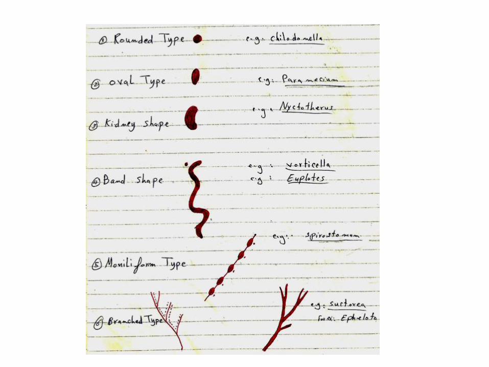
Nucleus types Species examples
Rounded type such which occur in chilodinella
Oval type such which occur in paramecium
Kidney shaped such which occur in Nyctotherus
Moniliform type occur in Spirostomum, Stentor
Branched type occur in Suctorea such as Ephelota
Band shaped Occur in Euplotes, vorticella
Forms of Macronucleus: In general the volumes of macronuclei increase with the size of ciliates and this indicate to the metabolic activity of it. There are many types of macronucleus such as :
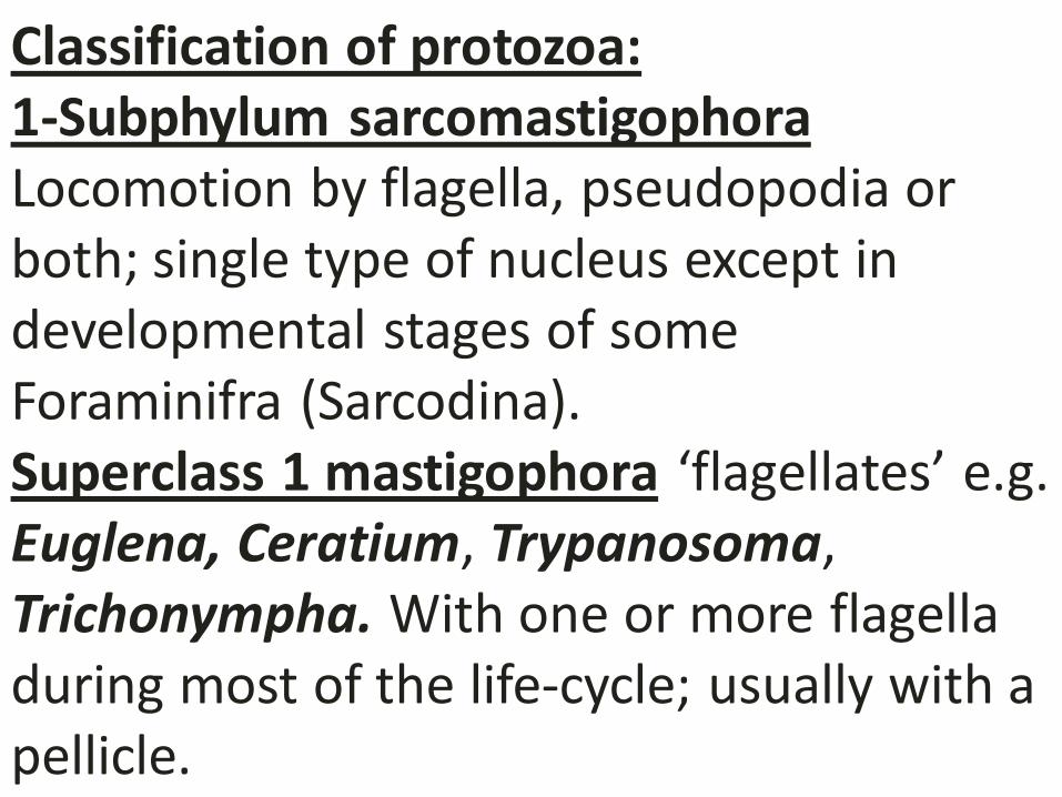
Classification of protozoa: 1-Subphylum sarcomastigophora Locomotion by flagella, pseudopodia or both; single type of nucleus except in developmental stages of some Foraminifra (Sarcodina). Superclass 1 mastigophora ‘flagellates’ e.g. Euglena, Ceratium, Trypanosoma, Trichonympha. With one or more flagella during most of the life-cycle; usually with a pellicle.
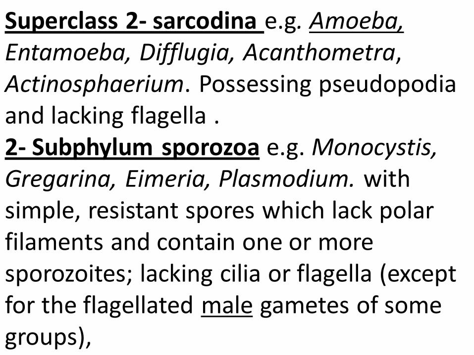
Superclass 2- sarcodina e.g. Amoeba, Entamoeba, Difflugia, Acanthometra, Actinosphaerium. Possessing pseudopodia and lacking flagella . 2- Subphylum sporozoa e.g. Monocystis, Gregarina, Eimeria, Plasmodium. with simple, resistant spores which lack polar filaments and contain one or more sporozoites; lacking cilia or flagella (except for the flagellated male gametes of some groups),
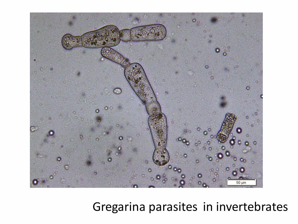
Gregarina parasites in invertebrates
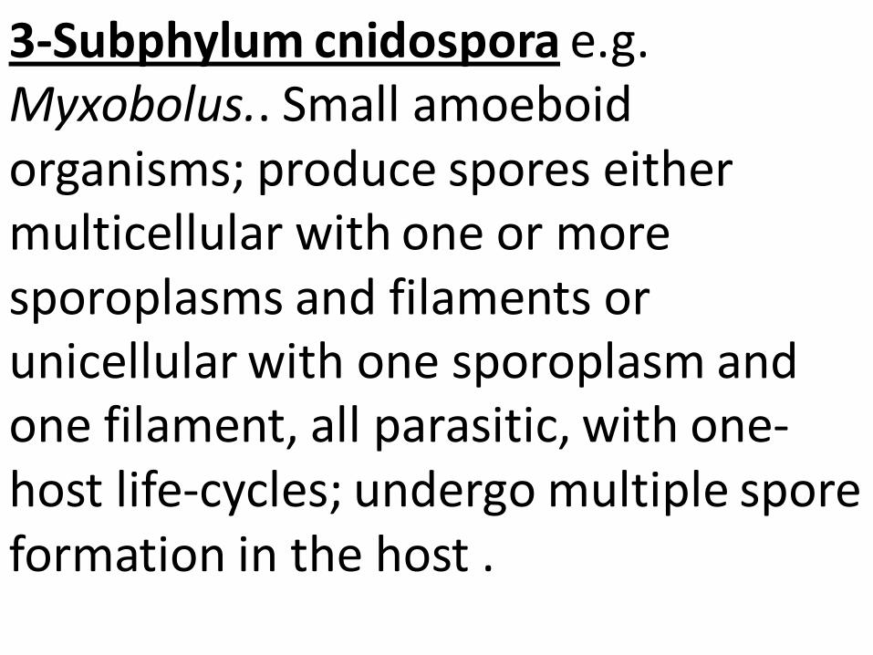
3-Subphylum cnidospora e.g. Myxobolus.. Small amoeboid organisms; produce spores either multicellular with one or more sporoplasms and filaments or unicellular with one sporoplasm and one filament, all parasitic, with one-host life-cycles; undergo multiple spore formation in the host .
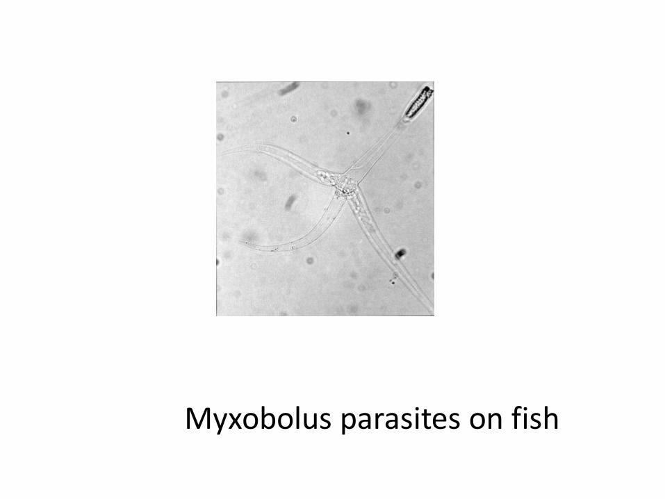
Myxobolus parasites on fish
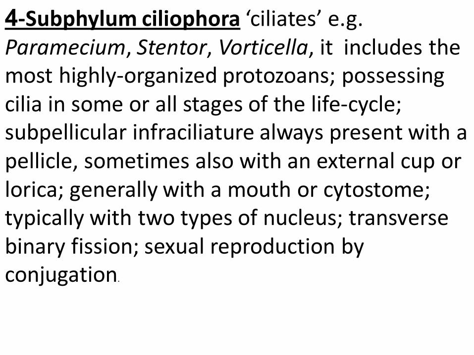
4-Subphylum ciliophora ‘ciliates’ e.g. Paramecium, Stentor, Vorticella, it includes the most highly-organized protozoans; possessing cilia in some or all stages of the life-cycle; subpellicular infraciliature always present with a pellicle, sometimes also with an external cup or lorica; generally with a mouth or cytostome; typically with two types of nucleus; transverse binary fission; sexual reproduction by conjugation.
Nutrition (Feeding): 1- Autotrophic Nutrition In this type organic compounds are synthesized from inorganic materials by the organism. a-In chemotrophic – N – (Chemoautotrophic) Energy necessary for the synthesis may be obtains from chemical processes. b-In phototrophic –N- (Holophylic) Energy of light is used (Photosynthesis) and this method occurs in the colored photomastigophora .
2-Heterotrophic Nutrition In this type the food of the organism contains organic material built up by autotrophs. The animal are unable to synthesize organic compounds to use as food . It includes:
a-Osmotrophic Nutrition In this type the protozoa absorb their organic food by osmosis through the body surface and thus no special organelles are required. “Osmosis Process” smaller molecules are generally more easily passed than larger molecules
b-permeation or saprozoic Nutrition : The organic food molecules are taken by active transport involving the consumption of energy. This process occurs in many parasitic protozoa particularly the sporozoa and in some free living colorless flagellates. Some parasites like cnidosporodia seem to liquify the cells and tissues of the host by their enzymes before they absorb their substances. The process takes place through any point of the body
c-Pinocytosis Nutrition: -In this process the organic food molecules are taken inside by some microscopic vesicle cut off from the membrane into the interior of the body. -The process takes place through definite point of the body and thus it is different from osmotrophy in which the liquid may pass in continuously through the whole body surface. The significance of the process seems to be the ability of the organism to ingest large macromolecules which cannot pass through ordinary osomosis or permeation)
-Pinocytosis is stimulated by various active substances in the medium especially proteins and amino acids. -Pinocytosis has been studied in many species whose nutrition has been thought to be only osmotrophic. For example: Some species of plasmodium were found by E.M to ingest portions of the cytoplasm of their host red cells obtaining haemoglobim and other proteins, however there is a permanent micropore in these stages of the parasite.
d-Phagotrophic Nutrition ( Holozoic or Zootrophic N ) This similar of animals and includes: 1- Food capture 2- Ingestion 3-
Digestion 4- Egestion
With the development of specialized organelles it is adopted by the majority of the free living and many parasitic protozoa.
1, 2 Food capture, Ingestion
The locomotory organelles usually serve for the capture of the food, but additional structures may occurs.
In the sarcodina and many phagotrophic Flagellates :
Food is captured by pseudopodia and ingested at any point of the body surface. -Vampyrella (protomyxida) penetrate the cellulose wall of the filament of algae and sucks out their protoplasmic contains. -In Trichonympha small pieces of wood are ingested at a marked area at the posterior end.
In Certain Flagellates and typical ciliates:
Ingestion is limited to a definite cytostome which is primitively at the anterior end and often opens into a cytopharynx passing deep into the endoplasm. The flagella help in bringing about the food particles to the cytostome. In choanoflagellates, the collar is a further device for concentrating food.
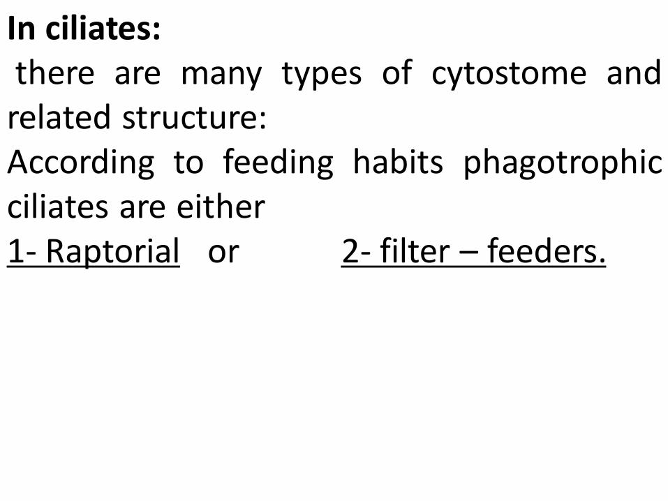
In ciliates: there are many types of cytostome and related structure: According to feeding habits phagotrophic ciliates are either 1- Raptorial or 2- filter – feeders.
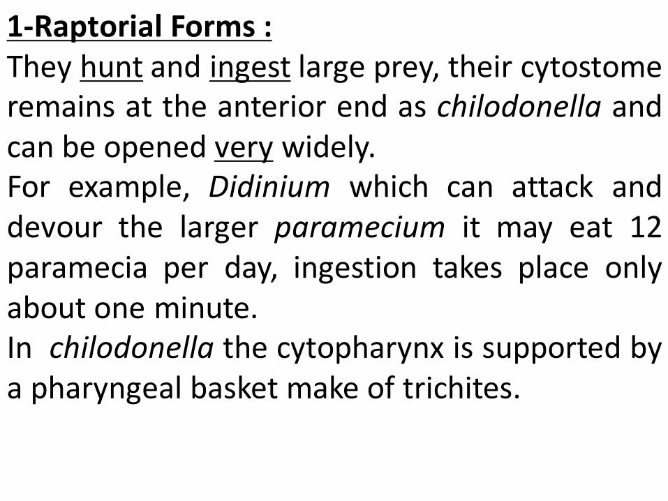
1-Raptorial Forms : They hunt and ingest large prey, their cytostome remains at the anterior end as chilodonella and can be opened very widely. For example, Didinium which can attack and devour the larger paramecium it may eat 12 paramecia per day, ingestion takes place only about one minute. In chilodonella the cytopharynx is supported by a pharyngeal basket make of trichites.
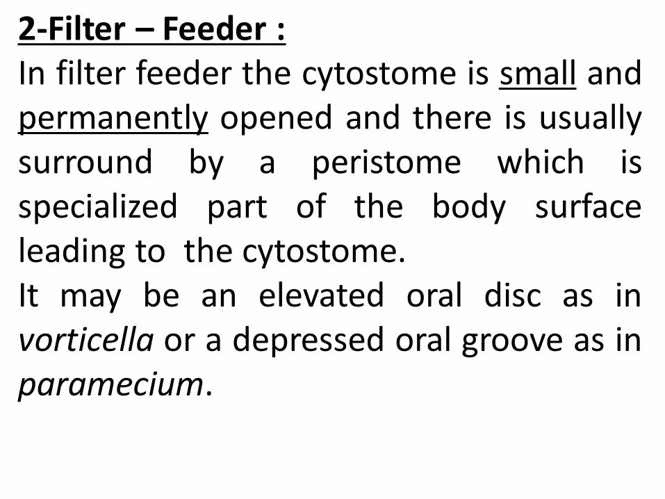
2-Filter – Feeder : In filter feeder the cytostome is small and permanently opened and there is usually surround by a peristome which is specialized part of the body surface leading to the cytostome. It may be an elevated oral disc as in vorticella or a depressed oral groove as in paramecium.
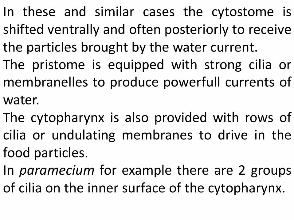
In these and similar cases the cytostome is shifted ventrally and often posteriorly to receive the particles brought by the water current. The pristome is equipped with strong cilia or membranelles to produce powerfull currents of water. The cytopharynx is also provided with rows of cilia or undulating membranes to drive in the food particles. In paramecium for example there are 2 groups of cilia on the inner surface of the cytopharynx.
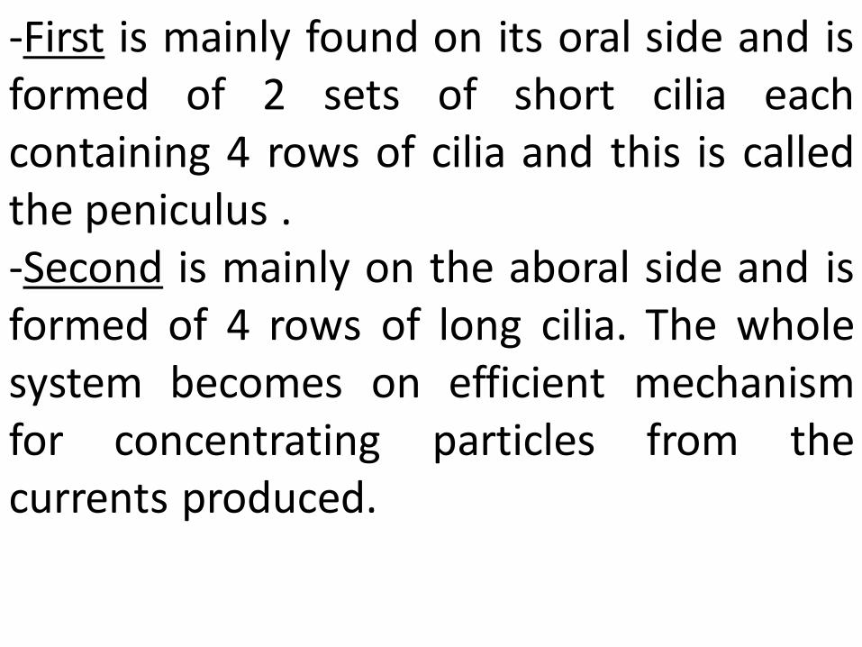
-First is mainly found on its oral side and is formed of 2 sets of short cilia each containing 4 rows of cilia and this is called the peniculus . -Second is mainly on the aboral side and is formed of 4 rows of long cilia. The whole system becomes on efficient mechanism for concentrating particles from the currents produced.
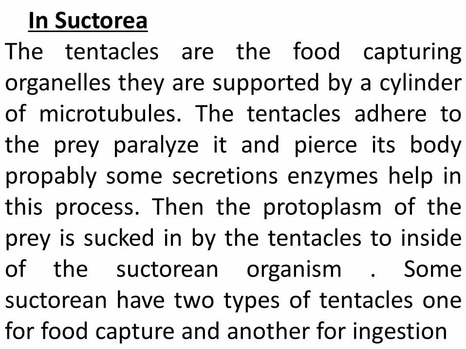
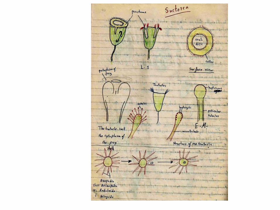
In Suctorea The tentacles are the food capturing organelles they are supported by a cylinder of microtubules. The tentacles adhere to the prey paralyze it and pierce its body propably some secretions enzymes help in this process. Then the protoplasm of the prey is sucked in by the tentacles to inside of the suctorean organism . Some suctorean have two types of tentacles one for food capture and another for ingestion
3- Digestion Ingested food together with some liquid is enclosed in food vacuoles in forms with shells, large food particles are digested in the pseudopodia outside the main body.
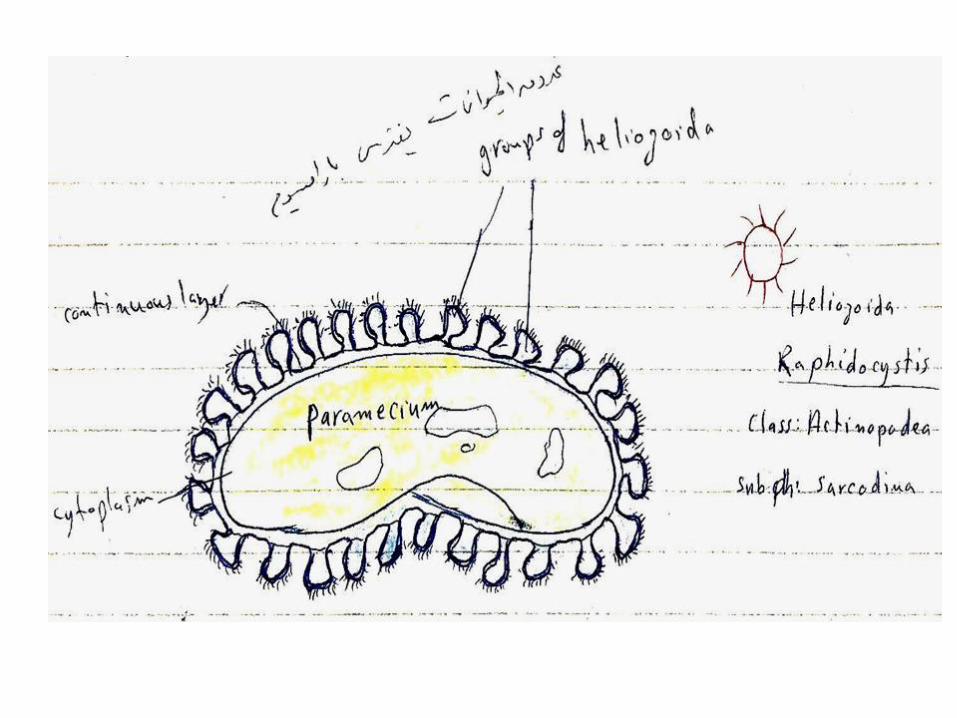
-In actionopodea, the axial filaments of the axopodia many locally disappear and food is ingested and pass to inside. -A group of heliozoans like Raphidocystis may attach a large prey as paramycium and parts of their protoplasm may fuse and form a continuous layer around the captured food.
In the amoeba and similar forms the food vacuoles are carried about irregularly coures in the endoplasm, but in the ciliates vacuoles are moved along in definite course by endoplasmic current. This process is known as cyclosis. The contents of the food vacuoles usually pass through on early acidic and a latter alkaline phases produced by the endoplasmic secretion. Digested food is absorbed by the surrounding endoplasm and micropinocytosis round the surface of the vacuole plays some part in absorption of digested food
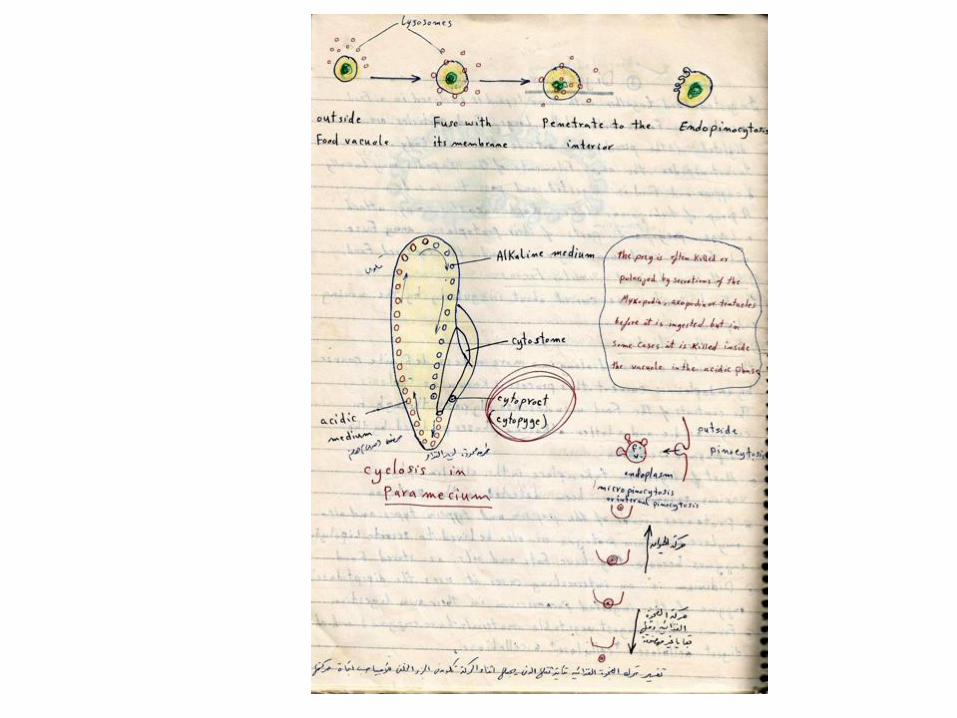
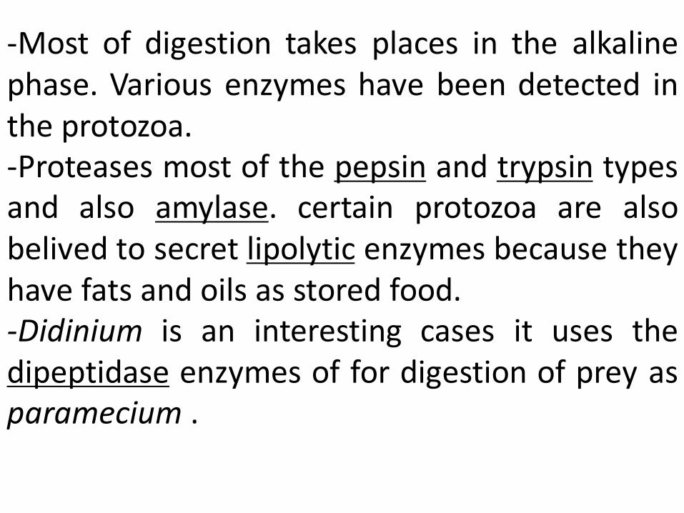
-Most of digestion takes places in the alkaline phase. Various enzymes have been detected in the protozoa. -Proteases most of the pepsin and trypsin types and also amylase. certain protozoa are also belived to secret lipolytic enzymes because they have fats and oils as stored food. -Didinium is an interesting cases it uses the dipeptidase enzymes of for digestion of prey as paramecium .
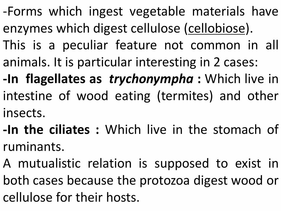
-Forms which ingest vegetable materials have enzymes which digest cellulose (cellobiose). This is a peculiar feature not common in all animals. It is particular interesting in 2 cases: -In flagellates as trychonympha : Which live in intestine of wood eating (termites) and other insects. -In the ciliates : Which live in the stomach of ruminants. A mutualistic relation is supposed to exist in both cases because the protozoa digest wood or cellulose for their hosts.
4- Egestion of waste : - Indigestible material after digestion is egested. In the Sarcodina egestion takes place at any point at the temporary posterior end during movement, But In Flagellates and ciliates egestion takes place at a definite point known as cytopyge or cytoproct.
3- Mixotrophic nutrition: -Many protozoa feed themselves by more than one method at the same or different times. The relative importance of one method or another largely depends on environmental conditions.
Some colored flagellates ingested solid food i.e. they are both phagotrophic and phototrophic, while many of them are often osmotrophic. Even in typical phototrophic forms.they need vitamins especially vitamin B12 and other organic growth factors in the media in which they live.
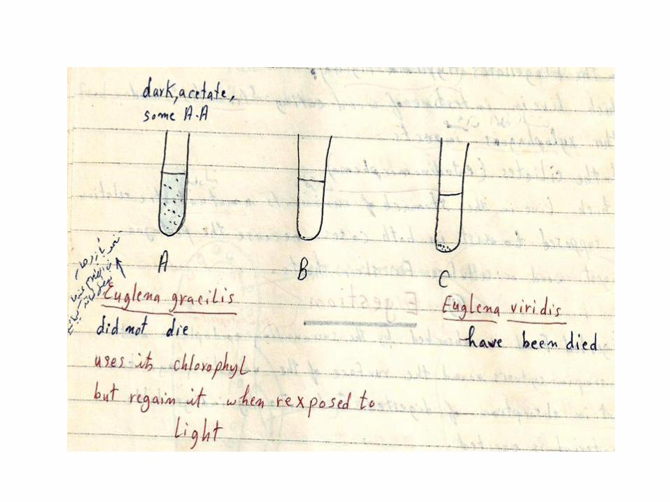
- many protozoa which were known to be only osmotrophic or phagotrophic have been found to feed also by pinocytosis ( the case of tetrahymena ) -In Euglena many species of it have difficulty in utilizing nitrates and grow better of nitrogen is provided as ammonia while others need amino acids and peptones. Moreover Euglena gracilis can flourish in the dark if provided with acetate as a source of energy and carbon and also certain amino acids. In this case it usually not uses its chlorophyll but can regain it when exposed again to light.
Osmoregulation/excretion
Many protozoans, particularly fresh-water
species, possess one or more fluid-filled
contractile vacuoles whose number, structure and
position vary with the group or species, which
may be fixed in position, and which may empty as
in flagellates and ciliates through a distinct pore in
the pellicle or, as in amoebae, anywhere on the
cell surface. In species with a pellicle the ejection
spot for solid wastes is the cell anus or cytoproct
(cytopyge as ) in Ciliophora.
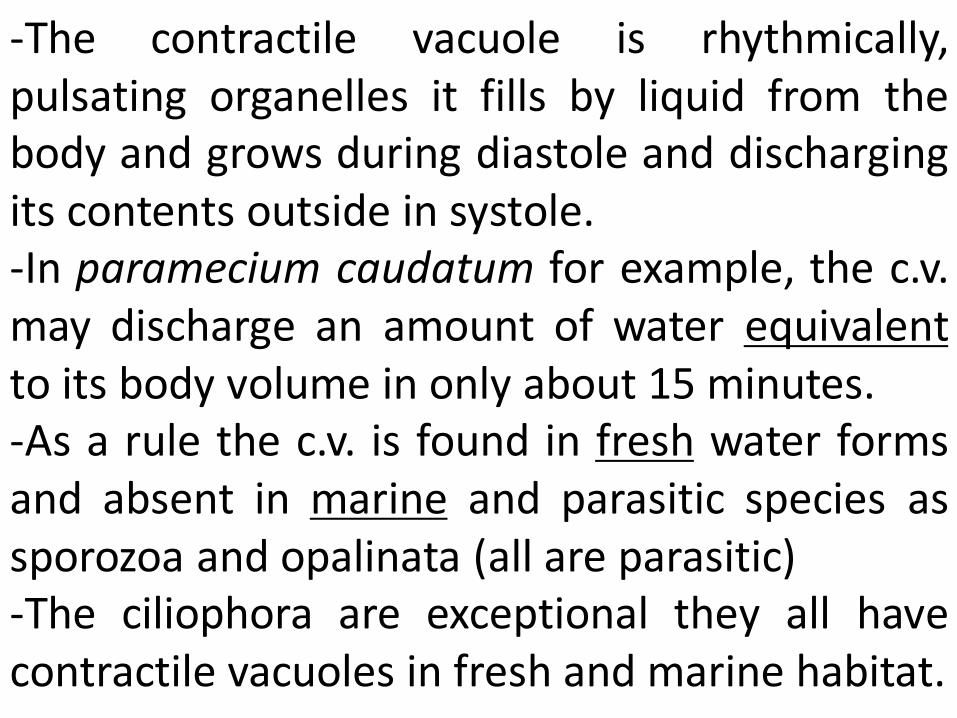
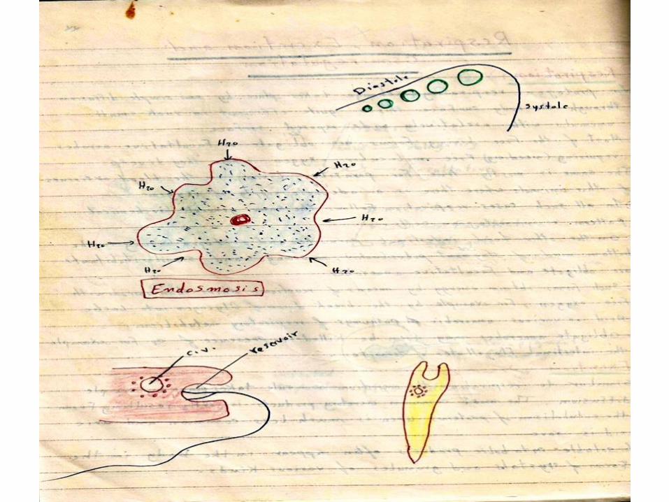
-The contractile vacuole is rhythmically, pulsating organelles it fills by liquid from the body and grows during diastole and discharging its contents outside in systole. -In paramecium caudatum for example, the c.v. may discharge an amount of water equivalent to its body volume in only about 15 minutes. -As a rule the c.v. is found in fresh water forms and absent in marine and parasitic species as sporozoa and opalinata (all are parasitic) -The ciliophora are exceptional they all have contractile vacuoles in fresh and marine habitat.

Number, Distribution and structures of C.V
-The number of c.v. varies in the various species usually one or two sometimes more up to one hundred. - Their position, structure are vary in the various groups:
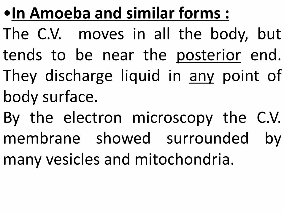
•In Amoeba and similar forms : The C.V. moves in all the body, but tends to be near the posterior end. They discharge liquid in any point of body surface. By the electron microscopy the C.V. membrane showed surrounded by many vesicles and mitochondria.
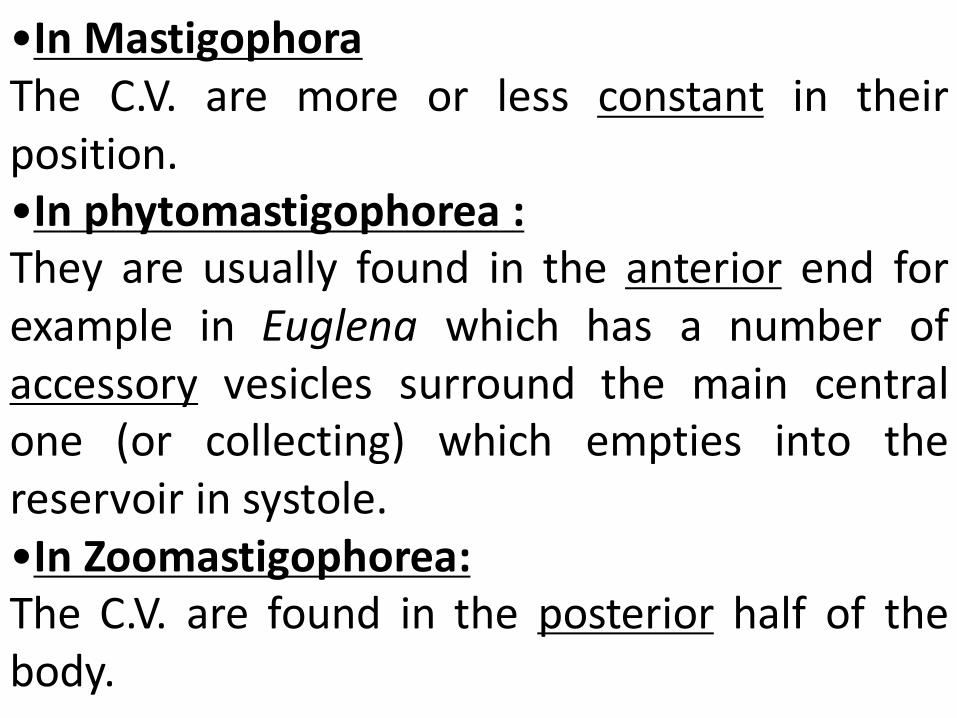
•In Mastigophora The C.V. are more or less constant in their position. •In phytomastigophorea : They are usually found in the anterior end for example in Euglena which has a number of accessory vesicles surround the main central one (or collecting) which empties into the reservoir in systole. •In Zoomastigophorea: The C.V. are found in the posterior half of the body.
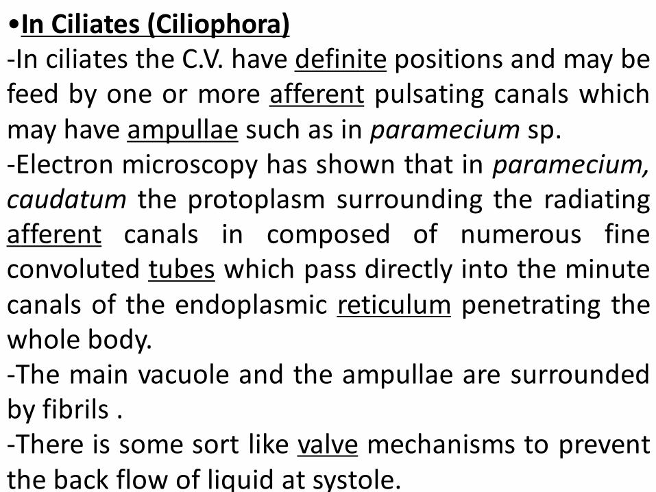
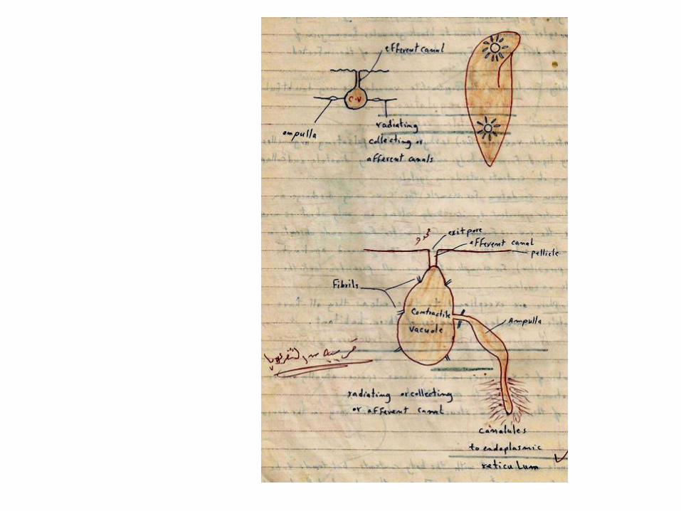
•In Ciliates (Ciliophora) -In ciliates the C.V. have definite positions and may be feed by one or more afferent pulsating canals which may have ampullae such as in paramecium sp. -Electron microscopy has shown that in paramecium, caudatum the protoplasm surrounding the radiating afferent canals in composed of numerous fine convoluted tubes which pass directly into the minute canals of the endoplasmic reticulum penetrating the whole body. -The main vacuole and the ampullae are surrounded by fibrils . -There is some sort like valve mechanisms to prevent the back flow of liquid at systole.
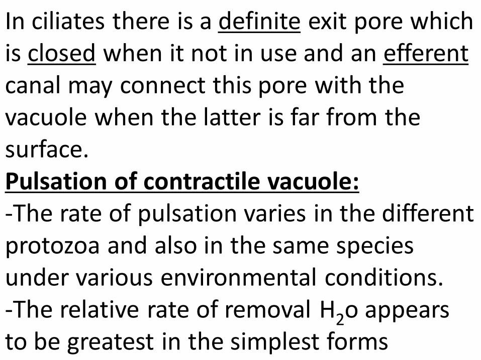
In ciliates there is a definite exit pore which is closed when it not in use and an efferent canal may connect this pore with the vacuole when the latter is far from the surface. Pulsation of contractile vacuole: -The rate of pulsation varies in the different protozoa and also in the same species under various environmental conditions. -The relative rate of removal H2o appears to be greatest in the simplest forms
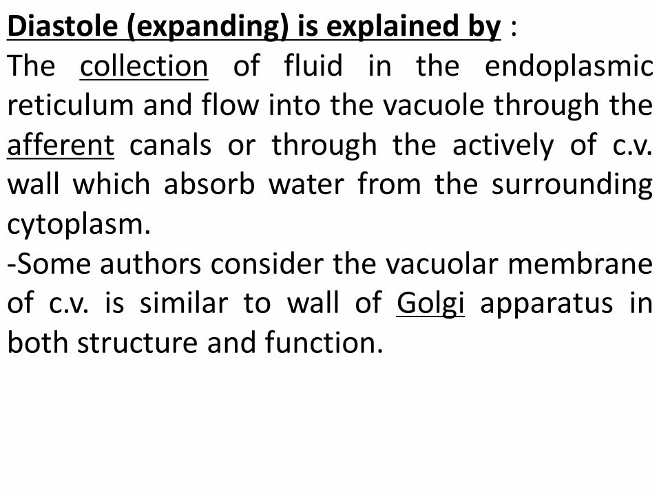
Diastole (expanding) is explained by : The collection of fluid in the endoplasmic reticulum and flow into the vacuole through the afferent canals or through the actively of c.v. wall which absorb water from the surrounding cytoplasm. -Some authors consider the vacuolar membrane of c.v. is similar to wall of Golgi apparatus in both structure and function.

Systole (contraction)is explained by: The contraction of the vacuolar membrane of c.v. or fibrils surrounding it are stimulated by some catalyst . In vitro isolated c.v. of amoeba is contract if ATP and Mg ++ are added to the media . Also the contraction stimulated if the pressure of dilated cytoplasm around the formed vacuole is low or when the c.v. move toward the outer surface or to the exit pore .
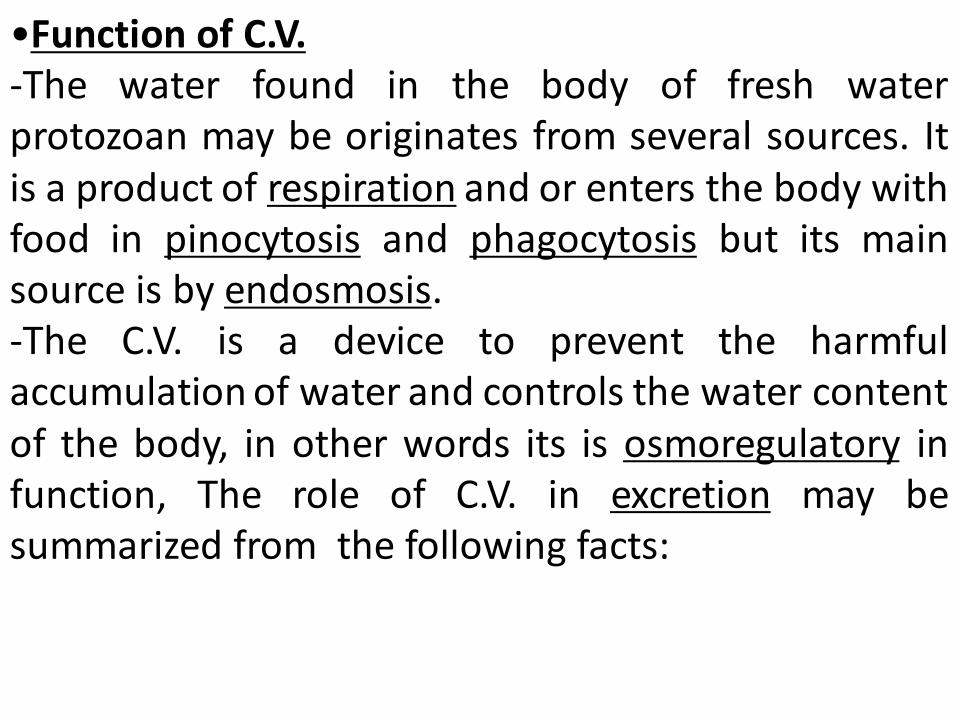
•Function of C.V. -The water found in the body of fresh water protozoan may be originates from several sources. It is a product of respiration and or enters the body with food in pinocytosis and phagocytosis but its main source is by endosmosis. -The C.V. is a device to prevent the harmful accumulation of water and controls the water content of the body, in other words its is osmoregulatory in function, The role of C.V. in excretion may be summarized from the following facts:
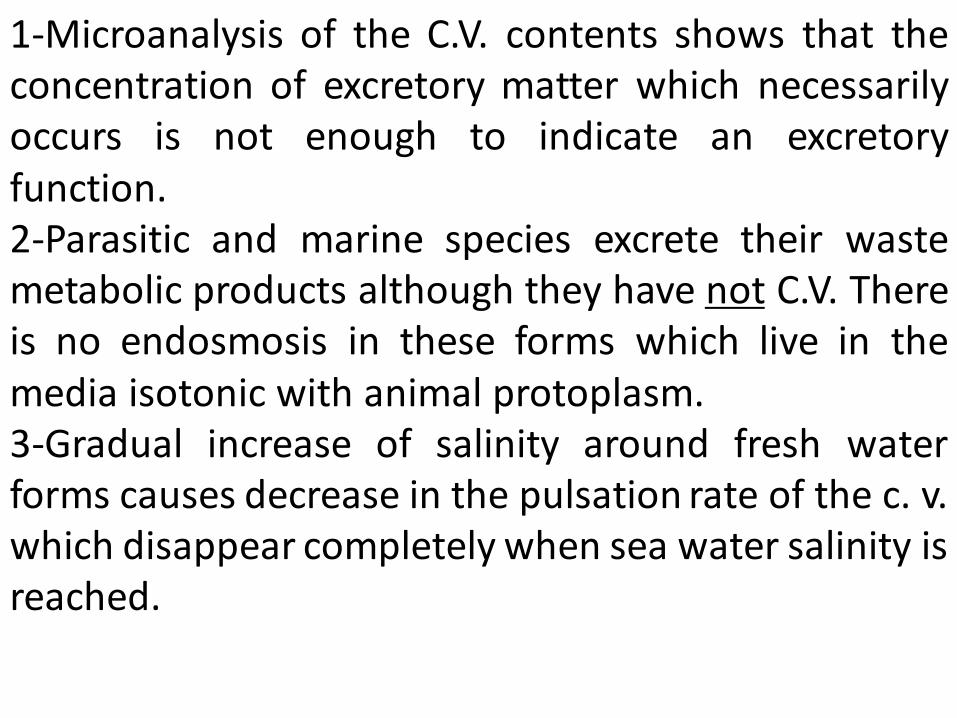
1-Microanalysis of the C.V. contents shows that the concentration of excretory matter which necessarily occurs is not enough to indicate an excretory function. 2-Parasitic and marine species excrete their waste metabolic products although they have not C.V. There is no endosmosis in these forms which live in the media isotonic with animal protoplasm. 3-Gradual increase of salinity around fresh water forms causes decrease in the pulsation rate of the c. v. which disappear completely when sea water salinity is reached.
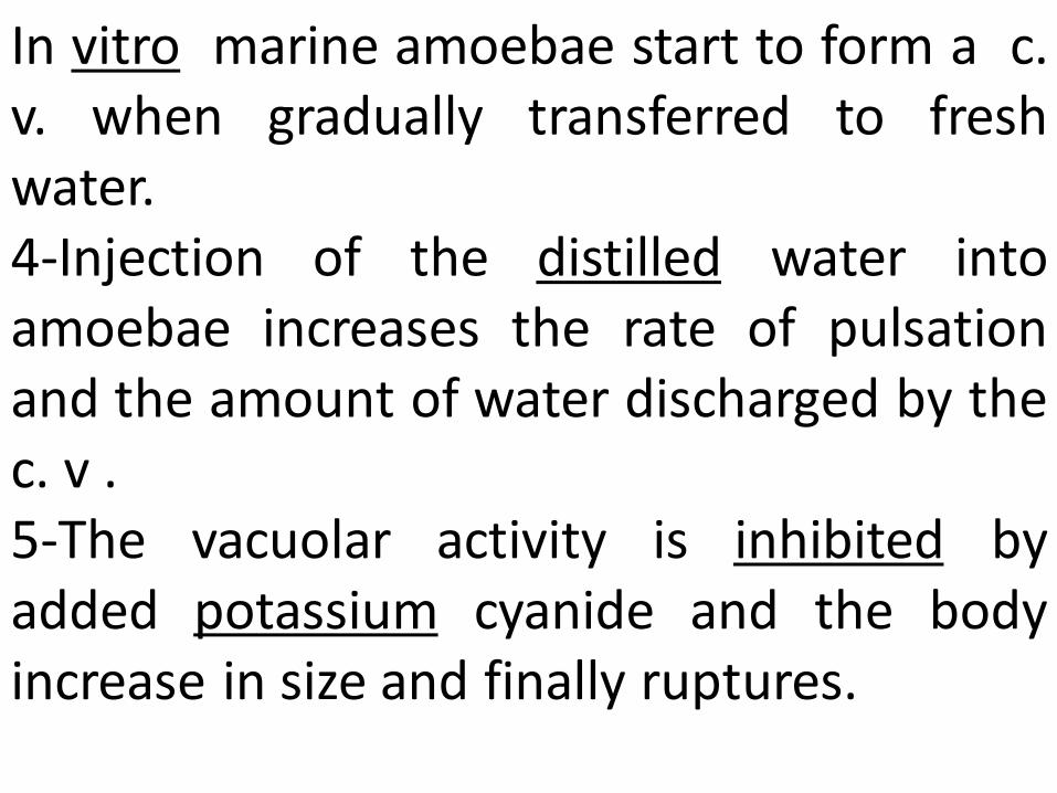
In vitro marine amoebae start to form a c. v. when gradually transferred to fresh water. 4-Injection of the distilled water into amoebae increases the rate of pulsation and the amount of water discharged by the c. v . 5-The vacuolar activity is inhibited by added potassium cyanide and the body increase in size and finally ruptures.
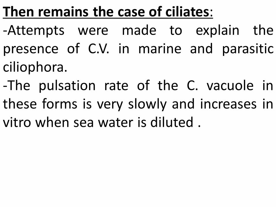
Then remains the case of ciliates: -Attempts were made to explain the presence of C.V. in marine and parasitic ciliophora. -The pulsation rate of the C. vacuole in these forms is very slowly and increases in vitro when sea water is diluted .
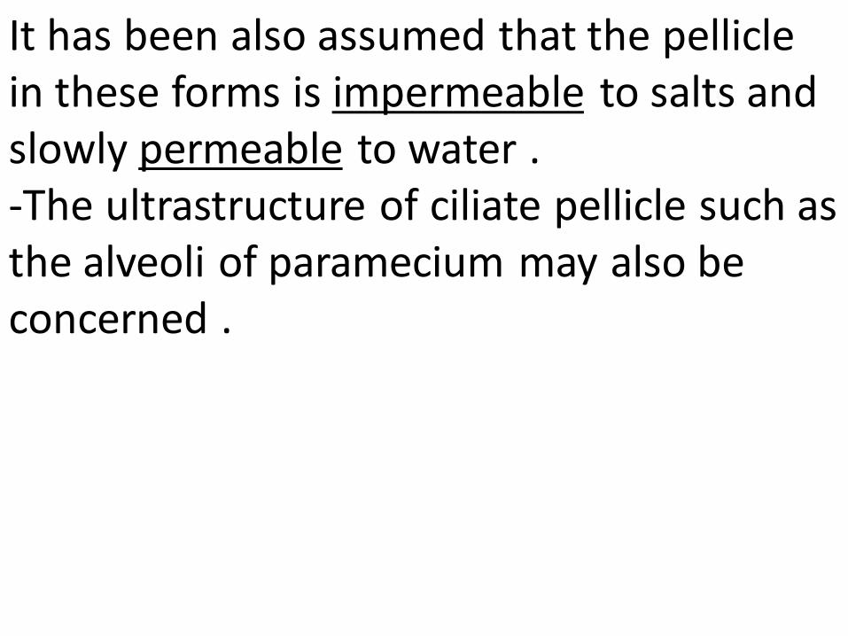
It has been also assumed that the pellicle in these forms is impermeable to salts and slowly permeable to water . -The ultrastructure of ciliate pellicle such as the alveoli of paramecium may also be concerned .
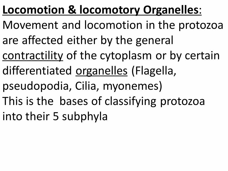
Locomotion & locomotory Organelles: Movement and locomotion in the protozoa are affected either by the general contractility of the cytoplasm or by certain differentiated organelles (Flagella, pseudopodia, Cilia, myonemes) This is the bases of classifying protozoa into their 5 subphyla
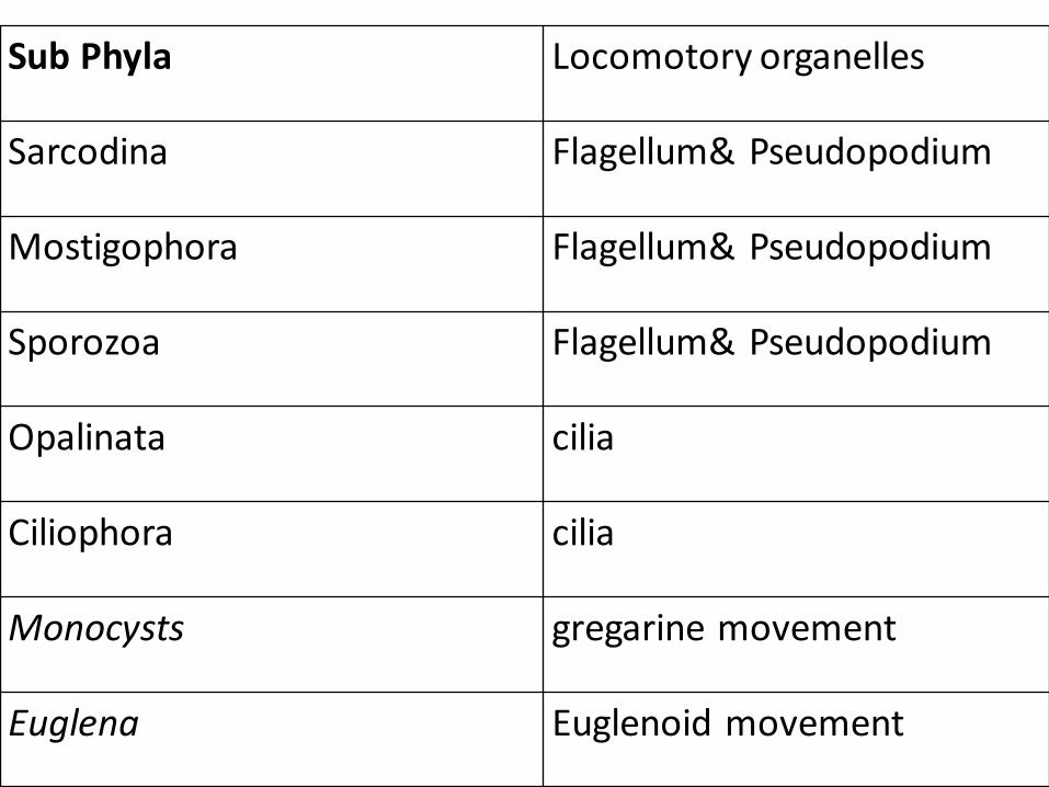
Sub Phyla Locomotory organelles
Sarcodina Flagellum& Pseudopodium
Mostigophora Flagellum& Pseudopodium
Sporozoa Flagellum& Pseudopodium
Opalinata cilia
Ciliophora cilia
Monocysts gregarine movement
Euglena Euglenoid movement
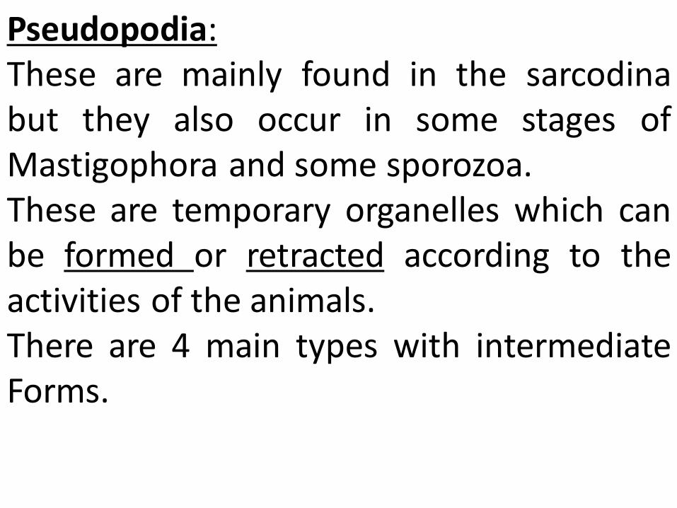
Pseudopodia: These are mainly found in the sarcodina but they also occur in some stages of Mastigophora and some sporozoa. These are temporary organelles which can be formed or retracted according to the activities of the animals. There are 4 main types with intermediate Forms.
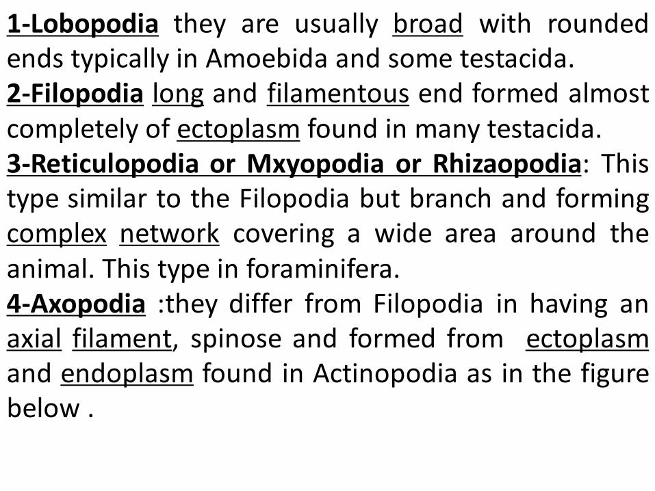
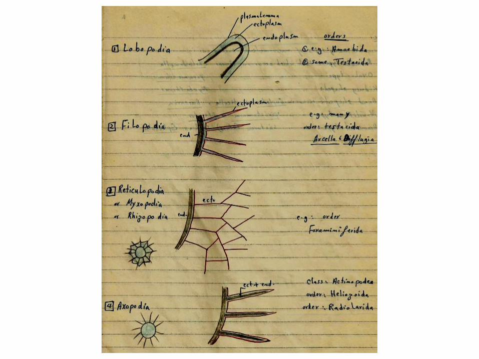
1-Lobopodia they are usually broad with rounded ends typically in Amoebida and some testacida. 2-Filopodia long and filamentous end formed almost completely of ectoplasm found in many testacida. 3-Reticulopodia or Mxyopodia or Rhizaopodia: This type similar to the Filopodia but branch and forming complex network covering a wide area around the animal. This type in foraminifera. 4-Axopodia :they differ from Filopodia in having an axial filament, spinose and formed from ectoplasm and endoplasm found in Actinopodia as in the figure below .
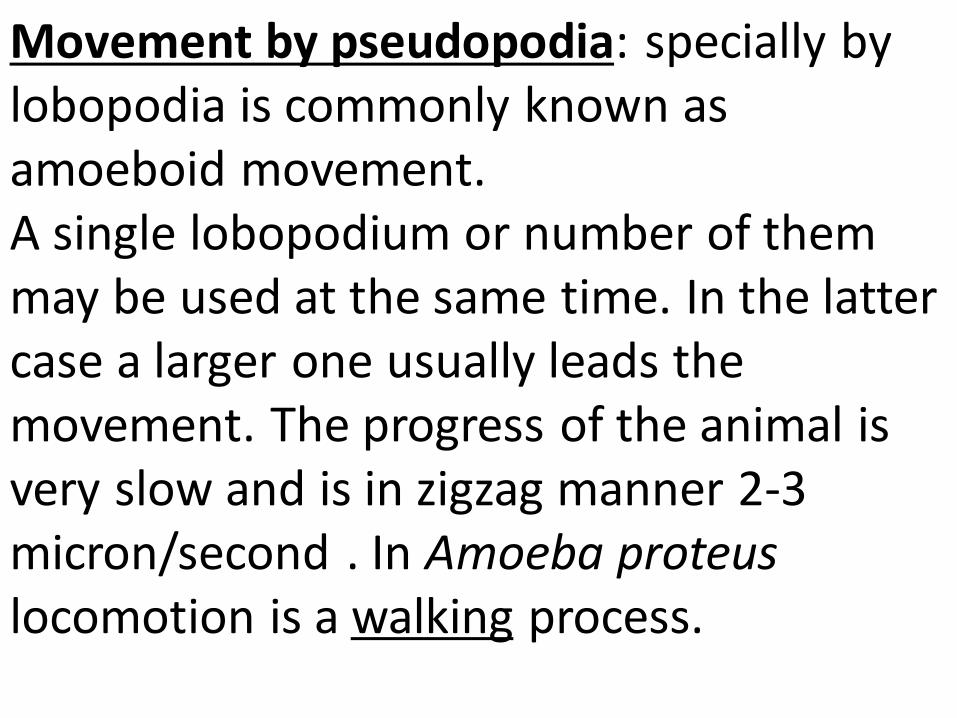
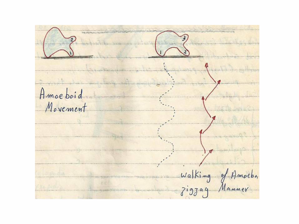
Movement by pseudopodia: specially by lobopodia is commonly known as amoeboid movement. A single lobopodium or number of them may be used at the same time. In the latter case a larger one usually leads the movement. The progress of the animal is very slow and is in zigzag manner 2-3 micron/second . In Amoeba proteus locomotion is a walking process.
Foraminifera can creep by their myxopodia (reticulopodia) is similar manner, the progress by axopodia is very limited. Both myxpodia and axopodia are formed mostly in floating organisms and they are used primarly for food capture.
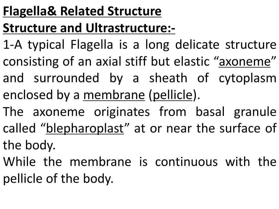
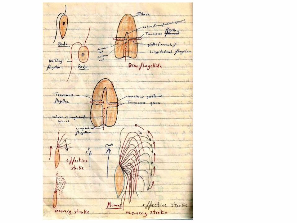
Flagella& Related Structure
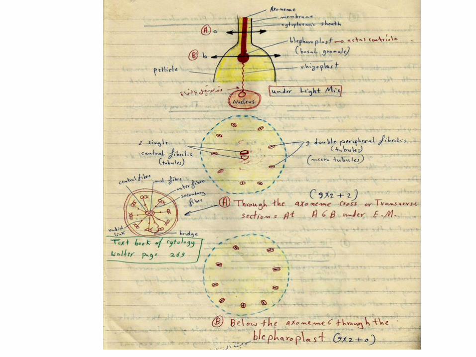
Structure and Ultrastructure:- 1-A typical Flagella is a long delicate structure consisting of an axial stiff but elastic “axoneme” and surrounded by a sheath of cytoplasm enclosed by a membrane (pellicle). The axoneme originates from basal granule called “blepharoplast” at or near the surface of the body. While the membrane is continuous with the pellicle of the body.
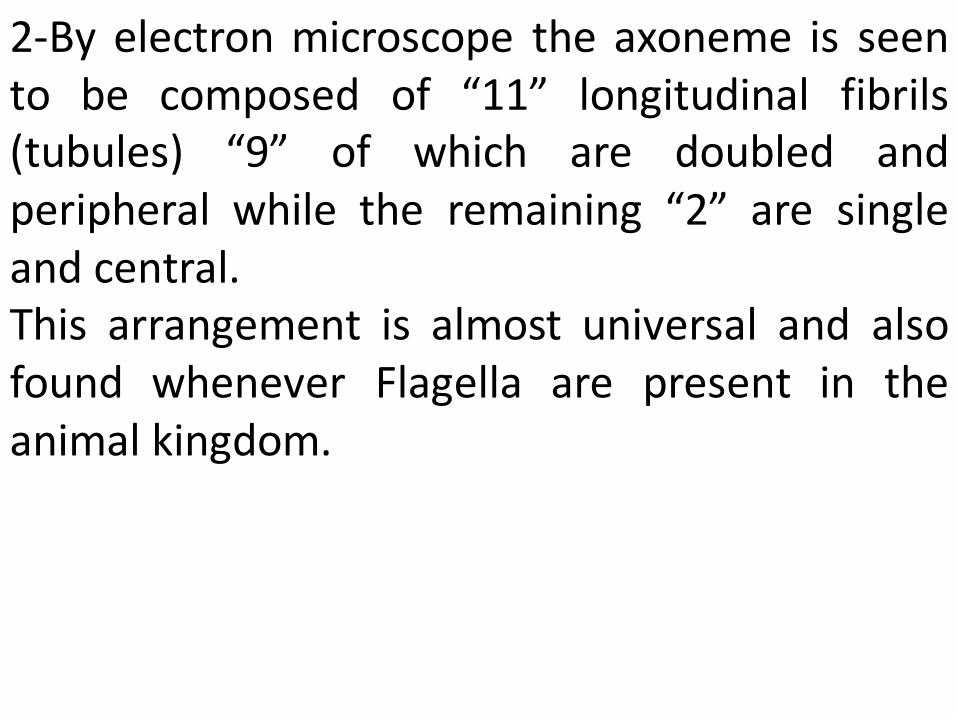
2-By electron microscope the axoneme is seen to be composed of “11” longitudinal fibrils (tubules) “9” of which are doubled and peripheral while the remaining “2” are single and central. This arrangement is almost universal and also found whenever Flagella are present in the animal kingdom.
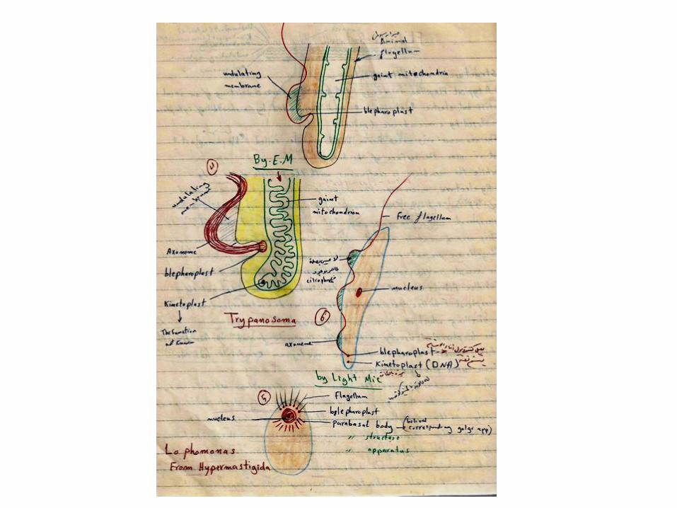
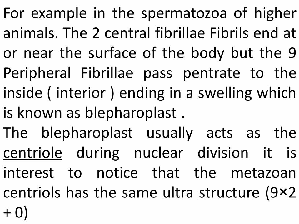
For example in the spermatozoa of higher animals. The 2 central fibrillae Fibrils end at or near the surface of the body but the 9 Peripheral Fibrillae pass pentrate to the inside ( interior ) ending in a swelling which is known as blepharoplast . The blepharoplast usually acts as the centriole during nuclear division it is interest to notice that the metazoan centriols has the same ultra structure (9×2 + 0)
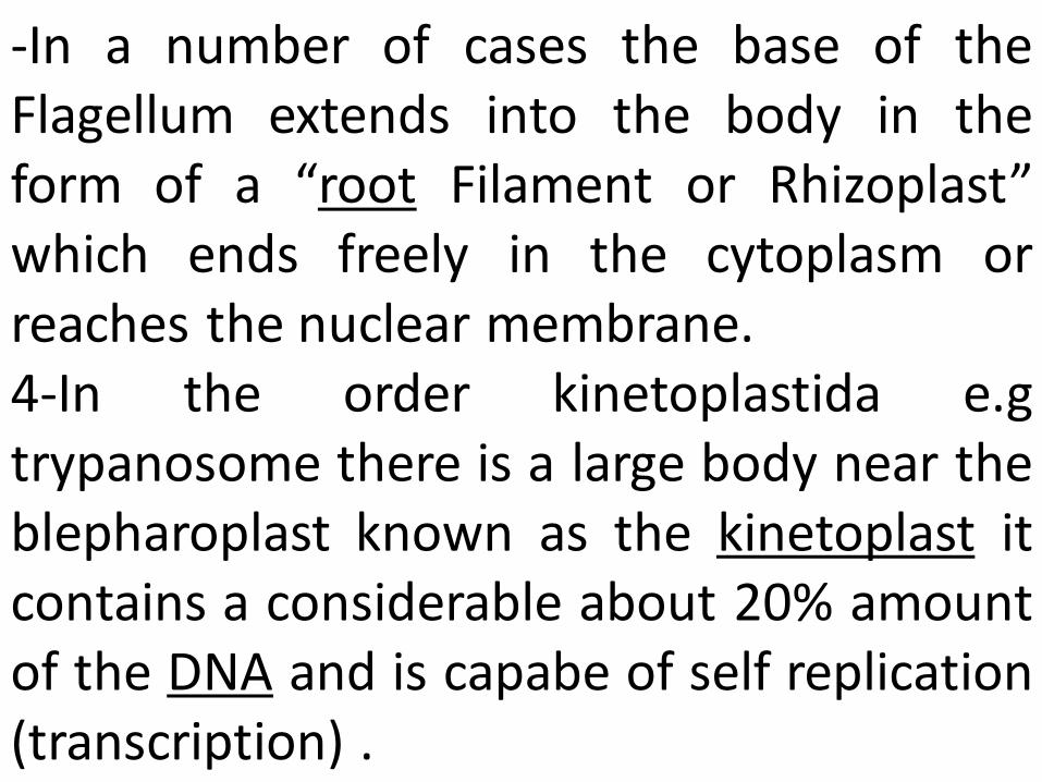
-In a number of cases the base of the Flagellum extends into the body in the form of a “root Filament or Rhizoplast” which ends freely in the cytoplasm or reaches the nuclear membrane. 4-In the order kinetoplastida e.g trypanosome there is a large body near the blepharoplast known as the kinetoplast it contains a considerable about 20% amount of the DNA and is capabe of self replication (transcription) .
A modified enlarged region of the single giant mitochondrian are found in these Flagellates because the kinetoplast is closely associated with the bleopharoplast it is thought that it is concerning which supplying energy for the activity of Flagellum. But the real function of the kinetoplast is not known so at present time it is an important subject for research.
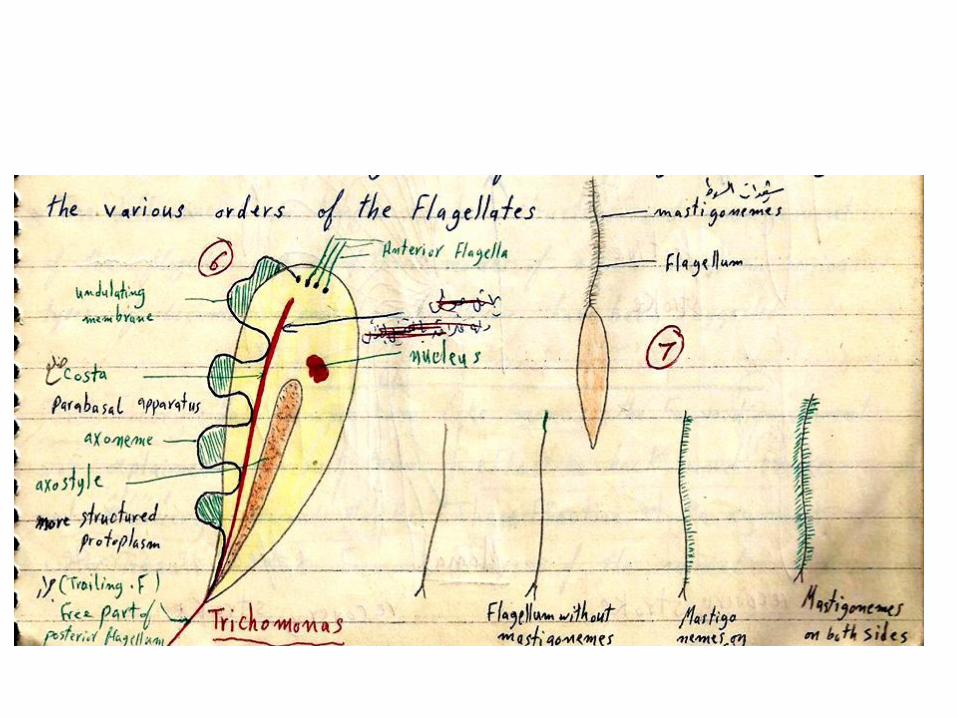
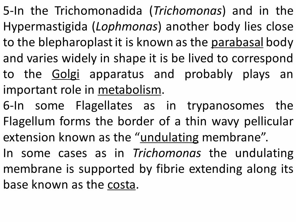
5-In the Trichomonadida (Trichomonas) and in the Hypermastigida (Lophmonas) another body lies close to the blepharoplast it is known as the parabasal body and varies widely in shape it is be lived to correspond to the Golgi apparatus and probably plays an important role in metabolism. 6-In some Flagellates as in trypanosomes the Flagellum forms the border of a thin wavy pellicular extension known as the “undulating membrane”. In some cases as in Trichomonas the undulating membrane is supported by fibrie extending along its base known as the costa.
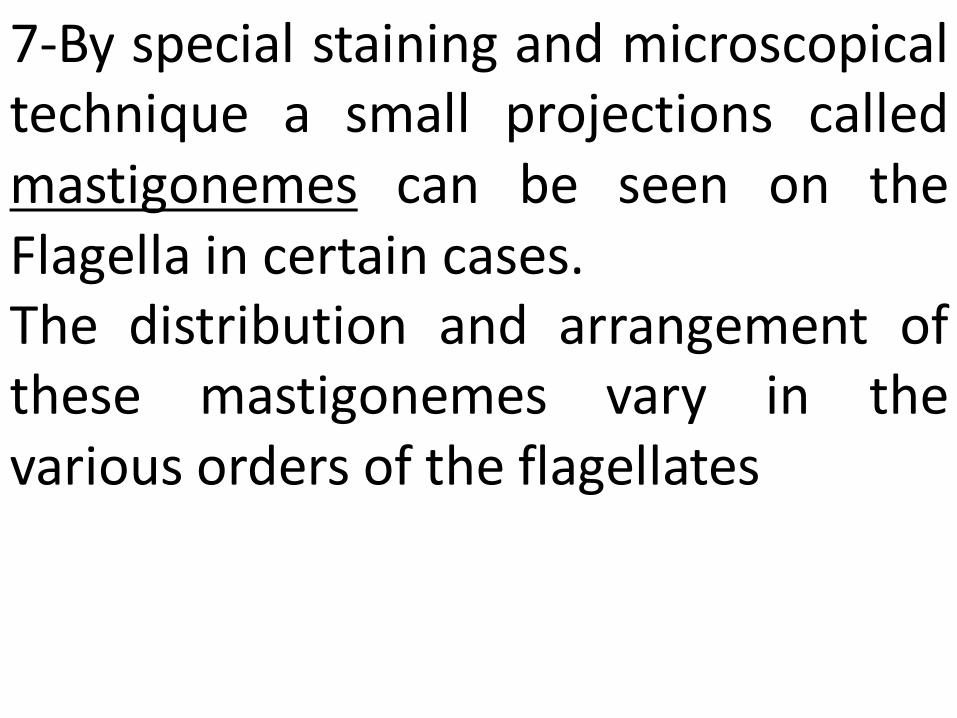
7-By special staining and microscopical technique a small projections called mastigonemes can be seen on the Flagella in certain cases. The distribution and arrangement of these mastigonemes vary in the various orders of the flagellates
Number & Distribution of Flagella
As a rule, the number of Flagella in the animal is small from 1-8, but in Hypermastigida they are numerous (e.g. Trichonympha).
•When there is more than one the Flagella often differ in size, structure and function, one of them maybe directed backwards as a trailing Flagella as in Bodo, or attached to an undulating membrane, for example Trypanosoma and Trichomonas. •In typical dinoflagellates there are 2- Flagella: •A transverse one around the body. A–longitudinal one usually directed posteriorly