Embed Size (px)
Citation preview

Knee is like a round ball on a flat surface
Ligaments provide most of the support to the knees
Little structure or support from the bones

First-degree: mild minimal signs and symptoms, minimal functional loss and resolves in a few days.
second-degree: moderate- partial structural disruption, swollen tender, may show some signs of instability. Performance deficit for up to 6 weeks.
Third-degree: severe extensive structural disruption, extensive swelling, severe pain, joint unstable. Performance deficit. Minimum 6-8 weeks.

Third-degree: Grade I: less than a 0.5-cm opening of the joint surfaces
Grade II: a 0.5- to 1-cm opening of the joint surface
grade III: a rupture larger than a 1-cm opening

Ligaments are slow to heal due to their hypovascular nature.
Pathologically ligaments are a type of dense connective tissue, 90% type I collagen, 9% type III collagen and 1% fibroblast cells

FACTORS : Degree of spain What? Where? Age Demand Assosiated injury

RICE Bracing Strengthening Functional brace

Repair : surgical treatment of acute injuries(Optimal surgical dissection and repair become increasingly difficult beyond 7 to 10 days after injury)
Reconstruction : usually refers to surgical treatment of ligamentous laxity several months after injury


An ACL injury (either grade I, II or III) can occur during the following:
Sudden hyperextension of the knee. Body weight twisting across the knee joint
causing a shearing force while the foot is still planted on the ground.
Sudden deceleration.
The ACL provides both mechanical stability and proprioceptive feedback to the knee.
Restrains anterior translation of the tibia on the femur.
Prevents hyper-extension of the knee. Secondary stabilizer to valgus stress Controls rotation of the tibia on femur in the
last 30 degrees of knee extension. (part of the locking mechanism)

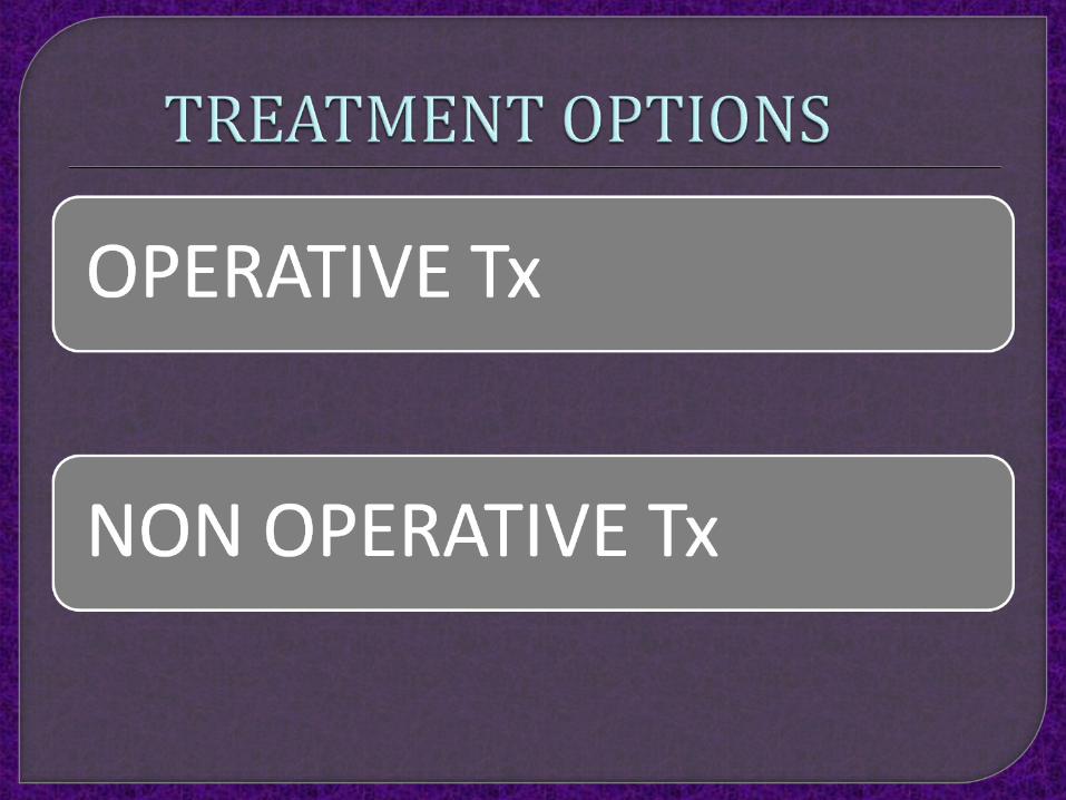
Operative vs. Non-operative• Demand level• Age• lifestyle• Other lesions
The decision to reconstruct an ACL tear should be based not only on the presence of symptomatic instability, but also on the lifestyle and activity level of the patient.
Age is’nt base of guide line for reconstruction
because the more important factor is the overall level of activity.
Consequently, age itself should not be a contraindication to ACL reconstruction.
Symptomatic patients with a more sedentary lifestyle and those who are willing to modify their level of activity can be considered for nonoperative treatment,

TIMING: No swelling
Good leg control
Full ROM (full hyperextension)
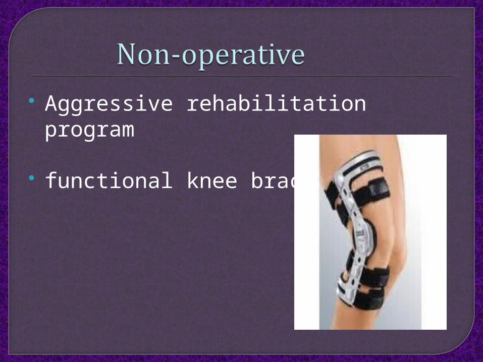
Aggressive rehabilitation program
functional knee brace

Healing is good: Blood supply
Relatively wide surface area
Association with other secondary stabilizers
Extra-articular location.
Shockwave
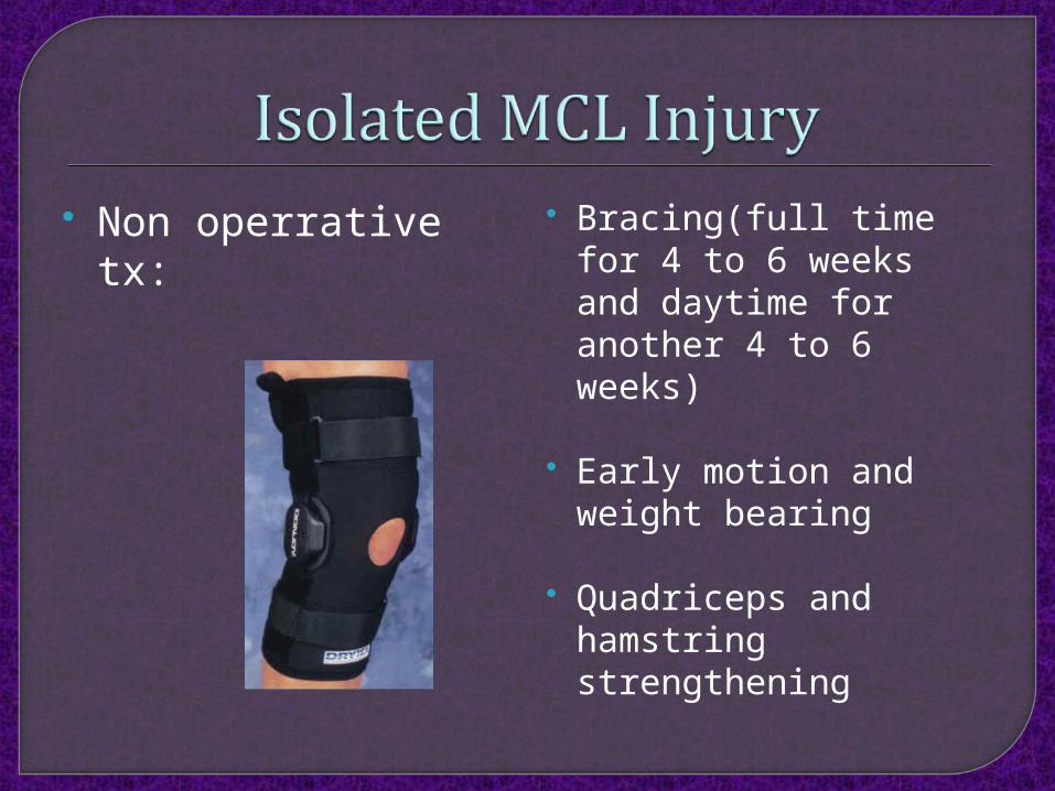
Non operrative tx: Bracing(full time for 4 to 6 weeks and daytime for another 4 to 6 weeks)
Early motion and weight bearing
Quadriceps and hamstring strengthening

Operative tx : Large bony avulsions identified on radiographs
Stener-type lesions of the distal MCL
patients with persistent functional valgus instability after nonoperative treatment

Nonoperative treatment of the MCL ACL reconstruction For chronic ACL tears with residual valgus
instability, simultaneous reconstruction of the ACL and MCL.
ACL/PCL/MCL injuries with reconstruction of all injured ligaments

PCL injuries are present in up to 3% of knee injuries in the general population and as many as 37% of knee injuries in trauma patients with acute hemarthrosis.
PCL injury typically results following an excessive posteriorly directed force on the tibia

Non operative Tx : Low-grade isolated PCL injuries
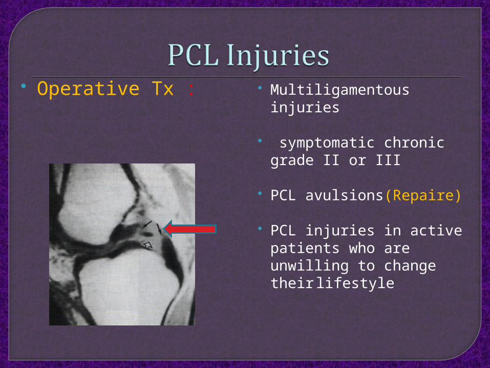
Operative Tx : Multiligamentous injuries
symptomatic chronic grade II or III
PCL avulsions(Repaire)
PCL injuries in active patients who are unwilling to change their lifestyle

Less commonly injured than the cruciate ligaments or the medial knee ligament complex.
Associated posterolateral corner injuries provide a potential source of residual instability following anterior cruciate ligament and posterior cruciate ligament reconstruction
Can lead to reconstruction graft failure
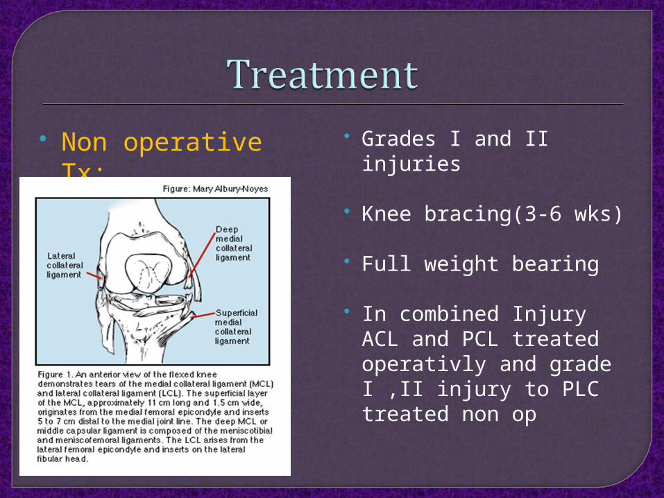
Non operative Tx: Grades I and II injuries
Knee bracing(3-6 wks)
Full weight bearing
In combined Injury ACL and PCL treated operativly and grade I ,II injury to PLC treated non op

Operatve Tx: Grade III injuries
Combined ACL ,PCL,PLC concurrent repair or reconstruction
Repair and Reconstruction

Intrasubstance repairs of the fibular collateral ligament and popliteus have not fared well and therefore should not be performed.
Other structures of the PLC areamenable to intrasubstance repair. These include the coronary ligament of the lateral meniscus, meniscofemoral and meniscotibial ligaments, and fibers of the popliteomeniscal ligaments

Reconstruction better and had fewer failures (9% vs. 37%) than the repair
These include nonanatomic and anatomic techniques.
Operative management provides improved outcomes compared with nonoperative
Early surgical management (within 3 weeks) is better