Embed Size (px)
Citation preview

اللهاللهبسم بسم الرحمن الرحيمالرحمن الرحيم
Interpretation of urine cytology
Nashwa Emara M.D.,phdASS. Prof. Pathology
Function
• Majority of UT malignancies are urothelial CA.
• The main function of urine cytology is diagnosis of UC.
IndicationsDiagnosis of symptomatic patients Diagnosis of symptomatic patients (hematuria).(hematuria).
Screen high risk patients (industrial Screen high risk patients (industrial chemicals, metals, etc.)chemicals, metals, etc.)
Follow-up patients with UT neoplasia.Follow-up patients with UT neoplasia.
Complementary to cystoscopy and biopsy: Complementary to cystoscopy and biopsy: detect small and hidden lesions (diverticuli, detect small and hidden lesions (diverticuli, ureters, renal pelvis)..ureters, renal pelvis)..
Urine cytology is the most reliable method Urine cytology is the most reliable method for detecting urothelial CIS (>biopsies).for detecting urothelial CIS (>biopsies).
Types of SpecimensVoided urineVoided urine (avoid 1st morning (avoid 1st morning specimens) specimens)
Catheterized urineCatheterized urine (in Females) (in Females)
Washings/Brushings Washings/Brushings
Superior to voided urine but localized, Superior to voided urine but localized, may not sample upper urinary tract may not sample upper urinary tract and urethra and urethra
Ileal conduit urineIleal conduit urine
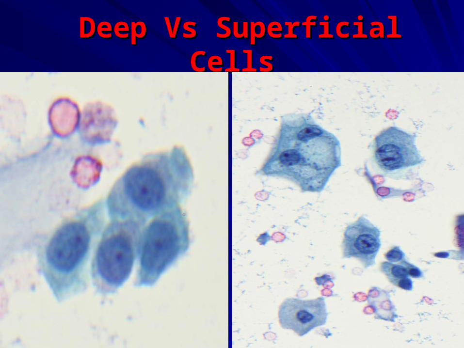
Deep Vs Superficial CellsDeep Vs Superficial Cells
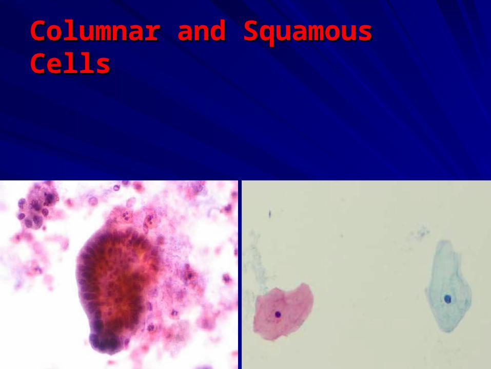
Columnar and Squamous CellsColumnar and Squamous Cells
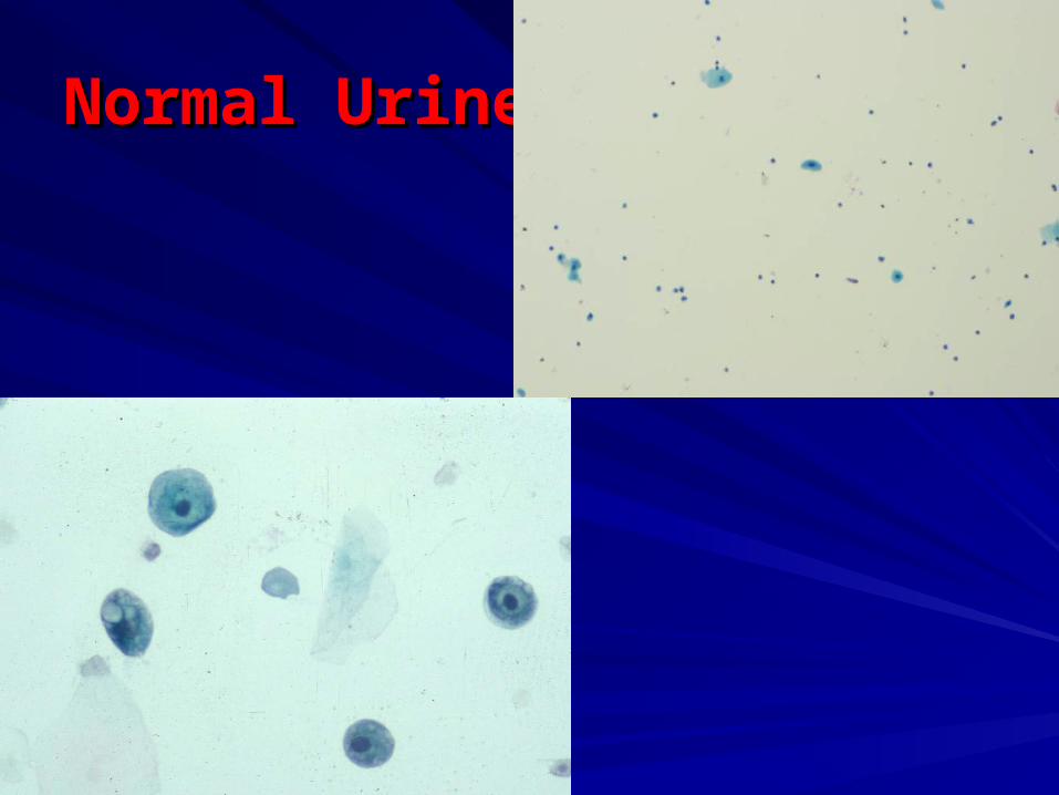
Normal Urine CytologyNormal Urine Cytology
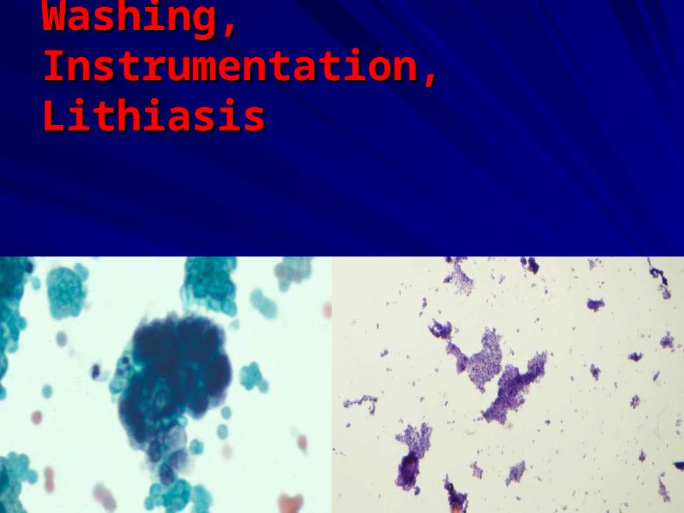
Washing, Instrumentation, Washing, Instrumentation, LithiasisLithiasis
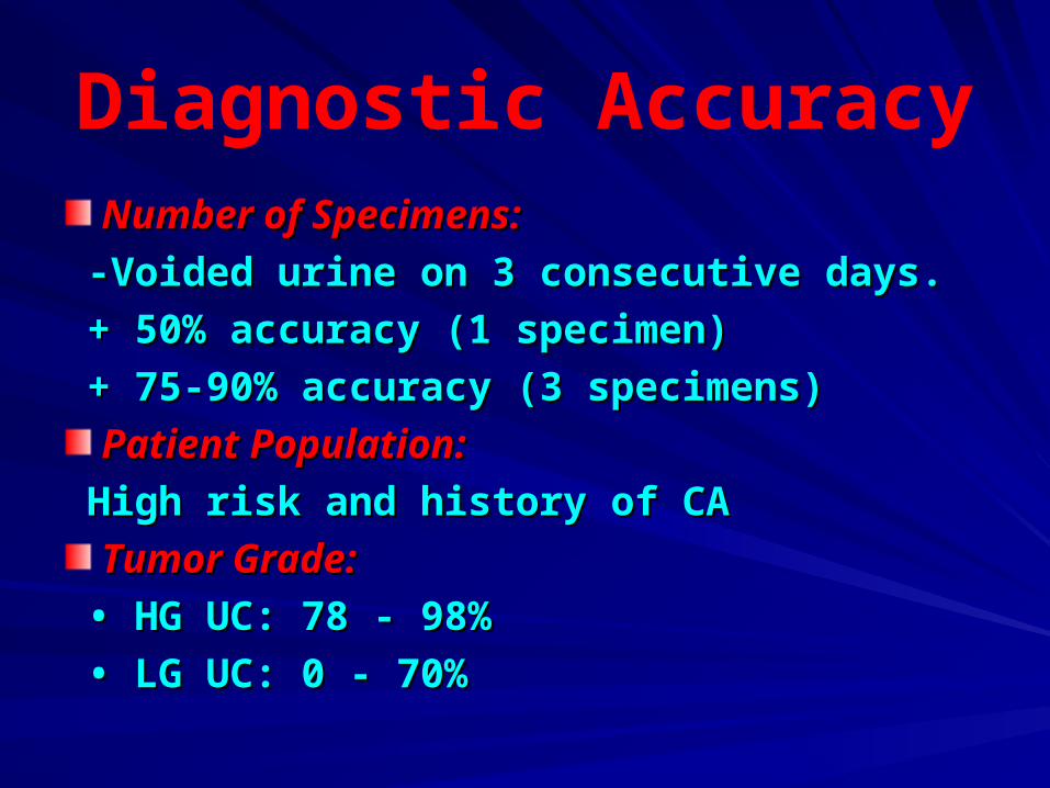
Diagnostic AccuracyNumber of Specimens:Number of Specimens:
-Voided urine on 3 consecutive days.-Voided urine on 3 consecutive days.
+ 50% accuracy (1 specimen)+ 50% accuracy (1 specimen)
+ 75-90% accuracy (3 specimens)+ 75-90% accuracy (3 specimens)
Patient Population:Patient Population:
High risk and history of CAHigh risk and history of CA
Tumor Grade:Tumor Grade:
• • HG UC: 78 - 98%HG UC: 78 - 98%
• • LG UC: 0 - 70%LG UC: 0 - 70%
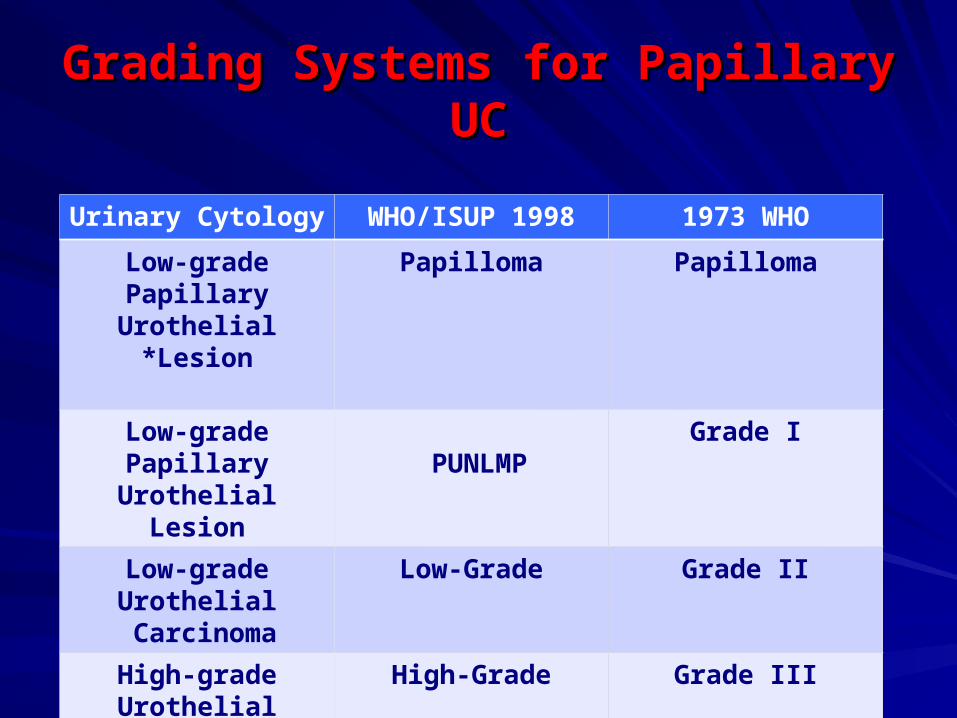
Grading Systems for Papillary UCGrading Systems for Papillary UC
1973 WHO1998 WHO/ISUPUrinary Cytology
PapillomaPapillomaLow-grade Papillary Urothelial Lesion*
Grade IPUNLMP
Low-grade Papillary Urothelial Lesion
Grade IILow-GradeLow-grade Urothelial Carcinoma
Grade IIIHigh-GradeHigh-grade Urothelial Carcinoma
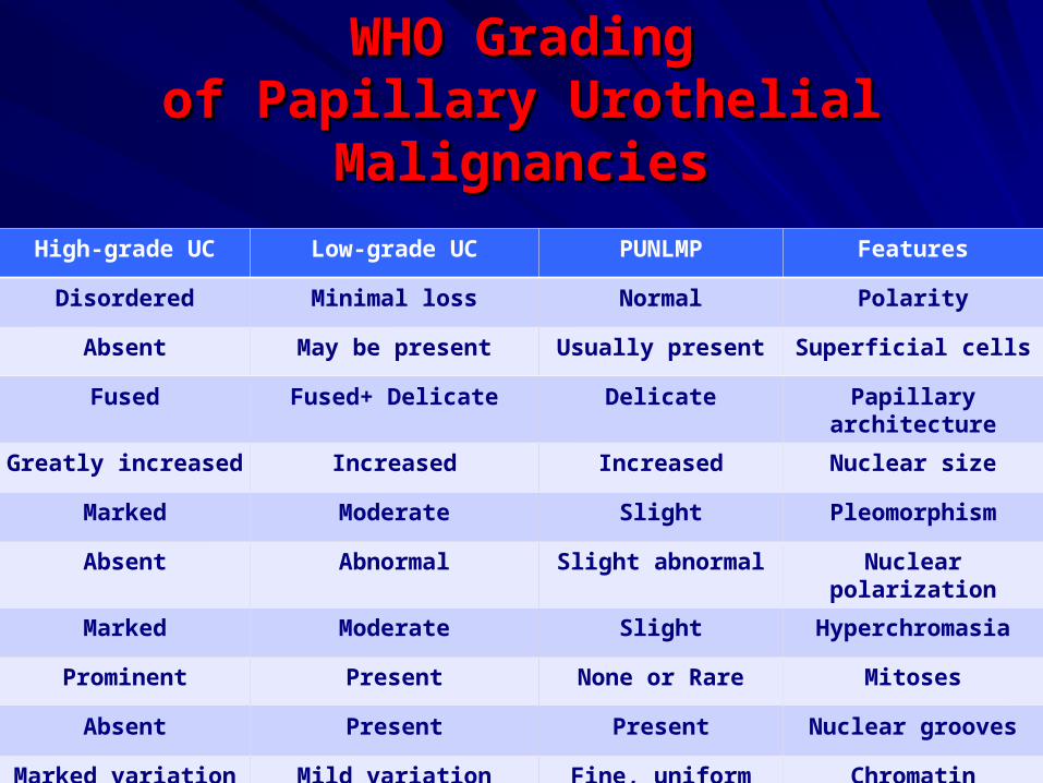
WHO GradingWHO Gradingof Papillary Urothelial Malignanciesof Papillary Urothelial Malignancies
FeaturesPUNLMPLow-grade UCHigh-grade UC
PolarityNormalMinimal lossDisordered
Superficial cellsUsually presentMay be presentAbsent
Papillary architectureDelicateFused+ DelicateFused
Nuclear sizeIncreasedIncreasedGreatly increased
PleomorphismSlightModerateMarked
Nuclear polarizationSlight abnormalAbnormalAbsent
HyperchromasiaSlightModerateMarked
MitosesNone or RarePresentProminent
Nuclear groovesPresentPresentAbsent
ChromatinFine, uniformMild variationMarked variation
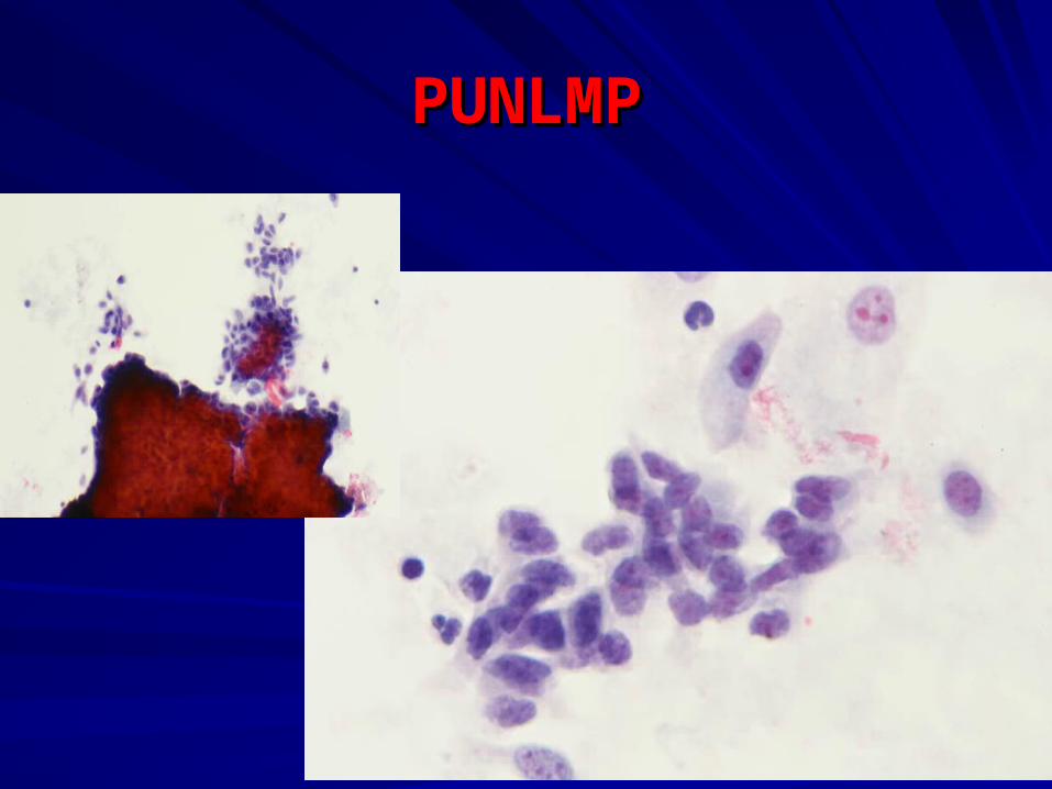
PUNLMPPUNLMP
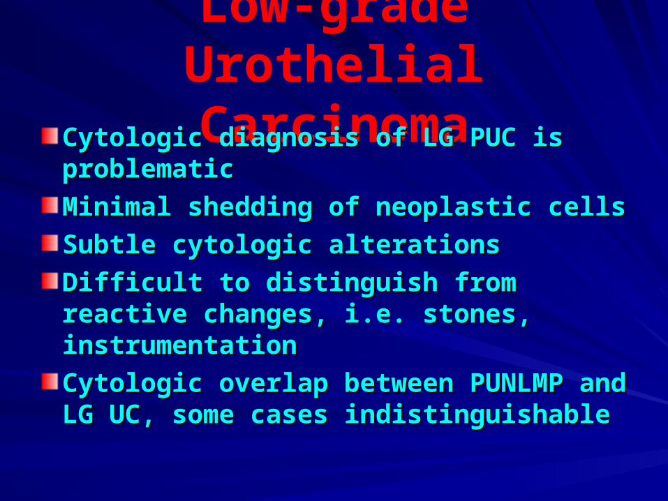
Low-grade Urothelial CarcinomaCytologic diagnosis of LG PUC is Cytologic diagnosis of LG PUC is
problematicproblematic
Minimal shedding of neoplastic cellsMinimal shedding of neoplastic cells
Subtle cytologic alterationsSubtle cytologic alterations
Difficult to distinguish from reactive Difficult to distinguish from reactive changes, i.e. stones, instrumentation changes, i.e. stones, instrumentation
Cytologic overlap between PUNLMP and Cytologic overlap between PUNLMP and LG UC, some cases indistinguishable LG UC, some cases indistinguishable
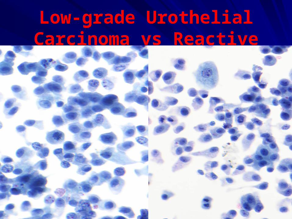
Low-grade Urothelial Carcinoma vs Reactive
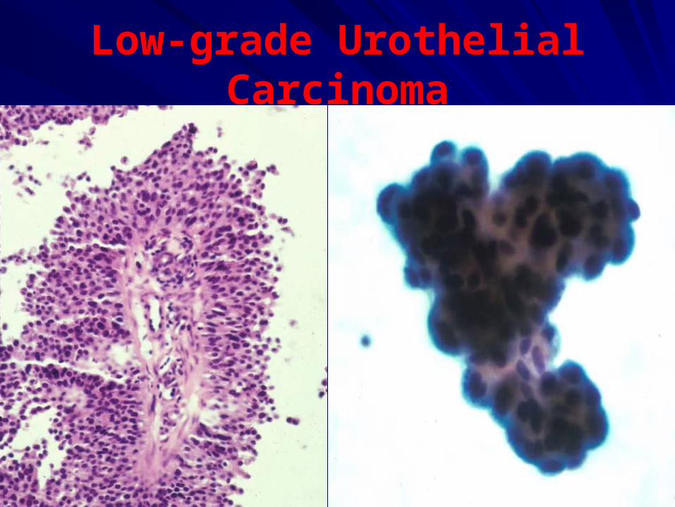
Low-grade Urothelial Carcinoma
Diff. Diag. of LGUC
Reactive/reparative changesReactive/reparative changes
Instrumentation effectInstrumentation effect
LithiasisLithiasis
Upper urinary tract samplingUpper urinary tract sampling
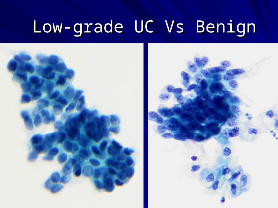
Low-grade UC Vs BenignLow-grade UC Vs Benign
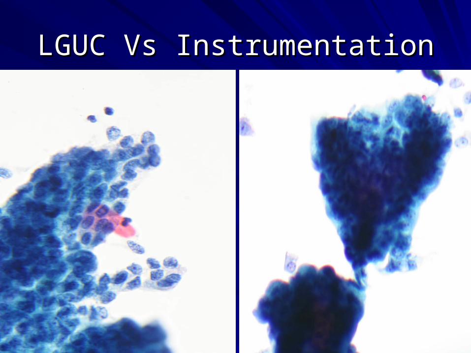
LGUC Vs InstrumentationLGUC Vs Instrumentation
Instrumentation Effect
Catheterized urine & bl. wash specimens.Catheterized urine & bl. wash specimens.
Large pseudopapillary groups and 3D Large pseudopapillary groups and 3D clusters.clusters.
Nuclear overlap and crowding.Nuclear overlap and crowding.
Low N/C ratio. Low N/C ratio.
Finely granular chromatin with even Finely granular chromatin with even distribution.distribution.
Well defined cytoplasmic borders.Well defined cytoplasmic borders.
Nuclear palisading at periphery of clusters Nuclear palisading at periphery of clusters with abundant cytoplasm.with abundant cytoplasm.
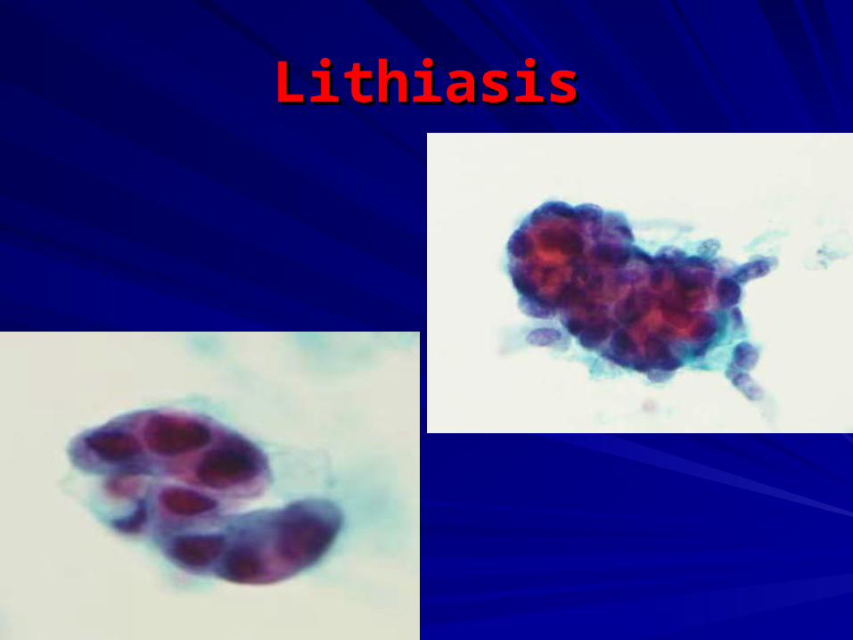
LithiasisLithiasis
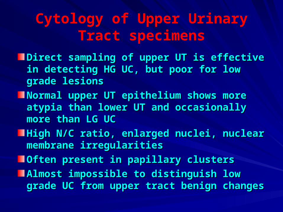
Cytology of Upper Urinary Tract specimens
Direct sampling of upper UT is effective in Direct sampling of upper UT is effective in detecting HG UC, but poor for low grade detecting HG UC, but poor for low grade lesionslesions
Normal upper UT epithelium shows more Normal upper UT epithelium shows more atypia than lower UT and occasionally more atypia than lower UT and occasionally more than LG UCthan LG UC
High N/C ratio, enlarged nuclei, nuclear High N/C ratio, enlarged nuclei, nuclear membrane irregularitiesmembrane irregularities
Often present in papillary clustersOften present in papillary clusters
Almost impossible to distinguish low grade Almost impossible to distinguish low grade UC from upper tract benign changes UC from upper tract benign changes
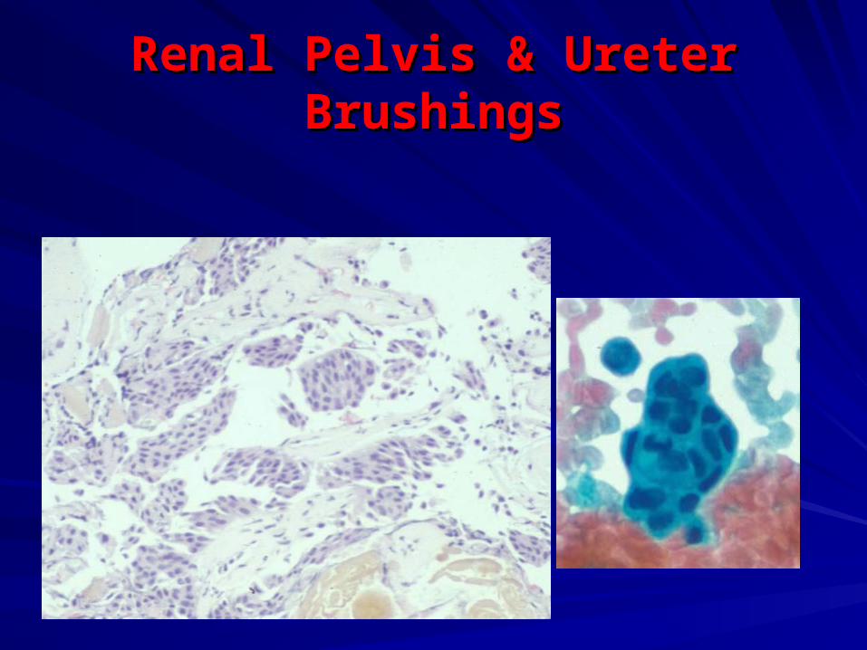
Renal Pelvis & Ureter BrushingsRenal Pelvis & Ureter Brushings
High-grade Urothelial Carcinoma
Often invasive, 70 mortality.Often invasive, 70 mortality.
Can not reliably separate CIS from Can not reliably separate CIS from invasive high-grade UC.invasive high-grade UC.
High diagnostic accuracy of cytology:High diagnostic accuracy of cytology:
- Sensitivity 80 %.- Sensitivity 80 %.
- Specificity > 95%.- Specificity > 95%.
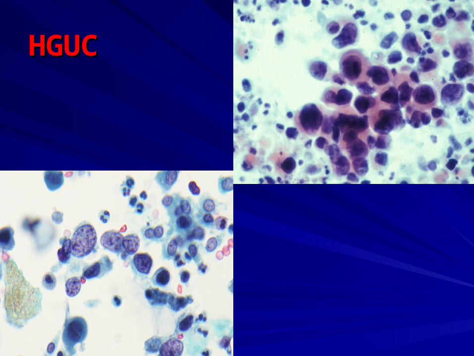
HGUCHGUC
Diff. Diag. of HGUC
Viral infectionViral infection
Therapy effectTherapy effect
Degenerative and reactive Degenerative and reactive changeschanges
Upper urinary tract specimensUpper urinary tract specimens
StonesStones
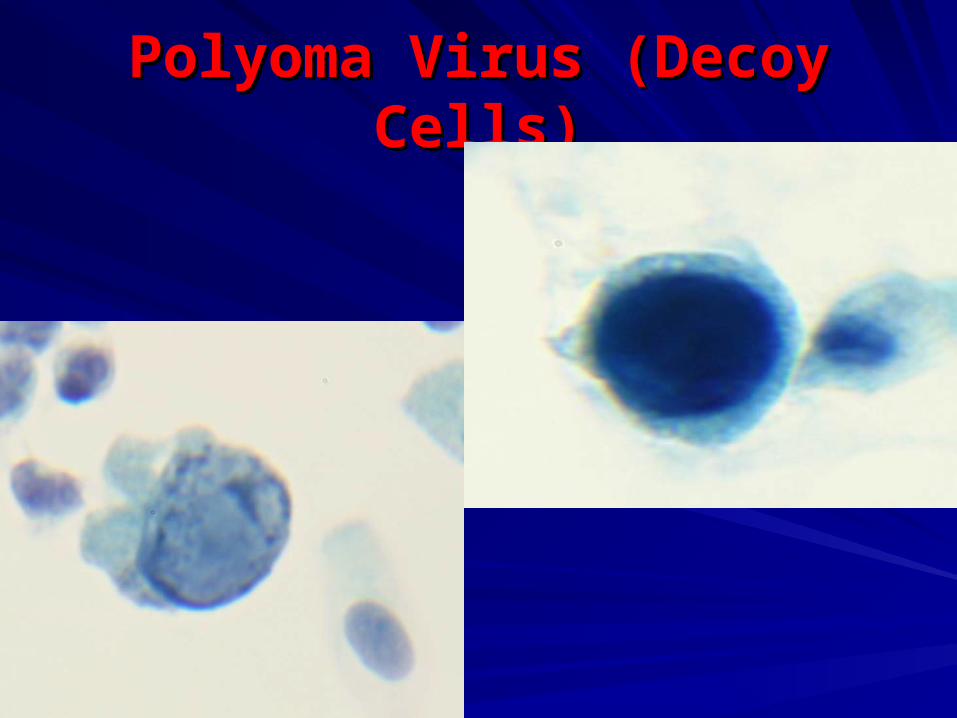
Polyoma Virus (Decoy Cells)Polyoma Virus (Decoy Cells)
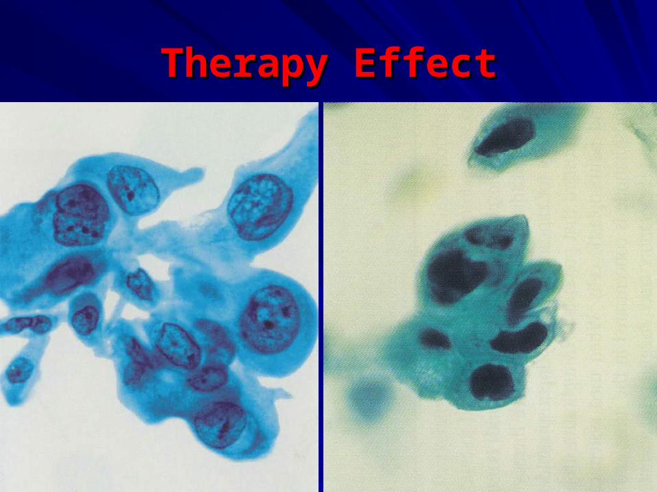
Therapy EffectTherapy Effect
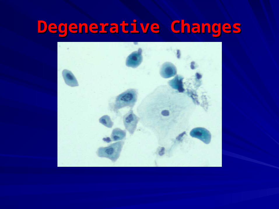
Degenerative ChangesDegenerative Changes
Diagnostic categories
NegativeNegative
Atypical, rule out LGUC Atypical, rule out LGUC /PUNLMP/PUNLMP
Suspicious for HG UC/ Suspicious for HG UC/ malignancymalignancy
HG UC/ other HG UC/ other malignanciesmalignancies(Murphy)(Murphy)
Summary Urothelial neoplasms can be separated into Urothelial neoplasms can be separated into 2 main categories:2 main categories:
– –Low grade neoplasia (PUNLMP and LG UC).Low grade neoplasia (PUNLMP and LG UC).
– –High grade UC.High grade UC.
Urine cytology best applied to HG UC.Urine cytology best applied to HG UC.
Cytology less helpful for detecting and Cytology less helpful for detecting and monitoring LG neoplasms.monitoring LG neoplasms.
– –Not major limitation.Not major limitation.
– –LG neoplasms rarely aggressive and can be LG neoplasms rarely aggressive and can be readily detected by cystoscopy.readily detected by cystoscopy.
N.B.:N.B.: Ancillary techniques are highly sensitive Ancillary techniques are highly sensitive poorly specific, not for routine use poorly specific, not for routine use

GOOD LUCKGOOD LUCK..…..…