Embed Size (px)
Citation preview

*AD-A280 359
sIcuOT.- ..REPORT DOCUMENTATION PAGE
A, I T r" ATION " ,. FIESTRICTIVE MARKINGS
LE N UNCLASSIFIED/A
2& St C"Prf"4 TY3. OiSTAIBUTIONIAVA4LABILITY OP AEPORIT
i ,Approved for publio release;
2b-' P rCATr OOR CMEOULE distribution 1M 9l e d6. PERFORMING ORG NIZATION REPORT NUMIERIS) 5. MONITORING ORGANIZATION REPORT NUMSERIS)
AMUR-1. 94 0346;
Ga. NAME OP PERFORMING ORGANIZATION . OFFICE SYbOL,. 7&. NAME OF MONITORING ORGANIZATION
Indiana University AFOSR/LG& ADDRESS iCity. State dad ZIP Code) lb. AOORESS (City. State end ZIP Co"e
4601 Central Avenue Building 410Columbus IN 47203 Bolling AFB DC 20332-6448
da. NAME OF PUNOING/SPONSO0ING ob. OFFICE SYMBOL 9. PROCUREMENT INSTRUMENT IOENTIFICATION NUMBER
ORGANI ZATION ifI .p"iablei
AFOSR NL AFOSR - F49620-93-1-0297DEF
ft. ADORESS icily. State and ZIP Codul 10. SOURCE OF FUNOING NO&.
kko O.AQ.-CCK lcoc- zolA-e ?:2&S PROGRAM PROJECT TASK WORK U.NITBuilding 410 ELEMENT NO. NO. NO. O
Bolling AFB DC 20332-644811. TITLE ,(include SeCurft Clawafeicaon) Two-dimensional prote •n 61102F 2312 A5
•t~D rconition in cmia nvieitv
12. PERSONAL AUTHORISIFrank A. Witzmann, Ph.D.
13&. TYPE OP REPORT MLb TIME COVERED j4. DATE OP REPORT (Yr.. Mo.. Day) S PAGE COUNT
Annual PROM I Apr 93To30 Mar O94 Apr 20 4216. SUPPLEMENTAR Y NOTATION
17. COSATI COOES ,I& SUBJECT TERMS (Coufiuui on reverse it aces"ry and identfy by b•€ number)
FILDO GRtouP UsB. aR.- rat liver, rat kidney, rat testis, perfluorcarboxylic acidperoxisome proliferator, 2D protein electrophoresis, image
l l l •analy si s. Ir r l e ence . nattern re nnan it ion - ,
19. ABSTRACT (Conlidaue on M-1 a4 if neeNmy and id~d by111 M 011 0um114r
This report summarizes the progress made in the development of a two-dimensionil protdatabase for toxicity screening and mechanistic determination. Various chemicallydistinct peroxisome proliferators were compared with regard to their effect on the 2Dprotein pattern of various target tissues in the rodent. Protein alterations, novelidentifications, and future directions are described.
,4 6 14 15494-18490 W =caAr SET
2&. DISTRI*w I, .....w..- . ;TRACT 21. ABSTRACT SECURITY CLASSIFICATION
IUNCLASSIPID/UNLMITI'DIMSME AS RPT. 0 OTIC USERS -1 unclassified
22a. NAME OP RESPONSIBLE INDIVIDUAL 22b. TELEPIIONE NUMBER , 22c. OFFICE SYMBOLlinclude Amt Codea
Dr. Walt Kozumbo 202-767-5021 NL
DO FORM 1473, 83 APR EDITION OF 72 IS ,S... T• 0 MAY 73,2,,,7, LASIICAoN oI .,es ,,

t
Report AFOSR F49620-93-1-0297DEF A ~Rh94 0348i
Akproved for publi o releaOsdistribution utxD,•"O4o
TWO-DIMENSIONAL PROTEIN PATTERN RECOGNITION IN CHEMICAL TOXICITY
Frank A. Witzmann, Ph.D.Department of BiologyIndiana University-Purdue University at IndianapolisColumbus Campus4601 Central AvenueColumbus IN 47203
20 April 1994
Annual Report for Period 1 Apr 93 through 30 Mar 94
Accesiol For APrepared for: NTIS .,C.C I-
Dr. Walt Kozumbo U'.xl DDirectorate of Life Sciences Juitifc • ...........................AFOSRBuilding 410 By .........................................Boiling AFB DC 20332-6448 ODitributiO!' I
Availability Codes
AvaiD atialorDist special

t
Witzmann - Report 93-942
INTRODUCTION
The efforts of this laboratory during the research period have been directed at developing a predictivetoxicological approach based on target tissue protein pattern recognition. Through interaction with theToxicology Division of the Armstrong Laboratory and the unique capabilities we possess, our laboratory hascontinued the development and expansion of a two-dimensional protein pattern database for each of threerodent target tissues. By using large-scale two-dimensional electrophoresis of proteins (2D-PAGE)combined with computerized image analysis, we have used methods for comparing computer-recognized 2D-protein pattern alterations in various target tissues induced by specific chemical agents. These alterationshave been used to indicate and comparatively assess hepatotoxicity, nephrotoxicity, and testicular toxicity bydetecting specific patterns of protein alteration.
The effectiveness of the large-scale 2D-PAGE technique used in this laboratory results from theresolution of (in the case of the rat liver) nearly 1,500 cellular proteins in a single sample, first based on theircontent of acidic and basic amino acids (isoelectric focusing) and second by molecular weight (SDSelectrophoresis)(Andersen et aL., 1981; George and Andersen, 1986; Olson and Andersen, 1983). Incombination, these two separation techniques produce a two dimensional protein pattern unique for eachtissue or group of cells tested. Individual proteins within the pattern can be analyzed for alterations in volume(density), charge, and molecular weight. Changes in volume or spot density reflect alterations in a protein'sabundance and suggest up- or down-regulation of the genome or altered protein turnover rates. Chargemodifications suggest either posttranslational modification such as phosphorylation, ribosylation, conjugationor amino acid substitutions resulting from point mutations in the genome.
Regardless of the type of changes observed, a well-resolved 2D protein pattern, or 2D protein map(fingerprint), provides a pattern realistically containing 1000-2000 proteins and is thus a significant source ofinformation regarding the health/activity of a particular cell/tissue type and its response to toxic insult. Withthe application of computerized imaging of the protein patterns generated by 2D-PAGE, its use in patternrecognition for animal model target tissues, and the recent advent of microsequencing proteins isolated from2D gel patterns (Hughes et aL., 1992), 2D-PAGE has become an even more powerful technique in toxicitytesting; one with increasingly well-documented potential (Anderson, 1990). For example, Hochstrasser et al.(1992) have reviewed the clinical (diagnostic) applications of high resolution, large-scale 2D electrophoresisand its use in the clinical analysis of human body fluids, blood cells, and various tissues. Correspondingly,the long-term objective of the present investigation is to generate a 2D protein database for toxicologictargets, one that can be used to analyze chemical effects both in vivo and in vitro.
This report details the progress made during the research period 1 April 1993 to 30 March 1994 ineach of the following areas:
O Perfluorocarboxylic acid toxicology (comparative peroxisome proliferator toxicity) - patternalterations in 2D protein maps
O Database Development-identification of proteins via peptide mass fingerprinting-MW estimation and standardization of master-pattern based on amino acid composition-isoelectric point estimation and standardization of master-pattern based on amino acidcomposition-inclusion of normal average abundances for each protein in the database-construction of rodent kidney and testis whole homogenate protein databases
o Improved resolution of all sample proteins with pI > 7.0 via first-dimension immobilized pHgradient electrophoresis (IPG-DALT)

Witzmann - Report 93-943
MATERIALS AND METHODS
Animal Care and Intoxication. Male Fisher-344 rats (225-250g) were obtained from CharlesRiver Breeding Labs, individually housed, and maintained on rat chow and water ad libitum. PFDA andPFOA, Aldrich Chemical Company (Milwaukee WI), were dissolved in propylene glycol and water, 1:1 byvolume, and concentration adjusted so that the dose volume did not exceed 0.5 ml. Rats were injectedintraperitoneally with the above solutions so that exposures were as follows: 2 rag (n--5), 20 mg (n=-5), and50 mg PFDA/kg body weight (n--9), single injection, animals sacrificed on day 8 of exposure; 50 mgPFDA/kg body weight (n=5), single injection, 30 days after exposure; and 150 mg PFOA/kg body weight(n=8), single injection, animals sacrificed on day 3 of exposure. Clofibrate (ethyl-a -p-chlorophenoxy-isobutyrate), Sigma Chemical Co. (St. Louis MO), was administered as neat oil, 250 mng clofibratelk bodyweight, single intraperitoneal injection on each of 3 successive days, animals sacrificed on day five ofexposure (n=10). DE-P, Aldrich Chemical Company (Milwaukee WI), was administered as neat oil via orallavage, 1200 mg/kg per day, animals sacrificed on day 5 of exposure (n=3). Matched control rats werevehicle injected and pair fed (PFC; n-10) while one group (Ad Lib; n7--6) served as free-eating controls. It isimportant to emphasize that the route and level of toxicant exposures described above have previously beenshown to result in maximal peroxisome proliferation with minimal lethality in male rats (Okita et al., 1993;George and Andersen, 1986).
Sample Preparation. After each exposure period, livers were surgically removed from theketamine/xylazine anesthetized rats and manually perfused with ice-cold saline to remove excess bloocL One0.5g piece was removed, minced, and homogenized in 8 volumes (4mL) of a lysis buffer containing 9M urea,4% NP-40, 2% DTE (dithioerythritol), and 2% ampholytes (Serva pH 9-11) pH 9.5 for ISO-DALTOelectrophoresis (Anderson, 1988). After solubilization at room temperature for 120min, all samples werecentrifuged at 100,000 x g for 30 min using a Beckman TL-100 ultracentrifuge to remove insoluble materialsand nucleic acid and the supernates stored at -700C. A second liver sample was removed from some PFDA-treated rats, homogenized in ice-cold 0.25M sucrose, and the microsomal fraction prepared by differentialcentrifugation (Tata, 1972). This fraction was then solubilized in lysis buffer, centrifuged, and stored asdescribed above.
Two-dimensional Electrophoresis. Using the Anderson ISO-DALTI (2D Electrophoresis) System(Anderson, 1988), 8-10 pl of the solubilized protein sample was placed on each of 20 first dimension gels(25 cm x 1.5 mm) containing 4% acrylamide (Protogel from National Diagnostics, Atlanta GA), 9M urea(BDH), 2% NP-40, 2% ampholyte (BDH pH 4-8) and electrophoresed for 32,000 VHr at room temperature.Each first dimension gel was then placed on a second-dimension DALT slab gel (20 cm x 25 cm x 1.5 mm)containing a linear 11-17% acrylamide gradient. Gradient slab gels were poured reproducibly using theANGEUIQUE computer-controlled gradient maker (Large Scale Biology Corp.). This system reduces run-to-run variability in the polyacrylamide gel concentration, an essential characteristic for protein pattern imageanalysis. Molecular weight standards (Sigma) were comigrated on the gel margin while internal chargestandardization was accomplished using carbamylated creatine kinase charge-train standards obtained fromPharmacia (Piscataway NJ). DALT gels were rim for approximately 18 hr at 150 V and 10 C and laterstained with Coomassie brilliant blue G-250 (Bio-Rad)(Neuhoff et al., 1988).
Protein patterns on replicate gels were electroblotted for immunological identification of Grp78/BiPand Hsp6O. Proteins were transferred onto PVDF membranes (Millipore) (in 49mm tris - 39mM glycinebuffer with 0.04%SDS and 20% methanol, pH 9.2) using the Bio-Rad Trans-Blot Semi-Dry transfer cell for37.5 VHr at room temperature. The membranes were washed and blocked with several exchanges of 2.5%powdered milk in PBS (0. 15M NaCl, 0.05M phosphate buffer, pH 7.2) and incubated with primary antibody(1:1000 in PBS with 0.1% w/v bovine serum albumin): either rabbit anti-Grp78/BiP (StressGen, Victoria BCCanada), which recognizes only Grp78/BiP in murine, rat, and hamster cell lines, or anti-hsp60 (StressGen).

Witzmann - Report 93-944
Identification of Grp78/BiP was confirmed by comigration of recombinant hamster Grp78 (StressGen). The2D coordinate position of Grp78/BiP relative to other proteins in the rat liver whole homogenate pattern haspreviously been established (Witzmann et al., 1994b) using methods identical to those described here. Blotsvmr then washed with 0.1% powdered milk in PBS, incubated with goat anti-rabbit peroxidase conjugatedlgG, and visualized using 4-chloronaphthol. Other stress proteins mentioned later in this report wereidentified by similar methods using StressGen antibodies and purified proteins.
Image Analysis. Stained gels and wet protein blot membranes were digitized at 125 micronresolution using an Ektron 1412 CCD scanner that produces 8 bit images in the optical density domain withup to 2048x2048 pixels although most images were 1838xl966 pixels. The gel images were processed on aDEC VAXStation 3100/76 workstation using the KEPLER software system (Large Scale Biology Corp.)with procedure PROC008. This procedure uses background and streak subtraction, erosion/dilation spotcutout, and 2D Gaussian fitting to generate a spotlist giving xy position, shape, and density information foreach detected spot. Groups of numerous sample gels corresponding to all the animal treatment groups wereassembled and matched to a standard master pattern F344LIVER_1 (Figure 1) for this particular experimentand set of running conditions. Individual gel patterns were scaled using a linear fit to the abundances of 389selected spots to compensate for variations in sample protein loading as previously described (Anderson etal., 1987). In effect, 389 protein spots (whose volume, shape, presence in each pattern, and coefficient ofvariation are consistent and occur in a narrow range of values throughout the experimental group patterns)were chosen and scaling factors for each pattern calculated prior to statistical comparison of the individualspots. Groupwise statistical comparisons (Student's t-Test) were made graphically and interactively and theresults displayed in montage format using the KPLA2 module. Graphical results of individual spot statisticsand spot maps were printed in postscript on a microLaser Plus printer (Texas Instr.) while raw gel images andspot profiles were printed using a 64 level grey-scale videoprinter (Codonics). Spot volume informationgenerated by Kepler" was exported to a PC (Gateway2000) for one-way ANOVA and the SNK multiplecomparison test using SigmaStat (Jandel).
The level of charge modification was determined by calculating the Charge Modification Index(CMU (Anderson et al., 1992) for each sample. This index describes the overall average number of chargesadded per protein molecule examined. Table 1 illustrates the CMI calculation of Grp78/BiP for a
Table 1. Charge Modification Index
.-7:W :MSN MSN MSN MSN MSN TOTAL1537 1536 1i 14 5
Protein Abundance (A) 1,048 2,153 22,000 39.300 82,600 147,101
Charge (C) -4 -3 -2 -1 0
Protein Charge, (A x C) -4.192 -6.459 -44.000 -39,300 0 -93,951
Charge Modification Index (CMI) f Total ChargeWsotal Protein= -93,9511147,101f -0.64
representative sample from the pair-fed control group; an identical calculation was conducted for Grp78/BiPand Hsp6O in each sample and the group means compared by one-way ANOVA as mentioned above and ispresented in the results section on page 8.

Witzmann - Report 93-945
Peptide-mass Fingerprinting. Protein identification via sequencing of peptides has traditionallybeen done via Edman chemistry. This process is requires tens of picomoles of purified sample and does notfunction well when analyzing proteins with either modified or unusual amino acids or those that have ablocked N-terminus. Recent developments in the time-of-flight mass spectrometry (TOF-MS) of matrix-assisted laser desorption/ionization (MALDI) allow accurate molecular mass determination of the peptideproducts of proteolytic cleavage. Sub-picomole amounts of a protein can now be proteolyticafly digested andthe masses of the peptide products accurately determined. The results of such proteolytic cleavages andpeptide mass determinations can be compared to a database of peptide masses and identified, provided theprotein has been sequenced and is in the database. We applied this innovative technology to proteinsseparated by 21)-PAGE in an effort to determine its efficacy with regard to our need for rapid yet accuratemethods for identifying the hundreds of proteins resolved in our electrophoretic system. In doing so, weassumed that proteins on 2D gels are significantly pure and can be isolated and digested to yield constitutivepeptides that can be isolated, masses determined and submitted to the MOlecular Weight SEarch (MOWSE)(Pappin et al. 1993) database for identification.
Tryptic digestion in situ. Various protein spots with known identity (cytochrome b5, superoxidedismutase, and FABP-L) were cut from each of 11 replicate 2D gels along with 11 blank acrylamide cutouts.These cutouts were destained overnight in 50% methanol, washed with H20 several times, weighed, and air-dried for 21rs. Using a scalpel blade, the 11 acrylamide cutouts for tach protein and blank were cut into finepieces (<1mm2) and rehydrated with a trypsin solution (2gg modified trypsin (PROMEGA) in 300gl 25mMNH4HCO 3 buffer pH 7.8) in which the estimated [E]:[S] was approximately 1:50. Digestion was conductedin closed microcentrifuge tubes for 24hr at 370C. Peptides were eluted from the digest with 2 X 7501tlwashes using 60% acetonitrile/0. 1% trifluoroacetic acid and intermittent vortexing. Following briefcentrifugation (13,000 x g), the 1.5ml eluent was speed-vacuumed to dryness and peptides resuspended with60% acetonitrile.
Tryptic digestion of hydrophobic (Immobilon-P) PVDF-bound proteins. Proteins resolved onstained or unstained 2D gels were electroblotted onto either Immobilon-P or Immobilon-CD PVDFmembranes (Millipore Corp.) using conventional western blotting techniques. Known protein spots werecutout, destained with 70% acetonitrile (Iml/spot), air-dried, and cut into small 1-2mm pieces. Themembrane pieces were placed in a 0.5ml Eppendorf tube and 2-4gtl of working incubation buffer was added(25mM NH4HCO 3, 1% octyl glucoside, 10% methanol, 5mM dithiothreitol, and modified trypsin [20gjg/ml;]to give an approximate [E]:[S] of 1:10). After the digests incubated for l8hr at 37*C, lOtL of ethanol wasadded to the tube to elute the peptides for lhr. The tubes were centrifuged for 15 minutes at 13,000 x g, andthe supemate collected. The wash and centrifugation were repeated using 50% ethanol and the supernatecollected. The supernates were pooled, lyophilized, and the peptides resuspended in 2-4gl of 10% methanol.
Tryptic digestion of cationically derivatized (Immobilon-CD) PVDF-bound proteins. PVDF-CD blots from unstained gels were reverse-stained, selected protein spots cut-out, and cut into 1-2mm pieces.Immobilon-CD was used because its cationic surface allows easier recovery of blotted proteins/peptides.The membrane pieces were placed in a 0.5ml Eppendorf tube and 4ttl of working incubation buffer wasadded (25mM NH 4HCO 3, 1% octyl glucoside, 5mM dithiothreitol, and modified trypsin [40ttg/ml;J to givean approximate [E]:[S] of 1:10), some incubated at room temperature, some at 370C. Following digestion,peptides were eluted with 10-20il of 50% formic acid in ethanol (v/v) for lhr and saved for mass specanalysis.
Matrix-assisted laser-desorption/ionization mass spectrometry of peptides (MALDI). Usingthe Finnegan LasermatOO laser-desorption mass spectrometer (located at Large Scale Biology Corp.,Rockville MD), the peptide masses eluted from the above protein digests were determined and analyzed. Inthis instrument, sample ions are generated by UV laser bombardment as a result of proton transfer frommatrix to the peptides. These sample ions are accelerated in an electric field and drift through a field-free

Witzmann - Report 93-946
region to a detector. The mass of each ion can be calculated based on its time of flight, i.e. the time it takes toreach the detector. With a sensitive detector and an efficient ionization technique, it is generally accepted thatproteins and especially peptides with molecular masses <IOOkD are readily desorbed/ionzed by MALDI andmasses accurately determined (Arnott et aL., 1993). 0.5ttl aliquots of the tryptic digests described abovewere placed on a stainless steel target along with 0.51tl of either 100mM sinapinic or 33mM a-cyano-4-hydroxy cinnamic acid and the target was air-dried. Data was collected and detected masses were displayedand archived as both hardcopy output and PC file storage. Known protein masses were submitted to theMOWSE database and compared to peptide mass fingerprints generated from the Swiss-Prot, ProteinIdentification Resource, and GenBank databases.
Estimation of pl and MW. Isoelectric point was calculated based on amino acid composition(Neidhardt et al, 1989; Anderson et al., 1991). To standardize the rat liver 2D protein map, pI wascalculated for the proteins we have conclusively identified and whose sequence is known (PIR/SWISS-PROT, DB Release 35). These included calmodulin (p1 3.85) to catalase (pI 7.45) as well as 20 others withpls distributed within that range. Using these pls with their corresponding x coordinates, a standard fittedcurve was calculated using Tablecurve Software (Jandel). The pl estimate for each protein in the databasewas then calculated based on this equation. A similar method was applied to molecular weight estimation.The same proteins used in pl estimation were used to calculate MW. The identified protein MW wasobtained directly from the PIR/SWISS-PROT databases where it is calculated from amino acid composition.As before, a standard fitting curve was calculated from MW and y coordinate data. Because we use an 11-171/o acrylamide gel gradient for 2nd dimension separation, the fitting curve was not limited by somepredetermined model. Rather, we selected the equation that described a curve that best fit the data from themany generated by Tablecurve. The y coordinate data from each protein in the database was then used toestimate MW.
RESULTS AND DISCUSSION
Peroxisome Proliferator Studies - BiP/Grp78 and Hsp6O. With the exception of Tables 1 and 2,all data are compiled in the Addendum. Figure I illustrates the master pattern F344LIVER 1, in which thestandard 2D pattern of over 1,400 whole homogenate proteins is displayed. This master pattern is essentiallya composite of all proteins detectable in the 61 samples that comprise this experiment and serves as areference for comparison of current and future experimental rat liver protein patterns generated under similarrunning conditions. Each circle or ellipse represents either a distinct protein or alternate forms of the sameprotein. Each protein spot is also arbitrarily assigned a master spot number (MSN) connoting that protein'sidentity in the database for F344LIVER. Although many proteins in this pattern are altered by the chemicaltreatments described, this report focuses on BiP/Grp78 (Fig. 1, upper left) and Hsp60 (below, right), whoseidentities and appearance as trains of charge variants were confirmed immunologically and by comigration ofpurified BiP/Grp78. In this electrophoretic system, proteins that ordinarily exist as "microheterogeneities"are assumed to have undergone some form of post-translational modification such as phosphorylation,glycosylation, deamidation/amidation or conjugation. These microheterogeneities are observed as trains ofspots with similar molecular weight trailing to the left (acidic) end of the slab. Typically, the most basiccomponent of this "charge train" is the "parent" or unmodified form. This unique feature of the 2D pattern isespecially conspicuous in serum protein patterns (Anderson and Anderson, 1991) where glycosylated proteinsare numerous.
While the master pattern in Figure I shows 5 distinct Grp78/BiP charge variants, only those 3 withthe most basic pl are normally expressed in the untreated liver. The additional 2 acidic charge variants ofGrp78 have been observed almost exclusively in PFDA-treated rats. Hsp60 normally appears as the threespots shown on the master pattern; no additional charge variants (neither acidic nor basic) have been

Witzrnann - Report 93-947
observed in our experiments.Modifications induced by PFDA exposure (50mg/kg, 8da) are illustrated by representative gel-
patterns in Figure 2A & B. In the pair-fed control protein pattern (Fig. 2A), Grp78/BiP exists as three and aminor fourth charge variants. In contrast, a typical pattern representing the PFDA (50mg/kg, 8da) treatmentgroup is also shown (Fig. 2B). This 2D pattern shows a distinct leftward shift in protein abundance, from themost basic parent species to more acidic forms. Other treatnenAt groups failed to exhibit this obvious shiftand are therefore not shown. In contrast to Grp78/BiP, Hsp6O appears to undergo a proportional induction ofall charge variants as a result of PFDA exposure and not a specific acidic charge-shift. Total proteinabundance of Grp78/BiP across the experimental groups (calculated by summing the individual abundancesof all charge forms) was slightly increased by the PFDA (50mg/kg, 8da) exposure only (F=4. 11, p<.00 1),while all experimental manipulations, with the exception of PFDA (2mk/kg), caused a significant induction oftotal Hsp60 abundance (F-15.52, P<.001). Hsp6O was thus chosen to reflect a protein quantitatively alteredby chemical exposure in distinct contrast to the qualitative alterations detected in Grp78/BiP.
The comparison of all treatment groups regarding abundance of charge variants per group is shownin Figure 3. The calculation of charge relative to the major, native form was based on the internal chargestandardization described above and analogous to that performed previously (Anderson, 1992). Based onthis standardization, native BiP/Grp78 has an apparent pI of approximately 4.84. In the case of PFDA, BiPwas clearly and significantly modified by the addition of negative charges rendering the charge variants moreacidic and reducing the abundance of the major, native form. Furthermore, this modification persistedvirtually undiminished at 30 days following the single exposure. PFDA also had a minor effect in this regardat lower exposures (2 and 20mg/kg) as did PFOA. In comparison, the classic peroxisome proliferatorsclofibrate and DEHP exerted less effect in this regard.
To determine the quantitative effect of the exposures on total BiP/Grp78 abundance, the s-xm of allcharge variants per gel pattern was calculated, statistical comparisons made, and the results illustrated inFigure 4. Significant differences (F=4. 111, p<.001) were observed between the following: PFDA (50mg/kg,8 da) vs. PFDA (2mg/kg), Ad lib, and clofibrate and PFDA (50mg/kg, 30 da) vs. PFDA (2mg/kg).
Microsomal patterns from control (MICROSOME_1), PFDA-treated (50mg/kg; 8 (LO03E),halothane (CF 3CHCIBr) hepatitis (LO03H), and a protein blot exposed to anti-TFA serum (L0031) areillustrated in Figure 5. This figure documents the identity of the trifluoroacetylated ER 80kDa protein fromhalothane-exposed rats with BiP/Grp78 (arrows). The major protein to the lower-left of BiP has recentlybeen identified (unpublished) as protein disulfide isomerase (PDI)(double arrow). Due to its partial sequencehomology with BiP and its own trifluoroacetylation (Martin et al., 1989), it is also weakly recognized by theanti-TFA serum in pattern L0031.
A comparison of the CMI from all treatment groups is shown in Table 2 (next page). Based on thiscalculation, 8 day exposure to PFDA (50mg/kg) was associated with a significant modification of Grp78/BiPby the addition of negative charges. Furthermore, this modification persisted virtually undiminished at 30days following the single exposure. PFDA also had a lesser, statistically insignificant effect at 20mg/kg asdid PFOA. Comparatively, clofibrate did not significantly alter CMI although the minor elevation (0.21)resembled that associated with PFOA (0.17), while DEHP exerted no effect whatsoever. With regard toHsp60, Table 2 confirms the lack of charge modification of this protein shown in Fig. 2. In fact, none of thetreatments elicited a change in Hsp60 CMI greater than 0.17.
This segment of the investigation evaluated the alterations in rat liver whole-homogenate 2D proteinpatterns associated with a group of structurally diverse peroxisome proliferators as indicators of specific invivo effects. Specifically, attention has been focuted on the calculable, as well as visible, chargemodifications induced exclusively in the endoplasmic reticular protein Grp78/BiP by a perfluorocarboxylicacid (PFDA) and those noticeably absent from another well known stress protein, Hsp60.
The statistically significant alteration in the small population of charge variants of Grp78/BiP

Witzmann - Report 93-948
associated only with high-dose PFDA exposure and the induction of aggregate Grp78/BiP supports the viewthat, while PFDA is a potent peroxisome proliferator (PP), its mechanism may be very different from that ofclassic PP's like clofibrate and DEHP. To interpret the meaning of the observed protein modifications andhow PFDA's toxic mechanism may be different, one must consider BiP's normal cellular function. Asmentioned earlier, BiP is believed to be involved in the processing of secretory proteins and the recognition,retention and degradation of misfolded or misassembled proteins in the ER. In carrying out this finction,BiP is phosphorylated (Hendershot et al., 1988) and ADP ribosylated (Leno and Ledford, 1990). It is alsoknown that both phosphorylation and ribosylation are associated with nonfunctional or inactive BiP(Hendershot et aL, 1988; Leustek et al., 1992). Thus the cell may regulate its protein secretory activity notnw-essarily by synthesizing more or less BiP but rather by post-translational modification of existing protein.Since the dephosphoprotein and deubosylated forms are the active forms, our observations of leftward, acidicshifts from the native, most basic charge form of BiP suggest that PFDA may cause the inactivation of BiPdespite increasing its abundance. This assumes that the observations are indeed ribosylation,phosphorylation, or both.
Table 2. Charge Modification Index (CMI) of Grp78/BiP and Hsp6O
Grp78/BIP H tp6OTreatment n G 78/BiP Treatment Hsp6O Treatment
Tramntn TMI - PFC CMI -PFC
Pair-fed Control (PFC) 6 -0.65 NA -0.49 NA
Ad lib fed Control 9 -0.62 0.03 -0.34 0.15
PFOA (150mg/kg) 8 -0.82 0.17 -0.32 0.17
PFDA (2mg/kg) 5 -0.75 0.1 -0.41 0.08
PFDA (20mg/kg) 5 -0.96 0.31 -0.46 0.03
PFDA (50mg/kg; 8da) 9 *.3.•.- -0.4 0.09
PFDA (50mg/lg;30da) 5 -0.45 0.04
Clofibrate 10 -0.86 0.21 -0.45 0.04
DEHP 3 -0.61 0.04 -0.43 0.06
Values shown above are group means; comparisons were made via one-way ANOVA (Grp78/BiP,*F=12.638, p<0.001; Hsp6O, F=3.433, ns.). Pairwise multiple comparisons of Grp78 byStudent-Newman-Keuls method (p<0.05) revealed that PFDA exposures, 50mg/kg, (8da and 30da)differed significantly from all other groups but not from each other. No other significant differenceswere observed.
It is unlikely that the present alterations are the result of adduct formation such as the methapyrileneeffects observed previously in F344 rat liver (Anderson et al., 1992) and in vitro (Richardson, et al., 1993).In those studies, methapyrilene-protein conjugation resulted in the appearance of as many as 6 additionalcharge variants of Hsp60 and a change in CMI of over 1.00 (-0.42 to -1.52). Regarding Grp78/BiP in thepresent study, a large number of new charge variants do not appear as a result of PFDA intoxication. Instead,

Witzmann - Report 93-949
an increase in the abundance of those occurring naturally and a significant decline in the abundance of the"parent" form occurs. This decline results in a comparatively modest elevation in CMI of 0.65-0.71 (-0.65 to-1.30 or -1.36), an acidic shift more representative of enhanced ribosylation or phosphorylation effects. Thatthis PFDA-induced effect should persist even 30 days after single-dose exposure relates to the inability of theliver to metabolize PFDA (Vanden Heuvel et al., 1991a and b) and that PFDA accumulates there more thanin any other tissue (George and Andersen, 1986).
Whether this peculiar PFDA effect on Grp78/BiP is 1) a part of a generalized stress response (i.e.oxidative stress), as suggested by the induction of total Hsp60, that results in elevated phosphorylation or(ribosylation) of BiP to stop the export of certain proteins from the hepatic ER or 2) a specific, primary effectattributable to PFDA intoxication is being studied and will be the subject of a pending report. Nevertheless,PFDA exposure results in distinctive changes in this important cellular protein in direct contrast to the otherPP's studied. These data also demonstrate that one need not necessarily look for a increase or decrease in theabundance of stress proteins such as those seen here and in other studies, but rather one can observe subtlequalitative changes that cannot be detected easily via other means.
Although PFDA's specific mechanism and peroxisome proliferative mechanisms in general have notbeen elucidated from these data, this study has confirmed our expectation that high-resolution 2Delectrophoresis in combination with image analysis can reveal subtle biochemical changes associated withimportant xenobiotic effects. As more proteins are identified in our database of rat liver 2D patterns andthose of others, it, will be possible to assess enzyme induction, modification, and protein conjugation and toexplore cellular metabolic pathways associated with specific intoxications (i.e. peroxisomal 0-oxidation).
The data presented conform to the criteria established for systematic use of 2D electrophoresis intoxicology (Anderson, 1990) in that we have detected a reproducible effect that is characterized by detectablechanges at the molecular level, changes that are specifically associated with a molecular effect that enablesone to differentiate various classes of mechanisms. Further investigation, already underway, will ensure thatthere is a firm basis for expecting that the molecular changes can be interpreted in a way that helps explainnot only the details, but also the significance of the events observed.
Peroxisome Proliferator Studies - other proteins not previously reported.Stress Proteins. Various members of the heat-shock and glucose-regulated protein families have
been identified in our rat liver protein database and their 2D coordinate positions are illustrated in Figures 6and 7. Stress protein regulation in response to peroxisome proliferator exposure is of particular interestbecause these proteins serve as reliable biomarkers of cell and tissue damage. Secondly, they represent agroup of proteins whose abundance and qualitative characteristics tend to vary as a group (i.e. patternalteration) and can be used to demonstrate the specific compartmental effects of xenobiotics. The effect ofexposui. to PFDA, PFOA, clofibrate and DEHP on these proteins is shown in Figures S and 9A-E. In thesefigures each bar represents an experimental sample. Groups of bars are separared by thin vertical lines. Theleftmost group is Ad Lib, second is Pair-fed, and so on. Induction of total Hsp60 and Grp78/BiP by PFDA(50mg/kg, 8da, i.e. the sixth group) already has been described. It also appears that up-regulation of stress-proteins occurs only in Grp78/BiP, Hsp60, and Grp75 in response to LD50 exposure to PFDA. Clofibrateand DEHP, both at peroxisome proliferative exposure levels, exhibited little or no stress protein induction.While this may be a poor group of proteins to use as an indicator of a generalized stress response, theobservations do suggest that structurally diverse PP's have very different secondary effects, some related to ageneralized stress effect and others not.
Senescence Marker Protein-30 (SMP30). MSN 62 (and a charge variant MSN 141) was identifiedas SMP30 via N-terminal amino acid sequencing on our master pattern. SMP30 is a soluble protein foundpredominantly in the liver and kidney of male rats, is widely conserved among higher animals, and decreasesandrogen-independently with aging. Its reported MW (33,387) and pI (4.9) (Fujita et al., 1992) are in close

Witzinann - Report 93-9410
agreement with our estimates MW 35,560 and pl 4.97. This novel protein has been suggested to have asignificant regulatory function manifested by changes in concentrations. Although its specific role isuncertain, it may have hydroxysteroid sulfotransferase activity. Figure 10 illustrates a 2D map of the proteinsin our database that have been identified thus far. MSN 62, SMP30 located just below and to the left of 0and y actin, is significantly down-regulated by PFOA, PFDA (at 20mg/kg >), and clofibrate (Fig. 11 & 12).DEHP results in a less significant reduction (attributable to the small n). Although the significance of thisdown-regulation is not yet clear, SMP30 may prove to be an excellent biomarker for this type of intoxication.
Alpha 2u Globulin (a 2u). Another protein significantly altered is a 2u-globulin. Tentativelyidentified by homologous position (Large Scale Biology, Corp.), this protein is significantly down-regulatedby pair-feeding (essentially food restriction) and nearly undetectable in the PFDA 50mg/kg, 8 or 5Oda,exposures (Fig. 13). In this respect, clofibrate and DEHP had no significant effect. Alpha 2u-globulin is a19,000 Da protein with an estimated pI of 5.48. It is produced in the liver of adult male rats under multi-hormonal control, is secreted into the general circulation, and generally declines with age. Circulating a 2u-globulin is endocytotically taken up by the kidney and catabolized. Exposure to various xenobiotics,including chlorinated hydrocarbons, results in accumulation of a 2u-globulin in the kidney cortex as hyalinedroplets, a phenomenon known as "a2u-globulin nephropathy" (Lock, 1993). However, hepaticconcentration of a 2u-globulin is not altered during such exposures. The reduction observed in a 2u-globulinabundance to at-or-below the limits of detection in the male rat liver by PFDA (but not clofibrate nor DEHP)is nearly identical to the effect on a 2u-globulin (MSN 110) observed in the kidney (Fig. 14). The down-regulation of a 2u-globulin in the liver evidently results in less accumulation in the kidney where itsabundance is normally more than ten-fold that in the liver (Lock, 1993). The underlying mechanism ofhepatic a 2u-globulin's decline is unknown. Nevertheless, the subsequent decline in renal a 2u-globulin islikely a function of hepatic events. Interestingly, this effect parallels the alteration observed in hepaticSMP30, another protein whose synthesis is subject to senescence. The involvement of immunoglobulinheavy-chain binding protein (BiP/Grp78) and its possible inactivation by PFDA (Witzmann et al.. 1994a &b) in the decrease in a 2u-globulin is being investigated. The relationship between these proteins and otherswith respect to the unique hepatotoxicity of PFDA is also under investigation.
Database Development. Identification of proteins via peptide mass fingerprinting. Ourattempts to conclusively identify proteins from 2D gels using MALDI-TOFMS was unsuccessful. Initialefforts using in vitro tryptic purified proteins (i.e. bovine hemoglobin) followed by mass spec analysis of thepeptides and submission of these data to the MOWSE database demonstrated that this approach providedexcellent peptide mass accuracy (Fig. 15). This figure illustrates the peptide masses obtained by trypticallydigesting bovine hemoglobin (actually a mixture of a and 1 chains) as described in the methods section.Results of the peptide mass search (Addendum B) show that out of 30 "hits", a chain was number 1 while Pchain was fifth. The last two pages show the peptides that actually matched. When the digestion wasattempted on various protein spots in acrylamide cutouts, results like those in Figure 16 were obtained. Thisfigure illustrates a mass spectrum of cytochrome b5 digest. While masses were certainly detected, confirmingtrypsin proteolysis, the masses were inaccurate with regard to those expected for cyt b,. Repeatedly, this andother known proteins gave spurious results. We concluded that residual SDS and acrylic acid co-eluted fromthe sample digest interfered ionization of the sample-matrix mixture on the mass spec target, thus quenchingthe signal and yielding inaccurate masses. Passing the eluted peptides through a hydrophobic C4 column andeluting them from the column resulted in little improvement. Conducting digests on proteins blotted ontoPVDF membranes gave slightly better results although there was little difference between these and theresults in Figure 16. Because of logistical considerations in conducting these experiments at LSB Corp. inMaryland and the intended scope of the project reported here, we have mutually decided not to pursue thesestudies further.

Witznann - Report 93-9411
MW and pl Estimates; standardization of the database. Figures 17 and 18 illustrate the fittingcurves and the equations for those curves used in calculating the estimated MW and pl for each protein in therat liver 2D protein database. As a result, each of the protein spots on the master pattern for liver was givenan estimated pI and MW. Table 3 shows a sample of the database, listing the proteins we currently identify(either confirmed or tentative). Our estimates are in excellent agreement with other rodent liver databasesand published individual protein data. While we acknowledge that these data are only estimates, the MW andpI values will be used to improve the identification of unknown protein spots by enabling us to identifyprobable gel locations and "narrow the playing field." The table also includes a data column listing theaverage "normal" abundance for a specific protein spot obtained under the standardized running conditions.This information will also be useful to screen probable spots for identification. In identifying the 2D locationof proteins known to exist in large abundance in the liver, those spots with very low abundance situated in theprobable gel coordinate location can thus be eliminated from consideration, and vice versa.
Construction of rodent kidney and testis whole homogenate protein databases. Figures 19 and20 illustrate the rat kidney whole homogenate 2D master pattern (F344KIDNEY 1) and the rat testis wholehomogenate 2D master pattern (F344TESTIS 1), respectively. These data have been generated using tissuesobtained from the identical animals that provided the liver samples. The 2D gels were run under virtuallyidentical conditions, stained via the same protocol, scanned at the identical resolution, and processed byKEPLER software in an identical manner. Thus the gel format and coordinates of the kidney and testissample patterns are rendered quite comparable to the well-studied liver patterns. In many cases, proteinscommon to two or all three of these tissues (i.e., actin, tubulins, a 2u-glcbulin, SMP30, and stress proteins toname a few) can be compared in assessing the toxic effect of the xenobiotic studied. Because the kidney andthe testis represent principle and secondary targets of many xenobiotics, protein pattern alterations studied inthese tissues, relative to those occurring in the liver, will broaden our understanding of xenobioticmechanisms.
IPG-DALT. Improving the resolution of basic proteins. Despite its proven utility, theconventional 2D technique used in our studies has inherent difficulty in generating an isoelectric focusinggradient beyond pH 7.0 with broad range BDH 4-8 ampholytes, despite the linearity of the 4-7 pH gradient.Consequently, proteins with alkaline pJ (>7.0, such as enoyl CoA hydratase and other notable enzymes) thatmight be altered by PFDA toxicity are not resolved via the conventional 2D system. One approach tovisualizing basic proteins has been to run NEPHGE gels in the first dimension followed by conventionalSDS-PAGE in the second (Witzmann et al., 1991 a & b). One can visually combine the results of aconventional 2D separation of a protein mixture with its 2D NEPHGE separation, side by side. It would beinherently difficult to image analyze such 2 gel sample separations given the unrealiabilty of both sampleapplication regions on the separate gel patterns. To overcome this problem and thus successfully analyze awider pH range on single 2D gels, we are currently using "pH Stabilized Gels", a Millipore product whichacts as a typical immobilized pH gradient gel (IPG) (i.e. Immobiline Dry-Plate IPG, Pharmacia) yet isphysically compatible with our ISO-DALT apparatus. The pH Stabilized Gels are 0.75mm tube gels incontrast to the Dry-Plate IPG strips used in horizontal systems (G6rg et al., 1986). The exact constituentsand their concentrations of the pH Stabilized Gels are proprietary information at this writing. We are testingtheir suitability in our system and thus have been informed that they are composed of the very same chemicalsthat are used in the production of conventional IPG strips (4% acrylamide, Fluka Immobilines (pH3- 10),urea, NP-40 detergent, and DTT). A representative gel pattern is shown in the videoprint (Fig. 21). Weintend to rerun all the samples from the liver study already reported and compare, via image analysis, theimprovements and the retrogression of 2D pattern quality.

Witzmann - Report 93-9412
CONCLUSION
This annual report has summarized the progress made during the research period I Apr 93 through30 Mar 94. With respect to the three objectives originally set forth in the grant proposal, significant progresshas been made in the first two, i.e. the characterization of perfluorocarboxylic acid toxicity with regard torodent liver, kidney and testis and comparison of the effects of other related xenobiotics. In this regard it hasbecome evident that, while PFDA, clofibrate, a-d DEHP have some toxicologic endpoints in common, theireffects on the liver's 2D protein pattern are rather diverse. Furthermore, the effects observed in the principaltarget are manifest in other tissues, such as the kidney and can be studied there as well. Several candidateproteins have been identified as potentially important biomarkers of hepatotoxicity.
We have also taken steps to improve the resolution of basic proteins using pH stabilized firstdimension gels, a second and principally technical objective. Later in the research period, IPG strips will beused as first dimension separation, and their use in a so-called IPG-DALT system will be assessed.
Current experiments and those planned for the next research period involve the expansion of thedatabase via protein identification, both in this lab as well as via collaboration; comparison of classicalperoxisome proliferators through rigorous dose-response design and a search for threshold-effect patternalterations; assessment of IPG, pH stabilized and conventional 2D separations; and a preliminary comparisonof in vivo vs. in vitro 2D Electrophoretic toxicologic analysis. With the latter, the third and final objective ofthis research project will be addressed.
Publications resulting from this effort:
Witzmann, F.A., B.M. Jarnot, and D.N. Parker. Induction of enoyl-CoA hydratase by LD50exposure to perfluorocarboxylic acids detected by two-dimensional electrophoresis. IicolgýLetelrs (in press) 1994.
Witzmann, F.A., B.M. Jarnot, D.N. Parker, and J.W. Clack. Modification of hepaticimmunoglobulin heavy chain binding protein (BiP/Grp78) following exposure to structurally diverseperoxisome proliferators. Einduz and Applie Toxiclogy (in press) 1994.
Witzmann, F.A. and B.M. Jamot. Charge modification in rodent hepatic Grp78/BiP followingexposure to structurally diverse peroxisome proliferators. Applied and Theoretical Electrophoresis(in press) 1994.
Papers presented at scientific meetings:
Application of 2D electrophoresis and image analysis to perfluorocarboxylic acid toxicology.Large Scale 2-D User's Conference and Workshop, September 8-10, 1993, Center for AdvancedBiotechnology Research, Rockville MD.
Witzmann, F.A., J.W. Clack, B.M. Jarnot, and D.N. Parker. Modifications in BiP followingexposure to structurally diverse peroxisome proliferators detected by two-dimensionalelectrophoresis. Presented at the Electrophoresis '93 Meeting, Wild Dunes Resort, Charleston SC,Nov 7-10, 1993

Witzmann - Report 93-9413
BIBLIOGRAPHY
Andersen, M.E., Baskin, G. and Rogers, A.M. The Toxicologist 1:16, 1981.Anderson, N.L. Two-dimensional Electrophoresis: Operation of the ISO-DALT System, Large Scale
Biology Press, Washington DC, pp. 3-15, 142, 1988.Anderson, N.L. In: New Horizons in Molecular Toxicology (G.S. Probst, Ed.), pp. 65-7 1, FASEB,
Bethesda MD. 1990.Anderson, N.L. and Anderson, N.G. Electrophoresis 12:883-906,1991.Anderson, N.L., Copple, D.C., Bendele, R-A., Probst, G.S., & F.C. Richardson. Fund App!. Toxicol. 18,
570-580, 1992.Anderson, N.L., Giere, F.A., Nance, S.L., Gemmefl, M.A., Tollaksen, S.L., & N.G. Anderson.
Fund. Appl. Toxicol. 8, 39-50, 1987.Anderson, N.L., Esquer-Blasco, R., Hofinann, J.P. and Anderson, N.G. Electrophoresis 12:907-930, 199 1.Arnott, D., Shabanowitz, J. and Hunt, D.F. Clin. Chem. 39:2005-2010, 1993.Fujita, T., Uchida, K. and Maruyama, N. Biochim. Biophys. Acta 1116:122-128, 1992.George, M.E. and Andersen, M.E. Toxicol. Appl. Pharmacol. 85:169-180, 1986.G6rg, A., Pastel, W., Gianther, S. and Weser, J., In: Electrophoresis '86, Dunn, M.J. (Ed.), VCH
Verlagsgesellschaft, Weinheim, pp. 435-449, 1986.Hendershot, L.M., Ting, J., and Lee, A.S. Mol. Cell. Biol. 8:4250-4256, 1988.Hochstrasser, D.F., Frutiger, S., Paquet, N. et al., Electrophoresis 13:992-1001, 1992.Hughes, G., Frutiger, S., Paquet, N. et al., Electrophoresis 13:707-714, 1992.Leno, G.H. and Ledford, B.E. FEBSLett. 276:29-33, 1990.Leustek, T., Arnir-Shapira, D., Toledo, H., Brot, N., and Weissbach, H. Cell. Mol. Biol. 38:1-10, 1992.Lock, E. A. In: Renal Disposition and Nephrotoxlcliy ofXenobiolics, Anders, M. W. (Ed.), Academic Press,
New York, pp.2 17-233, 1993.Martin, J. L., Kenna, J. G., Martin, B.M., and Pohl, L.R. Toxicologist 9:5, 1989.Neidhardt, F. C., Appleby, D.A., Sankar, P. Hutton, P, iind Phillips, T.A. Electrophoresis 10: 116-121, 1989.Neuhoff, V., Arold, N., Taube, D., and Ehrhardt, W. Electrophoresis 9:255-262, 1988.Okita, J.R, Castle, P.J., and Okita, R.T. J. Biochem. Toxicol. 8:135-144, 1993.Olson, C.T. and Andersen, M.E. Toxicol, App!. PharmacoL. 70:362-372, 1983.Pappin, D.J.C., Hojrup, P., and Bleasby, A.J. Current Biology 3:327-332, 1993.Richardson, F.C., Strom, S.C., Copple, .M., Bendele, R.A., Probst, G.S., & Anderson, N.L. Electrophoresis
14:157-161, 1993.Tata, J.R- In: Subcellular Components (G.D. Birnie, Ed.), pp. 185-214. Butterworths, London. 1972.Vanden Heuvel, J.P., Kuslikis, B.I., Van Rafeighein, M.J., and R-E. Peterson. D. Toxicol. App!. Pharmacol.
107:450-459, 1991a.Vanden Heuvel, J.P., Kuslikis, B.I., Van Rafeighem, M.J., and R-E. Peterson. J. Biochem. Toxicol. 6:83-92,
1991lbWitzmann, F.A. and Parker, D.N. Toxicol. Letters 57:29-36, 1991a.Witzmann, F., Jarnot, B., and Parker, D. Electrophoresis 12:687-688, 199 l b.Witzmann, F.A., Jarnot, B.M., and Clack, J.W. Appi. Theoret. Electrophoresis. in press, 1994a.Witzmann, F.A., Jarnot, B.M., Parker, D.N., and Clack, J.W. Fund. App!. Toxicol. in press, 1994b.

ADDENDUM A

FIGURE 1
*IO 0o10
,:. 0. o 0 0) 0•..' - o ,• 0 0.°.0
1 .o" o "
,,. oo 0g e s 0 )00 0 *0 < O -& 0' 3 0c QO., .t 0 "- : 0000o oU Uo•"O,0 o "
.0 00 0 0 0 00 0se 8 .. 00 0.0. . _ o8 " co 00(" -o 00 0 0I So 0 0 0 6
00 0 0 0 0* o 0 0 0 .o. I oo. 0 0 a-) ,o 0 g. o ° _0 00 o 0 o r o0..: 0. o "".. 0 0 " , . 0*"°0
O ct. 0to c. "0 0 0 . 00 0 0 0 0 0
0 " • . • 0 0. 0 00"go- 0 o000 0 0 .. • 0 *oo 0 0 0 0 0000 to: a.0 s. , .00 oo
Io o 0 0 0:0 0.0o • r °o~ o , ,: 0 . .. . cc 08 0 0
0 :0 . 0 o'do 0 C" 0 0 0o.U, C6 &V 00 O0 0 0 u0 C14
• .. 0" .0."0o00. # 0 oo .00 o •0 0: •: '_ . 0,.. o • ... 0o .0 D s o . * 0, o o °0o?
• " ' ** o- "* g.- o n oo 0
r *.o - . * 0 * 0 *000 0OO : 0v 00 0 0 7
*.0 000 .0 00 0 000* 0
". -- "? ' .. 0 * *I 0 0 0
00 000
o. 0SOO * o . 0^ Oo 0 00a 0 0 O
0 " 0 *o .0 -0;* . o . . Z, . 00
G 0 . 0 0 0
o a 0 0 0
•6 : 0 -o0 . 00 o 0 0""
0 . 0 .
oo too 00 0
• 0 0 . • , 0• O
0 00
0 8. oO-
0 of go 00oo0o.

FIqGURI•E 2A
V V
<" . .:.• } i fi: :i - ::!::::ii!:i.i:•!!: •: ::C!• :-
S€: I - . 4 :-.: . .. :.. ::.::: : '.A.*" ••• .: .'4
k .'
i -. '
V -- S ... : .
0-• :4
•,; Z.Z-
t.
I- i I{

TGUICL
4 4
'S.
N . ' S '4 C.'4 r Vt
4" 1*
I*C. "
"'N
.5 ��*.' 4 . 4'�
V ' *
1� V* � pJ C'
ci
'--"'C'
t . :�Th.�- k
�Cr -� -
C.
C.V. �

FIGURE 3
- -
Ecm L C?~ C?
< 0
8_ _ ` 0
0 00aw 0L- ~ I-
0 S
0
0 '~0
E8 8 8 CE
U..-L -0* 0 * 0
14
*) C? C'?0 ~ U)
Ia.-
eouepunqV

FIGURE 4
0000
0
T N
c3.
o
0
o Co0 C,
i0 0
0 0-
u--
oo 00-0

FIGURE 5
9CO 9 0 0
* * 006
0 0
$o 0 0 00
*~ 0 c 00 0 CO.*0
s 0*
000 .0 0 0
M.0 00 0 0 00 0
.0 0
0 3o0 0 0 0 * f00 00,0oa *0 *00 00(0
O %0 0 0 0 *00 0o 00 00
0 * 00 0 0 .010 a,0O 0
*a 0
* .~ 0
0
* 0 0
0
8 0
* ) 00

FIGURE 6
¾k'
Am&,
61 . .
44
......................................

FIGURE 7
0 . : O Q~* cOa0 0O 0
00 0A 0r
so 00 0 e0.0 0 *00 *v0
.6 119.00 0 000 0 0~0OJ Q~ 00
%W 00~S 00 0 '00
8bo8 V- 0000 0
Os 09 8 :., a 0 0 00 lb 090 00oocr 00ft 0000. 0 0 000 0 00
00 9 0 0.0 0 0 00 00fOt :~ 0%0V 0~* 0
*.*; .0'. 00 t
.1o %. ** . .0O0 :4 .: .*.000 0 0 0 0 a 0N0 0
A0.00 i0 0 o, qj o 0 .00
to * , 0o 0. o* 0 0 0 0
.0 0.000 0 00.0 . * 0 0 00
~ 00.00 0 0. *r. Of.* 0.*~*oo 0 CD 0 0
C: : 0 Cp 0 , 00 0 06 -
% .0 0- 9 008 0 . 0 00
0 * .0 *o00 00
0 0 a 00 oa 0 0 o*0 0. 0 to 00.0 0 00
00 0
0 0 00
00
0~ 8~o * 0000

11gm, &~ ilutrated below are the stres proteins and their charge variants and the effect of vatious exposures on theirabundnce In each numbered ame the number is the MSN of th&eatoin and the bar graphs reresent that proteinsabundance in each individual sample. Fm left to right in ech frame, the grmups ae separated by a vertical line asfollows. Ad Lib; Pair-fed; PFOA 150m•gk; PFDA 2mz/kz PFDA 20mgkg PFDA 50wm/kg8da; PFDA 50mg/g.30da. Cloibrate; and DEHP. The ftiangles appearing beneath some groups indicates a significant differice (P<0010)relative to the AdLib group via Studciws t-Test Proteins are identified as follows: MSN I = GRP94/EdmcIpsmin;MSN 32 & 55 Hsp90" MSN 19.28. and 106 - Grp75; MSN 100 & 123 = HSP7O, MSN 9.24 & 130in Hsc7O.
- 09
V-4-
000
- 04
-----_-1_.,,44 4-,.
0... ••._ :
4)
4 _5II
1----~""4 -
-1! -- "4

FIGURE 9A
0-
cc
0 L
La-
o i ,

FIGURE 9B
___
V-4
C--

A
FIGURE 9C
moMO
000
'- qi

FIGURE 9D
Lrto
-4
LCL
rlc->w

FIGURE 9E
-- ON
'I' C4C9E

FIGURE 10
10 0 0
V?.:.IYs 0 800000* 19.~ .0 0 00 0 0 *O .
t 0 0 0.0 00of * *c'*~I~~tIQ~ 0 000
U : . a 0~0Q O E 0 00 01** op Go 0* %. 6. 0~L
*t..) ''c0 % .8 008 41 ~~ 0 a V: 0 cb' ago 00 0:. o . 3.00 Ot'lo 0 0. .* *.S.
0 uo 00
101 0 o ~ * s . 0 0 0o* le0 0 0 00 0 00-. 3% *0 000
Aej 0 8, 4 ~0q . 88 % 0 0 0 0 0:~~~0 0a~~ 4:c *:0 o0 *d 1 o2 ,t 0
to z0 O 00
2 * K A~' ~ ' ~ ~0 0 0o*e0
9ý Os *.0
I _0 . 0 0 0 0
* 5.~" 00 0 * .. j**0' * 00 00* .70 0 cn
* .0. .0 .y es to 00 C40 0 0 00-
V. 00 00 0;
%. "00 0 0 . 0 000 "D 0 0
0 00o on0 0 00% 0 00
.00 000 0 0
*0 0 0)
0 ~ 00cp 0 00
0 a 0 0
.9- .. .. 00

I
FIGURE 11
S0
---

FIGURE 12
140000SMP30 (MSN62)
120000
) 100000 #z
az800 0 0
*D
W 60000I- *
L. 40000 *
20000
0T2 3 A 5 M7 8 9TREATMENTS

FIGURE 13
C'4

FIGURE 14
-- N-
/'

FIGURE 15
Spectrum printed at 16:40 Wed Sep 22 1993
Sample: 4hr digest - speedvac
Sample No: 0
Comment:
Operator: FAWAccount No:Data File: C:\LASERMATRDATAHBDIGEST.503Saved on: 16:19 Wed Sep 22 1993
Peak Detection File: C:\LASERMATIDETECTMDEFAULT.CALCalibration: External A: -0.049 BN: 0.629925
Mass 1: 0.000 Mass 2: 1348.600No of shots: 20 Laser Aim: 3 Laser Power: 33
Polarity: Positive Accelerating Voltage: 20004 Gain: 31 mV
100 617.37 .95 •.1271.9
95
90- 1533.6
5 "12377.41840.6
go 80" 182. ~4246.775- . .
751428.15
70
65 18 g--- '.2099.7
1094
55
50 804.22
45 2980.8
i40-35 141.52
30 j~278,8.825
120
Is
4 62400 600 10 000 1200 1400 1600 2000 2400 2800 3200 3600 4000 4400

FIGURE 16
Spectrum printed at 18:19 Tue Nov 30 1993
Sample: 3-1Sample No: 0
Comment:
Operator Witzmann
Account No:
Data File: C:\LASERMAT\DATA\NOV30.002Saved on: 17:51 Tue Nov 30 1993
Peak Detection File: C:\LASERMATRDETECTAPO.CALCalibration: External A: -0.049 BN: 0.629925
Mass 1: 0.000 Mass 2: 1348.600No of shots: 10 Laser Aim: 1 Laser Power: 40
Polarity: Positive Accelerating Voltage: 20017 Gain: 31mV
27 749.83
26
25
24
23
1644.322
706.3 963.3821 599.26 734. 1
20
19
930.917
1016.016 079.5
90 170.31930.115 3. 14757.1601
14 s*, •1132.j7.8, 2050.6
13 i 23h 7 730.,820.8 3
112 ~65.8
9 1
600 700 800 900 1000 1100 1200 1300 1400 1500 1600 1700 1900 2100 2300Mass (mfz)

FIGURE 17
0C0
888 * C aa/ / N*l~ ------- ------- ------- ------ ---------- ------- C
+ a
xN•-----.. . . . . . . . . . . . . . . . . . . .... .. ........--------- - ------- ---------L ------ C
00 0 C" a a a a
r-- UO) CN C0 r- Lr
* a/A a

FIGURE 18
U,
CDD
op
-, --\ ----
F III F \
LC \ \'c-
S..... -a..... a .... a .... a .... a .... a .... a
L- i
oO !£ r'-,- LnD c-o L Lo) Ln 4- Ln
Id

FIGURE 19
S • o 04 0
00 0• 0 0
0 0 0 , O * 000S0 0w * 0 0 00000 0 0
• o *do* o( 00 :o 00o 0 ce o•0 0 oo
0 v:0 0
0 0 0 0 0 00 0 0000
. * l; : " *.~.; • 00 0 o 0 00-"" *"....* ,," o8 U •o 0 • o • " 00 ""
0.00 0 r
. :- 0- o o 0u 0 0 9) O. 0 000 * *00 0.0 0 .60 00o
W9v op : 0.8 0 0:0
"S *" * " 0 0 0* * 0 c, "" 0o
. . . .. 0J .. so eo .. 0"o 0 0. -0 0 00(
S , .- .Oo0. 0 * .o 00" o o
4 .o a - 0v--
S 0 0o. .oo .8 o 0 0o 00* * , 0 a D. 0 - a0 0
•o o,,,** • O 'a0. o • 0 .o 0 a 0 0
0 0 000 00 00
'" .r: - 00 0 . "0
.o" .•. 0 00 °
000
•e~ ~ ,D e•O o0 rff~ : ~ : : :~. ~ 0 0 002
S , o; O I ".? o 0.. 0 0 0
• ~c -0.. " 0 0 .coo. _
O * 0 0 0 0
0 .t .* .&. % .o 0 a000 .... 0 o o 0. o 0 0 o 0
00 0 0
• o *. 00O. o o°0 0
: 00 001 00 .0 0. ci
00 0 0O0 000
0 00
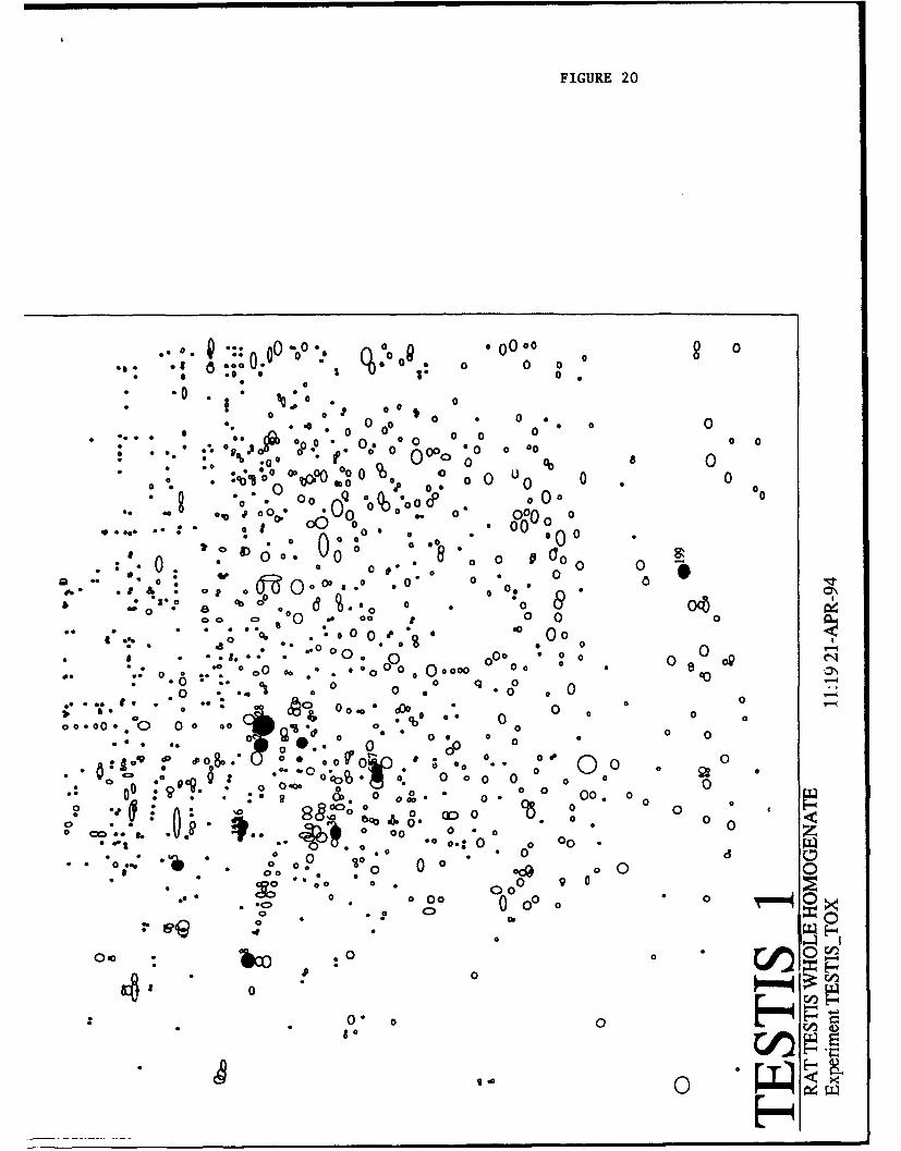
FIGURE 20
... • A : 00 o. o • O oo o00 OR 00 0 0 0
• * . o o 00 'o0 0 0
0 0 0 0 0
0 0~~0
0 0 " 0 o 0 0
• ... *. ga Ofli ,* 000, 00000
4 o" O 0 " O o 0 0 0 0 0* o " o o 0 0000 o 00
0 00 o 0 o0 0
o 00
0 "1 .-* -I f,( o •. o0 00:0* o, o O° 0 &
0 '° 0
00 2 oo 00 00
0:0 o• 0 00 p 00 0
-0 0 qD o00
0 ,b
:. 0 . 00 0 0 o 0 0 00•:";.-i0 :o>..- o 0 00000o0 00 08 -
a8 * . 0 0.. .o *o *0 0 o 0 0 0 0
So o . 0 .. . o• : c 00 0 0 0 0 OC 0
00 0
"" I'-.. " .o "o .-. .• o O. *oO .*o .0
4o~j, *: % Do 0
00.•. • •.-.0 0 00 :O • d 0 *O 0 0 0..o 0
:*..00 0 ce.0 0 0P 0" 4 * 0 00 - * 1 0 9,0, . 0 0
.00 40 ,,,'W 0 0 0 U'
0 06 00
&. .0 0 00 0 .* O
00 *0: 0 *cP 8 00 0 0
*0 9000000 00 0
ps , 0 0 0 0
0 :0op ga
... .. 0 oco... o 0• 0
0 00 0
.' o
00
0* o U • 51
o o o o
_ -H( BOoo o "-

~ASUL4 S. F344LIVER_1 Master Pattern Database (identified Proteins)
MSN kdenflication MW Y 1 X Volumfie Reference(name) cabC. coord. 0a1C. coord. Ev.cnrl
180 3s#-ylnddiduioIdsaydiog 36272 132e 7.30 2771 52409 imuwnnologic (Anderson atda., Eaectopitorsess 1901)103 340*I4qdrcgtftamid-diI"dodmio deliydrog. 38371 1323 7.04 2830 34376 immunaologi (Anderson at eL. Electrophorseew 1091)260 3&hdioroty-3-methyI~iutW CoA synthess 236 164.43 4.16 11M79 7691 immnuncolgic (Anderson atd.. Electophomeis. 1991)5158 34V3.mulh4nWotmzy C*A synthas. 72210 607.0 5.07 1418.3 1042 Waninunoogec (Anderson at *. E~colos~1001)263 3ruy3mhisilCoA synth-es 26304 1652.2 4.66 12613 7642 inromwuioogic (Anderson ad.L. Eisaftopitorss 1901)205 3-hti3mai3-hylgutaryl CoA syntiiese 26634 162786 4.76 11612 5156 imnmunoWoi (Anderson ofad. Selctrphossw 1801)360 34yduuty-3msdiylutsi CoA synth-s 54302 856.5 570 1844 6143 immunologic (Anderson at e.. Elsotropliomrse 1091)48 eode ooseb 42657 1118.1 . 5.06 1406.3 07553 homologous position (Anderson 41 e. Elecrophorsew. 1001)78 won (gamnsna) 42071 1111409 09 14315 26301 homologous position (Anidersonat ., Elsatrophoruuis 100)
296 slpt-2u globuin 10121 2136.3 546 '70(7 56046 homologous positon (Anderson atd&. Eloctrophor~sis 190)68 "Kiioprop sis A-I 24745 18261 5.32 15046 3370 homologous pFs- "o (Anderson atda., Boctophorevss 190)15645 spollpptagin A4 24326 18417.6 5.46 16017 11349 homologous position (Aunderson a a.. Elacophor~ses 1001)52 ATPoseFI alpha (Miloon4) 50602 92e 6.25 2182.2 125606 homologous position (Pavlos ts&iBDA, 1000)33 ATPase FI bola (Mkcon ) 4923 9%64 4.71 1130.2 2626 homologous poston Vderson at . Elecatphoreed. 1991)152 ATPas. Fl bela (Mitcon 1) 49480 9810 4.81 1218.3 .Z t2 homologous positon OAnderso aal- Elsaroporsi. 1IM)22 AT~omeF1 bela (Milcon 1) 49164 0506 4.76 11610 13b,;43 homologous position (Anderson at a. Ele rophoresw 1001)140 ulidmn 17706 22217 3.09 117.4 2696 homologous position (Andersoomatd.. Elactrophoresis IM0)
1532 uwltn 18246 2190.9 &:~390 1017 14421 homologous positon (Anderson atda.. Electrophaoreis 190)505 camkitcurn precursor 63181 7110 4.23 640LO 3P52 inimmnologic (Anderson at @L.Elcuposi. 1091)161 osreistvulin precursor 61117 7411 4.2 6365 7321 imun~onlogic (Anderson at st.. Electropluoresi 1001)2 osirsticuln precursor 6630 6696 iii 4.23 642.2 81703 inummamoogic (Aderson at si. Eledhopluomesi. 1001)51 alostlculi precursor 6806 6411 4.23 6410 15263 inmamologi (Anderson std@. Elecaoplorseei 1901)58 carbemo-1 phosphate synhase 163645 229.4 6.13 2110.0 1307 homologous position (Andervon ad.L. Eledtrophoresie 1901)
304 Cuod-lphos1Phate synIttise 165091 22&9 &00 20312 3350 homoloou position (Anderson atda., Eler~ophoesu 1001)139 carbarnoy phlosphate synthase 165203 227.0 6.07 2075.7 6468 homologous poiin(Anderson ad.a. Eledtrophoresui 1991)10 camnoyl phosphatesynthase 162948 230.6 6.19 2145.6 2416 homuologous positioni (Anderson tad.. Electrophoresi. 1091)7 ctubamoyl phosphate synthease 161027 2337 6.53 2347.0 150715 homologous position (Anderson ad. @Ladohr. 100)0 -*wbnoyl phosphate synthaes 161516 2319 6.30 22630. 120304 homologous position (Ander- ad.a. Eledbrophoreis. 199)3 carbernoyl phosphate synthase precursor 162068 2310 : 6.29 2205.0 146,478 homnologous poitio (Anderson std.. Electrophoresis, 1001)
11 xub" pos haesnheprcso 160522 2346 8.45 22067 15435 homologous position (Anderson ad..L Ehiearophoresis. 1001)
87 caue 5662 778,0 7.46 2668.5 111874 comigration of purified form (Molec. Anal. Lob.)7b atnls 30152 772.9 7.86 2674.9 60018 comigration of purified fain (Molec. Anat. Lob.)
96 cyoChroms b5 17326 2247.3 4.57 1000.8 02648 homologous;p*t (Anderson ad a.. Elsarophoresis. 1901)374 cynochrome b5 17321 224760 4.46 92Z8 6400 homologous peon(Anderson tad.. Elecwophorseis. 1901)300 feffy acid binding prolsin 14750 2400.4 6.00 2551.5 67573 wmuniuologic (Molec. AnsL Lab. vissiofrom Vanden Heuval)28 01p75 (Mibon:3) 69196 639.3 5.25 1547.5 15017 limmunologic (Mlot AnsI. Lob vim eblorom Sremssmo)
106 Gqo75 (Mitcon:3) 6033 637.8 5.16 1488.1 3308 iTmnsaologlo Not. AnsI. Lob via stofrom Sotressn)19 GOp75 (11114on:3) 60070 64068 5.38 16214 48734 imimunologi Pol. AnsI. Lavise afrom SVe.G")
116 Gip75 (Mitco:3) 70668 621.0 . 5.38 16212 6664 immunfologic (ldo. AnsI. Lob via stofrom Soasse(en)18 Gipi7ifli 71751 611.7 4.74 1167.3 26304 immunologic (WMtnnann ad a., Fund. Appl. Tot.. 1094)14 Gip76lwi 71465 614.7 4.77 1169.5 42583 irmunologic W~itlinenn ad a.. Fund. Appl. Tom.. 1994)
1531 Gip78iOI 72356 606.5 4.60 111(7 647 immu~nologic (Mkwn atn ad.. Fund. Appl. TaL. 1004)1536 alp71MeiP 72076 6064 4.72 1141.2 5007 immnunologic (Witamn satd.. Fund. Appi. Tax. 1994)
5 Gup78lB# 70965 820.0 4.50 1214.3 64302 immnunologic ffWitznwm atda.. Fund. Appl. T=L. 1004)4 GrpO4 (endoplosmmn) 10506 365.2 4.61 10534 15425 enneinologic MoI. AnsI. Lob via slo from StreseGen)I GrpQ4 (endoplasmin) 103957 390.2 4.58 1021.7 57834 ilnumnologic (Mol. AnsI. Lob via stofrom StreesGen)
20 Hsp80 (Mitcon:2) 5306 823.5 5.22 1827.0 114297 Orvnunelogc WMI AnsI. Lob via ab from SlrsesGen)50 Ilop6 (Mitoon:2) 56396 81(1 5.07 1410.5 7549 mnnnnoloinc (Md. AnsI. Lob viasslofrom StressGon)26 lHpO (MicOn:2) 56M 821.1 . 5.13 1465.5 40609 inununologac (Md Anst. Lob viasabfrom SosseGen)
123 Hsp70 66940 6658 6.30 15610 7506 inenunvologic (xmine with snt'hp70 from Stressena)130 Hep73 (Ha1c73) constitutive in unstressed 67475 659.2 5.04 1401.8 4713 inwriunologic (xmsdcwith anl-bp7olfrom StrsssGen)
0 Hsp73 (HWc3) conaiuliv in unstressed 66061 6651 5.14 1474.0 689645 immnniologic, (xsied wit~h nsp7fohm StmetGeii)24 Ave73 (Hsc73) conaitulive in unstressed 67215 6613 00 143165 31112 imuumnologic (used writh enI-sp7O from StraseGen)55 HapO 65613 4913 4.72 1149.6 9042 immunologic (MoI. AnsI. Lob vie ab from StreseGen)12 HspO 65261 495.5 4.79 1202.3 25459 imunsiologic (MCI. AnsI. Lob via ab from SitresGen)32 HepO 64777 400.2 4.75 1167.6 14525 immnnnnlogic (Mol. AnsI. Lob vieasbfrom SbessGen)
200 larbn 66121 675.7 4.93 131(8. 2205 homologous positin (Andersonadt .. Electrophorseis 1901)165 11mill 8 66260 674.0 .. 4.98 13514 3330 homoloous position (Anderson atd.. Electrophloresis. 190)76 larnu receptor protein 42585 11256 4.57 101(6 22461 sequence from gelspot (Mel-ue Innogenahc)
351 NADPH cytochrorns P450 reduclase 72213 607.0 5.13 1467.1 5472 homologous position (Anderson at W.. Elsctrophoreew 1901)122 NAOPH cytochoome P450 reductasse 2218 606s 5.17 1404.6 10430 homologous position (Anderson ata.. Elscrroporesis 1001)156 pro-8urnin 6563 661.7 0.30 22610 9377 homnologous position (Anderson ad a.. Electroploresis. 199)74 pro.*urnin 63001 6773 6.50 2327.9 59034 homologous position (Antderson atal. Eleaophoresei 190)40 prteeln disulide momersee 53685 866&2 4.60 1030.2 26170 immumnologic WMl AnsI. Lab vie atbfrom StreesGen)Is protein divullide momersee 53830 864.3 4.61 10516 679o5 innuanologinc "ao AnsI. Lab vie ablfrom StressGen)42 protein desuiklde nomerese 5366 667.5 4.64 1077.5 17373 imnounologic WMo AnsI. Lob vie eb from StasaGen)112 protein disulfide isomerase 53977 661.5 4.57 10114.5 7257 immunologic (Mot AnsI. Lab vie ab from StoseaGen)275 protein divullide isomerase 55117 840.68 . 4.61 1046.4 21670 immunologic (Mol. AnsI. Lab via abo from SbasseGen)330 Ilynavi acid cedmboyinse 129339 301.2 8.33 2229.6 1782 homologous position (Anderson at l., Eleatrophonesis. 199)III ppnavicWWcOdCsdWylsse 129379 3 8 .45 2297.0 12435 homologous position (Anderson ofal., Eiectrophcrssis 1901)43 pynrAvc acid carboxylaes 126664 5 (.38 2253.9 3001 homologous position (Anderson at el., Eloearopuoreseis 1901)166 pynr~c acid csbocxyloe' 126556 300.6 ~. 6.52 2337.3 7735 homologous position (Anderson at W., Elecarophoresie 100)141 10sensamos nsreor protein (SMP3O) 35546 1351.0 4.85 125668 23626 sequence from gel spot (Measfi5 Innogenaics)62 senmOsnors marker protein (SMP30) 35560 1351-5 4.97 1346.0 111435 sequence from gel spot (MW-^us Innoganet-c)45 snams abuminprecursor 85731 660.5 (.10 2004.5 59197 homologous position (Anderson ata.. Electophores*s 1901)506 sermmdxanin precursor 65699 680.9 5.97 2016.3 10356 homologous position (Anderson at al.. Eledtrophoresis, 1901)167 superosdds dinmutuse 16623 2276.6 8 .13 2111.7 143464 homologous position (Anderson rtid., Etecorophoresis. 190)267 equeowoide dismutase 36306 1325.6 6.76 2476.1 37004 homologous position (Anderson atd.. Electrophoreuia 199)301 kwnwm slt 53150 876.7 4.80 1275.0 7388 homologous position (Anderson al a.. Elsacrophoresiis. 1991)so ttbht5nIfI5 54317 855.2 . 4.66 1264.1 14328 homologous position (Anderson at ., Eleatrophoresie 199)124 itbxusiphs 53679 863.3 4.86 1279.0 13252 homnologous position (Anderson stad., Eleetrophoresis. 199)494 Imb~ii slphs 54197 857.4 . 4.84 1244.0 6261 homologous position (Anderson atda., Eotophoreiew 190)2D tsubui beta 53942 8662 4.75 1170.0 30411 homologous position (Anderson atal.,Electrophoresis, 109)

File. Opton Help
a -P

ADDENDUM B

MALDI Peptide-mass Determination:
Bovine Hemoglobin-alpha
MSM1W MQWSE SEQ AA
2980 2970 LLSHSLLVTLASHLPSDFTPAVHASLDK 100-1272376 2368 AVEHLDDLPGALSELSDLHAHK 69-901840 1834 TYFPHFDLSHGSAQVK 41-561534 1530 VGGHAAEYGAEALER 17-311276 1279 FLANVSTVLTSK 128-139
Bovine Hemoglobin-beta
MSM1w MQWSE SEQ AA
,2098 2090 FFESFGDLSTADAVMNNPK 40-581427 1423 EFTPVLQADFQK 120-1311276 1275 LLVVYPWTQR 30-391182 1177 VVAGVANALAHR 132-1431092 1097 VLDSFSNGMK 66-75952 950 AAVTAFWGK 8-16

#3 23-SEP-1993 09:25:00.34NEWMAILFrom: IN%"[email protected]"To: IN%[email protected]"CC:Subj:
Return-path: <[email protected]>Received: from mservl.dl.ac.uk by delphi.com (PMDF V4.2-11 #4520) id
<[email protected]>; Thu, 23 Sep 1993 09:24:52 EDTReceived: from s-ind2 (s-ind2.dl.ac.uk) by mservl.dl.ac.uk with SMTP idAA13246
(5.65c/DL-V3.2.1(DNS-IDE-pg) for <[email protected]> [email protected]); Thu, 23 Sep 1993 14:18:03 +0100
Received: by s-ind2 (920330.SGI/930727.MJE) for [email protected] idAA12309; Thu, 23 Sep 93 15:27:12 +0100
Date: Thu, 23 Sep 1993 15:27:12 +0100From: [email protected] (Mowse Server)To: [email protected]: <9309231427.AA12309@s-ind2>Content-transfer-encoding: 7BITPrecedence: first-classDatabase: /nfs/fabl/data2/owl/owlReagent: TrypsinTolerance: 10 daltonApparently-To: [email protected]
Sequence MW: 15200MW filter: 30%
SCAN of /nfs/fabl/data2/owl/owl using reagent TrypsinUSING fragment mws of:
42442980237620981840153414821427127611821092952805618442
No. of hits = 30 (MAX. ALLOWED)No. of database entries scanned = 658951 HBABOVIN HEMOGLOBIN ALPHA CHAIN. - BOS TAURUS (BOVINE).2 . JS0680 hypothetical protein 3 (gldA 5' region) - Bacillusstearotherm3 VAL3_TGMV AL3 PROTEIN. - TOMATO GOLDEN MOSAIC VIRUS (TGMV).4 X_HPBVY PROTEIN X. - HEPATITIS B VIRUS (SUBTYPE AYW).5 HBB_BOVIN HEMOGLOBIN BETA CHAIN. - BOS TAURUS (BOVINE).6 HBBABOSJA HEMOGLOBIN BETA-A CHAIN. - BOS JAVANICUS (WILD BANTENG).7 HBB_BOSGA HEMOGLOBIN BETA CHAIN. - BOS GAURUS FRONTALIS (GAYAL).8 PHAl_SYNPY C-PHYCOERYTHRIN CLASS I ALPHA CHAIN. - SYNECHOCOCCUS SP.(STRA9 HBE_RABIT HEMOGLOBIN EPSILON CHAIN (BETA-4). - ORYCTOLAGUS CUNICULUS(RA10 HBAIBOSMU HEMOGLOBIN ALPHA-I CHAIN. - BOS MUTUS GRUNNIENS (YAK).

11 S14886 Hypothetical protein 9 - Yeast (Hansenula polymorpha)12 . JT0902 chaperonin 60 beta - wheat (fragment)13 HBARANTA HEMOGLOBIN ALPHA CHAIN. - RANGIFER TARANDUS (REINDEER).14 . HBG_LEMFU HEMOGLOBIN GAMMA CHAIN. - LEMUR FULVUS FULVUS (BROWNLEMUR).15 S29555 Chalcone isomerase (EC 5.5.1.6) - Apple tree (fragment)16 S29554 Chalcone isomerase (EC 5.5.1.6) - Apple tree (fragment)17 YZFBECOLI VERY HYPOTHETICAL 18.0 KD PROTEIN IN FEPB 3iREGION. -ESCHERIC18 HBA2_BOSMU HEMOGLOBIN ALPHA-2 CHAIN. - BOS MUTUS GRUNNIENS (YAK).19 PL0018 Ig heavy chain V-D-J region (RP93) - mouse (fragment)20 S03527 Ig heavy chain V region - African clawed frog21 . LGB_PSOTE LEGHEMOGLOBIN. - PSOPHOCARPUS TETRAGONOLOBUS (GOA BEAN)(ASPAR22 S24697 Ig heavy chain V6 region - Human23 PHEAPORCR B-PHYCOERYTHRIN ALPHA CHAIN. - PORPHYRIDIUM CRUENTUM.24 PHEAPORSO B-PHYCOERYTHRIN ALPHA CHAIN. - PORPHYRIDIUM SORDIDUM.25 CBPAPIG CARBOXYPEPTIDASE A (EC 3.4.17.1) (FRAGMENT). - SUS SCROFA(PIG26 CEUIFN CEUIFN LOCUS CEUIFN 501 bp ss-mRNA MAM 07-DEC-1992 - Cervusel27 HBBLAMGL HEMOGLOBIN BETA CHAIN. - LAMA GLAMA (LLAMA), LAMA VICUGNA(VIC28 . HBB_CAMDR HEMOGLOBIN BETA CHAIN. - CAMELUS DROMEDARIUS (DROMEDARY)(ARAB29 . S41176 S41176 transactivator; For the protein sequence (NCBIgibbsq130 . S41175 S41175 transactivator; For the protein sequence (NCBIgibbsq1
1 HBABOVIN 2.893509e+05 15053 0.333HEMOGLOBIN ALPHA CHAIN. - BOS TAURUS (BOVINE).MW START END SEQ4244 NO MATCH2970 100 127 LLSHSLLVTLASHLPSDFTPAVHASLDK2368 69 90 AVEHLDDLPGALSELSDLHAHK2098 NO MATCH1834 41 56 TYFPHFDLSHGSAQVK1530 17 31 VGGHAAEYGAEALER1482 NO MATCH1427 NO MATCH1279 128 139 FLANVSTVLTSK1182 NO MATCH1092 NO MATCH952 NO MATCH805 NO MATCH618 NO MATCH442 NO MATCH
2 JS0680 1.699716e+05 18173 0.333hypothetical protein 3 (gldA 5' region) - Bacillus
stearothermophilusMW START END SEQ4253 48 81 AGLDYWEFVNIHYYDSLDHFFQQNQNGDFYYITK2980 NO MATCH2376 NO MATCH2098 NO MATCH1832 133 149 ALNLSNTAAILVYEALR1534 NO MATCH1473 86 97 YYTSYDFSDPSK1427 NO MATCH

1276 NO MATCH1182 NO MATCH1088 113 121 ELLAENEDR959 125 132 IPMTENVR805 NO MATCH618 NO MATCH442 NO MATCH
5 HBBBOVIN 5.948711e+04 15954 0.400HEMOGLOBIN BETA CHAIN. - BOS TAURUS (BOVINE).MW START END SEQ4244 NO MATCH2980 NO MATCH2376 NO MATCH2090 40 58 FFESFGDLSTADAVMNNPK1840 NO MATCH1534 NO MATCH1482 NO MATCH1423 120 131 EFTPVLQADFQK1275 30 39 LLVVYPWTQR1177 132 143 VVAGVANALAHR1097 66 75 VLDSPSNGMK950 8 16 AAVTAFWGK805 NO MATCH618 NO MATCH442 NO MATCH
6 HBBA_BOSJA 5.944985e+04 15964 0.400HEMOGLOBIN BETA-A CHAIN. - BOS JAVANICUS (WILD BANTENG).MW START END SEQ4244 NO MATCH2980 NO MATCH2376 NO MATCH2090 40 58 FFESFGDLSTADAVMNNPK1840 NO MATCH1534 NO MATCH1482 NO MATCH1423 120 131 EFTPVLQADFQK1275 30 39 LLVVYPWTQR1177 132 143 VVAGVANALAHR1098 66 75 VLDSFSDGMK950 8 16 AAVTAFWGK805 NO MATCH618 NO MATCH442 NO MATCH
7 HBB_BOSGA 5.936803e+04 15986 0.400HEMOGLOBIN BETA CHAIN. - BOS GAURUS FRONTALIS (GAYAL).MW START END SEQ4244 NO MATCH2980 NO MATCH2376 NO MATCH2090 40 58 FFESFGDLSTADAVMNNPK1840 NO MATCH1534 NO MATCH1482 NO MATCH1423 120 131 EFTPVLQADFQK1275 30 39 LLVVYPWTQR1177 132 143 VVAGVANALAHR1097 66 75 VLDSFSNGMK950 8 16 AAVTAFWGK805 NO MATCH