Embed Size (px)
Citation preview
© 2017. Published by The Company of Biologists Ltd.
Acoustic measurements of post-dive cardiac responses in southern elephant seals
(Mirounga leonina) during surfacing at sea
Louise DAY1, 2, Joffrey JOUMA’A1, Julien BONNEL2 and Christophe GUINET1
1. Centre d’Études Biologiques de Chizé-Centre National de la Recherche Scientifique (CEBC-
CNRS), 79360 Villiers-en-bois, France
2. ENSTA Bretagne, Lab-STICC (UMR CNRS 6285), 29200 Brest, France
Key-words: Mirounga leonina, heart rate, dive recovery, diving behaviour, passive acoustic
monitoring
Corresponding author’s email address: [email protected]
Jour
nal o
f Exp
erim
enta
l Bio
logy
• A
dvan
ce a
rtic
le
http://jeb.biologists.org/lookup/doi/10.1242/jeb.146928Access the most recent version at J Exp Biol Advance Online Articles. First posted online on 15 February 2017 as doi:10.1242/jeb.146928
Summary statement
This paper demonstrates the reliability of an acoustic method to extract and analyse the cardiac
function of free ranging southern elephant seals in order to study post-dive recovery.
Abstract
Measuring physiological data in free-ranging marine mammals remains challenging, owing to their
far-ranging foraging habitat. Yet, it is important to understand how these divers recover from effort
expended underwater, as marine mammals can perform deep and recurrent dives. Among them,
southern elephant seals (Mirounga leonina) are one of the most extreme divers, diving continuously
at great depth and for long duration while travelling over large distances within the Southern Ocean.
To determine how they manage post-dive recovery, we deployed hydrophones on four post-
breeding female southern elephant seals. Cardiac data were extracted from sound recordings when
the animal was at the surface breathing. Mean heart rate at the surface was 102.4 ± 4.9 beats.min-1
and seals spent on average 121 ± 20 s breathing. During these surface intervals, the instantaneous
heart rate is increasing with time. Elephant seals are supposed to drastically slow their heart rate
(bradycardia) while they are deep underwater, and increase it (tachycardia) during the ascent
towards the surface. Our finding suggests that tachycardia continues while the animal stays
breathing at the surface. Also, the measured mean heart rate at the surface was unrelated to the
duration and swimming effort of the dive prior to the surface interval. Recovery (at the surface)
after physical effort (underwater) appears to be related to the overall number of heart beats
performed at the surface, and therefore total surface duration. Southern elephant seals recover from
dives by adjusting the time spent at the surface rather than their heart rate.
Jour
nal o
f Exp
erim
enta
l Bio
logy
• A
dvan
ce a
rtic
le
Introduction
Diving marine mammals face a strong dilemma: their food resources are located at depth while they
need to restore oxygen supply at the surface. This specificity makes them particular because their
breath hold capability limits the time spent foraging. Hence, surfacing is essential to reconstitute
oxygen stores by breathing and restoring oxygen levels in muscles and organs. Oxygen stores are
higher for diving species than non-diving ones (Butler and Jones, 1997) and are located in the blood
and muscles (Hassrick et al., 2010; Kooyman et al., 1983). Bradycardia is the common response to
diving in marine mammals and diving seabirds (Ponganis, 2015), with, for instance, northern
elephant seal reducing its heart rate by 64% (Andrews et al., 1997). Regulation of heart rate, cardiac
output, and the degree of vasoconstriction and blood circulation shutdown is critical to the
management and utilization of oxygen stores.
Measures of physiological data are essential to understand diving mammal metabolism (Butler and
Jones, 1997). Ideally, physiological parameters should be recorded on free-ranging animals diving
voluntarily (Webb et al., 1998). However, accessing physiological data in situ, like cardiac response
at the surface, on free-ranging animals in the open ocean remains difficult. The pinnipeds share
their time both at sea and on land or ice (Harrison and Kooyman, 1968). This bimodal cycle
coupled to their large size makes them a unique system to study their physiological adaptations to
deep dives, because the deployment and recovery of loggers are eased on land (Costa et al., 2004).
Kooyman et al. (1968; 1971; 1973) were the first to access physiological data and calculate
metabolic rate using a respiratory chamber on free-ranging Weddell seals, Leptonychotes weddellii,
with the man-made ice-hole experiments under semi-natural conditions.
Due to difficulties to employ this technique in situ on other marine mammals species, methods
based on heart rate were used as a reliable indicator of field metabolic costs (Butler et al., 2004;
McPhee et al., 2003; Ropert-Coudert et al., 2012). For instance, Weimerskirch et al. (2000)
successfully used heart rate as a proxy of energy expenditure and instantaneous effort in flying
wandering albatrosses, with the highest heart frequencies observed while albatrosses were walking
on land and taking off. This method also provides advantages over doubly labelled water (DLW) in
pinnipeds, since it provides an estimation of the metabolic rate of specific activities, such as those
occurring during a dive cycle (Butler et al., 2004). The electric approach, which measures the
electrical signal of the heart, is the most common way to record heart rate in free-ranging diving
mammals (Ropert-Coudert et al., 2012; Webb et al., 1998).
Jour
nal o
f Exp
erim
enta
l Bio
logy
• A
dvan
ce a
rtic
le
Heart rate studies on elephant seals with an electric method showed that during diving, they
exhibited bradycardia. Heart rate rapidly decreased by 50-80% at the beginning of the dive and
remained low while the seal was submerged (Andrews et al., 1997). Hindell and Lea (1998)
recorded extreme bradycardia with heart rate reaching 2 beats.min-1 in 23 dives. Heart rate then
increased gradually as the seal rose to the surface (Andrews et al., 1997). Bradycardia, apnoea and
vasoconstriction of the peripheral system constitutes the dive response in pinnipeds (Harrison and
Kooyman, 1968). However, the electrical method requires the fixation of an electrode into the body
which can cause complications in the field (Ropert-Coudert et al., 2012).
Fletcher et al., 1996, using an acoustic approach, provided the first record of a respiratory rate at the
surface of translocated northern elephant seals (Mirounga angustirostris). Between breaths, putative
heart beats were distinguished and cardiac frequency extracted. Several acoustic studies confirmed
that this method could provide physiological data such as breath frequency or heart rate at the
surface (Burgess et al., 1998; Génin et al., 2015; Le Boeuf et al., 2000).
The aim of this study is to investigate the cardiac response at the surface to active dives in free-
ranging southern elephant seals (SES here after). SESs are a major predator in the Southern Ocean.
At sea, they dive repeatedly to around 500 m during 20-30 minutes with surface intervals lasting on
average 2 minutes but extreme depth records reached over 1,800 m (Hindell et al., 1991; McConnell
et al., 1992). They come back on land twice a year for mating during the southern spring, and
moulting in the summer, with a high-site fidelity (Fabiani et al., 2006).
Génin et al (2015) have shown that the number of breaths is tightly related to surfacing time and
mainly explained by dive duration and swimming effort made by SESs. Yet, in terms of recovery,
the cardiac function might play a major role. In this study, we intend to explore the recovery
behaviour of SES through examining variation in heart frequency. First, we investigate how the
instantaneous cardiac frequency evolves at the surface. Second, we study the relation between the
mean cardiac frequency during the surface interval, and the dive duration and foraging effort
performed by SESs during the previous dive.
Jour
nal o
f Exp
erim
enta
l Bio
logy
• A
dvan
ce a
rtic
le
Materials and methods
Ethics statement
All animals in this study were treated in accordance with the French Polar Institute (IPEV) ethical
and Polar Environment Committees guidelines. All scientific procedures conducted on SES had
been validated beforehand.
Deployment of devices and data collection
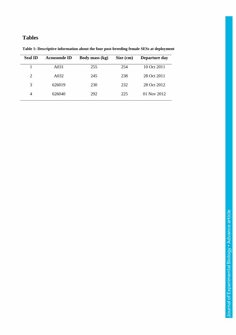
This study is based on data collected on four post-breeding SES females (Table 1): two equipped in
October 2011 and another two in October/November 2012 on Kerguelen Islands (49°20’S,
70°20’E). Individuals were captured, then anaesthetized using 1:1 combination of tiletamin and
zolazepam (Zoletil 100), which was injected intravenously (0.8 mg/100kg; McMahon et al., 2000).
They were then equipped with two devices glued on the head or the back of the individual, using
quick-setting epoxy (Araldite AW 2101, Ciba) after cleaning the fur with acetone. First, an Argos-
GPS satellite tag (Splash 10-F, Wildlife Computer, USA) was glued to the head of the seals. It
provided real-time position of the seals through the Argos system and also collected GPS location
data. Second, an autonomous acoustic/accelerometer/magnetometer and pressure logger named
AcousondeTM, model 3A (Acoustimetrics, Greeneridge Scences, Inc, USA) (Burgess, 2000;
Burgess et al., 1998) was fixed on the dorsal fur on the longitudinal axis, 10 cm behind the scapula.
AcousondesTM recorded at a sampling frequency of 6.3 kHz in 2011 and 12.2 kHz in 2012, with
both an acoustic sampling resolution of 16 bits. This difference in sampling rates does not interfere
in our study as cardiac and respiratory events occur in a frequency range inferior to 1 kHz. To save
battery power and storage space, it was programmed to record sound 3 h every 12 h in 2011 and 4 h
every 24 h in 2012. All devices provided measurements of time, location and depth at 1 Hz, as well
as the three-dimensional magnetic field strength and acceleration at a 5 Hz frequency. The
instruments sampled acoustic data until battery exhaustion, which occurred between 10 and 20 days
after deployment. All devices were retrieved once individuals returned ashore to moult after their
foraging trip in January/February following deployments. Seals were located on land using their
Argos position.
Jour
nal o
f Exp
erim
enta
l Bio
logy
• A
dvan
ce a
rtic
le
Acoustic data processing
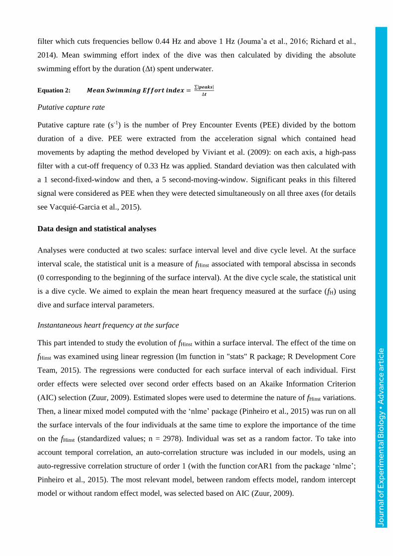
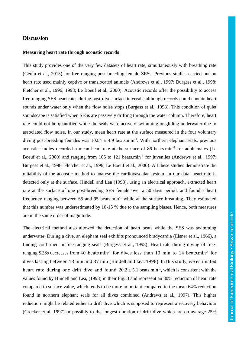
Cardiac occurrences detection
When the animal is surfacing, the water flow noise produced by swimming ceases and most of the
sound is due to breathing. Respiratory signals are contained between frequencies within the 0 and
700 Hz range. Between two respirations, spectrograms (time-frequency representation) showed
putative cardiac occurrences (Fletcher et al., 1996; Le Boeuf et al., 2000). Heart sounds are
expected to be dual due to the closure of mitral and the aortic valves (Burgess et al., 1998). The two
sounds are undistinguishable, as they occur too close together in time. Hence, cardiac occurrences
(a combination of the two valves’ sounds) are brief and regular temporal impulsions at frequencies
from 0 to 150 Hz (Burgess et al., 1998).
Acoustic recordings of surface intervals were visualized and analysed using the software Raven
(The Cornell Lab of Ornithology – Bioacoustics Research Program) to generate a spectrogram for
each surface interval. The same parameters were used for the computation of all spectrograms: a
window of Hann type and a size of 512 samples, an overlap of 50 % and a Discrete Fourier
Transform (DFT) calculated with 512 samples.
Each cardiac beat was determined using visual and auditory cues (Fig. 1). Hence, each occurrence is
characterised by its temporal abscissa. When two beats are consecutive, an instantaneous cardiac
frequency (beats.min-1) is calculated using the temporal difference between the two occurrences
(Eqn. 1).
Equation 1: 𝐈𝐧𝐬𝐭𝐚𝐧𝐭𝐚𝐧𝐞𝐨𝐮𝐬 𝐜𝐚𝐫𝐝𝐢𝐚𝐜 𝐟𝐫𝐞𝐪𝐮𝐞𝐧𝐜𝐲 (𝒇𝑯𝒊𝒏𝒔𝒕) =𝟔𝟎
𝒙𝒕+𝟏−𝒙𝒕
Some surface intervals contained high-amplitude noise from external sources (water splashes
possibly due to rough weather conditions or due to certain animal behaviours such as grooming). A
noisy acoustic environment prevents access to heart data. As such, a surface interval was only kept
for further analyses when a minimum of five cardiac beats were measured, so that the cardiac
frequency could be estimated with confidence. Cardiac frequency for a surface interval fH was
computed as the average of instantaneous heart frequency fHinst (Eqn. 1) during each surface
interval.
Surface interval duration
The beginning, end and duration of each surface interval, to the nearest second, were determined
using visual and auditory cues. A surface interval begins with the first respiration and ends with the
last one.
Jour
nal o
f Exp
erim
enta
l Bio
logy
• A
dvan
ce a
rtic
le
Dive cycle
A dive cycle is composed of a dive followed by a surface interval. SESs were considered to be
diving when they reached depths greater than 15 m, to avoid considering subsurface movements as
dives. Each dive was decomposed into three phases: descent, bottom and ascent. Each phase was
determined using a vertical speed criterion. Vertical speed was modelled with an order four
polynomial function adjusted on instantaneous vertical speed using a custom-written Matlab code
(Matlab software 8.1, The MathWorks, Natick, MA, USA). Ascent and descent phases were
identified as periods before or after surfacing where the modelled vertical speed exceeded 0.75 m.s-
1. Bottom phases were identified as periods between ascent and descent phases where the modelled
vertical speed remained below 0.75 m.s-1 (Jouma’a et al., 2016; Vacquié-Garcia et al., 2015).
Dive parameters
For each surface interval where fH (beats.min-1) was measured, data from the previous dive were
extracted from the AcousondeTM at a 5 Hz resolution. Statistics on each dive were then calculated to
give: maximum depth reached (m), dive duration (min), descent, bottom and ascent duration (min)
and the location (latitude and longitude) when the SES reached the surface. Elephant seals perform
dives where they passively descend through the water column over a large proportion of time
(Crocker et al., 1997). These passive ‘drift’ dives were identified based on the method designed by
Dragon et al., (2012) using the package “rbl” (Le Bras, 2016) in R. A dive was considered as a
passive drift dive when passive phases were detected with the following parameters: a minimum
duration of 50 s, an absolute roll superior to 90° and a drift rate (i.e. an absolute vertical speed)
ranging between -0.4 and 0.6 m.s-1.
Another important dive parameter is total acceleration. It can be decomposed into two types: static
and dynamic. Static acceleration is caused by earth’s gravitational pull whereas dynamic
acceleration results from the animal’s movements (body waves, tail strokes, head motions). The
static component corresponds to low frequencies and the dynamic one, to higher frequencies (Génin
et al., 2015; Richard et al., 2014).
Mean swimming effort index
Data provided by accelerometers were used to calculate absolute and mean swimming effort index
for each dive phase and for the entire dive (Eqn. 2). The lateral axis of the accelerometer contains
information on putative turning and rolling movements (static acceleration) and on flipper stroke
(dynamic acceleration) (Richard et al., 2014). Swimming effort was obtained by summing the
absolute values of the local extrema of the lateral axis of the acceleration filtered with a band-pass
Jour
nal o
f Exp
erim
enta
l Bio
logy
• A
dvan
ce a
rtic
le
filter which cuts frequencies bellow 0.44 Hz and above 1 Hz (Jouma’a et al., 2016; Richard et al.,
2014). Mean swimming effort index of the dive was then calculated by dividing the absolute
swimming effort by the duration (Δt) spent underwater.
Equation 2: 𝑴𝒆𝒂𝒏 𝑺𝒘𝒊𝒎𝒎𝒊𝒏𝒈 𝑬𝒇𝒇𝒐𝒓𝒕 𝒊𝒏𝒅𝒆𝒙 = ∑|𝒑𝒆𝒂𝒌𝒔|
∆𝒕
Putative capture rate
Putative capture rate (s-1) is the number of Prey Encounter Events (PEE) divided by the bottom
duration of a dive. PEE were extracted from the acceleration signal which contained head
movements by adapting the method developed by Viviant et al. (2009): on each axis, a high-pass
filter with a cut-off frequency of 0.33 Hz was applied. Standard deviation was then calculated with
a 1 second-fixed-window and then, a 5 second-moving-window. Significant peaks in this filtered
signal were considered as PEE when they were detected simultaneously on all three axes (for details
see Vacquié-Garcia et al., 2015).
Data design and statistical analyses
Analyses were conducted at two scales: surface interval level and dive cycle level. At the surface
interval scale, the statistical unit is a measure of fHinst associated with temporal abscissa in seconds
(0 corresponding to the beginning of the surface interval). At the dive cycle scale, the statistical unit
is a dive cycle. We aimed to explain the mean heart frequency measured at the surface (fH) using
dive and surface interval parameters.
Instantaneous heart frequency at the surface
This part intended to study the evolution of fHinst within a surface interval. The effect of the time on
fHinst was examined using linear regression (lm function in "stats" R package; R Development Core
Team, 2015). The regressions were conducted for each surface interval of each individual. First
order effects were selected over second order effects based on an Akaike Information Criterion
(AIC) selection (Zuur, 2009). Estimated slopes were used to determine the nature of fHinst variations.
Then, a linear mixed model computed with the ‘nlme’ package (Pinheiro et al., 2015) was run on all
the surface intervals of the four individuals at the same time to explore the importance of the time
on the fHinst (standardized values; n = 2978). Individual was set as a random factor. To take into
account temporal correlation, an auto-correlation structure was included in our models, using an
auto-regressive correlation structure of order 1 (with the function corAR1 from the package ‘nlme’;
Pinheiro et al., 2015). The most relevant model, between random effects model, random intercept
model or without random effect model, was selected based on AIC (Zuur, 2009).
Jour
nal o
f Exp
erim
enta
l Bio
logy
• A
dvan
ce a
rtic
le
Mean heart rate and number of beats at the surface
To investigate the contribution of previous diving behaviours on mean heart rate at the surface and
on the number of cardiac beats at the surface, we used linear mixed-effects models, also from the
package ‘nlme’. Values outside the 1.5 interquartile range were removed from the data. All
explanatory variables were centred and standardised at the population scale to keep individual
differences and allow comparisons between slope estimates. Time (in days) was included in the
model as we assumed a possible impact on the mean heart rate. To test linear and quadratic effects
of the time, both variables were included in the model. Individual was set as a random factor, and
an auto-regressive correlation structure was included (corAR1). As previously, the most
parsimonious model, between a random effect model, random intercept model or without random
effect model, was selected based on AIC (Zuur, 2009). Same models and same protocol were used
to explain the number of beats at the surface.
All statistical analyses were conducted using the software R (R Development Core Team, 2015).
For each model, normal distributions of the explained variable and of the residuals, and residuals’
homogeneity were checked up. All results are expressed as mean ± standard deviation for single
parameters. The significance level was set at p = 0.05.
Jour
nal o
f Exp
erim
enta
l Bio
logy
• A
dvan
ce a
rtic
le
Results
Foraging trips and overall diving behaviour
Each SES travelled eastward of Kerguelen Islands. AcousondesTM provided data for the first days of
foraging trips. We obtained 296 hours of sound recorded in 84 files of which 15 (53 hours) were
immediately put aside because the animal was still on land or data were too bad to be exploited. Of
the 243 hours left, there were 688 dive cycles and we kept the cycles that counted more than 5 heart
beats. 284 dive cycles were kept for this study. On average, seals dived for 18.4 ± 3.7 min with a
mean depth of 546 ± 159 m. Time spent at the surface recovering averaged 121 ± 19 s (i.e. 2 min 1
s; Table 2) with a maximum of 208 s (i.e. 3 min 28 s). Consequently, seals were submerged on
average 90.1 % of the time, ranging from 89.3 % for ind. 3 to 91.1 % for ind. 4. The four
individuals showed differences in their diving strategies. Ind. 1 performed deep long dives whereas
ind. 2 performed shallower and shorter dives. Ind. 3 had a greater number of prey event encounters
and higher putative capture rate compared to the three others (Table 3). The longest (33 min 12 s)
and the deepest (938.2 m) dives were both performed by ind. 1.
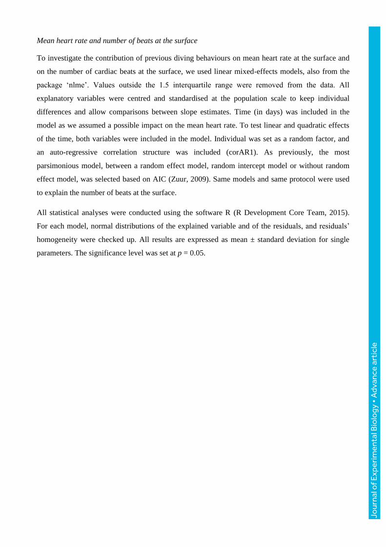
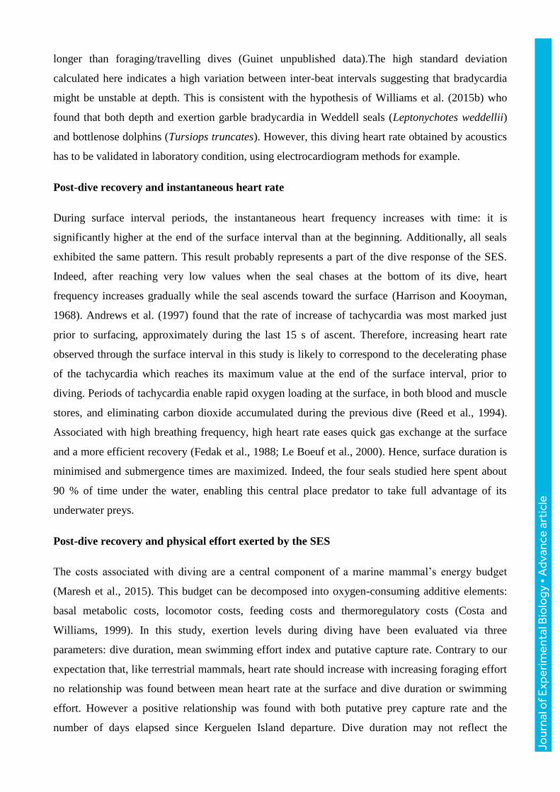
There was a strong negative relationship between dive duration and mean swimming effort index
per dive across the four individuals (Pearson’s correlation coefficient = -0.74, p ≤ 0.001; Fig. 2).
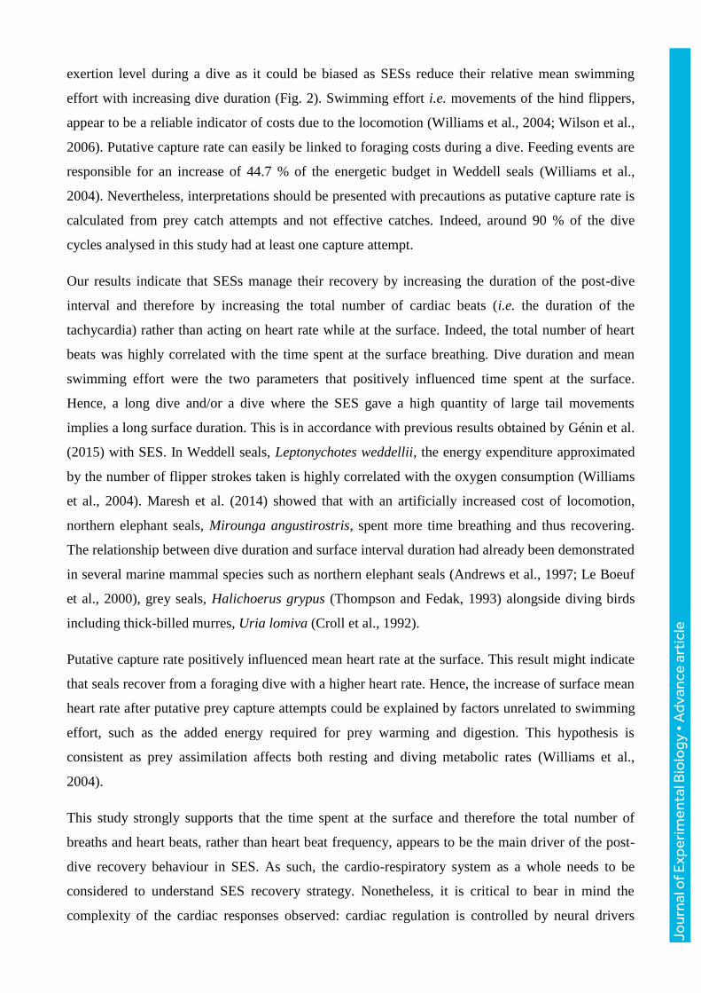
Instantaneous cardiac frequency during surface intervals
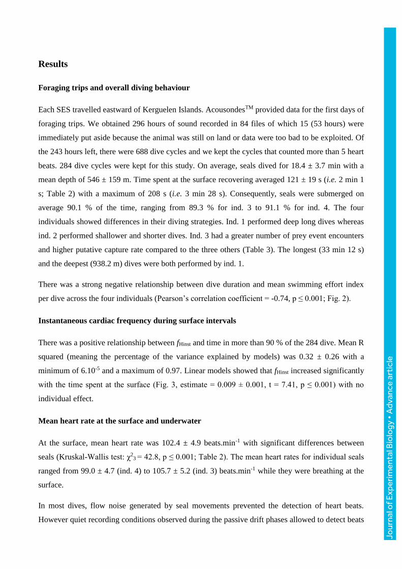
There was a positive relationship between fHinst and time in more than 90 % of the 284 dive. Mean R
squared (meaning the percentage of the variance explained by models) was 0.32 ± 0.26 with a
minimum of 6.10-5 and a maximum of 0.97. Linear models showed that fHinst increased significantly
with the time spent at the surface (Fig. 3, estimate = 0.009 ± 0.001, t = 7.41, p ≤ 0.001) with no
individual effect.
Mean heart rate at the surface and underwater
At the surface, mean heart rate was 102.4 ± 4.9 beats.min-1 with significant differences between
seals (Kruskal-Wallis test: χ23
= 42.8, p ≤ 0.001; Table 2). The mean heart rates for individual seals
ranged from 99.0 ± 4.7 (ind. 4) to 105.7 ± 5.2 (ind. 3) beats.min-1 while they were breathing at the
surface.
In most dives, flow noise generated by seal movements prevented the detection of heart beats.
However quiet recording conditions observed during the passive drift phases allowed to detect beats
Jour
nal o
f Exp
erim
enta
l Bio
logy
• A
dvan
ce a
rtic
le
(e.g. short impulse signals), in the frequency range between 0 and 40 Hz, which is likely be
attributed to heart beats. Drift dives were mainly observed in ind. 1. The low-frequency pattern
appeared during the whole passive drift event. A simple calculation of the frequency of occurrence
based on drift events exhibited a mean of 20.2 ± 5.1 beats.min-1, which represents an 80.3%
reduction in heart rate compared to surface measurements for that individual.
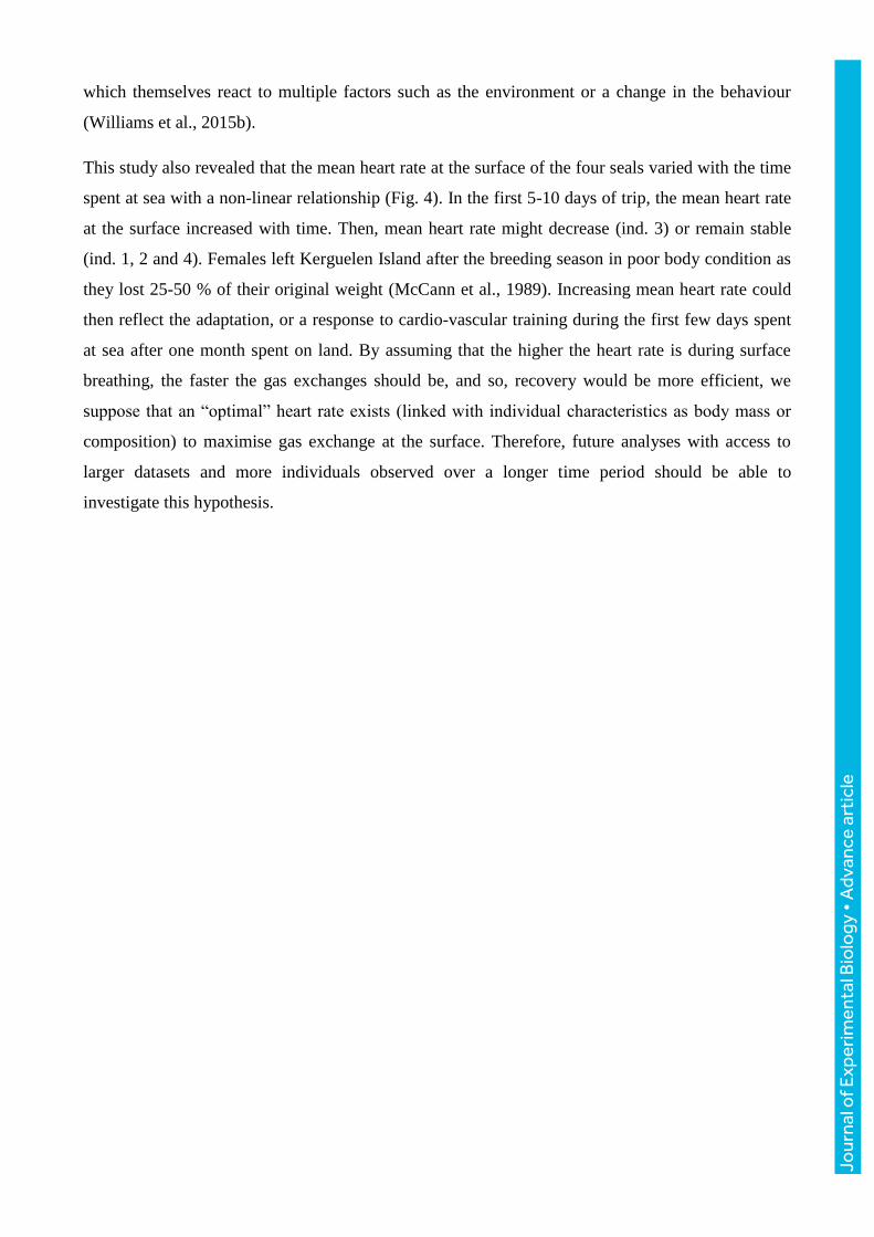
Mean surface heart rate in relation to dive parameters
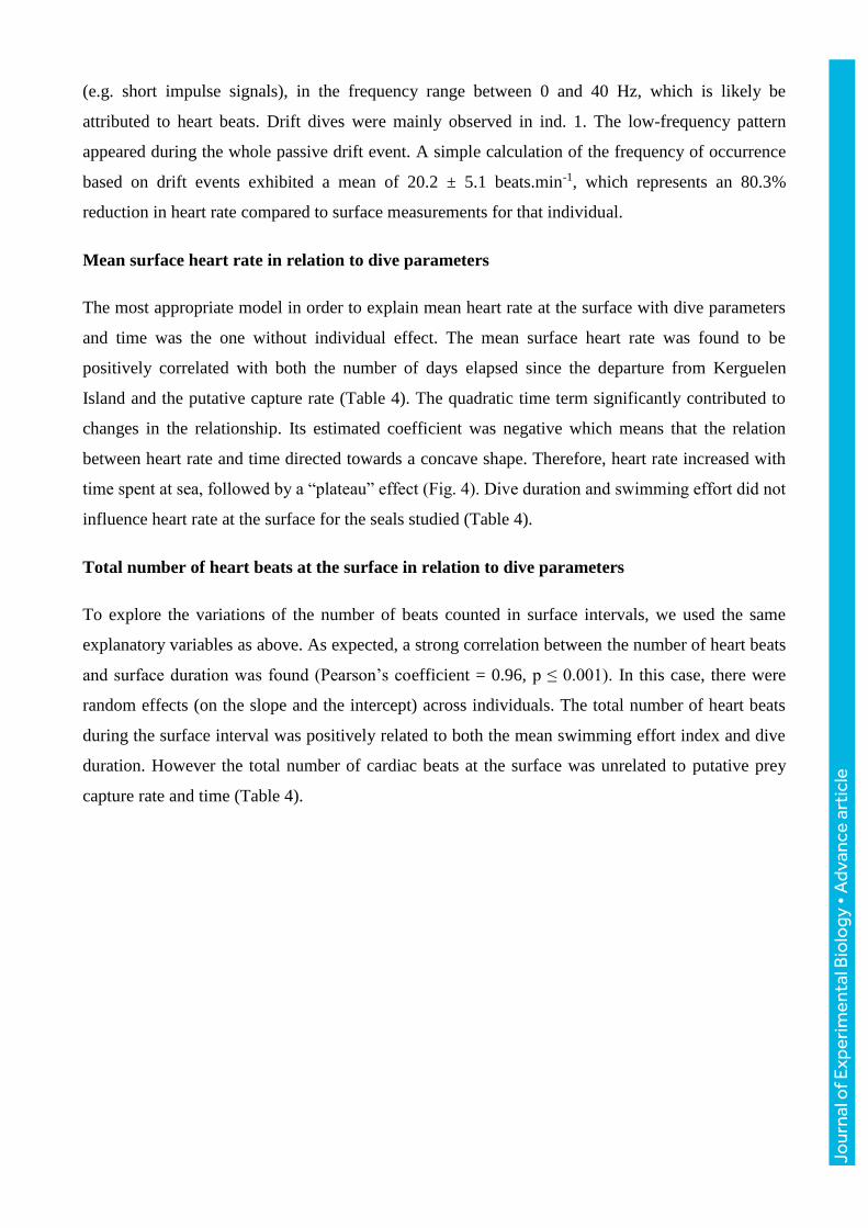
The most appropriate model in order to explain mean heart rate at the surface with dive parameters
and time was the one without individual effect. The mean surface heart rate was found to be
positively correlated with both the number of days elapsed since the departure from Kerguelen
Island and the putative capture rate (Table 4). The quadratic time term significantly contributed to
changes in the relationship. Its estimated coefficient was negative which means that the relation
between heart rate and time directed towards a concave shape. Therefore, heart rate increased with
time spent at sea, followed by a “plateau” effect (Fig. 4). Dive duration and swimming effort did not
influence heart rate at the surface for the seals studied (Table 4).
Total number of heart beats at the surface in relation to dive parameters
To explore the variations of the number of beats counted in surface intervals, we used the same
explanatory variables as above. As expected, a strong correlation between the number of heart beats
and surface duration was found (Pearson’s coefficient = 0.96, p ≤ 0.001). In this case, there were
random effects (on the slope and the intercept) across individuals. The total number of heart beats
during the surface interval was positively related to both the mean swimming effort index and dive
duration. However the total number of cardiac beats at the surface was unrelated to putative prey
capture rate and time (Table 4).
Jo
urna
l of E
xper
imen
tal B
iolo
gy •
Adv
ance
art
icle
Discussion
Measuring heart rate through acoustic records
This study provides one of the very few datasets of heart rate, simultaneously with breathing rate
(Génin et al., 2015) for free ranging post breeding female SESs. Previous studies carried out on
heart rate used mainly captive or translocated animals (Andrews et al., 1997; Burgess et al., 1998;
Fletcher et al., 1996; 1998; Le Boeuf et al., 2000). Acoustic records offer the possibility to access
free-ranging SES heart rates during post-dive surface intervals, although records could contain heart
sounds under water only when the flow noise stops (Burgess et al., 1998). This condition of quiet
soundscape is satisfied when SESs are passively drifting through the water column. Therefore, heart
rate could not be quantified while the seals were actively swimming or gliding underwater due to
associated flow noise. In our study, mean heart rate at the surface measured in the four voluntary
diving post-breeding females was 102.4 ± 4.9 beats.min-1. With northern elephant seals, previous
acoustic studies recorded a mean heart rate at the surface of 86 beats.min-1 for adult males (Le
Boeuf et al., 2000) and ranging from 106 to 121 beats.min-1 for juveniles (Andrews et al., 1997;
Burgess et al., 1998; Fletcher et al., 1996; Le Boeuf et al., 2000). All these studies demonstrate the
reliability of the acoustic method to analyse the cardiovascular system. In our data, heart rate is
detected only at the surface. Hindell and Lea (1998), using an electrical approach, extracted heart
rate at the surface of one post-breeding SES female over a 50 days period, and found a heart
frequency ranging between 65 and 95 beats.min-1 while at the surface breathing. They estimated
that this number was underestimated by 10-15 % due to the sampling biases. Hence, both measures
are in the same order of magnitude.
The electrical method also allowed the detection of heart beats while the SES was swimming
underwater. During a dive, an elephant seal exhibits pronounced bradycardia (Elsner et al., 1966), a
finding confirmed in free-ranging seals (Burgess et al., 1998). Heart rate during diving of free-
ranging SESs decreases from 40 beats.min-1 for dives less than 13 min to 14 beats.min-1 for
dives lasting between 13 min and 37 min (Hindell and Lea, 1998). In this study, we estimated
heart rate during one drift dive and found 20.2 ± 5.1 beats.min-1, which is consistent with the
values found by Hindell and Lea, (1998) in their Fig. 3 and represent an 80% reduction of heart rate
compared to surface value, which tends to be more important compared to the mean 64% reduction
found in northern elephant seals for all dives combined (Andrews et al., 1997). This higher
reduction might be related either to drift dive which is supposed to represent a recovery behaviour
(Crocker et al. 1997) or possibly to the longest duration of drift dive which are on average 25%
Jour
nal o
f Exp
erim
enta
l Bio
logy
• A
dvan
ce a
rtic
le
longer than foraging/travelling dives (Guinet unpublished data).The high standard deviation
calculated here indicates a high variation between inter-beat intervals suggesting that bradycardia
might be unstable at depth. This is consistent with the hypothesis of Williams et al. (2015b) who
found that both depth and exertion garble bradycardia in Weddell seals (Leptonychotes weddellii)
and bottlenose dolphins (Tursiops truncates). However, this diving heart rate obtained by acoustics
has to be validated in laboratory condition, using electrocardiogram methods for example.
Post-dive recovery and instantaneous heart rate
During surface interval periods, the instantaneous heart frequency increases with time: it is
significantly higher at the end of the surface interval than at the beginning. Additionally, all seals
exhibited the same pattern. This result probably represents a part of the dive response of the SES.
Indeed, after reaching very low values when the seal chases at the bottom of its dive, heart
frequency increases gradually while the seal ascends toward the surface (Harrison and Kooyman,
1968). Andrews et al. (1997) found that the rate of increase of tachycardia was most marked just
prior to surfacing, approximately during the last 15 s of ascent. Therefore, increasing heart rate
observed through the surface interval in this study is likely to correspond to the decelerating phase
of the tachycardia which reaches its maximum value at the end of the surface interval, prior to
diving. Periods of tachycardia enable rapid oxygen loading at the surface, in both blood and muscle
stores, and eliminating carbon dioxide accumulated during the previous dive (Reed et al., 1994).
Associated with high breathing frequency, high heart rate eases quick gas exchange at the surface
and a more efficient recovery (Fedak et al., 1988; Le Boeuf et al., 2000). Hence, surface duration is
minimised and submergence times are maximized. Indeed, the four seals studied here spent about
90 % of time under the water, enabling this central place predator to take full advantage of its
underwater preys.
Post-dive recovery and physical effort exerted by the SES
The costs associated with diving are a central component of a marine mammal’s energy budget
(Maresh et al., 2015). This budget can be decomposed into oxygen-consuming additive elements:
basal metabolic costs, locomotor costs, feeding costs and thermoregulatory costs (Costa and
Williams, 1999). In this study, exertion levels during diving have been evaluated via three
parameters: dive duration, mean swimming effort index and putative capture rate. Contrary to our
expectation that, like terrestrial mammals, heart rate should increase with increasing foraging effort
no relationship was found between mean heart rate at the surface and dive duration or swimming
effort. However a positive relationship was found with both putative prey capture rate and the
number of days elapsed since Kerguelen Island departure. Dive duration may not reflect the
Jour
nal o
f Exp
erim
enta
l Bio
logy
• A
dvan
ce a
rtic
le
exertion level during a dive as it could be biased as SESs reduce their relative mean swimming
effort with increasing dive duration (Fig. 2). Swimming effort i.e. movements of the hind flippers,
appear to be a reliable indicator of costs due to the locomotion (Williams et al., 2004; Wilson et al.,
2006). Putative capture rate can easily be linked to foraging costs during a dive. Feeding events are
responsible for an increase of 44.7 % of the energetic budget in Weddell seals (Williams et al.,
2004). Nevertheless, interpretations should be presented with precautions as putative capture rate is
calculated from prey catch attempts and not effective catches. Indeed, around 90 % of the dive
cycles analysed in this study had at least one capture attempt.
Our results indicate that SESs manage their recovery by increasing the duration of the post-dive
interval and therefore by increasing the total number of cardiac beats (i.e. the duration of the
tachycardia) rather than acting on heart rate while at the surface. Indeed, the total number of heart
beats was highly correlated with the time spent at the surface breathing. Dive duration and mean
swimming effort were the two parameters that positively influenced time spent at the surface.
Hence, a long dive and/or a dive where the SES gave a high quantity of large tail movements
implies a long surface duration. This is in accordance with previous results obtained by Génin et al.
(2015) with SES. In Weddell seals, Leptonychotes weddellii, the energy expenditure approximated
by the number of flipper strokes taken is highly correlated with the oxygen consumption (Williams
et al., 2004). Maresh et al. (2014) showed that with an artificially increased cost of locomotion,
northern elephant seals, Mirounga angustirostris, spent more time breathing and thus recovering.
The relationship between dive duration and surface interval duration had already been demonstrated
in several marine mammal species such as northern elephant seals (Andrews et al., 1997; Le Boeuf
et al., 2000), grey seals, Halichoerus grypus (Thompson and Fedak, 1993) alongside diving birds
including thick-billed murres, Uria lomiva (Croll et al., 1992).
Putative capture rate positively influenced mean heart rate at the surface. This result might indicate
that seals recover from a foraging dive with a higher heart rate. Hence, the increase of surface mean
heart rate after putative prey capture attempts could be explained by factors unrelated to swimming
effort, such as the added energy required for prey warming and digestion. This hypothesis is
consistent as prey assimilation affects both resting and diving metabolic rates (Williams et al.,
2004).
This study strongly supports that the time spent at the surface and therefore the total number of
breaths and heart beats, rather than heart beat frequency, appears to be the main driver of the post-
dive recovery behaviour in SES. As such, the cardio-respiratory system as a whole needs to be
considered to understand SES recovery strategy. Nonetheless, it is critical to bear in mind the
complexity of the cardiac responses observed: cardiac regulation is controlled by neural drivers
Jour
nal o
f Exp
erim
enta
l Bio
logy
• A
dvan
ce a
rtic
le
which themselves react to multiple factors such as the environment or a change in the behaviour
(Williams et al., 2015b).
This study also revealed that the mean heart rate at the surface of the four seals varied with the time
spent at sea with a non-linear relationship (Fig. 4). In the first 5-10 days of trip, the mean heart rate
at the surface increased with time. Then, mean heart rate might decrease (ind. 3) or remain stable
(ind. 1, 2 and 4). Females left Kerguelen Island after the breeding season in poor body condition as
they lost 25-50 % of their original weight (McCann et al., 1989). Increasing mean heart rate could
then reflect the adaptation, or a response to cardio-vascular training during the first few days spent
at sea after one month spent on land. By assuming that the higher the heart rate is during surface
breathing, the faster the gas exchanges should be, and so, recovery would be more efficient, we
suppose that an “optimal” heart rate exists (linked with individual characteristics as body mass or
composition) to maximise gas exchange at the surface. Therefore, future analyses with access to
larger datasets and more individuals observed over a longer time period should be able to
investigate this hypothesis.
Jour
nal o
f Exp
erim
enta
l Bio
logy
• A
dvan
ce a
rtic
le
Conclusion
This study indicates that SESs manage their post dive recovery by modulating their post dive
surface duration, and therefore the number of breaths and heart beats, rather than acting on their
breathing rate (Genin et al. 2015) or their heart rate as we found in this study.
Sound recording can be a powerful tool as it provides the simultaneous detection of breathing and
heart frequencies, allowing the investigation of the cardio-respiratory system in its entirety (Génin
et al., 2015; Le Boeuf et al., 2000). Both physiological data are essential to study post-dive recovery
of marine mammals and seabirds. However, the main limitation is that we access the heart rate only
when SESs are breathing at the surface. A study on harbour seals (Phoca vitulina) suggested that
the mean heart rate of the complete dive cycle (i.e. dive and surface) could be easily explained by
the percent of dive time and link to oxygen consumption (Fedak et al., 1988). An improvement in
data collection is essential to fully exploit the possibilities of the acoustic method. A major
breakthrough would be to trigger audio recording based on external events of interest (e.g. using
acceleration data). This would save battery and allow long-term dataset. Examples of interest
include recording acoustic data when the animal is at the surface (to study surface heart rate) and/or
during drift dive (to study underwater heart rate).
In addition, sound records can also be used to explore other aspects of the elephant seal behaviour
and environment. They allow the collection of abiotic sounds, such as those generated by wind and
rain, which represent a great interest for oceanographers as the Southern Ocean is difficult to
observe. Acoustics offer many possibilities, and non-invasive bio-logging data collection could
easily be improved by concertation between the users and the research teams in the future.
Jo
urna
l of E
xper
imen
tal B
iolo
gy •
Adv
ance
art
icle
Acknowledgements
The authors thank all the fieldworkers for field work at the Kerguelen Islands that helped to collect
data and the French Polar Institute (Institut Paul Emile Victor; IPEV) for the financial and logistical
support. This study is part Antarctic research program 109 (led by H. Weimerskirch and the
observatory Mammifères Explorateurs du Milieu Océanique, MEMO SOERE CTD 02) supported
by the French Polar Institute. This work was carried out in the framework of the ANR
Blanc MYCTO-3D-MAP, ANR VMC IPSOS-SEAL programs, CNES-TOSCA program
(“Éléphants de mer océanographes”), and the DGA/MRIS “PAM Mobile” program. Finally, we
wish to thank Samantha Cox for the English support.
Competing interests
No competing interests declared.
Author contribution
Conceptualization: C.G.; Methodology: L.D., J.J. and J.B.; Validation: J.B., J.J.; Formal analysis
and investigation: L.D. and J.J.; Writing - original draft preparation: L.D; Writing - review and
editing: L.D., J.J., C.G. and J.B.; Resources: C.G. and J.B.; Supervision: C.G. and J.B.; Funding
acquisition: C.G.
Funding
The authors thank the Total Foundation and the DGA/MRIS for financial support.
Data availability
Data are available under request. Jo
urna
l of E
xper
imen
tal B
iolo
gy •
Adv
ance
art
icle
References
Andrews, R. D., Jones, D. R., Williams, J. D., Thorson, P. H., Oliver, G. W., Costa, D. P. and
Boeuf, B. J. L. (1997). Heart rates of northern elephant seals diving at sea and resting on the beach.
Journal of Experimental Biology 200, 2083–2095.
Burgess, W. C. (2000). The bioacoustic probe: A general‐purpose acoustic recording tag. The
Journal of the Acoustical Society of America 108, 2583–2583.
Burgess, W. C., Tyack, P. L., Le Boeuf, B. J. and Costa, D. P. (1998). A programmable acoustic
recording tag and first results from free-ranging northern elephant seals. Deep Sea Research Part II:
Topical Studies in Oceanography 45, 1327–1351.
Butler, P. J. and Jones, D. R. (1997). Physiology of diving of birds and mammals. Physiological
Reviews 77, 837–899.
Butler, P. J., Green, J. A., Boyd, I. L. and Speakman, J. R. (2004). Measuring metabolic rate in
the field: the pros and cons of the doubly labelled water and heart rate methods. Functional Ecology
18, 168–183.
Costa, D. P. and Williams, T. M. (1999). Marine mammal energetics. Biology of marine
mammals. Smithsonian Institution Press, Washington, DC 176–217.
Costa, D. P., Kuhn, C. E., Weise, M. J., Shaffer, S. A. and Arnould, J. P. Y. (2004). When does
physiology limit the foraging behaviour of freely diving mammals? International Congress Series
1275, 359–366.
Crocker, D. E., Boeuf, B. J. L. and Costa, D. P. (1997). Drift diving in female northern elephant
seals: implications for food processing. Can. J. Zool. 75, 27–39.
Croll, D. A., Gaston, A. J., Burger, A. E. and Konnoff, D. (1992). Foraging Behavior and
Physiological Adaptation for Diving in Thick-Billed Murres. Ecology 73, 344–356.
Dragon, A., Bar-Hen, A., Monestiez, P. and Guinet, C. (2012). Horizontal and vertical
movements as predictors of foraging success in a marine predator. Marine Ecology Progress Series
447, 243–257.
Elsner, R., Franklin, D. L., Citters, R. L. V. and Kenney, D. W. (1966). Cardiovascular Defense
against Asphyxia. Science 153, 941–949.
Jour
nal o
f Exp
erim
enta
l Bio
logy
• A
dvan
ce a
rtic
le
Fabiani, A., Galimberti, F., Sanvito, S. and Hoelzel, A. R. (2006). Relatedness and site fidelity at
the southern elephant seal, Mirounga leonina, breeding colony in the Falkland Islands. Animal
Behaviour 72, 617–626.
Fedak, M. A., Pullen, M. R. and Kanwisher, J. (1988). Circulatory responses of seals to periodic
breathing: heart rate and breathing during exercise and diving in the laboratory and open sea. Can.
J. Zool. 66, 53–60.
Fletcher, S., Boeuf, B. J. L., Costa, D. P., Tyack, P. L. and Blackwell, S. B. (1996). Onboard
acoustic recording from diving northern elephant seals. The Journal of the Acoustical Society of
America 100, 2531–2539.
Génin, A., Richard, G., Jouma’a, J., Picard, B., El Ksabi, N., Vacquié Garcia, J. and Guinet,
C. (2015). Characterization of postdive recovery using sound recordings and its relationship to dive
duration, exertion, and foraging effort of southern elephant seals (Mirounga leonina). Marine
Mammal Science 31, 1452–1470.
Harrison, R. J. and Kooyman, G. L. (1968). General physiology of the pinnipedia. In The
Behavior and physiology of pinnipeds, pp. 211–296. Appleton-Century-Crofts.
Hassrick, J. L., Crocker, D. E., Teutschel, N. M., McDonald, B. I., Robinson, P. W., Simmons,
S. E. and Costa, D. P. (2010). Condition and mass impact oxygen stores and dive duration in adult
female northern elephant seals. Journal of Experimental Biology 213, 585–592.
Hindell, M. A. and Lea, M. (1998). Heart Rate, Swimming Speed, and Estimated Oxygen
Consumption of a Free‐Ranging Southern Elephant Seal. Physiological Zoology 71, 74–84.
Hindell, M., Slip, D. and Burton, H. (1991). The Diving Behavior of Adult Male and Female
Southern Elephant Seals, Mirounga-Leonina (Pinnipedia, Phocidae). Aust. J. Zool. 39, 595–619.
Jouma’a, J., Le Bras, Y., Richard, G., Vacquié-Garcia, J., Picard, B., El Ksabi, N. and Guinet,
C. (2016). Adjustment of diving behaviour with prey encounters and body condition in a deep
diving predator: the Southern Elephant Seal. Functional Ecology 30, 636–648.
Kooyman, G. L. (1968). An Analysis of Some Behavioral and Physiological Characteristics
Related to Diving in the Weddell Seal. In Biology of the Antarctic Seas III, pp. 227–261. American
Geophysical Union.
Kooyman, G. L., Kerem, D. H., Campbell, W. B. and Wright, J. J. (1971). Pulmonary function
in freely diving Weddell seals, Leptonychotes weddelli. Respiration Physiology 12, 271–282.
Jour
nal o
f Exp
erim
enta
l Bio
logy
• A
dvan
ce a
rtic
le
Kooyman, G. L., Kerem, D. H., Campbell, W. B. and Wright, J. J. (1973). Pulmonary gas
exchange in freely diving weddell seals Leptonychotes weddelli. Respiration Physiology 17, 283–
290.
Kooyman, G. L., Castellini, M. A., Davis, R. W. and Maue, R. A. (1983). Aerobic diving limits
of immature Weddell seals. J Comp Physiol B 151, 171–174.
Le Boeuf, B. J., Crocker, D. E., Grayson, J., Gedamke, J., Webb, P. M., Blackwell, S. B. and
Costa, D. P. (2000). Respiration and heart rate at the surface between dives in northern elephant
seals. Journal of Experimental Biology 203, 3265–3274.
Le Bras, Y. (2016). rbl: Biologging tools for diving predators in R. R package version 0.1.30.
https://github.com/SESman/rbl.
Maresh, J. L., Simmons, S. E., Crocker, D. E., McDonald, B. I., Williams, T. M. and Costa, D.
P. (2014). Free-swimming northern elephant seals have low field metabolic rates that are sensitive
to an increased cost of transport. Journal of Experimental Biology 217, 1485–1495.
Maresh, J., Adachi, T., Takahashi, A., Naito, Y., Crocker, D., Horning, M., Williams, T. and
Costa, D. (2015). Summing the strokes: energy economy in northern elephant seals during large-
scale foraging migrations. Movement Ecology 3, 22.
McCann, T. S., Fedak, M. A. and Harwood, J. (1989). Parental investment in southern elephant
seals, Mirounga leonina. Behav Ecol Sociobiol 25, 81–87.
McConnell, B. J., Chambers, C. and Fedak, M. A. (1992). Foraging ecology of southern elephant
seals in relation to the bathymetry and productivity of the Southern Ocean. Antarctic Science 4,
393–398.
McMahon, C. R., Burton, H., Slip, D., McLean, S. and Bester, M. (2000). Field immobilisation
of southern elephant seals with intravenous tiletamine and zolazepam. Veterinary Record 146, 251–
254.
McPhee, J. M., Rosen, D. A. S., Andrews, R. D. and Trites, A. W. (2003). Predicting metabolic
rate from heart rate in juvenile Steller sea lions Eumetopias jubatus. Journal of Experimental
Biology 206, 1941–1951.
Ponganis, P. J. (2015). Diving Physiology of Marine Mammals and Seabirds. Cambridge
University Press.
Jour
nal o
f Exp
erim
enta
l Bio
logy
• A
dvan
ce a
rtic
le
Pinheiro, J., Bates, D., DebRoy, S., Sarkar, D. and R Core Team (2015). Linear and Nonlinear
Mixed Effects Models. R package version 3.1-122.
R Development Core Team (2015). R: A language and environment for statistical computing. R
Foundation for Statistical Computing, Vienna, Austria.
Reed, J. Z., Chambers, C., Fedak, M. A. and Butler, P. J. (1994). Gas exchange of captive freely
diving grey seals (Halichoerus grypus). Journal of Experimental Biology 191, 1–18.
Richard, G., Vacquié-Garcia, J., Jouma’a, J., Picard, B., Génin, A., Arnould, J. P. Y., Bailleul,
F. and Guinet, C. (2014). Variation in body condition during the post-moult foraging trip of
southern elephant seals and its consequences on diving behaviour. Journal of Experimental Biology
217, 2609–2619.
Ropert-Coudert, Y., Kato, A., Grémillet, D. and Crenner, F. (2012). Bio-logging: recording the
ecophysiology and behaviour of animals moving freely in their environment. In Sensors for
ecology, pp. 17–41.
Thompson, D. and Fedak, M. A. (1993). Cardiac responses of grey seals during diving at sea.
Journal of Experimental Biology 174, 139–154.
Vacquié-Garcia, J., Guinet, C., Dragon, A., Viviant, M., El Ksabi, N. and Bailleul, F. (2015).
Predicting prey capture rates of southern elephant seals from track and dive parameters. Marine
Ecology Progress Series 541, 265–277.
Viviant, M., Trites, A. W., Rosen, D. A. S., Monestiez, P. and Guinet, C. (2009). Prey capture
attempts can be detected in Steller sea lions and other marine predators using accelerometers. Polar
Biol 33, 713–719.
Webb, P. M., Costa, D. P., Boeuf, B. J. L. and Andrews, R. D. (1998). Heart Rate and Oxygen
Consumption of Northern Elephant Seals during Diving in the Laboratory. Physiological Zoology
71, 116–126.
Weimerskirch, H., Guionnet, T., Martin, J., Shaffer, S. A. and Costa, D. P. (2000). Fast and
fuel efficient? Optimal use of wind by flying albatrosses. Proceedings of the Royal Society of
London B: Biological Sciences 267, 1869–1874.
Williams, T. M., Fuiman, L. A., Horning, M. and Davis, R. W. (2004). The cost of foraging by a
marine predator, the Weddell seal Leptonychotes weddellii: pricing by the stroke. Journal of
Experimental Biology 207, 973–982.
Jour
nal o
f Exp
erim
enta
l Bio
logy
• A
dvan
ce a
rtic
le
Williams, T. M., Fuiman, L. A., Kendall, T., Berry, P., Richter, B., Noren, S. R., Thometz, N.,
Shattock, M. J., Farrell, E., Stamper, A. M., et al. (2015a). Exercise at depth alters bradycardia
and incidence of cardiac anomalies in deep-diving marine mammals. Nat Commun 6, 6055.
Williams, T. M., Bengtson, P., Steller, D. L., Croll, D. A. and Davis, R. W. (2015b). The
Healthy Heart: Lessons from Nature’s Elite Athletes. Physiology 30, 349–357.
Wilson, R. P., White, C. R., Quintana, F., Halsey, L. G., Liebsch, N., Martin, G. R. and Butler,
P. J. (2006). Moving towards acceleration for estimates of activity-specific metabolic rate in free-
living animals: the case of the cormorant: Activity-specific metabolic rate in free-living animals.
Journal of Animal Ecology 75, 1081–1090.
Zuur, A. F. ed. (2009). Mixed effects models and extensions in ecology with R. New York, NY:
Springer.
Jour
nal o
f Exp
erim
enta
l Bio
logy
• A
dvan
ce a
rtic
le
Tables
Table 1: Descriptive information about the four post-breeding female SESs at deployment
Seal ID Acousonde ID Body mass (kg) Size (cm) Departure day
1 A031 255 254 10 Oct 2011
2 A032 245 238 28 Oct 2011
3 626019 230 232 28 Oct 2012
4 626040 292 225 01 Nov 2012
Jour
nal o
f Exp
erim
enta
l Bio
logy
• A
dvan
ce a
rtic
le
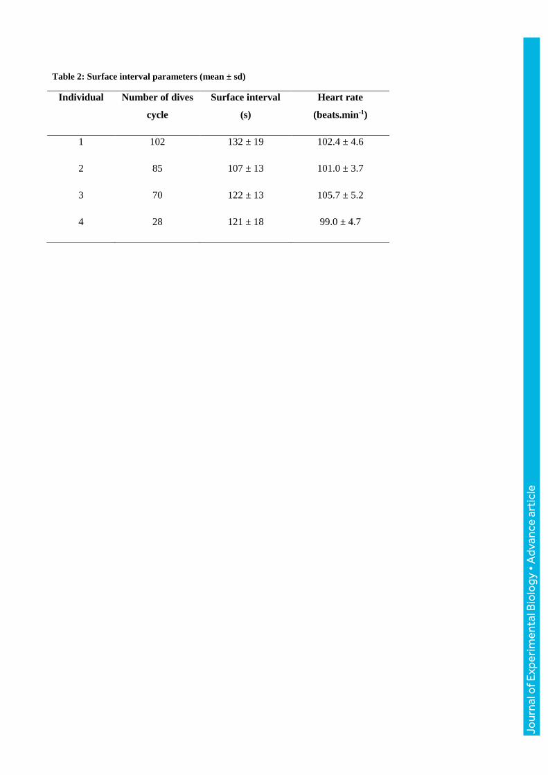
Table 2: Surface interval parameters (mean ± sd)
Individual Number of dives
cycle
Surface interval
(s)
Heart rate
(beats.min-1)
1 102 132 ± 19 102.4 ± 4.6
2 85 107 ± 13 101.0 ± 3.7
3 70 122 ± 13 105.7 ± 5.2
4 28 121 ± 18 99.0 ± 4.7
Jour
nal o
f Exp
erim
enta
l Bio
logy
• A
dvan
ce a
rtic
le
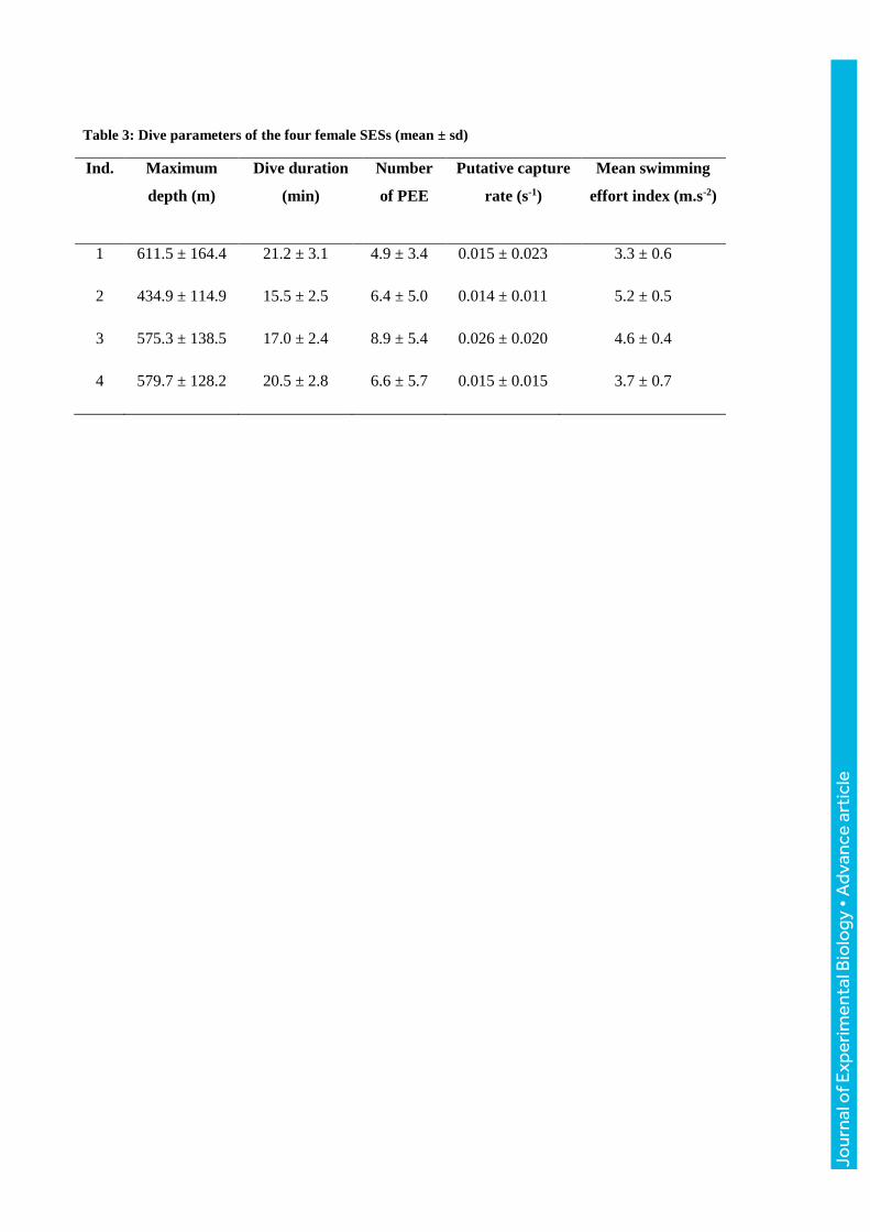
Table 3: Dive parameters of the four female SESs (mean ± sd)
Ind. Maximum
depth (m)
Dive duration
(min)
Number
of PEE
Putative capture
rate (s-1)
Mean swimming
effort index (m.s-2)
1 611.5 ± 164.4 21.2 ± 3.1 4.9 ± 3.4 0.015 ± 0.023 3.3 ± 0.6
2 434.9 ± 114.9 15.5 ± 2.5 6.4 ± 5.0 0.014 ± 0.011 5.2 ± 0.5
3 575.3 ± 138.5 17.0 ± 2.4 8.9 ± 5.4 0.026 ± 0.020 4.6 ± 0.4
4 579.7 ± 128.2 20.5 ± 2.8 6.6 ± 5.7 0.015 ± 0.015 3.7 ± 0.7
Jour
nal o
f Exp
erim
enta
l Bio
logy
• A
dvan
ce a
rtic
le
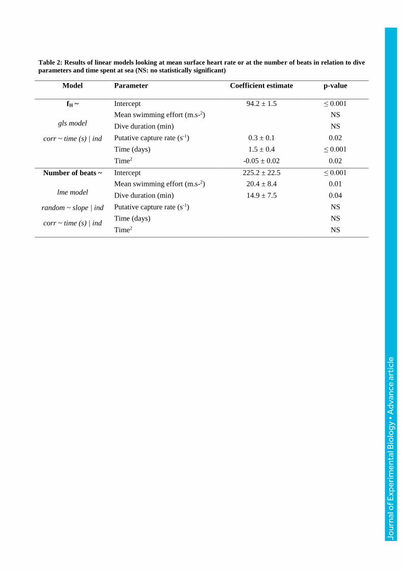
Table 2: Results of linear models looking at mean surface heart rate or at the number of beats in relation to dive
parameters and time spent at sea (NS: no statistically significant)
Model
Parameter Coefficient estimate p-value
fH ~
gls model
corr ~ time (s) | ind
Intercept 94.2 ± 1.5 ≤ 0.001
Mean swimming effort (m.s-2) NS
Dive duration (min) NS
Putative capture rate (s-1) 0.3 ± 0.1 0.02
Time (days) 1.5 ± 0.4 ≤ 0.001
Time2 -0.05 ± 0.02 0.02
Number of beats ~
lme model
random ~ slope | ind
corr ~ time (s) | ind
Intercept 225.2 ± 22.5 ≤ 0.001
Mean swimming effort (m.s-2) 20.4 ± 8.4 0.01
Dive duration (min) 14.9 ± 7.5 0.04
Putative capture rate (s-1) NS
Time (days) NS
Time2 NS
Jour
nal o
f Exp
erim
enta
l Bio
logy
• A
dvan
ce a
rtic
le
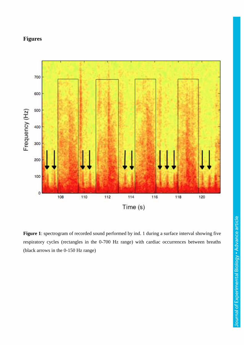
Figures
Figure 1: spectrogram of recorded sound performed by ind. 1 during a surface interval showing five
respiratory cycles (rectangles in the 0-700 Hz range) with cardiac occurrences between breaths
(black arrows in the 0-150 Hz range)
Jour
nal o
f Exp
erim
enta
l Bio
logy
• A
dvan
ce a
rtic
le
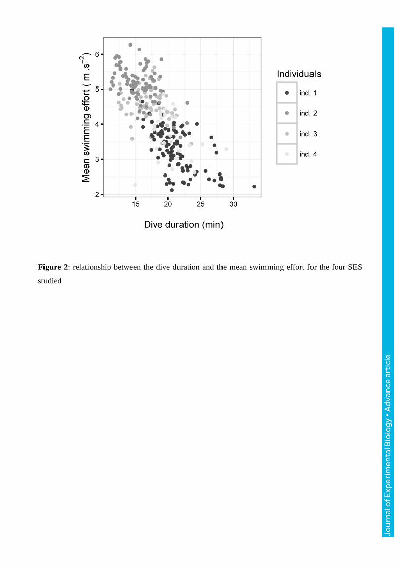
Figure 2: relationship between the dive duration and the mean swimming effort for the four SES
studied
Jour
nal o
f Exp
erim
enta
l Bio
logy
• A
dvan
ce a
rtic
le
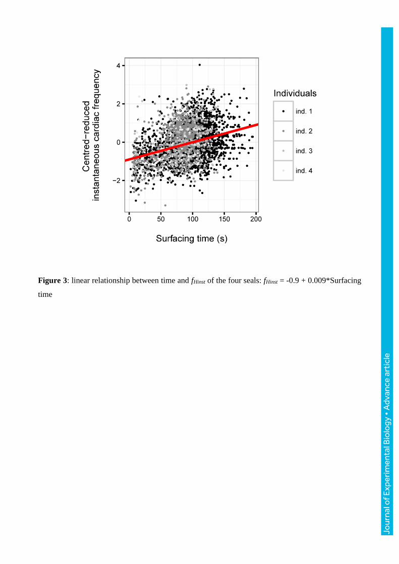
Figure 3: linear relationship between time and fHinst of the four seals: fHinst = -0.9 + 0.009*Surfacing
time
Jour
nal o
f Exp
erim
enta
l Bio
logy
• A
dvan
ce a
rtic
le
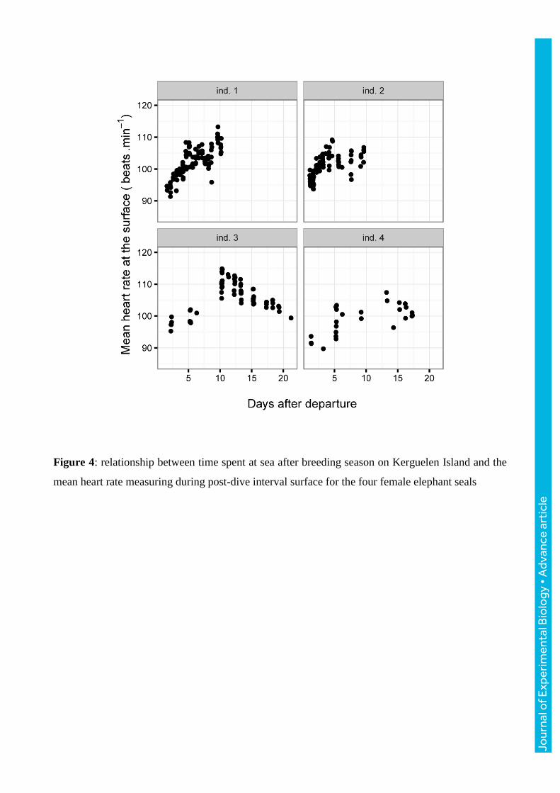
Figure 4: relationship between time spent at sea after breeding season on Kerguelen Island and the
mean heart rate measuring during post-dive interval surface for the four female elephant seals
Jour
nal o
f Exp
erim
enta
l Bio
logy
• A
dvan
ce a
rtic
le