Embed Size (px)
Citation preview
RETROPERITONEAL TERATOMAS – A DIAGNOSTIC DILEMMA
Authors : Dr Virendra S Athavale Associate Professor, Dept of General Surgery Padm. Dr D Y Patil Medical College & Research Centre, Pune, India. Email : [email protected]
Co- Authors : Dr Murtuza A Calcuttawala Associate Professor, Dept of General Surgery Padm. Dr D Y Patil Medical College & Research Centre, Pune, India. Email : [email protected]
Dr Dakshayani S Nirhale Professor, Dept of General Surgery. Padm. Dr D Y Patil Medical College & Research Centre, Pune, India. Email : [email protected] Dr Manashree Sankhe Resident, Dept of General surgery. Padm. Dr D Y Patil Medical College & Research Centre, Pune, India. Email : [email protected]
Abstract:
Dermoid cysts (benign cystic mature teratomas) are congenital tumors consisting of derivatives from the ectoderm, endoderm and mesoderm germ cell layers. A teratoma is considered to be a non-seminomatous germ cell tumour and is typically located in either the sacrococcygeal region or in the gonads. Retroperitoneal teratomas are commonly identified in early childhood, but are rarely reported in adults (1, 2). They constitute less than 4% of all extra-gonadal teratomas with less than 120 cases having been reported, and only partly described in the retroperitoneum of adults (3). We report here a case of a histologically unusual retroperitoneal tumour detected on magnetic resonance imaging during the workup of low backache in a 55-year-old male. The evaluation and treatment of this condition and a review of the literature are included in this paper.
Case Report:
A 55-year-old Indian male presented with a complaint of low backache radiating to right lower limb and vague abdominal discomfort localized to umbilicus since 3 months. There was no associated history of vomiting, bowel or bladder complaints, weight loss or
tingling and numbness in the lower limbs. His general physical examination revealed averagely built & nourished, active male with no jaundice, pallor, cyanosis, clubbing and lymphadenopathy. Abdominal examination revealed a soft, diffuse, intra-abdominal, non-tender, vague palpable mass in umbilical and right lumbar region. There was no

organomegaly. Bowel sounds were normal. Rest of the physical examination was unremarkable. Spine examination was normal with no spine tenderness or deformity. Laboratory investigations were within normal limits.
Ultrasonography of abdomen & pelvis was suggestive of a lobulated lesion of 9.5 x 7.6 x 5.2 cm size arising from right psoas muscle, displacing lower pole of right kidney.
In view of the backache which failed to improve even with continuous medication and physiotherapy, radiograph of lumbo-sacral spine as well as further investigation of MRI (Magnetic Resonance Imaging) lumbar spine with whole spine screening was done.
X Ray LS spine revealed there was destruction of the lateral margin of the right pedicle of L4 vertebra with evidence of a retroperitoneal mass with two discrete areas of calcification.
Illustration - 1
MRI lumbar spine with whole spine screening showed a mass arising from the right psoas muscle with multiple foci of calcifications measuring 11 x 6 x 6.2 cm causing compression of right pelvic ureteric junction. There was also scalloping of the right lateral margin of L2 vertebra due to the mass with no neurological compromise suggestive of chronic psoas abscess.
Illustration -1

Illustration - 2
Illustration - 3
CT(Computer Tomography) scan of abdomen and pelvis showed a well-defined solid mass measuring 11.2 x 6.0 x6.8 cm noted in retroperitoneum on right side in relation to anterior surface of right psoas causing extrinsic compression and anterior displacement of adjoining right upper ureter with resultant fullness of pelvicalyceal system. Also, Medially, it was abutting right lateral margin of IVC with obliteration of intervening fat plane and scalloping of right lateral cortex of L3 vertebral body was noted-findings suggestive of possibly a neoplastic etiology - ? Liposarcoma.

illustration - 4
Illustration - 5
With all the above investigations and provisional diagnosis of a retroperitoneal mass, possibly a neoplastic lesion- neurogenic or liposarcoma, a decision of excision of mass was made.
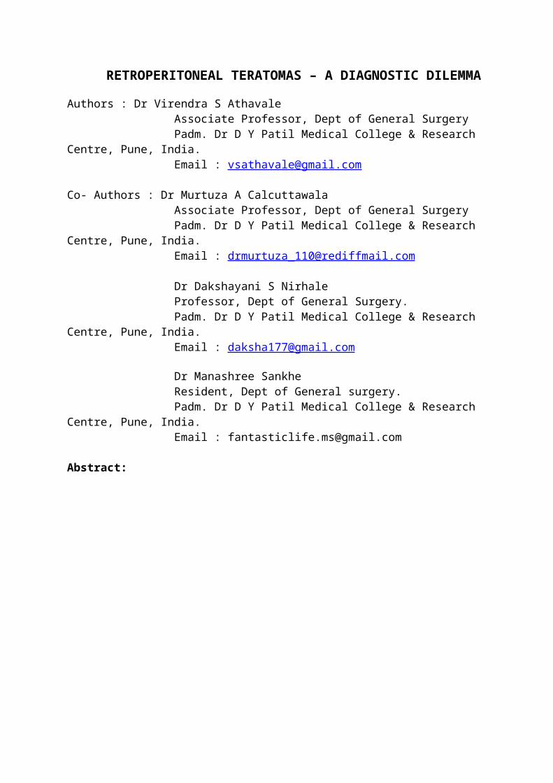
Operative findings : Under General Anaesthesia, upper midline vertical incision was taken. After mobilization of the right colon, a well circumscribed, encapsulated, cystic swelling of about 11x 6 x 6 cm was seen closely connected to the inferior vena cava. The cyst was freed intra-abdominally, paying special attention to inferior vena cava, kidney vessels and ureter. No enlarged lymph nodes were detected. Haemostasis was achieved. Abdomen was closed in layers, keeping a drain. Patient tolerated the procedure well. Post-operatively the patient had an unremarkable recovery.

Illustration - 6
Retroperitoneal mass closely connected to the inferior vena cava was seen
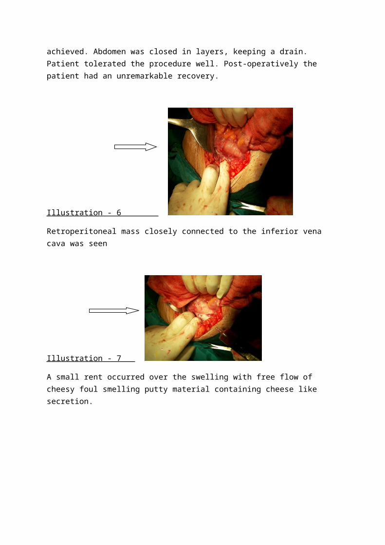
Illustration - 7
A small rent occurred over the swelling with free flow of cheesy foul smelling putty material containing cheese like secretion.
Illustration - 8
Operative specimen showing retroperitoneal cystic mass about 11 cm in greatest dimensions is seen.
Histopathological Report : Cystic mass shows an inner lining of squamous epithelium of the epidermis. The outer layer consists of thick fibrocollagenous tissue with multifocal areas of lymphocytic infiltration. The cavity showed presence of lamellated type of keratinous material. The features are consistent with a dermoid cyst with secondary inflammatory changes – Retroperitoneum.
Illustration - 10
Illustration - 11
Follow – up: The patient was followed up till 1 year & is doing well with no symptoms of backache. Follow up Ultrasound Abdomen and pelvis showed no evidence of recurrence.
Discussion : A teratoma is a true tumour or neoplasm composed of multiple tissues of kinds foreign to the part in which it arises. Macroscopically there are two types: Cystic teratoma: usually benign, contains yellowish liquid material , composed of fully developed tissue. Solid teratoma: generally malignant, have a varied aspect, formed of fibrous, fatty, cartilaginous and bone tissue consists of immature embryonic tissue (4) Retroperitoneal dermoid is rare and usually develops, in childhood(5)
The order of frequency of teratoma localisation is : Ovarian, Testicular, Anterior Mediastinal, with retroperitoneal localisation occurring least of all(6). Symptoms of

Retroperitoneal Teratoma ( RPT ) are variable. In benign cases there is never an alteration in the patient’s general condition. In malignant forms the initial clinical picture may be normal, but there are often general symptoms or disturbances due to compression. Complications are rare(7). However, as benignity cannot be ascertained; the tumour must be removed surgically. Tissue adherence, which has been observed with malignant and benign lesions, may hinder complete removal or require extended surgery. Patients who have had benign teratomas surgically removed have an excellent prognosis(8)
The operative management of RPTs, especially those with rupture, may be complex and challenging. Despite their benign nature, the lesions can attenuate and surround major vessels, making resection difficult. Preoperative imaging has been known to be offer limited help in demonstrating the position of the major vessels (9). A computed tomography (CT) scan or magnetic resonance image (MRI) can identify various components of these tumours, including bone, soft-tissue density structures, adipose tissue, and sebaceous and serous-type fluids. These imaging studies also can display the precise location, morphology, and adjacent structures of the tumour, which provide better preoperative planning and increased likelihood of complete removal of the tumour with less iatrogenic damage.(10)
Surgical resection remains the mainstay of therapy for mature teratomas and is required for definitive diagnosis (11) Benign tumours, when resected, yield a 5-year survival rate of 100%. A long-term study showed that complete surgical resection is associated with the best survival rates for primary retroperitoneal tumours (12) However it may be difficult to make a preoperative diagnosis and the surgical excision could be a challenging task. (13)
Source of Funding – None
Conflict of Interest – None

References :
1. Luo CC, Huang CS and Chu SM: Retroperitoneal teratomas in infancy and childhood. Surg Int. 2005; 21:536–540.
2. Gatcombe HG, Assikis V and Kooby D: Primary retroperitoneal teratomas: a review of the literature. J Surg Oncol. 2004; 86:107–113.
3. Lukanovic A; Patrelli TS: Retroperitoneal mass with ischiorectal fossa extension: diagnosis, clinical features and surgical approach. A literature review starting from a rare clinical case of primary retroperitoneal dermoid cyst. Eur J Gynaecol Oncol. 2010; 31(6):709-13.
4. Willis RA: Pathology of tumors. St. Louis, Mosby, 1948; 940: 430-4505. Pack GT, Tabah EJ: Collective review; primary retroperitoneal tumors; a study of
120 cases. Surggynecobstet 1954; 99:209-231, 19546. Engel RM, Elkins RC, Fletcher BD: Retroperitoneal teratoma. Review of the
literature and presentation of an unusual case. Cancer 1968; 22: 1068-1073.7. Caroli J, Hepp J, Phocas E, et al: Teratome retroperitoneal perforedans le canal
hepatique gauche etdansI’estomac. Contribution aI’etude des complications des teratomasretroperitoneaux. Sem Hop Paris 1963; 39: 1499 – 1508.
8. Shekappa C, Malagimani, Yashwanth C.N, et al: A CASE REPORT - RETROPERITONEAL DERMOID. Journal of Evolution of Medical and Dental Sciences 2013;36(2):6791-6795
9. Jones NM, Kiely EM.Retroperitoneal teratomas-potential for surgical misadventure. J Pediatr Surg 2008; 43: 184-6
10. H. Liu, L. Wanmeng, Y. Wenlong, and Q. Youfei, “Giant retroperitoneal teratoma in an adult,” The American Journal of Surgery, vol. 193, no. 6, pp. 736–737, 2007.
11. H. G. Gatcombe, V. Assikis, D. Kooby, and P. A. S. Johnstone, “Primary retroperitoneal teratomas: a review of the literature,” Journal of Surgical Oncology, vol. 86, no. 2, pp. 107–113, 2004.
12. W. Pinson, S. G. ReMine, W. S. Fletcher, and J. W. Braasch, “Long-term results with primary retroperitoneal tumors,” Archives of Surgery, vol. 124, no. 10, pp. 1168–1173, 1989.
13. Sarin YK. Peritonitis caused by rupture of infected retroperitoneal teratoma. APSP J Case Rep 2012; 3:2.


![[ADMXXXX Session | Year] · Web viewProfessor name, contact , class hours and locations, prerequites](https://img.pdfslide.us/doc/110x75/5fa18ac3b41bf0376f6509ef/admxxxx-session-year-web-view-professor-name-contact-class-hours-and-locations.jpg)