Embed Size (px)
Citation preview

Delivered by Ingenta toGuest User
IP 200246784Thu 14 Jun 2007 133050
RESEARCHARTICLE
Copyright copy 2007 American Scientific PublishersAll rights reservedPrinted in the United States of America
Journal ofBiomedical Nanotechnology
Vol 3 203ndash208 2007
Antibacterial Effect of Silver NanoparticlesProduced by Fungal Process on Textile Fabrics and
Their Effluent Treatment
Nelson Duraacuten12lowast Priscyla D Marcato1 Gabriel I H De Souza2Oswaldo L Alves3 and Elisa Esposito2
1Biological Chemistry Laboratory Instituto de Quiacutemica Universidade Estadual de CampinasCEP 13084862 Caixa Postal 6154 Campinas SP Brazil
2Biological Chemistry and Biotechnology Laboratory Center Environmental SciencesUniversidade de Mogi das Cruzes Mogi das Cruzes SP Brazil
3Solid State Chemistry Laboratory Instituto de Quiacutemica Universidade Estadual de CampinasCEP 13084862 Caixa Postal 6154 Campinas SP Brazil
Microorganisms play an important role in toxic metal remediation through reduction of metal ionsStudies demonstrated that silver ions may be reduced extracellularly using Fusarium oxysporum togenerate stable gold or silver nanoparticles in water These particles can be incorporated in severalkinds of materials such as cloths These cloths with silver nanoparticles are sterile and can be usefulin hospitals to prevent or to minimize infection with pathogenic bacteria such as Staphylococcusaureus In this work the extracellular production of silver nanoparticles by F oxysporum and itsantimicrobial effect when incorporated in cotton fabrics against S aureus were studied In additionall effluent was bioremediated using treatment with C violaceum The results showed that cottonfabrics incorporated with silver nanoparticles displayed a significant antibacterial activity againstS aureus The effluent derived from the process was treated with C violaceum and exhibited anefficient reduction in the silver nanoparticles concentration In conclusion it was demonstrated theapplication of biological synthesis to silver nanoparticles production and its incorporation in clothsproviding them sterile properties Moreover to avoid any damage to the environment the effluentcontaining silver nanoparticles can be treated with cyanogenic bacterial strains
Keywords Nanoparticles Silver Antimicrobial Metal Ion Microbiology TEM
1 INTRODUCTION
Recently the development of resistant or even multi-resistant pathogens has become a major problem forinstance Staphylococcus aureus resistance to methicillinand Candida albicans resistance to fluconazole have to bementioned1 On the other hand the introduction of newlydevised wound dressing has been a major breakthrough inthe management of wounds or infections In order to pre-vent or reduce infection a new generation of dressing incor-porating antimicrobial agents like silver was developed2
It is well known that silver ions and silver-based com-pounds are highly toxic to microorganisms Thus sil-ver ions have been used in many kinds of formulations3
and recently it was shown that hybrids of silver nano-particles with amphiphilic hyperbranched macromolecules
lowastAuthor to whom correspondence should be addressed
exhibit effective antimicrobial surface coating4 The wounddressing impregnated with colloidal silver (Contreet-Hreg)resulted in a strong decrease of pathogen-specific alter-ations in infected epithelium The delivery of silver toinfected keratinocytes in a moist healing environmentimproves the benefitrisk ratio as compared to wounddressing without silver1 Similar results with E coli wereobtained with silver nanoparticles3
Nanometer sized silver particles synthesized by inertgas condensation or co-condensation techniques showedantibacterial activity against E coli The antibacterial effi-ciency of the nanoparticles was investigated by introducingthe particles into a media containing E coli and it wasfound that they exhibited antibacterial effect at low concen-trations In addition it was observed a relationship betweenthe antibacterial properties and the total surface area of thenanoparticles Smaller particles with a larger surface areawere more efficient in the antibacterial activity tests5
J Biomed Nanotechnol 2007 Vol 3 No 2 1550-703320073203006 doi101166jbn2007022 203
Delivered by Ingenta toGuest User
IP 200246784Thu 14 Jun 2007 133050
RESEARCHARTICLE
Antibacterial Effect of Silver Nanoparticles Produced by Fungal Process on Textile Fabrics Duraacuten et al
In the last few decades there has been increased inter-est in reducing the availability of commercial textile con-taining antibacterial agents due to environmental pollutionSince silver is a good antibacterial agent and non-toxic andnatural inorganic metal it appears as an interesting mate-rial to be used in different kind of textile fibers In thisdirection polypropylenesilver nanocomposite fibers wereprepared and the antibacterial tests showed that the fiberscontaining silver nanoparticles in core-part (inside thefiber) had no nearly significant antibacterial activity How-ever the fibers having silver nanoparticles (30 nm size) insheath-part showed excellent antibacterial effects67 Tex-tile fabrics with antibacterial efficacy were easily achievedusing nanosized colloidal silver particles (2ndash5 nm size)by padding process on cotton and polyesters These fab-rics showed laundering durability against S aureus andK pneumoniae8 Similar results were achieved with nano-sized colloidal silver particles on polyester nonwovensThe growth of bacteria colonies was absolutely inhibitedwith only 10 ppm colloidal silver when the mean diameterof the silver particles was 2ndash5 nm Consequently a smallerparticle size yielded better bacteriostasis on silver-paddednonwoven fabrics9
Silver nanoparticles can be coated onto polyurethanefoams in diverse forms This material can be washed sev-eral times without any loss of nanoparticles The perfor-mance of the material as an antibacterial water filter wasstudied and no bacterium (E coli) was detected in the out-put water when the input water had a bacterial load of 105
to 106 CFUml10
Many synthetic procedures for silver nanoparticles areavailable but a narrow and controlled size preparationseems difficult to obtain because depend of the adjustedthe concentration of reacting chemicals and controlled thereaction environment11 Colloidal metal particles can beobtained by chemical synthesis but these methods usetoxic chemicals in the synthesis protocol which raisesgreat concern for environmental reasons12 Consequentlyresearchers have turned to biological synthesis becausethrough this biological synthesis obtaining particles withgood control on the size distribution than the other meth-ods The nanoparticles could also be stabilized directlyin the process by proteins13 Although it is known thatmicroorganisms such as bacteria yeast and fungi playan important role in the remediation of toxic metalsthrough reduction of the metal ions only recently thisapproach was considered interesting as nanofactories14 Inthis respect the biosynthesis of inorganic nanomaterialsusing eukaryotic organisms such as fungi was achievedwith the intracellular production of silver nanoparticles byVerticillium strains12 Recently it was found that aqueoussilver ions may be reduced extracellularly using the fungusF oxysporum to generate silver nanoparticles in water15
The mechanistic aspects were very recently described13
and this process occurs probably by conjugation of reduc-tase action and by electron shuttle quinones Our aims in
this research were to compare different impregnation pro-cesses published before with silver nanoparticle biosynthe-sized by the fungus F oxysporum Also important is ourecotoxicological concern by recovering the silver nano-particles generated in the process using a biotechnologicalapproach involving Chromobacterium violaceum which isable to metabolize or store metal ions in order to avoidany environment damage16 As recently described C vio-laceum produces around 1ndash4 mM free cyanide17 and itis able to metabolize several metals as cyanide complexAmong these metals are gold1819 nickel17 and silver20
2 MATERIALS AND METHODS
21 Silver Nanoparticles Preparation
The F oxysporum strain used was 07 SD fromESALQ-USP Genetic and Molecular Biology Laboratory-Piracicaba SP Brazil The fungal inoculum was preparedin 2 malt extract and 05 yeast extract at 28 C inPetri dishes The liquid fungal growth was carried outin the presence of 05 yeast extract at 28 C for 6days The biomass was filtrated and resuspended in ster-ile water The biosynthesis of silver nanoparticles wascarried out as following approximately 10 g of F oxys-porum biomass was taken in a conical flask containing100 ml of distilled water kept for 72 h at 28 C andthen the aqueous solution components were separated byfiltration In this solution (fungal filtrate) AgNO3 (10minus3
M) was added and the system was kept for several hoursat 28 C Periodically aliquots of the reaction solutionwere removed and the absorption was measured in aUV-Vis spectrophotometer (Agilent 8453mdashdiode array) at440 nm The silver nanoparticles were characterized byTransmission Electron Microscopy (TEM) and Elemen-tal Spectroscopy Imaging (ESI) Bright field images andthe elemental distribution within silver nanoparticles wereobtained using a Carl Zeiss CEM-902 transmission elec-tron microscope (80 KeV) equipped with a Castaing-Henry-Ottensmeyer energy filter spectrometer within thecolumn For the examination of the silver particle onedrop of the particle dispersion was deposited on carbon-coated parlodion films supported in 300 mesh copper grids(Ted Pella)
Elemental images were obtained for the relevant ele-ments found in this sample using monochromatic elec-trons corresponding to the silver K-edge sulfur L23-edgeand nitrogen L3-edge The energy-selecting slit was set at367 plusmn 6 keV for Ag 165 plusmn 6 eV for S and 400 plusmn 6 eVfor N The images were recorded by a Proscan high-speedslow-scan CCD camera and processed in the AnalySis 30system
The size of silver nanoparticles was measured by X-RayDiffraction (XRD model XD3A from Shimadzu) withnickel-filtered Cu-K radiation (40 KV 30 mA) at anangle of 2 from 5 to 50 The scan speed was 002min
204 J Biomed Nanotechnol 3 203ndash208 2007
Delivered by Ingenta toGuest User
IP 200246784Thu 14 Jun 2007 133050
RESEARCHARTICLE
Duraacuten et al Antibacterial Effect of Silver Nanoparticles Produced by Fungal Process on Textile Fabrics
and the time constant was 2 s The size was calculatedthrough of the Scherrerrsquos equation
D = Kcor cos with cor = 2sample minus2
ref12
where D is the average crystal size K is the Scherrer coef-ficient (089) is the X-ray wavelength (= 1542 Aring) Braggrsquos angle (2 = 251) cor the corrected of the fullwidth at half-maximum (FWHM) in radians and sample
and ref are the FWHM of the reference and sample peaksrespectively21
22 Silver Nanoparticles Loading on Cotton Fabrics
Cotton fabrics were washed sterilized and dried beforeuse Experiments were performed on samples with maxi-mum dimensions of 5 cm times 5 cm The final filtrate (100ml 240 ppm) obtained above was treated by ultracentrifu-gation for 5 minutes and half of the filtrated (superior part)was eliminated to concentrate the silver nanoparticles Inorder to impregnate cotton fabrics (5 cm times 5 cm) thesewere submersed in an Erlenmeyer (50 ml) and shaking at600 rpm for 24 h and dried at 70 C The percentage ofsilver nanoparticles incorporated in the cotton fabrics wasmeasured by X-ray fluorescence (XRF) (Shimatzu ModEDX 7000) Source Rh 50 kV and 15 kV
23 Antibacterial Activity
The antibacterial behavior of the fabrics were evaluatedagainst Staphylococcus aureus (ATCC 6538) a Gram-positive bacterium The cotton fabrics were inoculated onagar plates inoculated with S aureus The inoculum was13ndash16 105ml After 24 h the plates were sterilized andthe cotton fabrics were analyzed by Scanning ElectronMicroscopy (SEM) and Energy Dispersive Spectroscopy(EDS) at a voltage of 20 kV after previously coated withAuPd under vacuum
In order to study the antimicrobial activity of the fab-rics squares of 1 cm of each fabric were prepared in asep-tic manner Each square was placed in a sterile vial andthe fabrics subjected to pretreatment with 800 l distilledwater for 10 min Tryptone soy broth (22 ml) was thenadded to each vial to make up to a total volume of 3 ml Analiquot (10 l) of S aureus suspension was added to eachvial (16times 105ml) containing the fabrics Control brothswith and without bacterial inoculation were also includedThe vials were then incubated with agitation at 35 C220 rpm Aliquots of 10 l broth were sampled at 24 hand serial dilution for the aliquots were prepared in brothDuplicate aliquots (50 l) of the serially diluted sampleswere spread on to plates The plates were incubated at35 C and bacterial counts were performed The bacte-riostatic activity was evaluated after 24 h and calculatedpercent reduction of bacteria Using the following equa-tion R= AminusBAtimes100 Where R= the reduction
rate A = the number of bacterial colonies from untreatedfabrics and B = the numbers of bacterial colonies fromtreated fabrics
24 Microbial Treatment
The cotton fabrics previously loaded with silver nano-particles were washed several times (as it could be usedin a laundromat) and the effluent was treated as followsa suspension of Chromobacterium violaceum CCT 3496previously grown for 12 h at 30 C in an orbital shakerat 120 rpm in 05 D-glucose 05 peptone 02 yeastextract and 003 tryptophan22 The suspension wereinoculated into 100 ml liquid collected from 5 consecu-tive water washes of the cotton fabrics loaded with silvernanoparticles The concentration of C violaceum inocu-lated were 102 105 e 108 CFUml Three inoculated flaskswere incubated at 30 C for 24 h in an orbital shaker at120 rpm The silver nanoparticles were measured in theeffluent before and after C violaceum treatment The bac-terial biomass was analyzed by SEM and EDS techniqueas described before
3 RESULTS AND DISCUSSION
31 Preparation and Characterization
The Erlenmeyer flasks with the fungal filtrate had apale yellow color before the addition of Ag+ ions whichchanged to a brownish color on completion of the reactionwith Ag+ ions for 28 h The appearance of a brownishcolor in solution containing the biomass is a clear indica-tion of the formation of silver nanoparticles in the reac-tion mixture13 Time-dependent increase in the intensityof the plasmon resonance (440 nm) was observed in the
0 25 50 75 100 125 150 175
00
01
02
03
04
05
Abs
orba
nce
Time (h)
Fig 1 Intensity absorbance of the plasmon resonance (440 nm) infunction of time of reaction in an aqueous solution of 10minus3 M AgNO3
with the fungal filtrate
J Biomed Nanotechnol 3 203ndash208 2007 205
Delivered by Ingenta toGuest User
IP 200246784Thu 14 Jun 2007 133050
RESEARCHARTICLE
Antibacterial Effect of Silver Nanoparticles Produced by Fungal Process on Textile Fabrics Duraacuten et al
40 nm
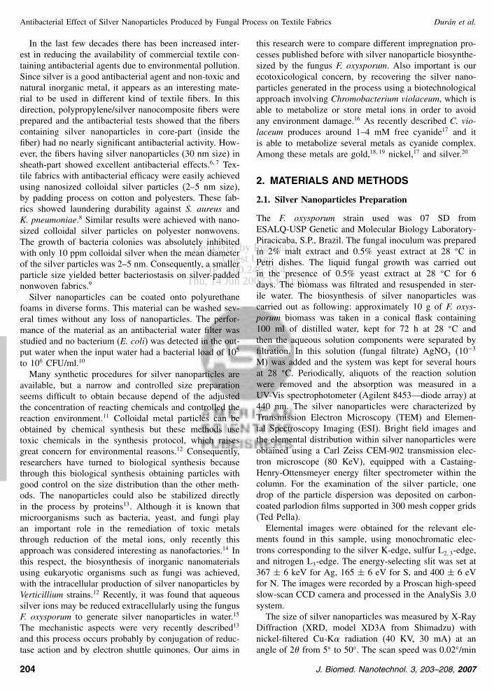
Fig 2 TEM bright field image of the silver nanoparticles
F oxysporum reaction vessels (Fig 1) confirming the sil-ver nanoparticles formation
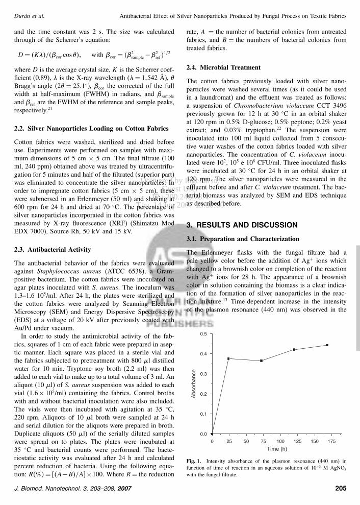
The TEM micrograph (Fig 2) showed spherical sil-ver nanoparticles with size of 16 nm calculated by XRDthrough of the Scherrerrsquos equation These nanoparticleswere analyzed by elemental spectroscopy imaging (ESI)(Fig 3) The Figure 3(A) shows the bright field image ofthe silver nanoparticles and Figures 3(B) (C) and (D) showthe ESI maps of this same region for Ag N and S atomsrespectively As can be seen in the maps particles wereformed by silver and the presence of the N (Fig 3(C))and S (Fig 3(D)) atoms around the silver nanoparticles are
(A) (B)
(D)(C)
Fig 3 (A) Bright field image of the silver nanoparticles (B) ESI mapfor Ag atoms (C) ESI map for N atoms and (D) ESI map for S atoms
(A) (B)
(C)
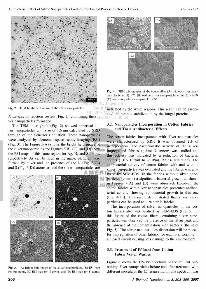
Fig 4 SEM micrographs of the cotton fiber (A) without silver nano-particles (control) times75 (B) without silver nanoparticles (control) times1400(C) containing silver nanoparticles times60
indicated by the white regions This result can be associ-ated the particle stabilization by the fungal proteins
32 Nanoparticles Incorporation in Cotton Fabricsand Their Antibacterial Effects
The cotton fabrics incorporated with silver nanoparticleswere characterized by XRF It was obtained 2 ofincorporation The bacteriostatic activity of the silver-impregnated fabrics against S aureus was studied andthis activity was indicated by a reduction of bacterialcounts (16 times 105ml to lt10ml 999 reduction) Theantibacterial activity of cotton fabrics with and withoutsilver nanoparticles was evaluated and the fabrics was ana-lyzed by SEM-EDS In the fabrics without silver nano-particles (control) a significant bacterial growth as shownin Figures 4(A) and (B) were observed However thecotton fabrics with silver nanoparticles presented antibac-terial activity showing no bacterial growth in this one(Fig 4(C)) This result demonstrated that silver nano-particles can be used to turn sterile fabrics
The incorporation of silver nanoparticles in the cot-ton fabrics also was verified by SEM-EDS (Fig 5) Inthis figure of the cotton fibers containing silver nano-particles was observed the presence of the silver peak andthe absence of the contamination with bacteria (the insetFig 5) The silver nanoparticles dispersion will be reusedfor impregnation of other fabrics for example working ina closed circuit causing less damage to the environment
33 Treatment of Effluent from CottonFabric Water Washes
Figure 6 shows the UV-Vis spectrum of the effluent con-taining silver nanoparticles before and after treatment withdifferent inocula of the C violaceum In this spectrum was
206 J Biomed Nanotechnol 3 203ndash208 2007
Delivered by Ingenta toGuest User
IP 200246784Thu 14 Jun 2007 133050
RESEARCHARTICLE
Duraacuten et al Antibacterial Effect of Silver Nanoparticles Produced by Fungal Process on Textile Fabrics
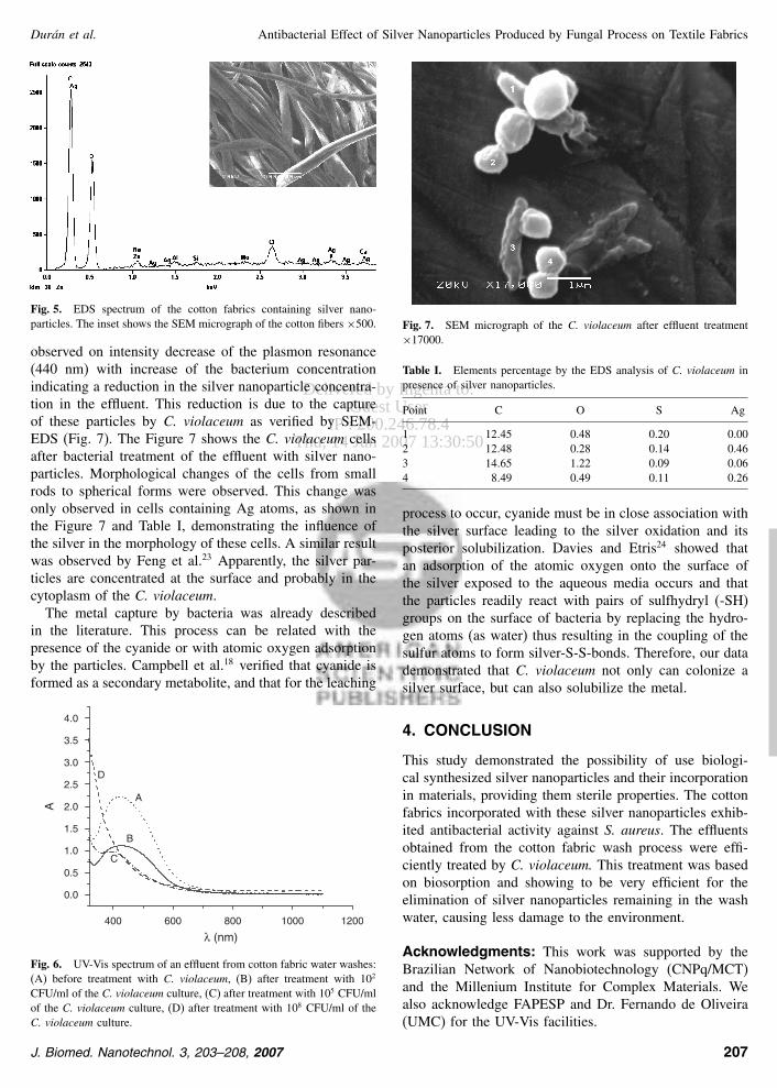
Fig 5 EDS spectrum of the cotton fabrics containing silver nano-particles The inset shows the SEM micrograph of the cotton fibers times500
observed on intensity decrease of the plasmon resonance(440 nm) with increase of the bacterium concentrationindicating a reduction in the silver nanoparticle concentra-tion in the effluent This reduction is due to the captureof these particles by C violaceum as verified by SEM-EDS (Fig 7) The Figure 7 shows the C violaceum cellsafter bacterial treatment of the effluent with silver nano-particles Morphological changes of the cells from smallrods to spherical forms were observed This change wasonly observed in cells containing Ag atoms as shown inthe Figure 7 and Table I demonstrating the influence ofthe silver in the morphology of these cells A similar resultwas observed by Feng et al23 Apparently the silver par-ticles are concentrated at the surface and probably in thecytoplasm of the C violaceum
The metal capture by bacteria was already describedin the literature This process can be related with thepresence of the cyanide or with atomic oxygen adsorptionby the particles Campbell et al18 verified that cyanide isformed as a secondary metabolite and that for the leaching
400 600 800 1000 1200
00
05
10
15
20
25
30
35
40
D
C
B
A
λ (nm)
A
Fig 6 UV-Vis spectrum of an effluent from cotton fabric water washes(A) before treatment with C violaceum (B) after treatment with 102
CFUml of the C violaceum culture (C) after treatment with 105 CFUmlof the C violaceum culture (D) after treatment with 108 CFUml of theC violaceum culture
Fig 7 SEM micrograph of the C violaceum after effluent treatmenttimes17000
Table I Elements percentage by the EDS analysis of C violaceum inpresence of silver nanoparticles
Point C O S Ag
1 1245 048 020 0002 1248 028 014 0463 1465 122 009 0064 849 049 011 026
process to occur cyanide must be in close association withthe silver surface leading to the silver oxidation and itsposterior solubilization Davies and Etris24 showed thatan adsorption of the atomic oxygen onto the surface ofthe silver exposed to the aqueous media occurs and thatthe particles readily react with pairs of sulfhydryl (-SH)groups on the surface of bacteria by replacing the hydro-gen atoms (as water) thus resulting in the coupling of thesulfur atoms to form silver-S-S-bonds Therefore our datademonstrated that C violaceum not only can colonize asilver surface but can also solubilize the metal
4 CONCLUSION
This study demonstrated the possibility of use biologi-cal synthesized silver nanoparticles and their incorporationin materials providing them sterile properties The cottonfabrics incorporated with these silver nanoparticles exhib-ited antibacterial activity against S aureus The effluentsobtained from the cotton fabric wash process were effi-ciently treated by C violaceum This treatment was basedon biosorption and showing to be very efficient for theelimination of silver nanoparticles remaining in the washwater causing less damage to the environment
Acknowledgments This work was supported by theBrazilian Network of Nanobiotechnology (CNPqMCT)and the Millenium Institute for Complex Materials Wealso acknowledge FAPESP and Dr Fernando de Oliveira(UMC) for the UV-Vis facilities
J Biomed Nanotechnol 3 203ndash208 2007 207
Delivered by Ingenta toGuest User
IP 200246784Thu 14 Jun 2007 133050
RESEARCHARTICLE
Antibacterial Effect of Silver Nanoparticles Produced by Fungal Process on Textile Fabrics Duraacuten et al
References and Notes
1 M Schaller J Laude H Bodewaldt G Hamm and H C KortingToxicity and antimicrobial activity of a hydrocolloid dressing con-taining silver particles in an ex vivo model of cutaneous infectionSkin Pharmacol Physiol 17 31 (2004)
2 H Q Yin R Langford and R E Burrell Comparative evaluation ofthe antimicrobial activity of ACTICOAT antimicrobial barrier dress-ing J Burn Care Rehabil 20 195 (1999)
3 I Sondi and B Salopek-Sondi Silver nanoparticles as antimicrobialagent a case study on E coli as a model for Gram-negative bacteriaJ Colloid Interf Sci 275 177 (2004)
4 C Aymonier U Scholotterbeck L Antonietti P ZachariasR Thomann J C Tiller and S Mecking Hybrids of silvernanoparticles with amphiphilic hyperbranched macromoleculesexhibiting antimicrobial properties Chem Commun 24 3018(2002)
5 C Baker A Pradhan L Pakstis D J Pochan and S I Shah Syn-thesis and antibacterial properties of silver nanoparticles J NanosciNanotechnol 5 244 (2005)
6 S Y Yeo H J Lee and S H Jeong Preparation of nanocompos-ite fibers for permanent antibacterial effect J Mater Sci 38 2143(2003)
7 S Y Yeo and S H Jeong Preparation and characterization ofpolypropylenesilver nanocomposite fibers Polymer Intern 52 1053(2003)
8 H J Lee S Y Yeo and S H Jeong Antibacterial effect of nano-sized silver colloidal solution on textile fabrics J Mater Sci 382199 (2003)
9 H J Lee and S H Jeon Bacteriostasis of nanosized colloidal silveron polyester nonwovens Text Res J 74 442 (2004)
10 P Jain and T Pradeep Potential of silver nanoparticles-coatedpolyurethane foam as an antibacterial water filter Biotechnol Bio-eng 90 59 (2005)
11 M Kowshik S Ashtaputre S Kharrazi W Vogel J Urban S KKulkarni and K M Paknikar Extracellular systhesis of silver nano-particles by a silveritolerant yeast strain MKY3 Nanotechnology 1495 (2003)
12 M Sastry A Ahmad N I Islam and R Kumar Biosynthesis ofmetal nanoparticles using fungi and actinomycete Curr Sci 85 162(2003)
13 N Duraacuten P D Marcato O L Alves G I H De Souza andE Esposito Mechanistic aspects of biosynthesis of silver nano-particles by several Fusarium oxysporum strains J Nanobiotechnol3 8 (2005) DOI1011861477-3155-3-8
14 D Fortin and T J Beveridge Mechanistic routes towards biomineralsurface development Biomineralisation From biology to biotechnol-ogy and medical application edited by E Baeuerlein Wiley-VCHVerlag Germany (2000) p 294
15 A Ahmad P Mukherjee S Senapati D Mandal M I KhanR Kumar and M Sastry Extracellular biosynthesis of silver nano-particles using the fungus Fusarium oxysporum Colloid Surf BBiointerf 28 313 (2003)
16 N Duraacuten and C F M Menck Chromobacterium violaceumA review of pharmacological and industrial perspectives Crit RevMicrobiol 27 201 (2001)
17 M A Faramarzi M Stagars E Pensini W Krebs and H BrandlMetal solubilization from metal-containing solid materials bycyanogenic Chromobacterium violaceum J Biotechnol 113 321(2004)
18 S C Campbell G J Olson T R Clark and G McFeter Biogenicproduction of cyanide and its application to gold recovery J IndMicrobiol Biotechnol 26 134 (2001)
19 A D Smith and R J Hunt Solubilization of gold by Chromo-bacterium violaceum J Chem Technol Biotechnol 35B 110 (1985)
20 L Zeire B V Bronk Y Shabtai J Czeacutegeacute and S Efrima Silvermetal induced surface enhanced Raman of bacteria Colloid SurfA-Physicochem Eng Asp 208 357 (2002)
21 H Borchert E V Shevchenko A Robert I Mekis A KornowskiG Gruumlbel and H Weller Determination of Nanocrystal SizesA Comparison of TEM SAXS and XRD Studies of HighlyMonodisperse CoPt3 Particles Langmuir 21 1931 (2005)
22 D Rettori and N Duraacuten Production extraction and purificationof violacein an antibiotic pigment produced by Chromobacteriumviolaceum World J Microbiol Biotechnol 14 685 (1998)
23 Q L Feng J Wu G Q Chen F Z Cui T N Kim and J OKim A mechanistic study of the antibacterial effect of silver ionson Escherichia coli and Staphylococcus aureus J Niomed MaterRes 52 662 (2000)
24 R L Davies and S F Etris The development and functions of silverin water purification and disease control Catalysis Today 36 107(1997)
Received 12 January 2007 RevisedAccepted 27 March 2007
208 J Biomed Nanotechnol 3 203ndash208 2007
Delivered by Ingenta toGuest User
IP 200246784Thu 14 Jun 2007 133050
RESEARCHARTICLE
Antibacterial Effect of Silver Nanoparticles Produced by Fungal Process on Textile Fabrics Duraacuten et al
In the last few decades there has been increased inter-est in reducing the availability of commercial textile con-taining antibacterial agents due to environmental pollutionSince silver is a good antibacterial agent and non-toxic andnatural inorganic metal it appears as an interesting mate-rial to be used in different kind of textile fibers In thisdirection polypropylenesilver nanocomposite fibers wereprepared and the antibacterial tests showed that the fiberscontaining silver nanoparticles in core-part (inside thefiber) had no nearly significant antibacterial activity How-ever the fibers having silver nanoparticles (30 nm size) insheath-part showed excellent antibacterial effects67 Tex-tile fabrics with antibacterial efficacy were easily achievedusing nanosized colloidal silver particles (2ndash5 nm size)by padding process on cotton and polyesters These fab-rics showed laundering durability against S aureus andK pneumoniae8 Similar results were achieved with nano-sized colloidal silver particles on polyester nonwovensThe growth of bacteria colonies was absolutely inhibitedwith only 10 ppm colloidal silver when the mean diameterof the silver particles was 2ndash5 nm Consequently a smallerparticle size yielded better bacteriostasis on silver-paddednonwoven fabrics9
Silver nanoparticles can be coated onto polyurethanefoams in diverse forms This material can be washed sev-eral times without any loss of nanoparticles The perfor-mance of the material as an antibacterial water filter wasstudied and no bacterium (E coli) was detected in the out-put water when the input water had a bacterial load of 105
to 106 CFUml10
Many synthetic procedures for silver nanoparticles areavailable but a narrow and controlled size preparationseems difficult to obtain because depend of the adjustedthe concentration of reacting chemicals and controlled thereaction environment11 Colloidal metal particles can beobtained by chemical synthesis but these methods usetoxic chemicals in the synthesis protocol which raisesgreat concern for environmental reasons12 Consequentlyresearchers have turned to biological synthesis becausethrough this biological synthesis obtaining particles withgood control on the size distribution than the other meth-ods The nanoparticles could also be stabilized directlyin the process by proteins13 Although it is known thatmicroorganisms such as bacteria yeast and fungi playan important role in the remediation of toxic metalsthrough reduction of the metal ions only recently thisapproach was considered interesting as nanofactories14 Inthis respect the biosynthesis of inorganic nanomaterialsusing eukaryotic organisms such as fungi was achievedwith the intracellular production of silver nanoparticles byVerticillium strains12 Recently it was found that aqueoussilver ions may be reduced extracellularly using the fungusF oxysporum to generate silver nanoparticles in water15
The mechanistic aspects were very recently described13
and this process occurs probably by conjugation of reduc-tase action and by electron shuttle quinones Our aims in
this research were to compare different impregnation pro-cesses published before with silver nanoparticle biosynthe-sized by the fungus F oxysporum Also important is ourecotoxicological concern by recovering the silver nano-particles generated in the process using a biotechnologicalapproach involving Chromobacterium violaceum which isable to metabolize or store metal ions in order to avoidany environment damage16 As recently described C vio-laceum produces around 1ndash4 mM free cyanide17 and itis able to metabolize several metals as cyanide complexAmong these metals are gold1819 nickel17 and silver20
2 MATERIALS AND METHODS
21 Silver Nanoparticles Preparation
The F oxysporum strain used was 07 SD fromESALQ-USP Genetic and Molecular Biology Laboratory-Piracicaba SP Brazil The fungal inoculum was preparedin 2 malt extract and 05 yeast extract at 28 C inPetri dishes The liquid fungal growth was carried outin the presence of 05 yeast extract at 28 C for 6days The biomass was filtrated and resuspended in ster-ile water The biosynthesis of silver nanoparticles wascarried out as following approximately 10 g of F oxys-porum biomass was taken in a conical flask containing100 ml of distilled water kept for 72 h at 28 C andthen the aqueous solution components were separated byfiltration In this solution (fungal filtrate) AgNO3 (10minus3
M) was added and the system was kept for several hoursat 28 C Periodically aliquots of the reaction solutionwere removed and the absorption was measured in aUV-Vis spectrophotometer (Agilent 8453mdashdiode array) at440 nm The silver nanoparticles were characterized byTransmission Electron Microscopy (TEM) and Elemen-tal Spectroscopy Imaging (ESI) Bright field images andthe elemental distribution within silver nanoparticles wereobtained using a Carl Zeiss CEM-902 transmission elec-tron microscope (80 KeV) equipped with a Castaing-Henry-Ottensmeyer energy filter spectrometer within thecolumn For the examination of the silver particle onedrop of the particle dispersion was deposited on carbon-coated parlodion films supported in 300 mesh copper grids(Ted Pella)
Elemental images were obtained for the relevant ele-ments found in this sample using monochromatic elec-trons corresponding to the silver K-edge sulfur L23-edgeand nitrogen L3-edge The energy-selecting slit was set at367 plusmn 6 keV for Ag 165 plusmn 6 eV for S and 400 plusmn 6 eVfor N The images were recorded by a Proscan high-speedslow-scan CCD camera and processed in the AnalySis 30system
The size of silver nanoparticles was measured by X-RayDiffraction (XRD model XD3A from Shimadzu) withnickel-filtered Cu-K radiation (40 KV 30 mA) at anangle of 2 from 5 to 50 The scan speed was 002min
204 J Biomed Nanotechnol 3 203ndash208 2007
Delivered by Ingenta toGuest User
IP 200246784Thu 14 Jun 2007 133050
RESEARCHARTICLE
Duraacuten et al Antibacterial Effect of Silver Nanoparticles Produced by Fungal Process on Textile Fabrics
and the time constant was 2 s The size was calculatedthrough of the Scherrerrsquos equation
D = Kcor cos with cor = 2sample minus2
ref12
where D is the average crystal size K is the Scherrer coef-ficient (089) is the X-ray wavelength (= 1542 Aring) Braggrsquos angle (2 = 251) cor the corrected of the fullwidth at half-maximum (FWHM) in radians and sample
and ref are the FWHM of the reference and sample peaksrespectively21
22 Silver Nanoparticles Loading on Cotton Fabrics
Cotton fabrics were washed sterilized and dried beforeuse Experiments were performed on samples with maxi-mum dimensions of 5 cm times 5 cm The final filtrate (100ml 240 ppm) obtained above was treated by ultracentrifu-gation for 5 minutes and half of the filtrated (superior part)was eliminated to concentrate the silver nanoparticles Inorder to impregnate cotton fabrics (5 cm times 5 cm) thesewere submersed in an Erlenmeyer (50 ml) and shaking at600 rpm for 24 h and dried at 70 C The percentage ofsilver nanoparticles incorporated in the cotton fabrics wasmeasured by X-ray fluorescence (XRF) (Shimatzu ModEDX 7000) Source Rh 50 kV and 15 kV
23 Antibacterial Activity
The antibacterial behavior of the fabrics were evaluatedagainst Staphylococcus aureus (ATCC 6538) a Gram-positive bacterium The cotton fabrics were inoculated onagar plates inoculated with S aureus The inoculum was13ndash16 105ml After 24 h the plates were sterilized andthe cotton fabrics were analyzed by Scanning ElectronMicroscopy (SEM) and Energy Dispersive Spectroscopy(EDS) at a voltage of 20 kV after previously coated withAuPd under vacuum
In order to study the antimicrobial activity of the fab-rics squares of 1 cm of each fabric were prepared in asep-tic manner Each square was placed in a sterile vial andthe fabrics subjected to pretreatment with 800 l distilledwater for 10 min Tryptone soy broth (22 ml) was thenadded to each vial to make up to a total volume of 3 ml Analiquot (10 l) of S aureus suspension was added to eachvial (16times 105ml) containing the fabrics Control brothswith and without bacterial inoculation were also includedThe vials were then incubated with agitation at 35 C220 rpm Aliquots of 10 l broth were sampled at 24 hand serial dilution for the aliquots were prepared in brothDuplicate aliquots (50 l) of the serially diluted sampleswere spread on to plates The plates were incubated at35 C and bacterial counts were performed The bacte-riostatic activity was evaluated after 24 h and calculatedpercent reduction of bacteria Using the following equa-tion R= AminusBAtimes100 Where R= the reduction
rate A = the number of bacterial colonies from untreatedfabrics and B = the numbers of bacterial colonies fromtreated fabrics
24 Microbial Treatment
The cotton fabrics previously loaded with silver nano-particles were washed several times (as it could be usedin a laundromat) and the effluent was treated as followsa suspension of Chromobacterium violaceum CCT 3496previously grown for 12 h at 30 C in an orbital shakerat 120 rpm in 05 D-glucose 05 peptone 02 yeastextract and 003 tryptophan22 The suspension wereinoculated into 100 ml liquid collected from 5 consecu-tive water washes of the cotton fabrics loaded with silvernanoparticles The concentration of C violaceum inocu-lated were 102 105 e 108 CFUml Three inoculated flaskswere incubated at 30 C for 24 h in an orbital shaker at120 rpm The silver nanoparticles were measured in theeffluent before and after C violaceum treatment The bac-terial biomass was analyzed by SEM and EDS techniqueas described before
3 RESULTS AND DISCUSSION
31 Preparation and Characterization
The Erlenmeyer flasks with the fungal filtrate had apale yellow color before the addition of Ag+ ions whichchanged to a brownish color on completion of the reactionwith Ag+ ions for 28 h The appearance of a brownishcolor in solution containing the biomass is a clear indica-tion of the formation of silver nanoparticles in the reac-tion mixture13 Time-dependent increase in the intensityof the plasmon resonance (440 nm) was observed in the
0 25 50 75 100 125 150 175
00
01
02
03
04
05
Abs
orba
nce
Time (h)
Fig 1 Intensity absorbance of the plasmon resonance (440 nm) infunction of time of reaction in an aqueous solution of 10minus3 M AgNO3
with the fungal filtrate
J Biomed Nanotechnol 3 203ndash208 2007 205
Delivered by Ingenta toGuest User
IP 200246784Thu 14 Jun 2007 133050
RESEARCHARTICLE
Antibacterial Effect of Silver Nanoparticles Produced by Fungal Process on Textile Fabrics Duraacuten et al
40 nm
Fig 2 TEM bright field image of the silver nanoparticles
F oxysporum reaction vessels (Fig 1) confirming the sil-ver nanoparticles formation
The TEM micrograph (Fig 2) showed spherical sil-ver nanoparticles with size of 16 nm calculated by XRDthrough of the Scherrerrsquos equation These nanoparticleswere analyzed by elemental spectroscopy imaging (ESI)(Fig 3) The Figure 3(A) shows the bright field image ofthe silver nanoparticles and Figures 3(B) (C) and (D) showthe ESI maps of this same region for Ag N and S atomsrespectively As can be seen in the maps particles wereformed by silver and the presence of the N (Fig 3(C))and S (Fig 3(D)) atoms around the silver nanoparticles are
(A) (B)
(D)(C)
Fig 3 (A) Bright field image of the silver nanoparticles (B) ESI mapfor Ag atoms (C) ESI map for N atoms and (D) ESI map for S atoms
(A) (B)
(C)
Fig 4 SEM micrographs of the cotton fiber (A) without silver nano-particles (control) times75 (B) without silver nanoparticles (control) times1400(C) containing silver nanoparticles times60
indicated by the white regions This result can be associ-ated the particle stabilization by the fungal proteins
32 Nanoparticles Incorporation in Cotton Fabricsand Their Antibacterial Effects
The cotton fabrics incorporated with silver nanoparticleswere characterized by XRF It was obtained 2 ofincorporation The bacteriostatic activity of the silver-impregnated fabrics against S aureus was studied andthis activity was indicated by a reduction of bacterialcounts (16 times 105ml to lt10ml 999 reduction) Theantibacterial activity of cotton fabrics with and withoutsilver nanoparticles was evaluated and the fabrics was ana-lyzed by SEM-EDS In the fabrics without silver nano-particles (control) a significant bacterial growth as shownin Figures 4(A) and (B) were observed However thecotton fabrics with silver nanoparticles presented antibac-terial activity showing no bacterial growth in this one(Fig 4(C)) This result demonstrated that silver nano-particles can be used to turn sterile fabrics
The incorporation of silver nanoparticles in the cot-ton fabrics also was verified by SEM-EDS (Fig 5) Inthis figure of the cotton fibers containing silver nano-particles was observed the presence of the silver peak andthe absence of the contamination with bacteria (the insetFig 5) The silver nanoparticles dispersion will be reusedfor impregnation of other fabrics for example working ina closed circuit causing less damage to the environment
33 Treatment of Effluent from CottonFabric Water Washes
Figure 6 shows the UV-Vis spectrum of the effluent con-taining silver nanoparticles before and after treatment withdifferent inocula of the C violaceum In this spectrum was
206 J Biomed Nanotechnol 3 203ndash208 2007
Delivered by Ingenta toGuest User
IP 200246784Thu 14 Jun 2007 133050
RESEARCHARTICLE
Duraacuten et al Antibacterial Effect of Silver Nanoparticles Produced by Fungal Process on Textile Fabrics
Fig 5 EDS spectrum of the cotton fabrics containing silver nano-particles The inset shows the SEM micrograph of the cotton fibers times500
observed on intensity decrease of the plasmon resonance(440 nm) with increase of the bacterium concentrationindicating a reduction in the silver nanoparticle concentra-tion in the effluent This reduction is due to the captureof these particles by C violaceum as verified by SEM-EDS (Fig 7) The Figure 7 shows the C violaceum cellsafter bacterial treatment of the effluent with silver nano-particles Morphological changes of the cells from smallrods to spherical forms were observed This change wasonly observed in cells containing Ag atoms as shown inthe Figure 7 and Table I demonstrating the influence ofthe silver in the morphology of these cells A similar resultwas observed by Feng et al23 Apparently the silver par-ticles are concentrated at the surface and probably in thecytoplasm of the C violaceum
The metal capture by bacteria was already describedin the literature This process can be related with thepresence of the cyanide or with atomic oxygen adsorptionby the particles Campbell et al18 verified that cyanide isformed as a secondary metabolite and that for the leaching
400 600 800 1000 1200
00
05
10
15
20
25
30
35
40
D
C
B
A
λ (nm)
A
Fig 6 UV-Vis spectrum of an effluent from cotton fabric water washes(A) before treatment with C violaceum (B) after treatment with 102
CFUml of the C violaceum culture (C) after treatment with 105 CFUmlof the C violaceum culture (D) after treatment with 108 CFUml of theC violaceum culture
Fig 7 SEM micrograph of the C violaceum after effluent treatmenttimes17000
Table I Elements percentage by the EDS analysis of C violaceum inpresence of silver nanoparticles
Point C O S Ag
1 1245 048 020 0002 1248 028 014 0463 1465 122 009 0064 849 049 011 026
process to occur cyanide must be in close association withthe silver surface leading to the silver oxidation and itsposterior solubilization Davies and Etris24 showed thatan adsorption of the atomic oxygen onto the surface ofthe silver exposed to the aqueous media occurs and thatthe particles readily react with pairs of sulfhydryl (-SH)groups on the surface of bacteria by replacing the hydro-gen atoms (as water) thus resulting in the coupling of thesulfur atoms to form silver-S-S-bonds Therefore our datademonstrated that C violaceum not only can colonize asilver surface but can also solubilize the metal
4 CONCLUSION
This study demonstrated the possibility of use biologi-cal synthesized silver nanoparticles and their incorporationin materials providing them sterile properties The cottonfabrics incorporated with these silver nanoparticles exhib-ited antibacterial activity against S aureus The effluentsobtained from the cotton fabric wash process were effi-ciently treated by C violaceum This treatment was basedon biosorption and showing to be very efficient for theelimination of silver nanoparticles remaining in the washwater causing less damage to the environment
Acknowledgments This work was supported by theBrazilian Network of Nanobiotechnology (CNPqMCT)and the Millenium Institute for Complex Materials Wealso acknowledge FAPESP and Dr Fernando de Oliveira(UMC) for the UV-Vis facilities
J Biomed Nanotechnol 3 203ndash208 2007 207
Delivered by Ingenta toGuest User
IP 200246784Thu 14 Jun 2007 133050
RESEARCHARTICLE
Antibacterial Effect of Silver Nanoparticles Produced by Fungal Process on Textile Fabrics Duraacuten et al
References and Notes
1 M Schaller J Laude H Bodewaldt G Hamm and H C KortingToxicity and antimicrobial activity of a hydrocolloid dressing con-taining silver particles in an ex vivo model of cutaneous infectionSkin Pharmacol Physiol 17 31 (2004)
2 H Q Yin R Langford and R E Burrell Comparative evaluation ofthe antimicrobial activity of ACTICOAT antimicrobial barrier dress-ing J Burn Care Rehabil 20 195 (1999)
3 I Sondi and B Salopek-Sondi Silver nanoparticles as antimicrobialagent a case study on E coli as a model for Gram-negative bacteriaJ Colloid Interf Sci 275 177 (2004)
4 C Aymonier U Scholotterbeck L Antonietti P ZachariasR Thomann J C Tiller and S Mecking Hybrids of silvernanoparticles with amphiphilic hyperbranched macromoleculesexhibiting antimicrobial properties Chem Commun 24 3018(2002)
5 C Baker A Pradhan L Pakstis D J Pochan and S I Shah Syn-thesis and antibacterial properties of silver nanoparticles J NanosciNanotechnol 5 244 (2005)
6 S Y Yeo H J Lee and S H Jeong Preparation of nanocompos-ite fibers for permanent antibacterial effect J Mater Sci 38 2143(2003)
7 S Y Yeo and S H Jeong Preparation and characterization ofpolypropylenesilver nanocomposite fibers Polymer Intern 52 1053(2003)
8 H J Lee S Y Yeo and S H Jeong Antibacterial effect of nano-sized silver colloidal solution on textile fabrics J Mater Sci 382199 (2003)
9 H J Lee and S H Jeon Bacteriostasis of nanosized colloidal silveron polyester nonwovens Text Res J 74 442 (2004)
10 P Jain and T Pradeep Potential of silver nanoparticles-coatedpolyurethane foam as an antibacterial water filter Biotechnol Bio-eng 90 59 (2005)
11 M Kowshik S Ashtaputre S Kharrazi W Vogel J Urban S KKulkarni and K M Paknikar Extracellular systhesis of silver nano-particles by a silveritolerant yeast strain MKY3 Nanotechnology 1495 (2003)
12 M Sastry A Ahmad N I Islam and R Kumar Biosynthesis ofmetal nanoparticles using fungi and actinomycete Curr Sci 85 162(2003)
13 N Duraacuten P D Marcato O L Alves G I H De Souza andE Esposito Mechanistic aspects of biosynthesis of silver nano-particles by several Fusarium oxysporum strains J Nanobiotechnol3 8 (2005) DOI1011861477-3155-3-8
14 D Fortin and T J Beveridge Mechanistic routes towards biomineralsurface development Biomineralisation From biology to biotechnol-ogy and medical application edited by E Baeuerlein Wiley-VCHVerlag Germany (2000) p 294
15 A Ahmad P Mukherjee S Senapati D Mandal M I KhanR Kumar and M Sastry Extracellular biosynthesis of silver nano-particles using the fungus Fusarium oxysporum Colloid Surf BBiointerf 28 313 (2003)
16 N Duraacuten and C F M Menck Chromobacterium violaceumA review of pharmacological and industrial perspectives Crit RevMicrobiol 27 201 (2001)
17 M A Faramarzi M Stagars E Pensini W Krebs and H BrandlMetal solubilization from metal-containing solid materials bycyanogenic Chromobacterium violaceum J Biotechnol 113 321(2004)
18 S C Campbell G J Olson T R Clark and G McFeter Biogenicproduction of cyanide and its application to gold recovery J IndMicrobiol Biotechnol 26 134 (2001)
19 A D Smith and R J Hunt Solubilization of gold by Chromo-bacterium violaceum J Chem Technol Biotechnol 35B 110 (1985)
20 L Zeire B V Bronk Y Shabtai J Czeacutegeacute and S Efrima Silvermetal induced surface enhanced Raman of bacteria Colloid SurfA-Physicochem Eng Asp 208 357 (2002)
21 H Borchert E V Shevchenko A Robert I Mekis A KornowskiG Gruumlbel and H Weller Determination of Nanocrystal SizesA Comparison of TEM SAXS and XRD Studies of HighlyMonodisperse CoPt3 Particles Langmuir 21 1931 (2005)
22 D Rettori and N Duraacuten Production extraction and purificationof violacein an antibiotic pigment produced by Chromobacteriumviolaceum World J Microbiol Biotechnol 14 685 (1998)
23 Q L Feng J Wu G Q Chen F Z Cui T N Kim and J OKim A mechanistic study of the antibacterial effect of silver ionson Escherichia coli and Staphylococcus aureus J Niomed MaterRes 52 662 (2000)
24 R L Davies and S F Etris The development and functions of silverin water purification and disease control Catalysis Today 36 107(1997)
Received 12 January 2007 RevisedAccepted 27 March 2007
208 J Biomed Nanotechnol 3 203ndash208 2007
Delivered by Ingenta toGuest User
IP 200246784Thu 14 Jun 2007 133050
RESEARCHARTICLE
Duraacuten et al Antibacterial Effect of Silver Nanoparticles Produced by Fungal Process on Textile Fabrics
and the time constant was 2 s The size was calculatedthrough of the Scherrerrsquos equation
D = Kcor cos with cor = 2sample minus2
ref12
where D is the average crystal size K is the Scherrer coef-ficient (089) is the X-ray wavelength (= 1542 Aring) Braggrsquos angle (2 = 251) cor the corrected of the fullwidth at half-maximum (FWHM) in radians and sample
and ref are the FWHM of the reference and sample peaksrespectively21
22 Silver Nanoparticles Loading on Cotton Fabrics
Cotton fabrics were washed sterilized and dried beforeuse Experiments were performed on samples with maxi-mum dimensions of 5 cm times 5 cm The final filtrate (100ml 240 ppm) obtained above was treated by ultracentrifu-gation for 5 minutes and half of the filtrated (superior part)was eliminated to concentrate the silver nanoparticles Inorder to impregnate cotton fabrics (5 cm times 5 cm) thesewere submersed in an Erlenmeyer (50 ml) and shaking at600 rpm for 24 h and dried at 70 C The percentage ofsilver nanoparticles incorporated in the cotton fabrics wasmeasured by X-ray fluorescence (XRF) (Shimatzu ModEDX 7000) Source Rh 50 kV and 15 kV
23 Antibacterial Activity
The antibacterial behavior of the fabrics were evaluatedagainst Staphylococcus aureus (ATCC 6538) a Gram-positive bacterium The cotton fabrics were inoculated onagar plates inoculated with S aureus The inoculum was13ndash16 105ml After 24 h the plates were sterilized andthe cotton fabrics were analyzed by Scanning ElectronMicroscopy (SEM) and Energy Dispersive Spectroscopy(EDS) at a voltage of 20 kV after previously coated withAuPd under vacuum
In order to study the antimicrobial activity of the fab-rics squares of 1 cm of each fabric were prepared in asep-tic manner Each square was placed in a sterile vial andthe fabrics subjected to pretreatment with 800 l distilledwater for 10 min Tryptone soy broth (22 ml) was thenadded to each vial to make up to a total volume of 3 ml Analiquot (10 l) of S aureus suspension was added to eachvial (16times 105ml) containing the fabrics Control brothswith and without bacterial inoculation were also includedThe vials were then incubated with agitation at 35 C220 rpm Aliquots of 10 l broth were sampled at 24 hand serial dilution for the aliquots were prepared in brothDuplicate aliquots (50 l) of the serially diluted sampleswere spread on to plates The plates were incubated at35 C and bacterial counts were performed The bacte-riostatic activity was evaluated after 24 h and calculatedpercent reduction of bacteria Using the following equa-tion R= AminusBAtimes100 Where R= the reduction
rate A = the number of bacterial colonies from untreatedfabrics and B = the numbers of bacterial colonies fromtreated fabrics
24 Microbial Treatment
The cotton fabrics previously loaded with silver nano-particles were washed several times (as it could be usedin a laundromat) and the effluent was treated as followsa suspension of Chromobacterium violaceum CCT 3496previously grown for 12 h at 30 C in an orbital shakerat 120 rpm in 05 D-glucose 05 peptone 02 yeastextract and 003 tryptophan22 The suspension wereinoculated into 100 ml liquid collected from 5 consecu-tive water washes of the cotton fabrics loaded with silvernanoparticles The concentration of C violaceum inocu-lated were 102 105 e 108 CFUml Three inoculated flaskswere incubated at 30 C for 24 h in an orbital shaker at120 rpm The silver nanoparticles were measured in theeffluent before and after C violaceum treatment The bac-terial biomass was analyzed by SEM and EDS techniqueas described before
3 RESULTS AND DISCUSSION
31 Preparation and Characterization
The Erlenmeyer flasks with the fungal filtrate had apale yellow color before the addition of Ag+ ions whichchanged to a brownish color on completion of the reactionwith Ag+ ions for 28 h The appearance of a brownishcolor in solution containing the biomass is a clear indica-tion of the formation of silver nanoparticles in the reac-tion mixture13 Time-dependent increase in the intensityof the plasmon resonance (440 nm) was observed in the
0 25 50 75 100 125 150 175
00
01
02
03
04
05
Abs
orba
nce
Time (h)
Fig 1 Intensity absorbance of the plasmon resonance (440 nm) infunction of time of reaction in an aqueous solution of 10minus3 M AgNO3
with the fungal filtrate
J Biomed Nanotechnol 3 203ndash208 2007 205
Delivered by Ingenta toGuest User
IP 200246784Thu 14 Jun 2007 133050
RESEARCHARTICLE
Antibacterial Effect of Silver Nanoparticles Produced by Fungal Process on Textile Fabrics Duraacuten et al
40 nm
Fig 2 TEM bright field image of the silver nanoparticles
F oxysporum reaction vessels (Fig 1) confirming the sil-ver nanoparticles formation
The TEM micrograph (Fig 2) showed spherical sil-ver nanoparticles with size of 16 nm calculated by XRDthrough of the Scherrerrsquos equation These nanoparticleswere analyzed by elemental spectroscopy imaging (ESI)(Fig 3) The Figure 3(A) shows the bright field image ofthe silver nanoparticles and Figures 3(B) (C) and (D) showthe ESI maps of this same region for Ag N and S atomsrespectively As can be seen in the maps particles wereformed by silver and the presence of the N (Fig 3(C))and S (Fig 3(D)) atoms around the silver nanoparticles are
(A) (B)
(D)(C)
Fig 3 (A) Bright field image of the silver nanoparticles (B) ESI mapfor Ag atoms (C) ESI map for N atoms and (D) ESI map for S atoms
(A) (B)
(C)
Fig 4 SEM micrographs of the cotton fiber (A) without silver nano-particles (control) times75 (B) without silver nanoparticles (control) times1400(C) containing silver nanoparticles times60
indicated by the white regions This result can be associ-ated the particle stabilization by the fungal proteins
32 Nanoparticles Incorporation in Cotton Fabricsand Their Antibacterial Effects
The cotton fabrics incorporated with silver nanoparticleswere characterized by XRF It was obtained 2 ofincorporation The bacteriostatic activity of the silver-impregnated fabrics against S aureus was studied andthis activity was indicated by a reduction of bacterialcounts (16 times 105ml to lt10ml 999 reduction) Theantibacterial activity of cotton fabrics with and withoutsilver nanoparticles was evaluated and the fabrics was ana-lyzed by SEM-EDS In the fabrics without silver nano-particles (control) a significant bacterial growth as shownin Figures 4(A) and (B) were observed However thecotton fabrics with silver nanoparticles presented antibac-terial activity showing no bacterial growth in this one(Fig 4(C)) This result demonstrated that silver nano-particles can be used to turn sterile fabrics
The incorporation of silver nanoparticles in the cot-ton fabrics also was verified by SEM-EDS (Fig 5) Inthis figure of the cotton fibers containing silver nano-particles was observed the presence of the silver peak andthe absence of the contamination with bacteria (the insetFig 5) The silver nanoparticles dispersion will be reusedfor impregnation of other fabrics for example working ina closed circuit causing less damage to the environment
33 Treatment of Effluent from CottonFabric Water Washes
Figure 6 shows the UV-Vis spectrum of the effluent con-taining silver nanoparticles before and after treatment withdifferent inocula of the C violaceum In this spectrum was
206 J Biomed Nanotechnol 3 203ndash208 2007
Delivered by Ingenta toGuest User
IP 200246784Thu 14 Jun 2007 133050
RESEARCHARTICLE
Duraacuten et al Antibacterial Effect of Silver Nanoparticles Produced by Fungal Process on Textile Fabrics
Fig 5 EDS spectrum of the cotton fabrics containing silver nano-particles The inset shows the SEM micrograph of the cotton fibers times500
observed on intensity decrease of the plasmon resonance(440 nm) with increase of the bacterium concentrationindicating a reduction in the silver nanoparticle concentra-tion in the effluent This reduction is due to the captureof these particles by C violaceum as verified by SEM-EDS (Fig 7) The Figure 7 shows the C violaceum cellsafter bacterial treatment of the effluent with silver nano-particles Morphological changes of the cells from smallrods to spherical forms were observed This change wasonly observed in cells containing Ag atoms as shown inthe Figure 7 and Table I demonstrating the influence ofthe silver in the morphology of these cells A similar resultwas observed by Feng et al23 Apparently the silver par-ticles are concentrated at the surface and probably in thecytoplasm of the C violaceum
The metal capture by bacteria was already describedin the literature This process can be related with thepresence of the cyanide or with atomic oxygen adsorptionby the particles Campbell et al18 verified that cyanide isformed as a secondary metabolite and that for the leaching
400 600 800 1000 1200
00
05
10
15
20
25
30
35
40
D
C
B
A
λ (nm)
A
Fig 6 UV-Vis spectrum of an effluent from cotton fabric water washes(A) before treatment with C violaceum (B) after treatment with 102
CFUml of the C violaceum culture (C) after treatment with 105 CFUmlof the C violaceum culture (D) after treatment with 108 CFUml of theC violaceum culture
Fig 7 SEM micrograph of the C violaceum after effluent treatmenttimes17000
Table I Elements percentage by the EDS analysis of C violaceum inpresence of silver nanoparticles
Point C O S Ag
1 1245 048 020 0002 1248 028 014 0463 1465 122 009 0064 849 049 011 026
process to occur cyanide must be in close association withthe silver surface leading to the silver oxidation and itsposterior solubilization Davies and Etris24 showed thatan adsorption of the atomic oxygen onto the surface ofthe silver exposed to the aqueous media occurs and thatthe particles readily react with pairs of sulfhydryl (-SH)groups on the surface of bacteria by replacing the hydro-gen atoms (as water) thus resulting in the coupling of thesulfur atoms to form silver-S-S-bonds Therefore our datademonstrated that C violaceum not only can colonize asilver surface but can also solubilize the metal
4 CONCLUSION
This study demonstrated the possibility of use biologi-cal synthesized silver nanoparticles and their incorporationin materials providing them sterile properties The cottonfabrics incorporated with these silver nanoparticles exhib-ited antibacterial activity against S aureus The effluentsobtained from the cotton fabric wash process were effi-ciently treated by C violaceum This treatment was basedon biosorption and showing to be very efficient for theelimination of silver nanoparticles remaining in the washwater causing less damage to the environment
Acknowledgments This work was supported by theBrazilian Network of Nanobiotechnology (CNPqMCT)and the Millenium Institute for Complex Materials Wealso acknowledge FAPESP and Dr Fernando de Oliveira(UMC) for the UV-Vis facilities
J Biomed Nanotechnol 3 203ndash208 2007 207
Delivered by Ingenta toGuest User
IP 200246784Thu 14 Jun 2007 133050
RESEARCHARTICLE
Antibacterial Effect of Silver Nanoparticles Produced by Fungal Process on Textile Fabrics Duraacuten et al
References and Notes
1 M Schaller J Laude H Bodewaldt G Hamm and H C KortingToxicity and antimicrobial activity of a hydrocolloid dressing con-taining silver particles in an ex vivo model of cutaneous infectionSkin Pharmacol Physiol 17 31 (2004)
2 H Q Yin R Langford and R E Burrell Comparative evaluation ofthe antimicrobial activity of ACTICOAT antimicrobial barrier dress-ing J Burn Care Rehabil 20 195 (1999)
3 I Sondi and B Salopek-Sondi Silver nanoparticles as antimicrobialagent a case study on E coli as a model for Gram-negative bacteriaJ Colloid Interf Sci 275 177 (2004)
4 C Aymonier U Scholotterbeck L Antonietti P ZachariasR Thomann J C Tiller and S Mecking Hybrids of silvernanoparticles with amphiphilic hyperbranched macromoleculesexhibiting antimicrobial properties Chem Commun 24 3018(2002)
5 C Baker A Pradhan L Pakstis D J Pochan and S I Shah Syn-thesis and antibacterial properties of silver nanoparticles J NanosciNanotechnol 5 244 (2005)
6 S Y Yeo H J Lee and S H Jeong Preparation of nanocompos-ite fibers for permanent antibacterial effect J Mater Sci 38 2143(2003)
7 S Y Yeo and S H Jeong Preparation and characterization ofpolypropylenesilver nanocomposite fibers Polymer Intern 52 1053(2003)
8 H J Lee S Y Yeo and S H Jeong Antibacterial effect of nano-sized silver colloidal solution on textile fabrics J Mater Sci 382199 (2003)
9 H J Lee and S H Jeon Bacteriostasis of nanosized colloidal silveron polyester nonwovens Text Res J 74 442 (2004)
10 P Jain and T Pradeep Potential of silver nanoparticles-coatedpolyurethane foam as an antibacterial water filter Biotechnol Bio-eng 90 59 (2005)
11 M Kowshik S Ashtaputre S Kharrazi W Vogel J Urban S KKulkarni and K M Paknikar Extracellular systhesis of silver nano-particles by a silveritolerant yeast strain MKY3 Nanotechnology 1495 (2003)
12 M Sastry A Ahmad N I Islam and R Kumar Biosynthesis ofmetal nanoparticles using fungi and actinomycete Curr Sci 85 162(2003)
13 N Duraacuten P D Marcato O L Alves G I H De Souza andE Esposito Mechanistic aspects of biosynthesis of silver nano-particles by several Fusarium oxysporum strains J Nanobiotechnol3 8 (2005) DOI1011861477-3155-3-8
14 D Fortin and T J Beveridge Mechanistic routes towards biomineralsurface development Biomineralisation From biology to biotechnol-ogy and medical application edited by E Baeuerlein Wiley-VCHVerlag Germany (2000) p 294
15 A Ahmad P Mukherjee S Senapati D Mandal M I KhanR Kumar and M Sastry Extracellular biosynthesis of silver nano-particles using the fungus Fusarium oxysporum Colloid Surf BBiointerf 28 313 (2003)
16 N Duraacuten and C F M Menck Chromobacterium violaceumA review of pharmacological and industrial perspectives Crit RevMicrobiol 27 201 (2001)
17 M A Faramarzi M Stagars E Pensini W Krebs and H BrandlMetal solubilization from metal-containing solid materials bycyanogenic Chromobacterium violaceum J Biotechnol 113 321(2004)
18 S C Campbell G J Olson T R Clark and G McFeter Biogenicproduction of cyanide and its application to gold recovery J IndMicrobiol Biotechnol 26 134 (2001)
19 A D Smith and R J Hunt Solubilization of gold by Chromo-bacterium violaceum J Chem Technol Biotechnol 35B 110 (1985)
20 L Zeire B V Bronk Y Shabtai J Czeacutegeacute and S Efrima Silvermetal induced surface enhanced Raman of bacteria Colloid SurfA-Physicochem Eng Asp 208 357 (2002)
21 H Borchert E V Shevchenko A Robert I Mekis A KornowskiG Gruumlbel and H Weller Determination of Nanocrystal SizesA Comparison of TEM SAXS and XRD Studies of HighlyMonodisperse CoPt3 Particles Langmuir 21 1931 (2005)
22 D Rettori and N Duraacuten Production extraction and purificationof violacein an antibiotic pigment produced by Chromobacteriumviolaceum World J Microbiol Biotechnol 14 685 (1998)
23 Q L Feng J Wu G Q Chen F Z Cui T N Kim and J OKim A mechanistic study of the antibacterial effect of silver ionson Escherichia coli and Staphylococcus aureus J Niomed MaterRes 52 662 (2000)
24 R L Davies and S F Etris The development and functions of silverin water purification and disease control Catalysis Today 36 107(1997)
Received 12 January 2007 RevisedAccepted 27 March 2007
208 J Biomed Nanotechnol 3 203ndash208 2007
Delivered by Ingenta toGuest User
IP 200246784Thu 14 Jun 2007 133050
RESEARCHARTICLE
Antibacterial Effect of Silver Nanoparticles Produced by Fungal Process on Textile Fabrics Duraacuten et al
40 nm
Fig 2 TEM bright field image of the silver nanoparticles
F oxysporum reaction vessels (Fig 1) confirming the sil-ver nanoparticles formation
The TEM micrograph (Fig 2) showed spherical sil-ver nanoparticles with size of 16 nm calculated by XRDthrough of the Scherrerrsquos equation These nanoparticleswere analyzed by elemental spectroscopy imaging (ESI)(Fig 3) The Figure 3(A) shows the bright field image ofthe silver nanoparticles and Figures 3(B) (C) and (D) showthe ESI maps of this same region for Ag N and S atomsrespectively As can be seen in the maps particles wereformed by silver and the presence of the N (Fig 3(C))and S (Fig 3(D)) atoms around the silver nanoparticles are
(A) (B)
(D)(C)
Fig 3 (A) Bright field image of the silver nanoparticles (B) ESI mapfor Ag atoms (C) ESI map for N atoms and (D) ESI map for S atoms
(A) (B)
(C)
Fig 4 SEM micrographs of the cotton fiber (A) without silver nano-particles (control) times75 (B) without silver nanoparticles (control) times1400(C) containing silver nanoparticles times60
indicated by the white regions This result can be associ-ated the particle stabilization by the fungal proteins
32 Nanoparticles Incorporation in Cotton Fabricsand Their Antibacterial Effects
The cotton fabrics incorporated with silver nanoparticleswere characterized by XRF It was obtained 2 ofincorporation The bacteriostatic activity of the silver-impregnated fabrics against S aureus was studied andthis activity was indicated by a reduction of bacterialcounts (16 times 105ml to lt10ml 999 reduction) Theantibacterial activity of cotton fabrics with and withoutsilver nanoparticles was evaluated and the fabrics was ana-lyzed by SEM-EDS In the fabrics without silver nano-particles (control) a significant bacterial growth as shownin Figures 4(A) and (B) were observed However thecotton fabrics with silver nanoparticles presented antibac-terial activity showing no bacterial growth in this one(Fig 4(C)) This result demonstrated that silver nano-particles can be used to turn sterile fabrics
The incorporation of silver nanoparticles in the cot-ton fabrics also was verified by SEM-EDS (Fig 5) Inthis figure of the cotton fibers containing silver nano-particles was observed the presence of the silver peak andthe absence of the contamination with bacteria (the insetFig 5) The silver nanoparticles dispersion will be reusedfor impregnation of other fabrics for example working ina closed circuit causing less damage to the environment
33 Treatment of Effluent from CottonFabric Water Washes
Figure 6 shows the UV-Vis spectrum of the effluent con-taining silver nanoparticles before and after treatment withdifferent inocula of the C violaceum In this spectrum was
206 J Biomed Nanotechnol 3 203ndash208 2007
Delivered by Ingenta toGuest User
IP 200246784Thu 14 Jun 2007 133050
RESEARCHARTICLE
Duraacuten et al Antibacterial Effect of Silver Nanoparticles Produced by Fungal Process on Textile Fabrics
Fig 5 EDS spectrum of the cotton fabrics containing silver nano-particles The inset shows the SEM micrograph of the cotton fibers times500
observed on intensity decrease of the plasmon resonance(440 nm) with increase of the bacterium concentrationindicating a reduction in the silver nanoparticle concentra-tion in the effluent This reduction is due to the captureof these particles by C violaceum as verified by SEM-EDS (Fig 7) The Figure 7 shows the C violaceum cellsafter bacterial treatment of the effluent with silver nano-particles Morphological changes of the cells from smallrods to spherical forms were observed This change wasonly observed in cells containing Ag atoms as shown inthe Figure 7 and Table I demonstrating the influence ofthe silver in the morphology of these cells A similar resultwas observed by Feng et al23 Apparently the silver par-ticles are concentrated at the surface and probably in thecytoplasm of the C violaceum
The metal capture by bacteria was already describedin the literature This process can be related with thepresence of the cyanide or with atomic oxygen adsorptionby the particles Campbell et al18 verified that cyanide isformed as a secondary metabolite and that for the leaching
400 600 800 1000 1200
00
05
10
15
20
25
30
35
40
D
C
B
A
λ (nm)
A
Fig 6 UV-Vis spectrum of an effluent from cotton fabric water washes(A) before treatment with C violaceum (B) after treatment with 102
CFUml of the C violaceum culture (C) after treatment with 105 CFUmlof the C violaceum culture (D) after treatment with 108 CFUml of theC violaceum culture
Fig 7 SEM micrograph of the C violaceum after effluent treatmenttimes17000
Table I Elements percentage by the EDS analysis of C violaceum inpresence of silver nanoparticles
Point C O S Ag
1 1245 048 020 0002 1248 028 014 0463 1465 122 009 0064 849 049 011 026
process to occur cyanide must be in close association withthe silver surface leading to the silver oxidation and itsposterior solubilization Davies and Etris24 showed thatan adsorption of the atomic oxygen onto the surface ofthe silver exposed to the aqueous media occurs and thatthe particles readily react with pairs of sulfhydryl (-SH)groups on the surface of bacteria by replacing the hydro-gen atoms (as water) thus resulting in the coupling of thesulfur atoms to form silver-S-S-bonds Therefore our datademonstrated that C violaceum not only can colonize asilver surface but can also solubilize the metal
4 CONCLUSION
This study demonstrated the possibility of use biologi-cal synthesized silver nanoparticles and their incorporationin materials providing them sterile properties The cottonfabrics incorporated with these silver nanoparticles exhib-ited antibacterial activity against S aureus The effluentsobtained from the cotton fabric wash process were effi-ciently treated by C violaceum This treatment was basedon biosorption and showing to be very efficient for theelimination of silver nanoparticles remaining in the washwater causing less damage to the environment
Acknowledgments This work was supported by theBrazilian Network of Nanobiotechnology (CNPqMCT)and the Millenium Institute for Complex Materials Wealso acknowledge FAPESP and Dr Fernando de Oliveira(UMC) for the UV-Vis facilities
J Biomed Nanotechnol 3 203ndash208 2007 207
Delivered by Ingenta toGuest User
IP 200246784Thu 14 Jun 2007 133050
RESEARCHARTICLE
Antibacterial Effect of Silver Nanoparticles Produced by Fungal Process on Textile Fabrics Duraacuten et al
References and Notes
1 M Schaller J Laude H Bodewaldt G Hamm and H C KortingToxicity and antimicrobial activity of a hydrocolloid dressing con-taining silver particles in an ex vivo model of cutaneous infectionSkin Pharmacol Physiol 17 31 (2004)
2 H Q Yin R Langford and R E Burrell Comparative evaluation ofthe antimicrobial activity of ACTICOAT antimicrobial barrier dress-ing J Burn Care Rehabil 20 195 (1999)
3 I Sondi and B Salopek-Sondi Silver nanoparticles as antimicrobialagent a case study on E coli as a model for Gram-negative bacteriaJ Colloid Interf Sci 275 177 (2004)
4 C Aymonier U Scholotterbeck L Antonietti P ZachariasR Thomann J C Tiller and S Mecking Hybrids of silvernanoparticles with amphiphilic hyperbranched macromoleculesexhibiting antimicrobial properties Chem Commun 24 3018(2002)
5 C Baker A Pradhan L Pakstis D J Pochan and S I Shah Syn-thesis and antibacterial properties of silver nanoparticles J NanosciNanotechnol 5 244 (2005)
6 S Y Yeo H J Lee and S H Jeong Preparation of nanocompos-ite fibers for permanent antibacterial effect J Mater Sci 38 2143(2003)
7 S Y Yeo and S H Jeong Preparation and characterization ofpolypropylenesilver nanocomposite fibers Polymer Intern 52 1053(2003)
8 H J Lee S Y Yeo and S H Jeong Antibacterial effect of nano-sized silver colloidal solution on textile fabrics J Mater Sci 382199 (2003)
9 H J Lee and S H Jeon Bacteriostasis of nanosized colloidal silveron polyester nonwovens Text Res J 74 442 (2004)
10 P Jain and T Pradeep Potential of silver nanoparticles-coatedpolyurethane foam as an antibacterial water filter Biotechnol Bio-eng 90 59 (2005)
11 M Kowshik S Ashtaputre S Kharrazi W Vogel J Urban S KKulkarni and K M Paknikar Extracellular systhesis of silver nano-particles by a silveritolerant yeast strain MKY3 Nanotechnology 1495 (2003)
12 M Sastry A Ahmad N I Islam and R Kumar Biosynthesis ofmetal nanoparticles using fungi and actinomycete Curr Sci 85 162(2003)
13 N Duraacuten P D Marcato O L Alves G I H De Souza andE Esposito Mechanistic aspects of biosynthesis of silver nano-particles by several Fusarium oxysporum strains J Nanobiotechnol3 8 (2005) DOI1011861477-3155-3-8
14 D Fortin and T J Beveridge Mechanistic routes towards biomineralsurface development Biomineralisation From biology to biotechnol-ogy and medical application edited by E Baeuerlein Wiley-VCHVerlag Germany (2000) p 294
15 A Ahmad P Mukherjee S Senapati D Mandal M I KhanR Kumar and M Sastry Extracellular biosynthesis of silver nano-particles using the fungus Fusarium oxysporum Colloid Surf BBiointerf 28 313 (2003)
16 N Duraacuten and C F M Menck Chromobacterium violaceumA review of pharmacological and industrial perspectives Crit RevMicrobiol 27 201 (2001)
17 M A Faramarzi M Stagars E Pensini W Krebs and H BrandlMetal solubilization from metal-containing solid materials bycyanogenic Chromobacterium violaceum J Biotechnol 113 321(2004)
18 S C Campbell G J Olson T R Clark and G McFeter Biogenicproduction of cyanide and its application to gold recovery J IndMicrobiol Biotechnol 26 134 (2001)
19 A D Smith and R J Hunt Solubilization of gold by Chromo-bacterium violaceum J Chem Technol Biotechnol 35B 110 (1985)
20 L Zeire B V Bronk Y Shabtai J Czeacutegeacute and S Efrima Silvermetal induced surface enhanced Raman of bacteria Colloid SurfA-Physicochem Eng Asp 208 357 (2002)
21 H Borchert E V Shevchenko A Robert I Mekis A KornowskiG Gruumlbel and H Weller Determination of Nanocrystal SizesA Comparison of TEM SAXS and XRD Studies of HighlyMonodisperse CoPt3 Particles Langmuir 21 1931 (2005)
22 D Rettori and N Duraacuten Production extraction and purificationof violacein an antibiotic pigment produced by Chromobacteriumviolaceum World J Microbiol Biotechnol 14 685 (1998)
23 Q L Feng J Wu G Q Chen F Z Cui T N Kim and J OKim A mechanistic study of the antibacterial effect of silver ionson Escherichia coli and Staphylococcus aureus J Niomed MaterRes 52 662 (2000)
24 R L Davies and S F Etris The development and functions of silverin water purification and disease control Catalysis Today 36 107(1997)
Received 12 January 2007 RevisedAccepted 27 March 2007
208 J Biomed Nanotechnol 3 203ndash208 2007
Delivered by Ingenta toGuest User
IP 200246784Thu 14 Jun 2007 133050
RESEARCHARTICLE
Duraacuten et al Antibacterial Effect of Silver Nanoparticles Produced by Fungal Process on Textile Fabrics
Fig 5 EDS spectrum of the cotton fabrics containing silver nano-particles The inset shows the SEM micrograph of the cotton fibers times500
observed on intensity decrease of the plasmon resonance(440 nm) with increase of the bacterium concentrationindicating a reduction in the silver nanoparticle concentra-tion in the effluent This reduction is due to the captureof these particles by C violaceum as verified by SEM-EDS (Fig 7) The Figure 7 shows the C violaceum cellsafter bacterial treatment of the effluent with silver nano-particles Morphological changes of the cells from smallrods to spherical forms were observed This change wasonly observed in cells containing Ag atoms as shown inthe Figure 7 and Table I demonstrating the influence ofthe silver in the morphology of these cells A similar resultwas observed by Feng et al23 Apparently the silver par-ticles are concentrated at the surface and probably in thecytoplasm of the C violaceum
The metal capture by bacteria was already describedin the literature This process can be related with thepresence of the cyanide or with atomic oxygen adsorptionby the particles Campbell et al18 verified that cyanide isformed as a secondary metabolite and that for the leaching
400 600 800 1000 1200
00
05
10
15
20
25
30
35
40
D
C
B
A
λ (nm)
A
Fig 6 UV-Vis spectrum of an effluent from cotton fabric water washes(A) before treatment with C violaceum (B) after treatment with 102
CFUml of the C violaceum culture (C) after treatment with 105 CFUmlof the C violaceum culture (D) after treatment with 108 CFUml of theC violaceum culture
Fig 7 SEM micrograph of the C violaceum after effluent treatmenttimes17000
Table I Elements percentage by the EDS analysis of C violaceum inpresence of silver nanoparticles
Point C O S Ag
1 1245 048 020 0002 1248 028 014 0463 1465 122 009 0064 849 049 011 026
process to occur cyanide must be in close association withthe silver surface leading to the silver oxidation and itsposterior solubilization Davies and Etris24 showed thatan adsorption of the atomic oxygen onto the surface ofthe silver exposed to the aqueous media occurs and thatthe particles readily react with pairs of sulfhydryl (-SH)groups on the surface of bacteria by replacing the hydro-gen atoms (as water) thus resulting in the coupling of thesulfur atoms to form silver-S-S-bonds Therefore our datademonstrated that C violaceum not only can colonize asilver surface but can also solubilize the metal
4 CONCLUSION
This study demonstrated the possibility of use biologi-cal synthesized silver nanoparticles and their incorporationin materials providing them sterile properties The cottonfabrics incorporated with these silver nanoparticles exhib-ited antibacterial activity against S aureus The effluentsobtained from the cotton fabric wash process were effi-ciently treated by C violaceum This treatment was basedon biosorption and showing to be very efficient for theelimination of silver nanoparticles remaining in the washwater causing less damage to the environment
Acknowledgments This work was supported by theBrazilian Network of Nanobiotechnology (CNPqMCT)and the Millenium Institute for Complex Materials Wealso acknowledge FAPESP and Dr Fernando de Oliveira(UMC) for the UV-Vis facilities
J Biomed Nanotechnol 3 203ndash208 2007 207
Delivered by Ingenta toGuest User
IP 200246784Thu 14 Jun 2007 133050
RESEARCHARTICLE
Antibacterial Effect of Silver Nanoparticles Produced by Fungal Process on Textile Fabrics Duraacuten et al
References and Notes
1 M Schaller J Laude H Bodewaldt G Hamm and H C KortingToxicity and antimicrobial activity of a hydrocolloid dressing con-taining silver particles in an ex vivo model of cutaneous infectionSkin Pharmacol Physiol 17 31 (2004)
2 H Q Yin R Langford and R E Burrell Comparative evaluation ofthe antimicrobial activity of ACTICOAT antimicrobial barrier dress-ing J Burn Care Rehabil 20 195 (1999)
3 I Sondi and B Salopek-Sondi Silver nanoparticles as antimicrobialagent a case study on E coli as a model for Gram-negative bacteriaJ Colloid Interf Sci 275 177 (2004)
4 C Aymonier U Scholotterbeck L Antonietti P ZachariasR Thomann J C Tiller and S Mecking Hybrids of silvernanoparticles with amphiphilic hyperbranched macromoleculesexhibiting antimicrobial properties Chem Commun 24 3018(2002)
5 C Baker A Pradhan L Pakstis D J Pochan and S I Shah Syn-thesis and antibacterial properties of silver nanoparticles J NanosciNanotechnol 5 244 (2005)
6 S Y Yeo H J Lee and S H Jeong Preparation of nanocompos-ite fibers for permanent antibacterial effect J Mater Sci 38 2143(2003)
7 S Y Yeo and S H Jeong Preparation and characterization ofpolypropylenesilver nanocomposite fibers Polymer Intern 52 1053(2003)
8 H J Lee S Y Yeo and S H Jeong Antibacterial effect of nano-sized silver colloidal solution on textile fabrics J Mater Sci 382199 (2003)
9 H J Lee and S H Jeon Bacteriostasis of nanosized colloidal silveron polyester nonwovens Text Res J 74 442 (2004)
10 P Jain and T Pradeep Potential of silver nanoparticles-coatedpolyurethane foam as an antibacterial water filter Biotechnol Bio-eng 90 59 (2005)
11 M Kowshik S Ashtaputre S Kharrazi W Vogel J Urban S KKulkarni and K M Paknikar Extracellular systhesis of silver nano-particles by a silveritolerant yeast strain MKY3 Nanotechnology 1495 (2003)
12 M Sastry A Ahmad N I Islam and R Kumar Biosynthesis ofmetal nanoparticles using fungi and actinomycete Curr Sci 85 162(2003)
13 N Duraacuten P D Marcato O L Alves G I H De Souza andE Esposito Mechanistic aspects of biosynthesis of silver nano-particles by several Fusarium oxysporum strains J Nanobiotechnol3 8 (2005) DOI1011861477-3155-3-8
14 D Fortin and T J Beveridge Mechanistic routes towards biomineralsurface development Biomineralisation From biology to biotechnol-ogy and medical application edited by E Baeuerlein Wiley-VCHVerlag Germany (2000) p 294
15 A Ahmad P Mukherjee S Senapati D Mandal M I KhanR Kumar and M Sastry Extracellular biosynthesis of silver nano-particles using the fungus Fusarium oxysporum Colloid Surf BBiointerf 28 313 (2003)
16 N Duraacuten and C F M Menck Chromobacterium violaceumA review of pharmacological and industrial perspectives Crit RevMicrobiol 27 201 (2001)
17 M A Faramarzi M Stagars E Pensini W Krebs and H BrandlMetal solubilization from metal-containing solid materials bycyanogenic Chromobacterium violaceum J Biotechnol 113 321(2004)
18 S C Campbell G J Olson T R Clark and G McFeter Biogenicproduction of cyanide and its application to gold recovery J IndMicrobiol Biotechnol 26 134 (2001)
19 A D Smith and R J Hunt Solubilization of gold by Chromo-bacterium violaceum J Chem Technol Biotechnol 35B 110 (1985)
20 L Zeire B V Bronk Y Shabtai J Czeacutegeacute and S Efrima Silvermetal induced surface enhanced Raman of bacteria Colloid SurfA-Physicochem Eng Asp 208 357 (2002)
21 H Borchert E V Shevchenko A Robert I Mekis A KornowskiG Gruumlbel and H Weller Determination of Nanocrystal SizesA Comparison of TEM SAXS and XRD Studies of HighlyMonodisperse CoPt3 Particles Langmuir 21 1931 (2005)
22 D Rettori and N Duraacuten Production extraction and purificationof violacein an antibiotic pigment produced by Chromobacteriumviolaceum World J Microbiol Biotechnol 14 685 (1998)
23 Q L Feng J Wu G Q Chen F Z Cui T N Kim and J OKim A mechanistic study of the antibacterial effect of silver ionson Escherichia coli and Staphylococcus aureus J Niomed MaterRes 52 662 (2000)
24 R L Davies and S F Etris The development and functions of silverin water purification and disease control Catalysis Today 36 107(1997)
Received 12 January 2007 RevisedAccepted 27 March 2007
208 J Biomed Nanotechnol 3 203ndash208 2007
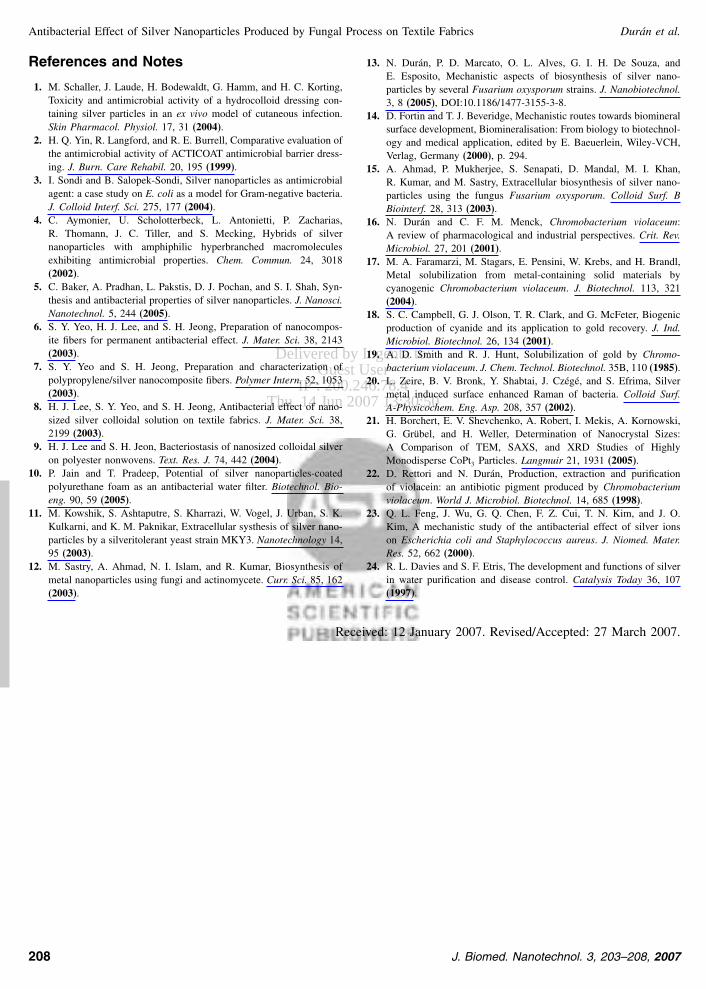