Embed Size (px)
Citation preview

Research
at the
de Duve Institute
and
Brussels Branch of the Ludwig Institute for Cancer Research
August 2009


de Duve Institute
Introduction 5
Miikka Vikkula 12
Frédéric Lemaigre 20
Annabelle Decottignies and Charles de Smet 25
Emile Vanschaftingen 31
Françoise Bontemps 37
Jean-François Collet 42
Guido Bommer 47
Mark Rider 50
Fred Opperdoes 56
Pierre Courtoy 62
Etienne Marbaix 69
Jean-Baptiste Demoulin 75
Jean-Paul Coutelier 80
Thomas Michiels 84
Pierre Coulie 89
LICR
Introduction 95
Benoît Van den Eynde 98
Pierre van der Bruggen 106
Nicolas Van Baren 114
Jean-Christophe Renauld 119
Stefan Constantinescu 125


5
The de Duve Institute

6
In 1974, when Christian de Duve founded the
Institute of Cellular Pathology (ICP), now rena-
med the de Duve Institute, he was acutely aware
of the constrast between the enormous progress
in biological sciences that had occurred in the 20
preceding years and the modesty of the medical
advances that had followed. He therefore crea-
ted a research institution based on the principle
that basic research in biology would be pursued
by the investigators with complete freedom, but
that special attention would be paid to the exploi-
tation of basic advances for medical progress. It
was therefore highly appropriate for the Institute
to be located on the campus of the Faculty of
Medicine of the University of Louvain (UCL).
This campus is located in Brussels. The Univer-
sity hospital (Clinique St Luc) is located within
walking distance of the Institute.
Benoît Van den Eynde
Emile Van Schaftingen
The main commitment of the members of
the de Duve Institute is research. Discovery is
the endpoint of their efforts and the only ele-
ment taken into account for their evaluation. The
Institute functions in symbiosis with the Faculty
of Medicine and many of its senior members
hold a Faculty position and have teaching ap-
pointments. The influx of doctoral students and
postdoctoral fellows from the University is also a
key element in the success
THE DE DUVE INSTITUTE: AN INTERNATIONAL BIOMEDICAL RESEARCH INSTITUTE

7
In 1978 the Ludwig Institute for Cancer Research decided to base its Belgian branch within the
walls of the de Duve Institute. A fruitful collaboration between the two Institutions has been pur-
sued since that time. Even though the two Institutes are completely independent, the collaboration
between the scientists of the de Duve Institute and the Ludwig Institute is extremely close and the
sharing of resources is considerable.
The de Duve Institute is managed by a directorate of three scientists, presently composed of
Emile Van Schaftingen, Benoît Van den Eynde, and Miikka Vikkula. The directorate is appointed by
the Board of directors, which comprises the Rector of the University of Louvain, one of the Pro-
rectors, the General Administrator of the University and the Dean of the Faculty of Medicine. Also
present in the Board of directors are eminent members of the business community.
About 170 researchers work in the de Duve
Institute and in the Ludwig Institute, assisted by
a technical and administrative staff of about 80
members. Despite this relatively small size, the
de Duve Institute has the ambition of pursuing
research projects of high quality under condi-
tions that allow original, long-term projects to be
pursued. The Institute has a limited endowment,
which is a source of key financing for priority is-
sues, such as the creation of new laboratories for
promising young researchers. We expect that the
quality of our researchers, supported by sound
organisational approaches, will enable the de
Duve Institute to stand at the forefront of Euro-
pean Research.Miikka Vikkula

8

9
DIRECTORATE
Emile VAN SCHAFTINGEN, Director
Miikka VIKKULA
Benoît VAN DEN EYNDE
BOARD OF DIRECTORS
Norbert MARTIN, President
Henri BEAUFAY
Luc BERTRAND
Thierry BOON-FALLEUR
Alfred BOUCKAERT
François CASIER
Bernard COULIE
Etienne DAVIGNON
Emmanuel de BEUGHEM
Christian de DUVE
Jean-François DENEF
Roland KEUNINGS
Anne-Marie KUMPS
Jean PETERBROECK
Maurice VELGE
SCIENTIFIC COUNCIL
Jean-Charles CEROTTINI, Lausanne Branch of the Ludwig Institute for Cancer Research, Switzerland
Philip COHEN, University of Dundee, UK
Daniel LOUVARD, UMR144 CNRS/Institut Curie, France
Gilbert VASSART, Université Libre de Bruxelles, Belgium
ADMINISTRATION AND GENERAL SERVICES
Philippe PENNINCKX, Finance and Admi-nistration Manager
Yolande de SELLIERS, External Relations Manager
Lydia HAUBOLD, Accountant
Delphine LEBBE, Executive Secretary
Alain BUISSERET, Technical Manager
Jean-Pierre SZIKORA, Informatics Support
Christian VAN LANGENHOVE, Workshop
Dan COMAN, Workshop
de Duve InstituteAvenue Hippocrate 74-75 1200 Brussels, Belgium
[T] +32 (02) 764 75 50[F] +32 (02) 764 75 73[W] www.deduveinstitute.be

10
DEVELOPMENT AND EXPANSION COUNCIL (DEC)
President : Baron BERTRAND
Honorary Presidents Me P. HOURDEAUBaron PETERBROECKBaron van der REST (†)Baron de TILLESSE, Honorary President of the Board of Directors
Honorary Vice-Presidents Mr A. LAGASSE de LOCHT (†)Baronne M. VELGE
Fellowship and/or Laboratory Sponsors Mr & Mrs A. BOUCKAERTMrs C. DELORIMrs G. DELORI (†)Mrs H. DELORIMr P. DELORIMrs L. DRESSE de LEBIOLESMrs Ph. FABRIComte B. de GRUNNEBaron van GYSEL de MEISEMr P. HOLLENFELTZ du TREUXMr & Mrs M. HUYNENMr & Mrs A. LACROIXMrs M. LHOISTMr & Mrs D. MATHIEUMr E. MOREL de BOUCLE ST DENISBaron & Baronne PETERBROECKMr M. RUIZ de ARCAUTEVicomte P. de SPOELBERCHDr J. TROQUETMrs J. VAN CAUWENBERGHEBaron M. VELGEMrs M. de VISSCHERFONDS JACQUES GOORUMICOREACKERMANS & VAN HAARENASCO INDUSTRIES
MembersMr E. van BARENMr J. BERTEAUMr & Mrs R. BOON-FALLEURMr. M. BRAGARDMr V. BRUNEAUMrs G. van CUTSEM-STAES POLETVicomtesse de DUVE (†)Baron S. EMSENSVicomte della FAILLE de WAERLOOSBaronne de GERLACHE de GOMERYMr P. GUILMOTMr V. JACOBS van MERLEN (†)Mr F. JAMARMr L. LHOISTComte J. de LIEDEKERKEDr J. MAILLETMrs O. MAIRLOTMr B. MATTLETMrs de MOREAU de GERBEHAYEMrs A. PIERONMr J. SAMAINMr & Mrs SOLVAY de LA HULPEBaronne H. van der STRATEN WAILLETDr E. VAN ASSCHEMr E. VAN CAMPENHOUTDr P. VAN HONSEBROUCKMr Ph. de VISSCHER
AXAINGBEKAERTCABLERIE D’EUPENCARLSBERG IMPORTERSCARMEUSECHEMITEXFONDATION M. et R. de HOVREGENZYME BELGIUMGBLGROUPE LHOISTPFIZERSIBEKA

11
ACKNOWLEDGMENTS
In 2008, the de Duve Institute has attracted major gifts from several foundations, companies and individuals who have been very generous. These sponsors are providing the resources that enable our scientists to better understand and treat diseases that afflict people around the world. Gifts are the lifeblood of new research initiatives and private resources are crucial in underwriting the costs of new laboratories. On an annual basis, fund-raising from private sources has increased during the past decade over levels achieved previously and now supports about 10 % of the Institute’s budget.
The appeal for sponsoring postdoctoral fellowships was also widely followed. In 2008 the Institute has been able to allocate the following fellowships, entirely supported by our donors :
The «Haas-Teichen» fellowship was attributed to Jhansi KOTA,
the «Pierre Lacroix» fellowship to Shreedhara GUPTA,
the «Maurange» fellowship to Sandrine MEDVES,
another fellowship was recently awarded by the Institute to Reece MARILLIER.
We express our gratitude to all who contributed to the financing of post-doctoral fellows and state-of-the art research laboratories at the de Duve Institute, ensuring that this institute will remain at the top of the field in biomedical research.
Luc BERTRAND, President of the Development and Expansion Council

12
Miikka VIKKULA, Member
Laurence BOON, Plastic Surgeon, Part-time Research AssociateMustapha AMYERE, Postdoctoral FellowPascal BROUILLARD, Postdoctoral FellowNisha LIMAYE, Postdoctoral FellowMichella GHASSIBE, Postdoctoral Fellow
Nicole REVENCU, Pediatrician, GeneticianArash GHALAMKARPOUR, M.D., Graduate StudentVirginie AERTS, Graduate StudentMélanie UEBELHOER, Graduate StudentThomas PALM, Graduate StudentLaurence DESMYTER, Graduate Student Anne VAN EGEREN, TechnicianDominique COTTEM, TechnicianAnnette MARCELIS, Technician (since 1.10.2008)Antonella MENDOLA, Technician (till 30.09.2008)Antonella MENDOLA, (since 1.10.2008) Research AssistantNicolas WARNIER, Part-time Technical AssistantCLAUDE MAUYEN, Part-time Technical AssistantLiliane NICULESCU, Secretary
The basic aim of our research is to get insights into the molecular mechanisms underlying a variety of disorders of cardiovascular and skeletal systems, as well as certain cancers. We are especially interested in evaluating the importance of genetic variation in human disease development. The precise cause of many disorders remains unknown, and current treatments are therefore aimed at alleviating symptoms. Identification of the primary causes as well as modulating factors would allow for the development of treatments that are more “curative” and specific. As this research is based on human DNA extracted from blood and tissue samples obtained from patients, the group works tightly together with several clinicians and multidiciplinary centers worldwide (e.g. Centre des Malformations Vasculaires, Cliniques universitaires St-Luc; Vascular Anomalies Center, Children’s Hospital, Boston, USA; Consultation des Angiomes, Hôpital Lariboisière, Paris, and Centre labiopalatin, Cliniques Universitaires St-Luc).
GENETICS OF HUMAN CARDIOVASCULAR ANOMALIES, CLEFT LIP PALATE AND CEREBRAL TUMORS
VENOUS MALFORMATIONS AND GLOMUVENOUS MALFORMA-TIONS (“GLOMANGIOMAS”)
P. Brouillard, V. Wouters, N. Limaye, M. Uebel-hoer, V. Aerts, M. Amyere, L.M. Boon and M. Vikkula, in collaboration with B.R. Olsen, Harvard Medical School, Boston, USA; J.B. Mulliken and S. Fishman, Children’s Hospital, Boston, USA; O.
Enjolras, Hôpital Lariboisière, Paris, France; A. Dompmartin, CHU, Caen, France
Venous malformations (VM) are bluish-purple cutaneous and mucosal vascular lesions. They are often congenital, but can appear later in life. They have a tendency to grow slowly with the growth of the child. Glomuvenous malformations (GVM, “glomangiomas”) are a special subtype of venous anomalies [1]. They

13
are clinically similar to VMs, yet our clinico-ge-netic study allowed for their clinical differen-tiation.
We previously discovered that rare, heredi-tary venous malformations can be caused by an activating mutation in the endothelial cell receptor tyrosine kinase TIE2/TEK. We em-ployed the DHPLC system, which allows for more efficient and sensitive screening for such mutations, and identified several novel activa-ting mutations amongst affected families (Fig 1; Wouters et al, in prep). We hypothesized that as the lesions are localized, a somatic second hit might be needed in the normal allele of the TIE2 gene, for lesions to develop. We obtained
proof for this from one lesion, in which the li-gand-binding region of the wild-type allele was deleted somatically, causing a local loss of its ability to function [2]. In addition, we discove-red that at least 50% of the far more common sporadic VMs are caused by somatic mutations in TIE2 [2]. All of the mutations discovered thus far are intracellular and cause receptor hy-
perphosphorylation in vitro, although much re-mains to be learnt as to precisely why this cau-ses lesions. Towards this end, we have begun to carry out functional analyses of the role of TIE2 in VM-pathogenesis, using a variety of in vitro and in vivo methods. These include the generation of mouse models of the anomaly, by “knock-in” substitution of the normal TIE2 allele with the most frequently mutated forms associated with inherited VMCM and sporadic VMs respectively; Affymetrix expression profi-ling is also being used in order to compare the effects of the wild-type receptor with those of different mutant forms.
In contrast to VMs, glomuvenous malforma-
tions (GVM) are mostly, if not always, inhe-rited. We discovered that GVM are caused by loss-of-function mutations in a gene we named glomulin. So far, we have identified 35 diffe-rent mutations in 133 families. As in VMs, we showed that GVM lesions appear locally be cause of the additional alteration of the second allele, likely in vascular smooth muscle cell pre-
Fig 1. Intracellular TIE2 mutations identified in VM. (A) Inherited changes in VMCM blood samples (left, in green) and somatic changes in sporadic VM lesions (right, in red). A somatic 2nd-hit deletion of the Ig2 ligand-binding domain (“Del”) was identified in a lesion from a VMCM patient carrying R849W. (B) The positions of the mutated residues in the intracellular region of TIE2, shown as a dimer.

14
cursors. To better understand the role of glo-mulin in normal and pathological conditions, we have created glomulin-deficient mice. While heterozygotes appear normal, homozygous knockouts are embryonic-lethal. To enable studies beyond this time-point, we generated RNAi conditional knockdown mice, in which glomulin inactivation is cre-inducible. When triggered during embryonic development, glo-mulin depletion is likewise lethal. We are cur-rently phenotyping these animals further. The-se mice provide new tools to continue the in vitro and in vivo characterization of glomulin expression and function.
LyMPHEDEMA
A. Ghalamkarpour, L.M. Boon and M. Vikkula in collaboration with K. Devriendt, KUL; D. Chitayat, Hospital for Sick Children, Toronto, Canada; K. Alitalo, Haartman Institute and Helsinki University, Finland.
Lymphedema is an external manifestation of lymphatic failure. It may be categorized as primary (idiopathic) or secondary (acquired) lymphedema. Primary hereditary lymphedema can occur at birth (Nonne-Milroy disease) or at puberty (Meige’s disease). It is extremely difficult to treat lymphedema. Patients have a lifetime problem with progressive swelling of extremities. We use genetic approaches to unra-vel the pathophysiology. In some families with Nonne-Milroy disease, missense inactivating mutations in the VEGFR3 gene were identified. We also recently found that VEGFR3 muta-tion can cause hydrops fetalis in Nonne-Milroy transmitting families. Moreover, some sporadic congenital primary lymphedemas are also ex-plained by a VEGFR3 mutation. We showed, for the first time, that recessive primary conge-nital lymphedema can be caused by a particular homozygous VEGFR3 mutation, which has a moderate effect on receptor function and can cause lymphedema only when both alleles are altered [3]. Mutations in the transcription fac-tor gene SOX18 were identified in families with autosomal recessive and dominant hypotricho-
sis-lymphedema-telangiectasia syndrome, and the forkhead transcription factor FOXC2 is mutated in some families with Meige disease, in association with distichiasis. We recently found that a proportion of sporadic fetal edema of unknown etiology is in fact attributable to mu-tations in the lymphedema-associated genes VEGFR3 and FOXC2 [4].
VASCULAR ANOMALIES AFFECTING CAPILLARIES
N. Revencu, N. Limaye, M. Amyere, L.M. Boon and M. Vikkula in collaboration with J.B. Mulliken, Children’s Hospital, Boston, USA; S. Watanabe, Showa University School of Medicine, Tokyo, Japan; A. Dompmartin, CHU de Caen, France; Virginia Sybert, University of Washington, Seattle, USA
Capillaries, the smallest blood vessels that connect arterioles to venules, can give rise to various anomalies, two of which are very common: 1) hemangioma, a benign, localized overgrowth of capillary-like vessels, and 2) ca-pillary malformation (CM; commonly known as portwine stain), a localized maldevelopment of capillary like vessels. Hemangiomas have a frequency of up to 12 % in 1-year-old children, and typically undergo a period of rapid expan-sion, followed by spontaneous regression. We have an extensive collection of samples from sporadic as well as rare familial forms of he-mangioma, and have begun to use Affymetrix high-density whole genome SNP arrays in or-der to carry out linkage, loss of heterozygosity and copy number analyses on them in an effort to identify causative genomic variants. Work done with collaborators has demonstrated that perturbations of the vascular endothelial growth factor (VEGF) signaling pathway can cause hemangioma pathogenesis [5].
CMs occur in 0.3% of newborns. Unlike hemangiomas, they stay throughout life if not treated. Certain capillary malformations affect other organs, such as the brain in the case of cerebral capillary malformations or CCMs. We discovered that inherited hyperkeratotic cuta-

15
neous capillary-venous malformations (HC-CVM) associated with CCM are caused by a mutation in the KRIT1 (Krev interaction trap-ped 1) gene, suggesting it is important not only for cerebral but also for cutaneous vasculature. In addition, a genome-wide linkage mapping on families with inherited capillary malformations led us to identify a linked locus CMC1. Scree-ning of positional functional candidate genes resulted in the identification of mutations in the RASA1 gene, a modifier of the Ras signa-ling pathway. This implies that RAS pathway modulators may serve as a novel therapy for these patients in the future. Ongoing studies have led to the identification of 54 additional families with RASA1 mutation, accounting for about 30% of those affected. This has allowed for a more precise clinical description of the clinical signs and symptoms associated with this newly recognized disorder that we have named Capillary malformation-arteriovenous malformation (CM-AVM) [6]. Importantly, ca-pillary lesions can be associated with deeper, more dangerous anomalies about 30% of the time; these include arteriovenous malforma-tions and fistulas (AVM/AVF), Parkes Weber, and Vein-of-Galen aneurysmal malformations, which warrant careful clinical management.
CARDIOPATHIES
M Amyere, I. Gutierrez-Roelens, and M. Vikkula, in collaboration with T. Sluysmans, C. Ovaert, St-Luc, UCL and M. Gewillig and K. Devriendt, KUL
The cardiovascular system can encounter developmental problems affecting the heart. These cardiac defects or cardiopathies vary from physiological septal defects to life-threa-tening complex malformations. Identification of genetic causes enables tight follow-up and preventive pacemaker implantation. CSX/NKx2.5 gene, an important transcription fac-tor for cardiac development, was mutated in three families, in which atrial septal defect is associated with progressive atrioventricular conduction defect. For families with hetero-taxia and situs inversus, whole-genome linkage
analysis was performed using 10K Affymetrix SNP-chips, which identified a possible locus for a causative gene.
CLEFT LIP AND PALATE
M. Ghassibé, L. Desmyter, N. Revencu, M. Vikkula, in collaboration with Y. Gillerot, B. Bayet, R. Vanwijck, Ch. Verellen-Dumoulin, N. Deggouj, St-Luc, UCL
Cleft lip and palate (CLP) is a congenital anomaly of complex etiology. Predisposition is governed by numerous genetic loci, in combi-nation with environmental factors. Clefts have an incidence of 1/700 births.
We collected DNA samples from a lar-ge number of patients affected with van der Woude syndrome, the most common cleft syn-drome, and showed that IRF6 is the major cau-sative gene in our Belgian cohort. Moreover, IRF6 is the gene responsible for popliteal pte-rygium syndrome. This study in turn led to se-veral collaborations that allowed us to carry out a genotype-phenotype correlation on hundreds of patients from different ethnic backgrounds. Results showed that IRF6 is mutated in 69% of VWS patients and 97% of PPS patients. Inte-restingly, mutation-distribution is non-random: 80% are localized in IRF6 exons 3, 4, 7 and 9 for VWS, and 72% in exon 4 for PPS patients. These findings are of great importance for cli-nical diagnosis, mutational screens and genetic counseling. We also demonstrated that IRF6 predisposes to non syndromic clefts in Europe. In parallel, we identified a new gene responsible for cleft palate only and Pierre Robin sequence. We are currently generating and phenotyping zebrafish and mouse models to understand the mechanism behind craniofacial development and cleft occurrence across species.

16
CEREBRAL TUMORS
T. Palm and M. Vikkula, in collaboration with C. Godfraind, Laboratory of Neuropathology, St-Luc, UCL
Morphological characterization and clas-sification of tumors is not always clear. Thus, better (molecular) criteria are needed. We are especially interested in two types of cerebral tu-mors: oligodendrogliomas and ependymomas. To better understand the molecular alterations leading to ependymomal oncogenesis, we per-formed microarray-based expression profiling on a series of 34 frozen ependymomas. Results of our profiling study are in concordance with the “oncology recapitulates ontology” hypo-thesis, in which genes implicated in stem cell fate decisions may be important for supporting cancer stem cells as well. Pathways activated in high grade ependymomas were consistent with the histological appearance of a more aggres-sive tumor phenotype [7]. Using array-CGH, we recognized a subgroup of supratentorial ependymomas affecting young adults, which are characterized by trisomy of chromosome 19.
Within the posterior fossa compartment, ependymomas cluster into three sub-groups. The first corresponds with ependymomas that are histologically of WHO grade II, the second with those of WHO grade III, and the third with a group of ependymomas of a bi-phasic appearance, combining regions of both grades. This sub-group shares gene-sets with tumors of both other groups, and in addition has a glycogen metabolism signature of its own. Whether these groups correspond to three dis-tinct tumoral entities, or demonstrate multifo-cal tumor progression remains to be investiga-ted (Palm et al, In Prep).
NEUROENDOCRINE TUMORS
A. Persu, Division of Cardiology, Saint-Luc, UCL; M. Amyere, A. Vanegeren, M. Vikkula, in colla-boration with P. Rustin, INSERM U676, Hôpital Robert Debré, Paris, France.
Pheochromocytomas and head and neck paragangliomas are neuroendocrine tumours derived from the neural crest. Paragangliomas are associated with parasympathetic ganglia and are usually non-secreting. By contrast, pheo-chromocytomas are derived from paraganglia associated with the orthosympathetic system and are characterized by increased secretion of catecholamines and paroxystic hypertension.
The current project aims to look at the na-ture and frequency of mutations in the known predisposing genes in pheochromocytoma and paraganglioma in Belgium and to detect possi-ble genotype-phenotype correlations. A multi-centric collaboration including the main acade-mic centers from Belgium has therefore been established.
The SDH genes code for the subunits of succinate deshydrogenase, at the crossroad of the mitochondrial respiratory chain and Krebs cycle. Three of the four subunits of succinate deshydrogenase, i.e. SDHD, SDHB, and more rarely SDHC, have been associated with paraganglioma and pheochromocytoma. Furthermore, SDHB mutations have been as-sociated with an increased risk of recurrence and malignancy in several European series. In our study, the prevalence of SDHB was unex-pectedly high in head and neck paraganglioma. Surprisingly, tumours associated with such mu-tations are mainly late-onset unilateral tumours without evidence of recurrence or malignancy [8]. We also described a family with a very rare presentation of severe head and neck paragan-glioma with liver and spine localization. No evidence of mutations was found in the known predisposing genes by dHPLC and/or SSCP. An in depth search for the genetic abnormality underlying this unusual form of paraganglioma

17
is currently under way.
ESSENTIAL HyPERTENSION
A. Persu (Division of Cardiology, Saint-Luc, UCL), N. Limaye, and M. Vikkula.
High blood pressure - commonly called hy-pertension - is found in almost 20 % of the adult population worldwide and affects 2 mil-lion Belgians. Despite the wide range of anti-hypertensive therapies available, blood pressure is adequately controlled in only 30-40% of hy-pertensive patients. In a large majority of cases, no specific cause is found (“essential hyperten-sion”) and high blood pressure is thought to reflect the interplay of lifestyle (obesity, high salt intake) and genetic factors; 30-50 % of blood pressure level is thought to be genetically determined. Despite this, conventional linkage and association studies have failed to establish the role of genetic variants in susceptibility.
In an effort to find genetic variations that account for a significant proportion of blood pressure heritability, and to study the interac-tions between known variants with mild to mo-derate effects, we set up a multicentric national genome wide association study (HYPERGEN) with the support of the Belgian Hypertension Committee. We aim to recruit at least 1000 hypertensive patients and 1000 normotensive subjects. Detailed phenotyping including renin and aldosterone dosages are obtained in all pa-tients, and genotyping will be performed using SNP chip technology.
HEMATOLOGICAL MALIGNANCIES
H. Antoine-Poirel, V. Havelange, F. Duhoux, M. Herman, G. Ameye, K. Bahloula, Human Genetics Center, St.Luc, UCL; with M. Vikkula
The genetic nature of hematological mali-gnancies (mainly leukemias, myelodysplasias, chronic myeloproliferative disorders, lympho-mas, myelomas) has been clearly established.
Despite this, we lack biomarkers for diagnosis, prognosis and treatment, for clinical manage-ment as well as for a better understanding of the genetic and epigenetic processes leading to tumorigenesis. Towards this end, we use a variety of techniques including conventional and molecular cytogenetics or FISH, molecular biology, and microarray approaches.
In an international therapeutic trial of children with mature B-cell lymphomas, we showed the adverse prognostic impact of chro-mosomal alterations of 13q and 7q, detected by cytogenetics. Furthermore, using genome-wide SNP array technology, we found that most 13q alterations lead to an amplification of the mi-croRNA 17-92 cluster, known to interact with the MYC oncogene, a finding confirmed with mature miRNA expression profiling. In addi-tion, we detected numerous cryptic genomic alterations including partial uniparental diso-mies. Their prognostic value is currently under study in collaborations within different thera-peutic trials across Europe.
SNP-CHIP PLATFORM
M. Amyere and M. Vikkula
We host the UCL DNA-oligonucleotide microarray platform (Affymetrix). This plat-form is currently used by several groups in the de Duve Institute and UCL for expression profiling as well as genotyping. In collaboration with Dr Jüppner from Harvard Medical Scho-ol, Boston, we genotyped a large family with a new form of hypophosphatemia and map-ped this autosomal recessive form (designated ARHP) to chromosome 4q21. This allowed for the identification of causative homozygous mutations in DMP1 (dentin matrix protein 1), which encodes a non-collagenous bone matrix protein expressed in osteoblasts and osteocy-tes [9]. In collaboration with G. Matthijs from KULeuven, we have been able to combine ge-notyping and expression profiling in one con-sanguineous family with a congenital glycosy-lation disorder. Autozygosity mapping along

18
with expression profile analysis allowed us to identify a new gene for CDG. In collaboration with Dr Vermeesch from KULeuven, we ge-notyped a large family with autosomal-domi-nant microtia. Copy number analysis led to the identification of five tandem copies of a copy number-variable region at chromosome 4p16, linked to the disease. With the same group, we establishes that chromosome instability is common during early human embryogenesis in study of 23 pre-implantation embryos from 9 fertile couples. Additionally, rearrangements such as segmental imbalances were observed in 70% of the 23 embryos tested. This explains low human fecundity and identifies post-zygo-tic chromosome instability as a leading cause of constitutional chromosomal disorders [10].
SELECTED PUBLICATIONS
1. Brouillard P, Vikkula M. Genetics causes of vascular malformations. Hum Mol Genet 2007;16(2): R140-9.
2. Limaye N, Wouters V, Uebelhoer M, Tuo-minen M, Wirkkala R, Mulliken JB, Eklund E, Boon LM, Vikkula M. Somatic mutations in angiopoietin receptor gene TEK cause solitary and multiple sporadic venous malformations. Nat Genet 2009;41(1):118-24.
3. Ghalamkarpour A, Holnthoner W, Saha-rinen P, Boon LM, Mulliken JB, Alitalo K, Vikkula M. Recessive primary congenital lym-phoedema caused by a VEGFR3 mutation. J Med Genet 2009; Epub ahead of print.
4. Ghalamkarpour A, Debauche C, Haan E, Van Regemorter N, Sznajer Y, Thomas D, Revencu N, Gillerot Y, Boon LM, Vikkula M. Sporadic In Utero Generalized Edema Cau-sed by Mutations in the Lymphangiogenic Genes VEGFR3 and FOXC2. J Pediatr 2009; Epub ahead of print.
5. Jinnin M, Medici D, Park L, Limaye N, Liu Y, Boscolo E, Bischoff J, Vikkula M, Boye E, Olsen BR. Suppressed NFAT-dependent VEGFR1 expression and constitutive VE-GFR2 signaling in infantile hemangioma. Nat Med 2008;14:1236-46.
6. Revencu N, Boon LM, Mulliken JB, En-jolras O, Cordisco MR, Burrows PE, Cla-puyt P, Hammer F, Dubois J, Baselga E, Brancati F, Carder R, Quintal JM, Dalla-piccola B, Fischer G, Frieden IJ, Garzon M, Harper J, Johnson-Patel J, Labrèze C, Martorell L, Paltiel HJ, Pohl A, Prendiville J, Quere I, Siegel DH, Valente EM, Van Hagen A, Van Hest L, Vaux KK, Vicente A, Weibel L, Chitayat D, Vikkula M. Parkes Weber syndrome, vein of Galen aneurysmal mal-formation, and other fast-flow vascular anomalies are caused by RASA1 mutations. Hum Mu-tat 2008;29(7):959-65.
7. Palm T, Figarella-Branger D, Chapon F, Lacroix C, Gray F, Scaravilli F, Vanden-broecke C, Ellison D, Salmon I, Vikkula M, Godfraind C. Expression profiling of epen-dymomas unravels localisation and tumor grade specific tumorigenesis. Cancer 2009; In Press.
8. Persu A, Amyere M, Gutierrez-Roelens I, Rustin P, Sempoux C, Lecouvet FE, Van Beers BE, Horsmans Y, De Plaen JF, MarcHamoir, Vikkula M. Rare presentation of familial paraganglioma without evidence of mutation in the SDH, RET and VHL genes: towards further genetic heterogeneity. J Hyper-tens 2009;27(1):76-82.
9. Lorenz-Depiereux B, Bastepe M, Benet-Pages A, Amyere M, Wagenstaller J, Muller-Barth U, Badenhoop K, Kaiser SM, Rittmaster RS, Shlossberg AH, Oli-vares JL, Loris C, Ramos FJ, Glorieux F, Vikkula M, Jüppner H, Ström TM. DMP1 mutations in autosomal recessive hypophosphate-mia implicate a bone matrix protein in the re-gulation of phosphate homeostasis. Nat Genet 2006;38(11):1248-50.

19
10. Vanneste E, Voet T, Le Caignec C, Ampe M, Konings P, Melotte C, Debrock S, Amyere M, Vikkula M, Schuit F, Fryns JP, Verbeke G, D’Hooghe T, Moreau Y, Ver-meesch JR. Chromosome instability is common in human cleavage-stage embryos. Nat Med 2009;15(5):577-83.
Miikka Vikkulade Duve InstituteAv. Hippocrate 74-75B - 1200 Brussels
[T] +32 02 764 74 96[F] +32 02 764 74 60[E] [email protected]

20
Frédéric P. LEMAIGRE, MemberPatrick JACqUEMIN, Associate Member
Rodolphe CARPENTIER, Postdoctoral fellowMarta COLLETTI, Postdoctoral fellow (from March 2009)Pierre-Paul PREVOT, Postdoctoral fellowPeggy RAYNAUD, Postdoctoral fellowAline ANTONIOU, Graduate student Eric HEINEN, Graduate student Ilaria LAUDADIO, Graduate student Sabrina MARGAGLIOTTI, Graduate student (till March 2009) Alexandru SIMION, Graduate student Sabine CORDI, Research TechnicianBianca PASTORELLI, Research Technician (from April 2009)Géraldine VAN DEN STEEN, Research Technician Freddy ABRASSART, Logistician Nicolas WARNIER, Animal Caretaker Vivien O’CONNOR, Secretary
LIVER AND PANCREAS DEVELOPMENT
The group studies the molecular and cellular mechanisms that govern differentiation and morphogenesis of the liver and pancreas, two organs which derive from the endoderm (primitive gut of the embryo). The fundamental knowledge gained by this work is essential for understanding the pathophysiology of organ malformations such as polycystic diseases, and for improving cell therapy of liver and pancreatic diseases, such as hepatic deficiencies (e.g. metabolic deficiencies and cirrhosis) and diabetes.
LIVER DEVELOPMENT
A. Antoniou, R. Carpentier, I. Laudadio, S. Margagliotti, P. Raynaud
The main cell types of the liver are the he-patocytes, which exert the metabolic functions of the organ, and the biliary cells which deli-neate the bile ducts. We study how the hepato-cytes and biliary cells differentiate and how bile ducts are formed in the embryo. Our preferred model organism to investigate liver develop-
ment is the mouse, and this includes generation and analysis of transgenic mouse lines.
The biliary tract consists of intrahepatic bile ducts which collect bile produced by the hepa-tocytes, and of extrahepatic ducts which drain bile from the liver to the intestine. Biliary cells, also called cholangiocytes, delineate the lumen of the bile ducts and modify the composition of bile. These cells, like hepatocytes, derive from liver progenitor cells called hepatoblasts. Our discovery of the Onecut transcription fac-tors Onecut-1 (OC-1/HNF-6), OC-2 and OC-

21
3, and the subsequent phenotypic characteriza-tion of HNF-6 and OC-2 knockout mice led to the identification of the first transcriptional network regulating bile duct development [1, 2]. Current efforts are devoted to the characte-rization of the transcription factors and signal transduction pathways that control bile duct development.
We have recently identified the transcription factor Sox9 as the earliest and most specific bi-liary cell marker in development. Using Sox9 in combination with hepatoblast markers, we analyzed the morphogenesis of the bile ducts and found that it occurs according to a new mode of tubulogenesis [3]. Biliary tubulogene-sis starts with formation of asymmetrical duc-tal structures, lined on one side (adjacent to the portal vein) by cholangiocytes and on the other side (adjacent to the liver parenchyma) by hepa-toblasts. When the ducts grow from the hilum to the periphery of the liver, the hepatoblasts lining the asymmetrical structures differentiate to cholangiocytes, thereby allowing formation of symmetrical ducts lined only by cholangio-cytes. This mode of tubulogenesis is unique as it is to our knowledge the only one characteri-zed by transient asymmetry (Figure 1).
The transcription factor network that dri-ves cholangiocyte morphogenesis and bile duct formation has been further investigated. By means of a liver-specific gene inactivation stra-tegy we found that Sox9 controls the timing of bile duct development. Within the biliary transcriptional network Sox9 is located downs-tream of HNF-6 and upstream of C/EBP-al-pha, two factors whose dysfunction is associa-ted with biliary cyst development. This work is currently pursued by the analysis of cyst for-mation in polycystic diseases, in the light of the new mechanism of tubulogenesis and of the role of Sox9. In addition, the function of Sox9 was found to be tightly linked with that of the Notch signaling pathway [4]. The latter is defi-cient in liver of patients affected with Alagille syndrome, a disease characterized by bile duct paucity and severe cholestasis.
We have shown that the Transforming Growth Factor-beta (TGF-beta) signaling sti-mulates differentiation of biliary cells. TGF-beta signaling is detectable in the liver as a gradient, with high signaling activity near the portal vein, where biliary cells differentiate, and lower signaling activity in the parenchyma, where hepatocytes differentiate. The results of this research are now used by collaborating teams who attempt to program in vitro diffe-rentiation of stem cells to hepatocytes for cell therapy of liver disease. Our efforts in 2008 were concentrated on the understanding of the role of TGF-beta signaling in the formation of bile ducts. We found that TGF-beta is essential at several steps of biliary development and is a regulator of the transient asymmetry that cha-racterizes bile duct development [3].
Our work also addresses the mechanisms of hepatocyte differentiation. We found that the Onecut factors HNF-6 and OC-2 are re-quired for liver expansion at the onset of liver development [5]. They are also critical for nor-mal differentiation of hepatic precursor cells to hepatocytes or cholangiocytes [2], and their level of expression during hepatocyte differen-tiation determines time-specific gene activation in the liver [6]. Current work focusses on the molecular mechanisms by which HNF-6 and OC-2 fine-tune gene expression at several sta-ges of hepatocyte differentiation.
PANCREAS DEVELOPMENT
M. Colletti, E. Heinen, P.-P. Prévot, A. Simion
In the embryo, the pancreas develops as an outgrowth of the endoderm which is the cell layer that delineates the primitive gut. Pan-creatic progenitors derived from the endoderm form two buds (dorsal and ventral) which later fuse to form a single organ. Within these buds the progenitor cells give rise, through a stepwise process, to endocrine cells that produce insulin, glucagon, somatostatin, pancreatic polypeptide

22
or ghrelin. The endocrine cells associate to form the islets of Langerhans. Our group in-vestigates the transcriptional mechanisms and signal transduction pathways that control how endoderm cells become pancreatic progenitors and how the latter develop into endocrine pre-cursor cells. This research is currently pursued using the mouse, including transgenic mice, as a model organism.
In 2008 we investigated how the transcrip-tion factors HNF-6 and OC-2 and FGF signa-ling control development of pancreatic proge-nitors from the endoderm, and the subsequent generation of endocrine precursor cells. The Onecut transcription factors HNF-6 and OC-2 exert redundant roles in pancreas morphoge-nesis and in differentiation of endocrine cells [7,8]. Work has progressed on the molecular mode of action of HNF-6 and OC-2, in parti-cular on the identification of microRNAs that regulate or are regulated by these factors.
FGF-10 is produced by the mesenchyme surrounding the developing pancreas and sus-tains development of the pancreas [9]. In 2008 we have investigated which receptors mediate FGF-10 signaling in pancreas by analysing mice
that are deficient in FGFR1b and/or FGFR2b. The results indicated that FGFR2b is critical for pancreas development and mediates the role of FGF-10 during early pancreas develo-pment.
The results of the research on Onecut fac-tors and FGF signaling are being translated in the design of cell therapy of diabetes by col-laborating teams who attempt to differenti-ate stem cells to insulin-producing cells. These collaborations include our participation to the EU-sponsored network BetaCellTherapy.
During pancreas development, the proge-nitor cells also give rise to exocrine and duc-tal cells. Our work addresses how pancreatic ducts are generated. These ducts drain the se-cretions from the pancreatic exocrine cells to the intestine and are delineated by ductal cells. We have shown that the Onecut factor HNF-6 controls a network of genes that is required for the formation of cilia at the apical pole of the ductal cells and for normal development of the ducts. In the absence of HNF-6, the ducts form cysts, much like in human polycystic di-seases [10].
ductal plate(cholangiocytes)
portal vein
Single-layered ductal plate Primitive ductal structure
Primitive ductal structureMature bile duct
portal mesenchyme
parenchymal layer(hepatoblasts)
portal layer(cholangiocytes)
basal lamina
Fig. 1. Bile duct development progresses from the hilum of the liver to the periphery of the lobes. The intrahepatic biliary tree is represented at a stage when ducts have reached maturity near the hilum (lower left), while the most peripheral structures are still in single-layered ductal plate configuration (upper left). Intermediate stages illustrate the progression from asymmetrical to radially symmetrical ducts (upper and lower right).

23
Work by our and other teams has provided evidence that ductal cells can transdifferentiate to pancreatic endocrine cells. Current work in-vestigates the transcriptional mechanisms that govern this transdifferentiation process.
CONCLUSIONS
Our work on the signaling pathways and transcription factors in developing liver and pancreas opens perspectives for understanding the pathophysiology of liver and pancreatic congenital diseases. The application of our findings to the programmed differentiation of cultured stem cells should help developing cell therapy of hepatic deficiencies and of pancrea-tic diseases such as diabetes.
SELECTED PUBLICATIONS
1. Clotman F, Lannoy VJ, Reber M, Cereghi-ni S, Cassiman D, Jacquemin P, Roskams T, Rousseau GG, Lemaigre FP. The onecut transcription factor HNF6 is required for normal development of the biliary tract. Development 2002;129:1819-28.
2. Clotman F, Jacquemin P, Plumb-Ru-dewiez N, Pierreux CE, Van der Smissen P, Dietz HC, Courtoy PJ, Rousseau GG, Lemaigre FP. Control of liver cell fate decision by a gradient of TGF beta signaling modulated by Onecut transcription factors. Genes Dev 2005;19:1849-54.
3. Antoniou A, Raynaud P, Cordi S, Zong Y, Tronche F, Stanger B, Jacquemin P, Pier-reux CE, Clotman F, Lemaigre FP. Intra-hepatic Bile Ducts Develop According to a New Mode of Tubulogenesis Regulated by the Trans-cription Factor SOX9. Gastroenterology 2009;136:2325-33.
4. Zong Y, Panikkar A, Xu J, Antoniou A, Raynaud P, Lemaigre F, Stanger BZ. Notch signaling controls liver development by regula-ting biliary differentiation. Development 2009;136:1727-39.
5. Margagliotti S, Clotman F, Pierreux CE, Beaudry J-B, Jacquemin P, Rousseau GG, Lemaigre FP. The Onecut transcription factors HNF-6/OC-1 and OC-2 regulate early liver expansion by controlling hepatoblast migration. Dev Biol 2007;311:579-89.
6. Beaudry J-B, Pierreux CE, Hayhurst GP, Plumb-Rudewiez N, Weiss MC, Rousseau GG, Lemaigre FP. Threshold levels of HNF-6 acting in synergy with HNF-4 and PGC-1a are required for time-specific gene expres-sion during liver development. Mol Cell Biol 2006;26:6037-46.
Fig. 2: The pancreas of mouse embryos that are wild-type or knockout for the FGF-10 receptor FGFR2b was studied at embryonic day 14.5 by whole mount immunostaining with an antibody against the pancreatic protein Pdx1. The ventral (vp) and dorsal (dp) parts of the pancreas can be distinguished. In FGFR2b-deficient embryos, the pancreas is severely hypoplastic and shows reduction in its branching pattern. Pancreatic endocrine cells can differentiate in these embryos albeit in reduced numbers (not shown on picture).

24
7. Jacquemin P, Durviaux SM, Jensen J, Godfraind C, Gradwohl G, Guillemot F, Madsen OD, Carmeliet P, Dewerchin M, Collen D, Rousseau GG, Lemaigre FP. Transcription factor hepatocyte nuclear factor 6 regulates pancreatic endocrine cell differentiation and controls expression of the proendocrine gene ngn3. Mol Cell Biol 2000;20:4445-54.
8. Vanhorenbeeck V, Jenny M, Cornut J-F, Gradwohl G, Lemaigre FP, Rousseau GG, Jacquemin P. Role of the Onecut transcription factors in pancreas morphogenesis and in pan-creatic and enteric endocrine differentiation. Dev Biol 2007;305:685-94.
9. Jacquemin P, Yoshitomi H, Kashima Y, Rousseau GG, Lemaigre FP, Zaret KS. An endothelial-mesenchymal relay pathway regulates early phases of pancreas development. Dev Biol 2006;290:189-99.
10. Pierreux CE, Poll AV, Kemp CR, Clotman F, Maestro MA, Cordi S, Ferrer J, Leyns L, Rousseau GG, Lemaigre FP. The trans-cription factor hepatocyte nuclear factor-6 controls the development of pancreatic ducts in the mouse. Gastroenterology 2006;130:532-41.
Frédéric Lemaigrede Duve InstituteUCL 75.29Av. Hippocrate 74-75B - 1200 Brussels
[T] +32 02 764 75 83[F] +32 02 764 75 07[E] [email protected][W] http://www.deduveinstitute.be/liver_and_pancreas_development.php

25
Anabelle DECOTTIGNIES, Associate MemberCharles DE SMET, Assistant Member
Axelle LORIOT, Postdoctoral FellowMarina MATTIUSSI, Graduate StudentGrégory PARVIZI, Graduate StudentGaëlle TILMAN, Graduate StudentFlorence FONTAINE, TechnicianSandrine LENGLEZ, Technician
GENETIC AND EPIGENETIC ALTERATIONS IN GENOMES
Preservation and regulation of genetic information is essential for proper cell function. Consequently, cells have evolved mechanisms of DNA repair, telomere maintenance, and epigenetic regulation of gene expression patterns. Deregulation of these processes contributes to the appearance and progression of cancer cells, which are characterized by genomic rearrangements and dysregulated gene expression patterns. Studies in our group examine how certain DNA repair processes lead to the insertion of mitochondrial DNA sequences into the nuclear genome. We are also exploring the mechanisms by which tumor cells maintain their telomeres to acquire unlimited proliferation potential. Furthermore, we have demonstrated that epigenetic alterations in tumors, involving loss of DNA methylation marks, can lead to the aberrant activation of a particular group of genes. We are currently investigating how epigenetic marks are established on these genes in embryonic cells, and how they become altered in tumor cells.
DNA DAMAGE REPAIR IN FISSION yEAST SchizoSaccharomyceS pombe
A. Decottignies, S. Lenglez
DNA repair processes have been well con-served throughout evolution, and yeast has proven to be a good model for their study. We use S. pombe to dissect the mechanisms of DNA double-strand break (DSB) repair, a type of genetic lesion arising after exposure to genotoxic agents or during DNA replication. Chromosomal and extrachromosomal DSBs can be induced experimentally in virtually any kind of cell. Such systems led to the dissection of the two major mechanisms of DNA repair: homologous recombination (HR) and non-
homologous end-joining (NHEJ). In the lab, fission yeast was used to investigate genetic re-quirements for microhomology-mediated end- joining (MMEJ), a third DNA repair process poorly characterized so far (1).
From yeast to mammals, different studies also reported the insertion of DNA fragments of various sources at experimentally-induced DSBs, including mitochondrial DNA (mtD-NA) in budding and fission yeast (2). Interes-tingly, recent studies reported the association of human genetic diseases with de novo inser-tions of mtDNA in the nuclear genome, inclu-ding a patient exposed to Chernobyl radiations. Moreover, systematic sequencing of eukaryotic nuclear genomes revealed the presence of nu-clear sequences of mitochondrial origin (NU-MTs) in chromosomes, suggesting that capture of mtDNA fragments at naturally occurring

26
DSBs took place during evolution, remodeling the nuclear genome. By analyzing fission yeast nuclear genome, we found a strong correlation between NUMT localization and chromoso-mal DNA replication origins. We are currently investigating the mechanisms responsible for this association.
TELOMERASE AND ALTERNATIVE MECHANISM(S) OF TELOMERE MAINTENANCE IN HUMAN CELLS
M. Mattiussi, G. Tilman and A Decottignies
Telomeres are specialized protein-DNA structures, which prevent chromosome ends from being recognized as DSBs. Synthesis of telomeric DNA sequences in replicating cells requires telomerase. Cancer cells often show an increased level of telomerase, and this contri-butes to their unlimited proliferation potential. In some cancers, however, telomeres are main-tained in the absence of telomerase activity by one or more mechanisms that are known as alternative lengthening of telomeres (ALT). ALT cell lines can also be obtained after in vitro immortalization of telomerase-negative human fibroblasts with SV40 T antigen. These two pa-thways of telomere maintenance are very dis-tinct phenotypically. In telomerase-expressing cells (TEL+), telomere length is very homoge-nous and telomeres are found at the end of all chromosomes. However, in ALT cells, telome-res are very heterogeneous in length and some chromatids lack telomeres (Fig. 1).
In addition to its role in telomere length maintenance, hTERT has been reported to play non-canonical roles in the cell, including modulation of expression of genes implicated in tumorigenesis, through mechanisms that are still largely unknown. We are interested in the study of genes that are distinctly regula-ted in ALT and TEL+ cells. In particular, we are investigating the role of telomerase in the modulation of TGF-b-dependent induction of extracellular matrix protein-encoding gene expression, like periostin (3), collagen or fibro-nectin in human dermal fibroblasts.
Figure 1. Telomere-specific fluorescence in situ hybridization (FISH) on metaphase chromosomes of telomerase-positive (TEL+) and ALT cancer cells (ALT). Telomeres are hybridized with a fluorescent telomeric probe (green) and DNA is stained with DAPI (blue). In ALT cells, telomeres are very heterogeneous, and even absent at some chromosome ends (arrowheads). ALT cells are further characterized by the presence of extrachromosomal telomeric DNA (arrow).
TEL+
ALT

27
DNA HyPOMETHyLATION AND ABERRANT GENE ACTIVATION IN CANCER
A. Loriot, G. Parvizi, C. De Smet
Genomic DNA in multiple species is mo-dified by the addition of a methyl group to cytosines in CpG dinucleotides. This herita-ble epigenetic modification is associated with transcriptional repression. Cell-type specific DNA methylation patterns are established du-ring embryonic development, and are generally maintained in adult somatic cells.
DNA methylation patterns often become altered in cancer cells. Alterations include hy-permethylation of selected promoters, leading to silencing of critical genes such as tumor suppressor genes, and hypomethylation of nu-merous other DNA sequences. We have shown that genome hypomethylation in tumors results in the activation of a group of germline-spe-cific genes, which use primarily DNA methy-lation for repression in somatic tissues (4). These genes, which were originally discovered because their activation in tumors leads to the expression of tumor-specific antigens, were named cancer-germline genes. To date, ~50 cancer-germline genes or gene families have been identified. Several of these were isolated in our group (5).
The process leading to hypomethylation of DNA sequences in tumors remains obscure. We undertook to address this issue by using MA-GEA1, the founding member of the cancer-germline group of genes, as a model. Detailed methylation analyses of the MAGEA1 genomic locus in expressing tumor cells, revealed pre-ferential hypomethylation within the 5’ region of the gene (6). Furthermore, transfection ex-periments with in vitro methylated MAGEA1 constructs, indicated that this site-specific hy-pomethylation relies on a historical event of DNA demethylation, and on the presence of appropriate transcription factors to protect the region against subsequent remethylation (6,7).
The proposed model of MAGEA1 demethyla-tion and activation during tumor development is illustrated in Figure 2. The factors that are responsible for the initial DNA demethylation process and for maintaining cancer-germline gene promoters unmethylated remain to be identified.
DNA METHyLATION CHANGES ASSOCIATED WITH CELL SENESCENCE AND IMMORTALIzATION
G. Tilman, A. Loriot, C. De Smet, A. Decottignies
In human and mouse cells, recent studies have shown that telomeric and subtelome-ric chromatin contains histone modifications that are commonly found in heterochromatin. Increasing evidence also indicates that chro-matin modifications at chromosome ends are important regulators of mammalian telome-res. In particular, alterations of either histone modifications in telomeric chromatin or of DNA methylation in subtelomeric regions are
Figure 2. Model for the stable activation of MAGEA1 in tumors.
MAGEA1
meCpG
CpG
Transient globalDNA demethylation
NORMAL
TUMOR
Binding oftranscription factors
Methylation activityrestored
Gene activated
Transcription factors
Transcriptional activatorsprevent local remethylation

28
associated with telomere length deregulation in mouse cells. In addition, a decreased subtelo-meric DNA methylation level in mouse cells was reported to be associated with increased homologous recombination between telomeric sequences (T-SCE for Telomeric Sister Chro-matid Exchange), a hallmark of human ALT cells.
This prompted us to evaluate the subte-lomeric DNA methylation level of human TEL+ and ALT cancer cell lines (8). We de-tected a significant hypomethylation of subte-lomeric DNA in ALT cancer cell lines when compared to TEL+ cell lines. However, subte-lomeric DNA was not hypomethylated in ALT cell lines derived from in vitro immortalization of human fibroblasts with SV40 T antigen, although T-SCE frequencies in the latter cells were similar to those in ALT cancer cells (8). We further showed that reducing T-SCE fre-quency of ALT cancer cells was not associated with subtelomeric DNA remethylation. Stri-kingly, subtelomeric DNA hypomethylation in ALT cancer cells was also associated with lower global DNA methylation. This observa-tion raises the interesting possibility that DNA demethylation in tumor cells may be linked to senescence or to the process that cells use to escape from senescence.
We are currently addressing the cellular me-chanisms underlying the differences in DNA methylation levels between ALT and TEL+ cancer cells. To this end, we are trying to re-produce, in vitro, the demethylation process that operates during tumorigenesis by overex-pressing RasV12 oncogene in human dermal fibroblasts. On one hand, RasV12 oncogene is known to induce cellular senescence of prima-ry fibroblasts through activation of the DNA damage response and, on the other hand, this oncogene leads to cellular transformation of p53/pRb-defective cells. As both senescence and transformation may be associated with ge-nomic DNA hypomethylation, we are investi-gating these two aspects of RasV12 expression. We also wish to compare the DNA methylation profiles of ALT and TEL+ cells that we expect
to arise from SV40 T and RasV12-expressing fibroblasts.
DNA METHyLATION OF CANCER-GERMLINE GENES IN HUMAN EMBRyONIC STEM CELLS
G. Parvizi, A. Loriot, C. De Smet
As new methylation patterns are established during early embryo development, embryonic stem (ES) cells provide a suitable experimental system for investigating the molecular mecha-nisms underlying this epigenetic reprogram-ming process. ES cells possess both DNA de-methylation and de novo methylation activities. Each of these opposing activities appears to be targeted to selected DNA sequences. The mechanisms underlying this targeting are still unclear, but likely involve sequence-specific DNA binding proteins and chromatin modi-fying enzymes. We recently initiated studies on the epigenetic regulation of cancer-germline genes in human ES cells.
We found cancer-germline genes to be re-pressed and methylated in human ES cells (ob-tained from Dr. D. Melton, Harvard University, MA), as well as in human embryonal carcinoma (EC) cells, the malignant counterparts of ES cells (9). This suggests that cancer-germline ge-nes are programmed for methylation-mediated silencing in human ES cells. Accordingly, trans-fection experiments indicated that human EC cells direct a potent repression activity towards the promoter of MAGEA1. This was associa-ted with progressive de novo methylation of the transgene. Our group is currently using the chromatin immunoprecipitation (ChIP) tech-nology to define histone modifications associa-ted with the silencing of cancer-germline genes in human ES/EC cells. This should help us to identify the factors that target DNA methyla-tion towards cancer-germline gene promoters in these cells. Loss of function of such factors may be a prerequisite for demethylation and ac-
29
tivation of cancer-germline genes in tumors.
MOUSE EMBRyONIC STEM CELLS AS A MODEL TO STUDy MAGEA1 DEMETHyLATION
A. Loriot, C. De Smet (In collaboration with O. De Backer, Molecular Physiology Research Unit, FUNDP, Namur)
We found mouse cancer-germline genes to be repressed and methylated in mouse ES cells. Surprisingly however, when in vitro methylated human cancer-germline sequences were trans-fected into mouse ES cells, the integrated trans-genes became demethylated (10). It appears therefore that mouse ES cells not only lack ap-propriate factors to induce de novo methylation of human cancer-germline transgenes, but in addition target the demethylation machinery to these sequences. This disparity between mouse and human cancer-germline genes is likely attributable to their poor sequence con-servation, especially within regulatory regions. Nevertheless, mouse ES cells offer a valuable experimental model to study the mechanisms of demethylation of human cancer-germli-ne sequences. To this end, we constructed a cDNA library from mouse ES cells, and trans-fected it into a recipient cell clone containing a selectable human MAGEA1 transgene. In this cellular clone, the transgene is methylated and repressed; its activation should therefore reveal the presence of a cDNA clone capable of inducing DNA demethylation. Transfec-tants with re-activated MAGEA1 transgenes are being isolated. Mouse ES-derived cDNAs contained in these transfectants will be identi-fied by direct PCR amplification with primers matching vector sequences, and their ability to induce demethylation will be confirmed by re-peating the transfection procedure described above.
DEVELOPING PREDICTIVE MARKERS OF RESPONSE TO CHEMOTHERAPy IN BREAST CANCER PATIENTS
F. Fontaine, C. De Smet (BruBreast project: in collaboration with C. Sotiriou and F. Fuks, ULB; J. De Grève, VUB)
Breast cancer is the most frequently en-countered type of cancer in women. Although several treatment options are available, one third of the patients eventually die from the disease. The currently used factors for predic-ting response to therapy are suboptimal and in-sufficient to explain the differences in survival. The BruBreast project aims to identify markers that would predict the response or resistance to anti-cancer treatment in individual patients with greater accuracy. Practically, the project is accomplished in the context of a multicen-tric clinical study (coordinated by the Institut Jules Bordet, ULB) aiming at analyzing gene expression profiles associated with response or resistance to epirubicine, one of the most active chemotherapies in breast cancer. We will determine if specific methylation marks are as-sociated with the differentially expressed genes. Our goal is to develop and validate a robust molecular detection kit based on gene expres-sion and methylation markers, which would predict resistance/response to treatment of breast cancer.
SELECTED PUBLICATIONS
1. Decottignies A. Microhomology-mediated end joining in fission yeast is repressed by pku70 and relies on genes involved in homologous recombina-tion. Genetics 2007;176:1403-15.
2. Decottignies A. Capture of extranuclear DNA at fission yeast double-strand breaks. Ge-netics 2005;17:1535-48.

30
3. Tilman G, Mattiussi M, Brasseur F, van Baren N, Decottignies A. Human periostin gene expression in normal tissues, tumors and melanoma: evidences for periostin production by both stromal and melanoma cells. Mol Cancer 2007;6:80.
4. De Smet C, Lurquin C, Lethé B, Marte-lange V, Boon T. DNA methylation is the primary silencing mechanism for a set of germ line- and tumor-specific genes with a CpG-rich promoter. Mol Cell Biol 1999;19:7327-35.
5. Loriot A, Boon T, De Smet C. Five new human cancer-germline genes identified among 12 genes expressed in spermatogonia. Int J Cancer 2003;105:371-76.
6. De Smet C, Loriot A, Boon T. Promoter-de-pendent mechanism leading to selective hypomethy-lation within the 5’ region of gene MAGE-A1 in tumor cells. Mol Cell Biol 2004;24:4781-90.
7. Loriot A, De Plaen E, Boon T, De Smet C. Transient down-regulation of DNMT1 methyltransferase leads to activation and stable hypomethylation of MAGE-A1 in melanoma cells. J Biol Chem 2006;281:10118-26.
8. Tilman G, Loriot A, Van Beneden A, Ar-noult N, Londono-Vallejo JA, De Smet C, Decottignies A. Subtelomeric DNA hy-pomethylation is not required for telomeric sister chromatid exchanges in ALT cells. Oncogene 2009;28:1682-93.
9. Loriot, A, Reister, S, Parvizi GK, Lysy PA, De Smet C. DNA methylation-associa-ted repression of cancer-germline genes in human embryonic and adult stem cells. Stem Cells 2009;27:822-24.
10. Loriot A, Sterpin C, De Backer O, De Smet C. Mouse embryonic stem cells induce targeted DNA demethylation within human MAGE-A1 transgenes. Epigenetics 2008;3:38-42.
Anabelle Decottigniesde Duve InstituteAv. Hippocrate 74-75B - 1200 Brussels
[T] +32 (02) 764 75 74[F] +32 (02) 764 75 07[E] [email protected][W] http://www.deduveinstitute.be/genetic_epige-netic.php
Charles De Smetde Duve InstituteAv. Hippocrate 74-75B - 1200 Brussels
[T] +32 (02) 764 74 75 23[E] [email protected]
31
Emile VAN SCHAFTINGEN, Member Maria VEIGA-da-CUNHA, Associate Member
Thierry de BARSY, Emeritus MemberYounes ACHOURI, Postdoctoral FellowFrançois COLLARD, Postdoctoral FellowJakub DROZAK, Postdoctoral FellowJuliette FORTPIED, Postdoctoral FellowStéphane JAISSON, Postdoctoral FellowElsa WIAME, Postdoctoral FellowAlice PREUMONT Graduate StudentGaëlle TAHAY, Graduate StudentGeneviève CONNEROTTE, TechnicianGaëtane NOËL, TechnicianCatherine PEEL, TechnicianDelphine LEBBE, SecretaryAlarice GATABAZI, AccountantKarim ACHERKI,Technical staff
PROTEIN REPAIR AND INBORN ERRORS OF METABOLISM
Our laboratory has a longstanding interest in the metabolism of carbohydrates and related compounds. The study of the mechanism of formation of an intriguing phosphate ester, fructose 3-phosphate, led us to identify fructosamine 3-kinase, an enzyme serving to remove sugar adducts from proteins. Other enzymes are potentially implicated in protein deglycation and we try to understand their role. Our group aims also at identifying enzymes that are potentially implicated in inborn errors of metabolism.
PROTEIN DEGLyCATION
J. Fortpied, S. Jaisson, M. Veiga-da-Cunha, E. Van Schaftingen
Chronic elevation of the blood glucose con-centration in diabetes appears to be responsible for the long-term complications of this disease. The link between the elevated concentration of glucose and the development of these com-plications is not clear. One of the theories on this link emphasizes the role of fructosami-nes. These are formed through a spontaneous reaction (known as ‘glycation’) of glucose with
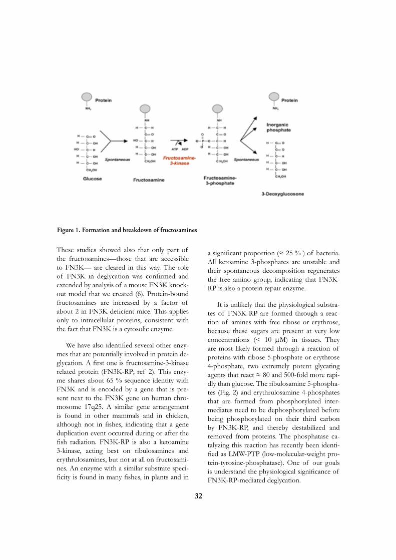
primary amines, followed by an Amadori rear-rangement. Fructosamine 3-kinase (FN3K) is a recently identified enzyme that phosphorylates both low-molecular-weight and protein-bound fructosamines (3). Fructosamine 3-phosphates are unstable, breaking down spontaneously to 3-deoxyglucosone, inorganic phosphate and the amino compound that originally reacted with glucose (Fig. 1).
That FN3K indeed acts as a ‘deglycating’ enzyme was first indicated by experiments in which erythrocytes were incubated ex vivo with an elevated concentration of glucose, with or without a competitive inhibitor of FN3K.
32
These studies showed also that only part of the fructosamines—those that are accessible to FN3K— are cleared in this way. The role of FN3K in deglycation was confirmed and extended by analysis of a mouse FN3K knock-out model that we created (6). Protein-bound fructosamines are increased by a factor of about 2 in FN3K-deficient mice. This applies only to intracellular proteins, consistent with the fact that FN3K is a cytosolic enzyme.
We have also identified several other enzy-mes that are potentially involved in protein de-glycation. A first one is fructosamine-3-kinase related protein (FN3K-RP; ref 2). This enzy-me shares about 65 % sequence identity with FN3K and is encoded by a gene that is pre-sent next to the FN3K gene on human chro-mosome 17q25. A similar gene arrangement is found in other mammals and in chicken, although not in fishes, indicating that a gene duplication event occurred during or after the fish radiation. FN3K-RP is also a ketoamine 3-kinase, acting best on ribulosamines and erythrulosamines, but not at all on fructosami-nes. An enzyme with a similar substrate speci-ficity is found in many fishes, in plants and in
a significant proportion (≈ 25 % ) of bacteria. All ketoamine 3-phosphates are unstable and their spontaneous decomposition regenerates the free amino group, indicating that FN3K-RP is also a protein repair enzyme.
It is unlikely that the physiological substra-tes of FN3K-RP are formed through a reac-tion of amines with free ribose or erythrose, because these sugars are present at very low concentrations (< 10 mM) in tissues. They are most likely formed through a reaction of proteins with ribose 5-phosphate or erythrose 4-phosphate, two extremely potent glycating agents that react ≈ 80 and 500-fold more rapi-dly than glucose. The ribulosamine 5-phospha-tes (Fig. 2) and erythrulosamine 4-phosphates that are formed from phosphorylated inter-mediates need to be dephosphorylated before being phosphorylated on their third carbon by FN3K-RP, and thereby destabilized and removed from proteins. The phosphatase ca-talyzing this reaction has recently been identi-fied as LMW-PTP (low-molecular-weight pro-tein-tyrosine-phosphatase). One of our goals is understand the physiological significance of FN3K-RP-mediated deglycation.
Figure 1. Formation and breakdown of fructosamines

33
NEUROMETABOLIC DISORDERS
Y Achouri, F. Collard, J. Drozak, S. Jaisson, G. Noël, K. Peel, G. Tahay, M. Veiga da Cunha, E. Wiame, E. Van Schaftingen
D- and L-2-hydroxyglutaric acidurias are distinct neurometabolic diseases characterized by the accumulation of abnormal amounts of either D- or L-2-hydroxyglutarate in cerebros-pinal fluid, blood and urine. Work in our lab has led to the elucidation of the metabolism of these compounds (Fig. 2). Both of them are converted to alpha-ketoglutarate by distinct FAD-linked dehydrogenases. The dehydroge-nase acting on L-2-hydroxyglutarate is bound to mitochondrial membranes and mutations in its gene are found in virtually all cases of L-2-hydroxyglutaric aciduria (8). The dehydro-genase acting on D-2-hydroxyglutarate is in the mitochondrial matrix and most likely transfers its electrons to the respiratory chain via elec-tron-transfer-flavoprotein (1). It is mutated in about 40 % of the patients with D-2-hydroxy-glutaric aciduria.
The formation of L-2-hydroxyglutarate is catalyzed by mitochondrial L-malate dehydro-genase (Fig. 3). This enzyme is not completely
specific for oxaloacetate : it also reduces, at a very low rate, alpha-ketoglutarate to L-2-hy-droxyglutarate. This activity is sufficient to ac-count for the daily formation of L-2-hydroxy-glutarate. Since L-2-hydroxyglutarate does not appear to have any role, but to have only toxic effects, L-2-hydroxyglutarate dehydrogenase is a ‘repair enzyme’ and L-2-hydroxyglutaric aci-duria is a disorder of metabolite repair. One of our aims is to produce a mouse model of L-2-hydroxyglutaric aciduria in order to confirm the origin of L-2-hydroxyglutarate and unders-tand the physiopathological mechanisms of this disease.
We are presently studying enzymes involved in the synthesis of brain-specific compounds, such as N-acetylaspartate, with the hope of elucidating as yet unresolved neurometabolic disorders.
Figure 2. Formation and breakdown of ribulosamines
Figure 3. Formation and breakdown of L-2-hydroxyglutarate

34
METABOLISM OF GLUCOSE 1,6-BISPHOSPHATE
P. Maliekal, M. Veiga-da-Cunha, E. Van Schaftingen
Glucose 1,6-bisphosphate (Glc-1,6-P2), a well known cofactor for phosphoglucomutase and other sugar phosphomutases, is ubiqui-tously present in tissues. Its concentration is particularly elevated in brain, where it reaches values of >100 µM, i.e. >1000-fold higher than the concentrations required to stimulate phos-phoglucomutase. Glc-1,6-P2 has been proposed to be an effector for several enzymes (Fig. 4). Phosphofructokinase and liver pyruvate kinase are both stimulated by this compound, whe-reas low Km hexokinases, 6-phosphogluconate dehydrogenase, and fructose-1,6-bisphospha-tase are inhibited. These effects have been de-monstrated in vitro, but under conditions that are not necessarily physiologically relevant. In addition, the occurrence of this regulation in intact cells has not been demonstrated.
We have recently reported the molecular identification of the enzymes that synthesize and degrade Glc-1,6-P2 (6,10). Glc-1,6-P2 is made from 1,3-bisphosphoglycerate and glu-cose 1-phosphate or glucose 6-phosphate by
Glc-1,6-P2 synthase, an enzyme particularly abundant in brain. We have recently identified this enzyme as PGM2L1 (phosphoglucomutase 2-like 1). It belongs to same family as phospho-glucomutase 1, the enzyme that interconverts glucose 6-phosphate and glucose 1-phosphate. Its closest mammalian homologue if PGM2, which serves to interconvert (deoxy)ribose 1-phosphate and (deoxy)ribose 5-phosphate. PGM2 also catalyzes the synthesis of Glc-1,6-P2, although with a lower Vmax than PGM2L1 and a much stronger inhibition by the reac-tion product Glc-1,6-P2. In comparison with PGM2, PGM2L1 is therefore better suited to provide cells with elevated concentrations of Glc-1,6-P2. This conclusion is supported by the finding that transfection of HEK293T cel-ls with PGM2L1 caused a ≈ 20-fold increase in the concentration of Glc-1,6-P2 whereas transfection with PGM2 did not affect the level of the bisphosphate ester.
Glc-1,6-P2 is degraded by glucose-1,6-bisphosphatase. The brain enzyme, which has been best characterized, is dependent for its activity on the presence of IMP, the concentration of which increases in anoxia. This effect is presumably responsible for the decrease in Glc-1,6-P2 concentration in brain during anoxia. We have recently identified this enzyme to PMM1 (phosphomannomutase 1), an enzyme that was initially thought to be a phosphomannomutase and which we knew to have a modest glucose-1,6-bisphosphatase activity. We have now shown that IMP, but not other nucleotides, stimulated by >100-fold the intrinsic glucose-1,6-bisphosphatase activity of recombinant PMM1 while inhibiting its phosphoglucomutase activity. No such effect is observed with PMM2. Transfection of HEK cells with PMM1 caused a marked decrease (>5-fold) in Glc-1,6-P2 in cells that were or were not cotransfected with PGM2L1. Furthermore, the concentration of Glc-1,6-P2 in wild-type mouse brain decreased with time after ischemia, whereas it did not change in PMM1-deficient mouse brain. Taken together, these data show that PMM1 corresponds to the IMP-stimulated glucose-1,6-bisphosphatase
Figure 4. Metabolism and potential regulatory role of glucose 1,6-bisphosphate in brain

35
and that this enzyme is responsible for the degradation of Glc-1,6-P2 in brain. One of our aims is presently to understand the meaning of the regulation of Glc-1,6-P2 concentration by IMP.
METABOLISM OF PSEUDOURIDINE
A. Preumont, E. Van Schaftingen, in collaboration with Jean-François Collet.
Pseudouridine, a non-classical nucleoside present in human urine as a degradation product of RNAs, is one of the few molecules having a glycosidic C-C bond. Through a database mining approach involving transcriptomic data, we have molecularly identified two enzymes that are involved in the metabolism of pseudouridine (Fig. 5) in uropathogenic Escherichia coli, the principal agent of urinary tract infections in humans (7). The first enzyme, coded by the gene yeiC, specifically phosphorylates pseudouridine to pseudouridine 5’-phosphate. Accordingly, yeiC(-) mutants are unable to metabolize pseudouridine, in contrast to wild-type E. coli UTI89.
The second enzyme, encoded by the gene yeiN belonging to the same operon as yeiC, catalyzes the conversion of pseudouridine 5’-phosphate to uracil and ribose 5-phosphate in a divalent cation-dependent manner. Remarka-bly, the glycosidic C-C bond of pseudouridine is cleaved in the course of this reaction, indi-cating that YeiN is the first molecularly identi-fied enzyme able to hydrolyze a glycosidic C-C bond. Though this reaction is easily reversible, the association of YeiN with pseudouridine ki-nase indicates that it serves physiologically to metabolize pseudouridine 5’-phosphate rather than to form it. YeiN is homologous to Ther-motoga maritima IndA, a protein with a new fold, which we also showed to act as a pseudouri-dine-5’-phosphate glycosidase.
Database mining indicates that most euka-ryotes possess homologues of pseudouridine kinase and pseudouridine-5’-phosphate glyco-sidase and that these are most often associated in a single bifunctional protein. The gene en-coding this bifunctional protein is absent from the genomes of man and other mammals, indi-cating that the capacity for metabolizing pseu-douridine has been lost late in evolution. The absence of pseudouridine metabolizing enzy-mes in man is consistent with the observation that this nucleoside is present in human urine and its urinary excretion is increased in cancer. Our present aim is to identify the mechanism by which pseudouridine is formed in mamma-lian cells.
SELECTED PUBLICATIONS
1. Achouri Y, Noël G, Vertommen D, Rider MH, Veiga-Da-Cunha M, Van Schaftingen E. Identification of a dehydrogenase acting on D-2-hydroxyglutarate. Biochem J 2004;381:35-42.
Figure 5. Metabolism of pseudouridine in Escherichia coli.

36
2. Collard F, Delpierre G, Stroobant V, Matthijs G, Van Schaftingen E. A mammalian protein homologous to fructosamine-3-kinase is a ketosamine-3-kinase acting on psicosamines and ribulosamines but not on fructosamines. Diabetes 2003;52:2888-95.
3. Delpierre G, Rider MH, Collard F, Stroobant V, Vanstapel F, Santos H, Van Schaftingen E. Identification, cloning, and he-terologous expression of a mammalian fructosa-mine-3-kinase. Diabetes 2000;49:1627-34.
4. Fortpied J, Gemayel R, Vertommen D, Van Schaftingen E. Identification of protein-ribulosamine-5-phosphatase as human low-mo-lecular-weight protein-tyrosine-phosphatase-A. Biochem J 2007;406:139-45
5. Hart CE, Race V, Achouri Y, Wiame E, Sharrard M, Olpin SE, Watkinson J, Bonham JR, Jaeken J, Matthijs G, Van Schaftingen E. Phosphoserine aminotransferase deficiency: a novel disorder of the serine biosynthesis pathway. Am J Hum Genet 2007;80:931-7.
6. Maliekal P, Sokolova T, Vertommen D, Veiga-da-Cunha M, Van Schaftingen E. Molecular identification of mammalian phosphopentomutase and glucose-1,6-bisphosphate synthase, two members of the alpha-D-phosphohexomutase family. J Biol Chem 2007;282:31844-51.
7. Preumont A, Snoussi K, Stroobant V, Collet JF, Van Schaftingen E. Molecular identification of pseudouridine-metabolizing enzymes. J Biol Chem 2008;283:25238-46.
8. Rzem R, Veiga-da-Cunha M, Noel G, Goffette S, Nassogne MC, Tabarki B, Scholler C, Marquardt T, Vikkula M, Van Schaftingen E. A gene encoding a putative FAD-dependent L-2-hydroxyglutarate dehydrogenase is mutated in L-2-hydroxyglutaric aciduria. Proc Natl Acad Sci USA 2004;101:16849-16854.
9. Veiga da-Cunha M, Jacquemin P, Delpierre G, Godfraind C, Theate I, Vertommen D, Clotman F, Lemaigre F, Devuyst O, Van Schaftingen E. Increased protein glycation in fructosamine 3-kinase-deficient mice. Biochem J 2006;399:257-64.
10. Veiga-da-Cunha M, Vleugels W, Maliekal P, Matthijs G, Van Schaftingen E. Mam-malian phosphomannomutase PMM1 is the brain IMP-sensitive glucose-1,6-bisphosphatase. J Biol Chem 2008;283:33988-93.
Emile Van Schaftingende Duve InstituteAv. Hippocrate 74-75B - 1200 Brussels[T] +32 02 764 75 64[F] +32 02 764 75 98[E] [email protected][W] http://www.deduveinstitute.be/repair_in-born_errors_of_metabolism.php

37
Françoise BONTEMPS, Associate MemberEric VAN DEN NESTE, Clinical Investigator
Georges VAN DEN BERGHE, Emeritus Member
Caroline SMAL, Postdoctoral FellowLaurent BASTIN-COYETTE, Graduate StudentEmeline de VIRON, Graduate StudentRachid AMSAILALE, Graduate StudentAngélique AERTS, Technician
Our group had a long-standing interest in purine metabolism, particularly adenine nucleotide metabolism, and its genetic defects. More recently, we expended our investigations on two therapeutic purine nucleoside analogues, 2-chlorodeoxyadenosine and fludarabine, which have revolutionized the treatment of indolent lymphoproliferative disorders. Despite this activity, clinical resistance to these drugs is frequently observed. The main objectives of our present studies are to unravel the mechanisms leading to resistance to nucleoside analogues and to find novel therapeutic strategies to counteract them, particularly in B-cell chronic lymphocytic leukaemia.
NUCLEOSIDE ANALOGUES IN LEUKAEMIA
In 1997, a collaborative study of the antileu-kaemic nucleoside analogues, 2-chlorodeoxya-denosine (CdA) and fludarabine (Fig. 1), was started with the Department of Haematology of the University Hospital Saint-Luc. These adenosine deaminase-resistant deoxyadeno-sine analogues display remarkable therapeutic properties in indolent lymphoid malignancies including hairy cell leukaemia and B-cell chro-nic lymphocytic leukaemia (B-CLL). Neverthe-less, resistance is also observed, and nucleoside analogues do not confer a survival advantage when compared to more conventional thera-pies such as alkylating agents. The aims of the project are to understand the mechanisms that lead to resistance to nucleoside analogues, and to improve their therapeutic efficacy by sear-ching for synergisms with other compounds.
CdA and fludarabine are prodrugs. To exert their antileukaemic effect, they have to be
phosphorylated by deoxycytidine kinase (dCK) into nucleoside analogue monophosphates, fol-lowed by conversion into di- and triphosphate derivatives. The latter are the active metabolites. They have been shown to inhibit various pro-cesses involved in DNA and RNA synthesis. Moreover, they can be incorporated into newly synthesised DNA, causing chain termination. Together, these actions result in arrest of DNA synthesis and in the progressive accumulation of DNA strand breaks, leading to apoptosis by mechanisms which are not yet entirely clear (1, for a review).
MECHANISMS OF ACTION
F. Bontemps, C. Smal, L. Bastin-Coyette, E. de Viron, A. Aerts, E. Van Den Neste
To improve our understanding of the me-

38
chanisms by which CdA induces apoptosis in B-CLL cells, we study its effects in EHEB cells, a continuous cell line derived from a patient with B-CLL. The EHEB cell line was found to be less sensitive (10- to 100-fold) to the nu-cleoside analogue CdA than other human lym-phoblastic cell lines. This can be explained by a lower intracellular accumulation of CdATP, the cytotoxic metabolite of CdA, due to a redu-ced dCK activity. Unexpectedly, DNA synthe-sis, measured by thymidine incorporation into DNA, was found to be increased, up to 2-fold, after a 24 h-incubation with CdA at concen-trations close to the IC50 (5 µM) (2). Analysis by flow cytometry, using double labelling with propidium iodide and bromodeoxyuridine, has shown that CdA, in EHEB cells, provokes an increase in the proportion of cells in S phase, synthesising actively DNA. These results con-trast with those reported in other leukaemic cell lines, like CCRF-CEM cells, in which CdA inhibits DNA synthesis. Kinetics and synchro-nisation experiments have shown that the in-crease in the proportion of cells in S-phase induced by CdA in EHEB cells was due to a stimulation of the progression of cells from G1 to S phase. Accordingly, we found that CdA enhances the activity of cyclin-dependent kinase 2 (Cdk2), a kinase that plays a major role in the progression of cells from G1 to S phase. This unexpected result has led us to examine the effects of CdA on proteins involved in the regulation of cell cycle, and to investigate
whether Cdk2 activation contributes to CdA-induced apoptosis in EHEB cells. Our further aim will be to elucidate how CdA and possibly other nucleoside analogues activate Cdk2.
In parallel, we have analysed the mechanisms of cell death induced by 2-chloroadenosine (2-CAdo) in various types of leukaemic B-cells. This analogue is not activated by dCK, but by adenosine kinase. We found that 2-CAdo was efficiently converted into 2-chloroATP, resul-ting in ATP depletion, as well as in inhibition of DNA, RNA and protein synthesis. 2-CAdo also caused activation of the intrinsic pathway of apoptosis, which was associated with a de-cline in Mcl-1 protein level, and p53-indepen-dent. Prevention of ATP loss by inhibition of AMP deaminase reduced apoptosis, indicating that ATP depletion plays an important role in 2-CAdo-induced apoptosis (3).
Finally, we have recently initiated microarray analyses to identify survival or death pathways that are activated in response to nucleoside analogue treatment. We intended to compare genes induced or repressed by nucleoside ana-logues in sensitive and refractory B-CLL pa-tients. We found that some genes were syste-matically induced in sensitive patients, and not in refractory patients. We are now analysing whether the induction of one or more of these genes play(s) a role in nucleoside analogue-in-duced cell death. This study is performed in
Fig. 1. Structures of deoxyadenosine and analogues

39
collaboration with Dr Knoops from the Lud-wig Institute at the de Duve Institute.
SEARCH FOR POTENTIATION OF ANTILEUKAEMIC EFFECT OF 2-CHLORODEOXyADENOSINE
E. Van Den Neste, C. Smal, F. Bontemps
In recent years, we have shown that com-bination of CdA with DNA-damaging agents, such as cyclophosphamide (CP) derivatives (4) or UV-light (5), resulted in synergistic cytotoxi-city in B-CLL lymphocytes, due to inhibition of DNA repair. The in vitro synergy between CdA and CP derivatives has provided the ra-tionale for a clinical trial of this combination, which gives encouraging results (6).
Lately, we have explored the possibility that CdA interacts with the mitogen-activated pro-tein kinases/extracellular signal-regulated ki-nase (MAPK/ERK) pathway. We have shown that CdA, at concentrations close to the IC50, activated the ERK pathway in EHEB cells. Be-cause activation of this pathway is assumed to exert anti-apoptotic effect, we combined CdA with inhibitors of the ERK pathway. The latter were found to enhance CdA-induced apopto-sis. These results suggest that the efficacy of CdA could be strengthened by combination with inhibitors of the ERK pathway (7).
REGULATION OF DEOXyCyTI-DINE KINASE ACTIVITy
C. Smal, R. Amsailale, A. Aerts, E. Van Den Neste, F. Bontemps
Since deoxycytidine kinase (dCK) activates numerous nucleoside analogues used in anti-cancer and antiviral therapy, knowledge of its regulation can be expected to allow optimiza-
tion of the activation of these analogues. Re-cently, it has been shown by others and by us that dCK activity can be increased by various genotoxic agents, including CdA, aphidicolin, etoposide, and UV-C irradiation. This activa-tion could not be explained by an allosteric ef-fect or by an increase of the protein amount. Therefore, a post-translational activation of dCK by intracellular signalling pathways was suggested. To unravel the mechanism of the activation of dCK, we first investigated the ef-fect of a variety of activators and inhibitors of protein kinases on dCK activity. We found that some of them could modify dCK activity in several types of leukaemic cells, strengthening the hypothesis of a phosphorylation/dephos-phorylation process as a mechanism of regu-lation of dCK activity. Most particularly, our results indicated that dCK could be down-re-gulated by the JAK/MAPK pathway and that it might be an in vivo target of protein phospha-tase 2A (8).
We then overexpressed dCK in HEK 293T cells and observed that the enzyme was label-led after incubation with [32P]orthophosphate, confirming that dCK is a phosphoprotein. Tandem mass spectrometry performed by Dr D. Vertommen and Prof. M.H. Rider from the Horm unit allowed the identification of four in vivo phosphorylation sites, Thr-3, Ser-11, Ser-15 and Ser-74 (Fig. 2). Site-directed mu-tagenesis demonstrated that Ser-74 phospho-rylation was crucial for dCK activity in HEK 293T cells, whereas phosphorylation of other identified sites did not seem essential (9). Phos-phorylation of Ser-74 was also observed on endogenous dCK in CCRF-CEM cells and in lymphocytes from B-CLL patients, in which the Ser-74 phosphorylation state was increased by several genotoxic agents (UV, etoposide, …) that enhanced dCK activity, and decreased by sorbitol that diminished dCK activity. Moreo-ver, interindividual variability in dCK activity in B-CLL lymphocytes could be related to its phosphorylation level on Ser-74 (10). To con-clude, our work has demonstrated that dCK activity largely depends on the phosphoryla-tion state of Ser-74 in human leukaemic lym-
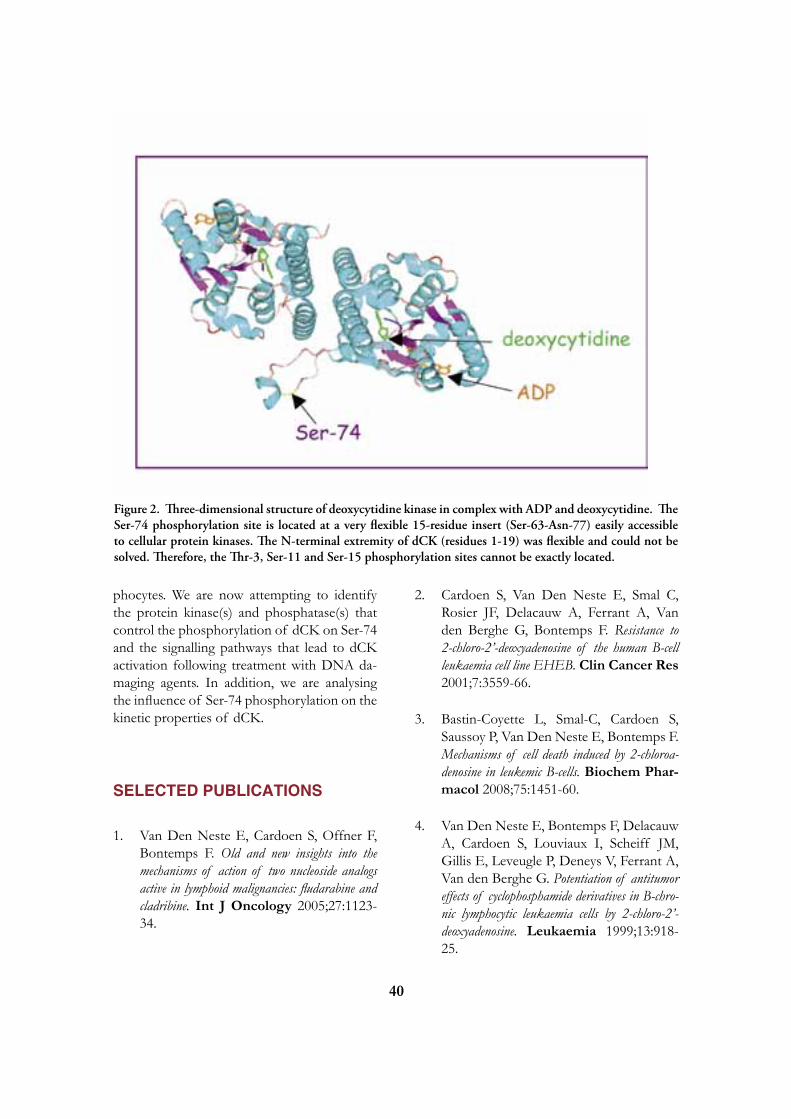
40
phocytes. We are now attempting to identify the protein kinase(s) and phosphatase(s) that control the phosphorylation of dCK on Ser-74 and the signalling pathways that lead to dCK activation following treatment with DNA da-maging agents. In addition, we are analysing the influence of Ser-74 phosphorylation on the kinetic properties of dCK.
SELECTED PUBLICATIONS
1. Van Den Neste E, Cardoen S, Offner F, Bontemps F. Old and new insights into the mechanisms of action of two nucleoside analogs active in lymphoid malignancies: fludarabine and cladribine. Int J Oncology 2005;27:1123-34.
2. Cardoen S, Van Den Neste E, Smal C, Rosier JF, Delacauw A, Ferrant A, Van den Berghe G, Bontemps F. Resistance to 2-chloro-2’-deoxyadenosine of the human B-cell leukaemia cell line EHEB. Clin Cancer Res 2001;7:3559-66.
3. Bastin-Coyette L, Smal-C, Cardoen S, Saussoy P, Van Den Neste E, Bontemps F. Mechanisms of cell death induced by 2-chloroa-denosine in leukemic B-cells. Biochem Phar-macol 2008;75:1451-60.
4. Van Den Neste E, Bontemps F, Delacauw A, Cardoen S, Louviaux I, Scheiff JM, Gillis E, Leveugle P, Deneys V, Ferrant A, Van den Berghe G. Potentiation of antitumor effects of cyclophosphamide derivatives in B-chro-nic lymphocytic leukaemia cells by 2-chloro-2’-deoxyadenosine. Leukaemia 1999;13:918-25.
Figure 2. Three-dimensional structure of deoxycytidine kinase in complex with ADP and deoxycytidine. The Ser-74 phosphorylation site is located at a very flexible 15-residue insert (Ser-63-Asn-77) easily accessible to cellular protein kinases. The N-terminal extremity of dCK (residues 1-19) was flexible and could not be solved. Therefore, the Thr-3, Ser-11 and Ser-15 phosphorylation sites cannot be exactly located.

41
5. Van Den Neste E, Cardoen S, Husson B, Rosier JF, Delacauw A, Ferrant A, Van den Berghe G, Bontemps F. 2-Chloro-2’-deoxyadenosine inhibits DNA repair synthesis and potentiates UVC cytotoxicity in chronic lym-phocytic leukaemia B lymphocytes. Leukaemia 2002;16:36-43.
6. Van Den Neste E, Louviaux I, Michaux JL, Delannoy A, Michaux L, Sonet A, Bosly A, Doyen C, Mineur P, André M, Straetmans N, Coche E, Venet C, Duprez T, Ferrant A. Phase I/II study of 2-chloro-2’-deoxyadenosine with cyclophosphamide in patients with pretreated B cell chronic lymphocytic leu-kaemia and indolent non-Hodgkin’s lymphoma. Leukaemia 2000;14:1136-42.
7. Smal C, Lisart S, Maerevoet M, Ferrant A, Bontemps F, Van Den Neste E. Pharmaco-logical inhibition of the MAPK/ERK pathway increases sensitivity to 2-chloro-2’-deoxyadenosine (CdA) in the B-cell leukaemia cell line EHEB. Biochem Pharmacol 2007;73:351-8.
8. Smal C, Cardoen S, Bertrand L, Delacauw A, Ferrant A, Van den Berghe G, Van Den Neste E, Bontemps F. Activation of deoxycytidine kinase by protein kinase inhibitors and okadaic acid in leukemic cells. Biochem Pharmacol 2004;68:95-103.
9. Smal C, Vertommen D, Bertrand L, Nta-mashimikiro S, Rider M, Van den Neste
E, Bontemps F. Identification of in vivo phos-phorylation sites on human deoxycitidine kinase. Role of Ser-74 in the control of enzyme activity. J Biol Chem 2006;281:4887-93.
10. Smal C, Van Den Neste E, Maerevoet M, Poiré X, Théate I, Bontemps F. Positive regulation of deoxycytidine kinase activity by phosphorylation of Ser-74 in B-cell chronic lymphocytic leukaemia lymphocytes. Cancer Lett 2007;253:68-73.
Françoise Bontempsde Duve InstituteAv. Hippocrate 74-75B - 1200 Brussels
[T] +32 (02) 764 75 68[F] +32 (02) 764 75 98[E] [email protected][W] http://www.deduveinstitute.be/nucleoside_analogues_in_leukaemia.php

42
Jean-Francois COLLET, Associate Member
Pauline LEVERRIER, Postdoctoral fellowVéronica Tamu Dufé, Postdoctoral fellowHayat EL HAJJAJI, Graduate StudentMatthieu DEPUYDT, Graduate StudentKatleen Denoncin, Graduate StudentCéline RECOUR, Technician Geneviève CONNEROTTE, Technician, part-time
PROTEIN FOLDING IN BACTERIA
To be active and stable, newly synthesized proteins have to fold correctly. However, reaching a proper three-dimensional structure is a real challenge for most proteins. This is especially true for proteins that are secre-ted to the cell envelope of Gram-negative bacteria. These proteins are assembled in the cytoplasm, are then transported across the inner-membrane and finally fold within the periplasm or the outer membrane, in an environment devoid of any obvious energy source. Moreover, the correct folding of many of these proteins requires the formation of a disulfide bond between two cysteine residues, which is a rate-limiting step of the folding process. Failure to fold secreted proteins properly affects the integrity of the bacterial cell envelope, which increases the sensitivity of bacteria to antimicrobial drugs.In our group, we study the pathways that govern the assembly of the cell envelope of Gram-negative bacteria. In particular, we are interested in the mechanisms of disulfide bond formation in the periplasm of Escheri-chia coli and in the chaperones that assist the folding of outer-membrane proteins.
OXIDATIVE PROTEIN FOLDING IN THE PERIPLASM
G. Connerotte, M. Depuydt, K. Denoncin, P. Leverrier, J.-F. Collet
In the gram-negative bacteria Escherichia coli, disulfide bonds are introduced in the pe-riplasm by the Dsb (Disulfide bond) proteins family [4].
The primary oxidant is the soluble protein DsbA. DsbA has a CXXC catalytic site motif present within a thioredoxin fold. The cystei-
ne residues of this motif are found oxidized in vivo. The disulfide bond of DsbA is very unstable and is rapidly transferred to secreted unfolded proteins. DsbA is then re-oxidized by the inner-membrane protein DsbB that trans-fers electrons from DsbA to the electron trans-port chain (Figure 1).
DsbA is a very powerful oxidant that appa-rently lacks proofreading activity. If the native disulfide bond pattern involves cysteine resi-dues that are non-consecutive in the amino-acid sequence, DsbA can form incorrect disulfides. The correction of these non-native disulfides is the role of a disulfide isomerization system.

43
This system involves a soluble periplasmic pro-tein disulfide isomerase, DsbC. Like DsbA, DsbC presents a thioredoxin fold and a CXXC catalytic site motif. In contrast to DsbA, the CXXC motif of DsbC is kept reduced in the periplasm. This allows DsbC to attack non-na-tive disulfides, a necessary step in the isomeri-zation reaction. The protein that keeps DsbC reduced is the inner-membrane protein DsbD. DsbD transfers reducing equivalents from the cytoplasmic thioredoxin system to the peri-plasm via a succession of disulfide exchange reactions. Recently, we showed that DsbC may also be acting as a stand-alone protein folding catalyst that is able to cycle from the reduced to the oxidized state upon substrate oxidation and substrate reduction, respectively [7].
The periplasm possesses another soluble Dsb protein, DsbG, but the function of this protein has remained elusive.
Current efforts in this field are devoted to the cha-racterization of the function of DsbG and the study of DsbA proteins from pathogenic bacteria.
Identification of the substrates of DsbG
We sought to find the function of DsbG in the periplasm by trapping it with its substrates. We produced a mutant protein in which the se-cond cysteine residue of the CXXC motif is re-placed by a serine. This version of the protein is still able to attack a substrate protein to form a mixed-disulfide intermediate. However, due to the absence of the second cysteine residue of the CXXC motif, this mixed disulfide bond cannot be resolved, allowing the purification of the complexes formed between DsbG and its substrates. We identified three proteins in complex with DsbG. The interaction between DsbG and these proteins is currently under in-vestigation.
Structural and biochemical characterization of three DsbA proteins from Neisseria meningitidis
Neisseria meningitidis is an invasive bacterial pathogen causing life-threatening in-fection in children worldwide. Host-pathogen interactions, and therefore virulence, depend on the correct folding of many surface-ex-posed proteins, which often requires disulfide bond formation. Uniquely among bacteria, the Neisseria meningitidis genome possesses three genes encoding active DsbAs: NmDsbA1, NmDsbA2 and NmDsbA3. NmDsbA1 and NmDsbA2 have been characterized as lipo-proteins involved in natural competence and in host interactive biology, while the function of NmDsbA3 remains unknown. In collaboration with Laurence Serre and Céline Lafaye (Greno-ble, France), we have characterized the neisse-rial enzymes biochemicaly and structurally. We found that the crystal structures of the Neisse-
Figure 1. Disulfide bond formation in the E. coli periplasm. Disulfides are introduced into folding proteins by DsbA. DsbA is re-oxidized by DsbB, an inner-membrane protein. Electrons flow from the reduced proteins to the CXXC motif of DsbA and then to DsbB. DsbA can form incorrect disulfides that are corrected by a protein disulfide isomerase DsbC. DsbC is kept reduced by DsbD, a membrane protein that transfers electrons from the cytoplasmic pool of NADPH to the periplasm. DsbD also transfers electrons to DsbG, a protein which resembles DsbC, but whose function is not known.
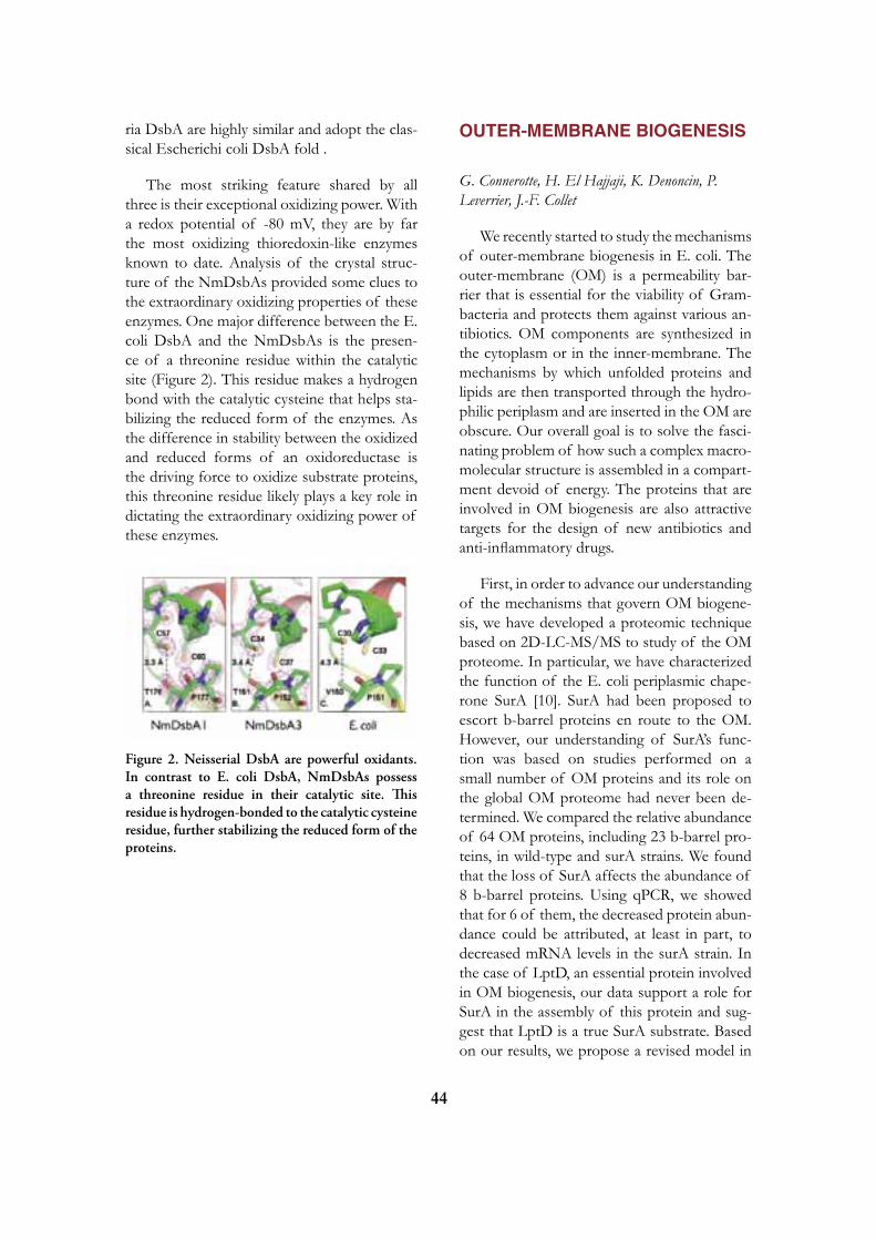
44
ria DsbA are highly similar and adopt the clas-sical Escherichi coli DsbA fold .
The most striking feature shared by all three is their exceptional oxidizing power. With a redox potential of -80 mV, they are by far the most oxidizing thioredoxin-like enzymes known to date. Analysis of the crystal struc-ture of the NmDsbAs provided some clues to the extraordinary oxidizing properties of these enzymes. One major difference between the E. coli DsbA and the NmDsbAs is the presen-ce of a threonine residue within the catalytic site (Figure 2). This residue makes a hydrogen bond with the catalytic cysteine that helps sta-bilizing the reduced form of the enzymes. As the difference in stability between the oxidized and reduced forms of an oxidoreductase is the driving force to oxidize substrate proteins, this threonine residue likely plays a key role in dictating the extraordinary oxidizing power of these enzymes.
OUTER-MEMBRANE BIOGENESIS
G. Connerotte, H. El Hajjaji, K. Denoncin, P. Leverrier, J.-F. Collet
We recently started to study the mechanisms of outer-membrane biogenesis in E. coli. The outer-membrane (OM) is a permeability bar-rier that is essential for the viability of Gram- bacteria and protects them against various an-tibiotics. OM components are synthesized in the cytoplasm or in the inner-membrane. The mechanisms by which unfolded proteins and lipids are then transported through the hydro-philic periplasm and are inserted in the OM are obscure. Our overall goal is to solve the fasci-nating problem of how such a complex macro-molecular structure is assembled in a compart-ment devoid of energy. The proteins that are involved in OM biogenesis are also attractive targets for the design of new antibiotics and anti-inflammatory drugs.
First, in order to advance our understanding of the mechanisms that govern OM biogene-sis, we have developed a proteomic technique based on 2D-LC-MS/MS to study of the OM proteome. In particular, we have characterized the function of the E. coli periplasmic chape-rone SurA [10]. SurA had been proposed to escort b-barrel proteins en route to the OM. However, our understanding of SurA’s func-tion was based on studies performed on a small number of OM proteins and its role on the global OM proteome had never been de-termined. We compared the relative abundance of 64 OM proteins, including 23 b-barrel pro-teins, in wild-type and surA strains. We found that the loss of SurA affects the abundance of 8 b-barrel proteins. Using qPCR, we showed that for 6 of them, the decreased protein abun-dance could be attributed, at least in part, to decreased mRNA levels in the surA strain. In the case of LptD, an essential protein involved in OM biogenesis, our data support a role for SurA in the assembly of this protein and sug-gest that LptD is a true SurA substrate. Based on our results, we propose a revised model in
Figure 2. Neisserial DsbA are powerful oxidants. In contrast to E. coli DsbA, NmDsbAs possess a threonine residue in their catalytic site. This residue is hydrogen-bonded to the catalytic cysteine residue, further stabilizing the reduced form of the proteins.

45
which only a subset of OM proteins depends on SurA for proper folding and insertion in the OM (Figure 3). We are currently using our powerful proteomic technique to characterize mutants that presents defects in OM integrity.
SELECTED PUBLICATIONS
1. Masip L, Pan JL, Haldar S, Penner-Hahn J, Delisa M, Georgiou G, Bardwell J, Collet J-F. An engineered pathway for the formation of protein disulfide bonds. Science, 2004;303:1185-9.
2. Collet J-F, Peisach D, Bardwell JCA, Xu Z. The crystal structure of TrxA(CACA): insights into the formation of a [2Fe-2S] iron-sulfur clus-ter in an E. coli thioredoxin mutant. Protein science, 2005;14:1863-9.
3. Hiniker A, Collet J-F, Bardwell JCA. Cop-per stress causes an in vivo requirement for the Escherichia coli disulfide isomerase DsbC. J Biol Chem, 2005;80:33785-91.
4. Messens J, Collet J-F. Pathways of disulfide bond formation in Escherichia coli. Int J Bio-chem Cell Biol, 2006;38:1050-62.
5. Hiniker A, Vertommen D, Bardwell JCA, Collet J-F. Evidence for conformational chan-ges within DsbD: Possible role for membrane-embedded proline residues. J Bacteriology, 2006;188:7317-20.
6. Brot N, Collet J-F, Johnson LC, Jonsson TJ, Weissbach H, Lowther WT. The thiore-doxin domain of Neisseria gonorrhoeae PilB can use electrons from DsbD to reduce downstream methionine sulfoxide reductases. J Biol Chem, 2006;281:32668-75.
7. Vertommen D, Depuydt M, Pan JL, Le-verrier P, Knoops L, Szikora JP, Messens J, Bardwell JCA, Collet J-F. The disulphide iso-merase DsbC cooperates with the oxidase DsbA in a DsbD-independent manner. Mol Micro-biol, 2008;67:336-49.
8. Messens J, Collet J-F, Van Belle K, Bro-sens E, Loris R, Wyns L. The oxidase DsbA folds a protein with a nonconsecutive disulfide. J Biol Chem, 2007;282:31302-07.
Figure 3. Transport of unfolded b-barrel proteins across the periplasm. Unfolded b-barrel proteins are escorted across the periplasm by periplasmic chaperones before reaching the OM where they are inserted by the Bam complex. There are 3 known periplasmic chaperones that interact with OMPs: SurA, Skp and DegP. Our results indicate that only a subset of OMPs strongly depend on SurA for biogenesis. We propose to divide the OMPs in three groups. Group 1 includes b-barrel proteins, such as LptD, that greatly depend on the presence of SurA for biogenesis. Group 2 includes b-barrel proteins such as the major OMPs OmpA, OmpF and LamB that preferentially interact with SurA for biogenesis but are also able to interact with the other periplasmic chaperones. Group 3 includes b-barrel proteins such as BamA and TolC that do not seem to require the presence of SurA for biogenesis.

46
9. El Hajjaji H, Dumoulin M, Matagne A, Colau D, Roos G, Messens J, Collet J-F. The zinc center influences the redox and thermo-dynamic properties of Escherichia coli thioredo-xin 2. J Mol Biol, 2009;386:60-71.
10. Vertommen D, Ruiz N, Leverrier P, Sil-havy T, Collet J-F. Characterization of the role of the Escherichia coli periplasmic chaperone SurA using differential proteomics. Proteo-mics, 2009;9:2432-43.
Jean-François Colletde Duve InstituteAv. Hippocrate 74-75B - 1200 Brussels
[T] +32 02 764 75 62[F] +32 02 764 75 98[E] [email protected][W] http://www.deduveinstitute.be/protein_fol-ding_in_bacteria.php

47
Guido T. Bommer, Scientist
Isabelle Gerin, Postdoctoral FellowOlivier Haumont, Technician
miRNAS IN PHySIOLOGy AND DISEASE
miRNAs are small non-protein-coding RNAs that can bind to mRNA transcripts of protein coding genes. Upon binding to these mRNAs, they inhibit their translation into proteins. However, each miRNA does not only recognize one target transcript, but rather numerous – in some cases several hundreds – of target transcripts. In addition, for many miRNAs, multiple different genes exist, that encode highly similar or identical mature miRNAs. The potential for combinatorial complexity and functional redundancy is therefore enormous.We have recently started to work in the unit BCHM focusing on two main topics.
ROLE OF miRNAS IN INTESTINAL DIFFERENTIATION
The intestine is required for the digestion and absorption of essential nutrients and wa-ter. In this process, its surface epithelium is ex-posed to one of the most toxic milieus of the whole body. It has to resist aggressive digestive juices, large pH changes, anaerobic bacteria and numerous toxic compounds. To resist this, its surface epithelium is completely renewed in less than 2 weeks. An intricate network of signaling pathways controls the proliferation and differentiation from intestinal stem cells to the mature cell types. We are studying the role of miRNAs in this differentiation process and how they control the development of colorec-tal cancer.

48
ROLE OF THE miRNAS DOWNSTREAM OF THE TUMOR SUPPRESSOR p53 IN CANCER
The p53 protein is a transcription factor that acts in the center of a signaling pathway that is activated by different forms of cellular stress. Upon exposure to ionizing radiation, oxidative stress, chemotherapeutic drugs or the activation of oncogenes, a cascade of kinases leads to the stabilization of the p53 protein.Depending on the cellular context, this leads to programmed cell death (apoptosis), cell cycle arrest or cellular senescence. Numerous pro-tein-coding transcriptional target genes of p53 have been described and seem to contribute to the observed effects: Upregulation of proteins like p21 induce cell cycle arrest, while proteins like BAX and PUMA lead to an increase in apoptosis. Overall, p53 function is inactivated in the majority of human cancer cases thereby allowing cancer cells to proliferate and survive in an uncontrolled manner.
We have previously identified the miRNA34 family as direct p53 target genes. Notably, this family of miRNAs seems to cooperate with protein-coding p53 target genes (see figure be-low):
On the one hand, miRNA34 can bind to the 3’UTR and downregulate translation of the transcripts encoding the cell cycle progression genes Cyclin E2 and CDK6, and thereby rein-force cell cycle arrest induced by p21. On the other hand, inhibition of the translation of the anti-apoptotic protein Bcl2 makes cells more susceptible to the pro-apoptotic action of BAX and PUMA.
Presently, we are further investigating the role of miRNA34 family members in tumor development in vivo and in vitro.

49
REFERENCES
1. Bommer GT, Fearon ER. Role of c-Myc in Apc mutant intestinal phenotype: case closed or time for a new beginning? Cancer Cell 2007;11:391-4.
2. Bommer GT, Gerin I, Feng Y, Kaczo-rowski AJ, Kuick R, Love RE, Zhai Y, Giordano TJ, Qin ZS, Moore BB, et al. p53-mediated activation of miRNA34 can-didate tumor-suppressor genes. Curr Biol 2007;17:1298-1307.
3. Bommer GT, Jager C, Durr EM, Baehs S, Eichhorst ST, Brabletz T, Hu G, Frohlich T, Arnold G, Kress DC, et al. DRO1, a gene down-regulated by oncogenes, mediates growth inhibition in colon and pancreatic cancer cells. J Biol Chem 2005;280:7962-75.
4. Feng Y, Bommer GT, Zhai Y, Akyol A, Hinoi T, Winer I, Lin HV, Cadigan KM, Cho KR, Fearon ER. Drosophila split ends homologue SHARP functions as a positive re-gulator of Wnt/beta-catenin/T-cell factor signa-ling in neoplastic transformation. Cancer Res 2007;67:482-91.
5. Herbst A, Bommer GT, Kriegl L, Jung A, Behrens A, Csanadi E, Gerhard M, Bolz C, Riesenberg R, Zimmermann W, et al. ITF-2 is disrupted via allelic loss of chromosome 18q21 and ITF-2B expression is lost at the adenoma-carcinoma transition. Gastroentero-logy 2009:in press.
6. Winer IS, Bommer GT, Gonik N, Fearon ER. Lysine residues Lys-19 and Lys-49 of beta-catenin regulate its levels and function in T cell fac-tor transcriptional activation and neoplastic trans-formation. J Biol Chem 2006;281:26181-7.
Guido Bommerde Duve InstituteUCL 75.39Av. Hippocrate 74-75B - 1200 Brussels
[T] +32 02 764 74 35[F] +32 02 764 75 98[E] [email protected][W] http://www.deduveinstitute.be/mirnas_in_physiology_and_disease.php

50
Mark RIDER, Member
Louis HUE, Emeritus ProfessorBrice SID, Assistant MemberDidier VERTOMMEN, Assistant Member Lisa MIRANDA, Graduate StudentLaurent BULTOT, Graduate StudentYang LIU, Graduate StudentCatheline PLAIDEAU, Graduate StudentMarie-Agnès GUEUNING, Technician (half-time)Nusrat HUSSAIN, TechnicianLiliane MAISIN, Technician (half-time)Steve CALBERSON, Technician Vivien O’CONNOR, Secretary (half-time)Freddy ABRASSART, Technical Staff (part-time)
SIGNAL TRANSDUCTION AND PROTEIN PHOSPHORyLATION
Research in our group focuses on the role of protein phosphorylation in the control of cell function, with special emphasis on the control of metabolism by nutrients, hormones and various stresses. As a model sys-tem, we studied 6-phosphofructo-2-kianse (PFK2)/fructose-2,6-bisphosphatase (FBPase-2) and the control of its activity by various protein kinases. This bifunctional enzyme catalyzes the synthesis and degradation of fructose 2,6-bisphosphate, a potent stimulator of glycolysis that was discovered in this Institute by Van Schaftingen, Hue and Hers in 1980. Our previous work on the regulation of heart PFK-2 activity by phos-phorylation led to the study of the insulin and AMP-activated protein kinase (AMPK) signalling cascades, which are now our main research interests.
INSULIN SIGNALLING
L. Toussaint, D. Vertommen, L. Hue, M.H. Rider, in collaboration with B. Hemmings, Basel and D. Alessi, Dundee
Activation of heart PFK-2 by insulin
Insulin stimulates heart glycolysis by in-creasing glucose transport and by activating PFK-2. This in turn leads to a rise in fructose 2,6-bisphosphate (1). Recombinant heart PFK-2 isozyme is a substrate of protein kinases of the insulin signalling pathways, such as protein kinase B (PKB), also known as Akt, which is
believed to mediate most metabolic effects of insulin. Our previous studies suggested that PFK-2 activation by insulin was dependent on PDK-1, a protein kinase upstream of PKB. Our first results using a dominant-negative PKB in HEK293 transfections suggested that PKB did not participate in heart PFK-2 acti-vation by insulin. Therefore, we examined the role of two other insulin-stimulated protein ki-nases, namely protein kinase C zeta (PKCzeta) and SGK3 (serum- and glucocorticoid-regu-lated protein kinase-3) in insulin-stimulated PFK-2 activation. Co-transfection of HEK 293T cells with SGK3 siRNA or with a kinase-inactive PKCzeta construct (2) did not affect PFK-2 activation, suggesting that these protein kinase are not required for PFK-2 activation

51
by insulin. Therefore, we re-evaluated the role of PKB. In HEK293 cells co-transfected with heart PFK-2 and total PKB siRNA, insulin-induced PFK-2 activation was abrogated. Our results with PKB beta-knockout mice indicated that this isoform is not required for heart PFK-2 activation by insulin. Moreover, PKB alpha silencing using the siRNA approach indicated that this PKB isoform is likely to be responsi-ble for heart PFK-2 activation by insulin (ma-nuscript in preparation).
Figure 1 summarizes the protein kinases from different signalling pathways that con-verge to phosphorylate heart PFK-2.
AMP-ACTIVATED PROTEIN KINASE
B. Sid, D. Vertommen, L. Miranda, L. Bultot, Y. Liu, C. Plaideau, L. Hue, M.H. Rider, in collabo-ration with N. Morel, UCL, Brussels, D.G. Hardie, Dundee, D. Alessi, Dundee, D. Neumann, Zurich, J. Jenssen, Oslo, K. Storey, Ottawa, B. Viollet, Paris and M.P. Walsh, Calgary
The AMP-activated protein kinase (AMPK) acts as a sensor of cellular energy status. AMPK is activated by an increase in the AMP/ATP ratio as it occurs during hypoxia or after ex-posure of cells to inhibitors of the mitochon-drial respiratory chain, such as oligomycin. In
certain cells, AMPK can also be activated by 5-aminoimidazole-4-carboxamide (AICA)-riboside, which enters cells to be phospho-rylated into ZMP, an analogue of AMP, or by the A769662 Abbott compound. The role of AMPK in the cell is to maintain ATP by stimu-lating ATP-producing pathways and inhibiting energy-consuming biosynthetic pathways. We contributed to the development of this concept by the discovery of new substrates of AMPK. We demonstrated that the activation of PFK-2 by AMPK participates in the stimulation of heart glycolysis by ischaemia (3). Similarly, we showed that the inactivation of elongation fac-tor 2 (eEF2) by AMPK explains at least in part the inhibition of protein synthesis by anoxia (4 and see below). We are currently engaged in identifying new substrates of AMPK.
AMPK activation inhibits protein synthesis
Protein synthesis, in particular peptide chain elongation, consumes a large proportion of intracellular ATP. We previously showed that in hepatocytes, AMPK activation was as-sociated with protein synthesis inhibition. This was due to eEF2 inactivation via AMPK-indu-ced phosphorylation and activation of eEF2 kinase (eEF2K), the kinase that phosphoryla-tes eEF2 at its inactivating Thr56 site, provi-ding a novel mechanism for the inhibition of protein synthesis (4). In a model of electrical stimulation of rat epitrochlearis skeletal mus-cles where protein synthesis was inhibited du-ring contraction, Thr56 eEF2 phosphorylation increased rapidly, but this was not related to AMPK activation. Moreover, the inhibition of protein synthesis during contraction could not be explained by inhibition of the mammalian target of rapamycin (mTOR)/p70 ribosomal S6 kinase (p70S6K)/4E-BP1 pathway (Fig. 2). The inhibition of protein synthesis by contrac-tion can be explained by an increase in eEF2 phosphorylation at the onset of contraction, probably secondary to a rise in calcium (5).
Fig. 1. Protein kinases of signalling pathways that converge on heart PFK-2. The numbering of residues refers to the bovine H1 isoform.

52
AMPK activation in animals adapting to extreme energy stress
Freezing is one of most serious environ-mental stresses faced by living organisms. We investigated whether AMPK could play a role in metabolic re-sculpting that occurs in animals that adapt to extreme energy stress. AMPK was proposed to play an important role in freeze-
tolerance in frogs (6) and anoxia-tolerance in turtles (in press).
Control of ion transport by AMPK
We investigated whether AMPK would control the NKCC1 (Na+-K+-2Cl-) co-trans-porter. NKCC1 was phosphorylated in vitro by AMPK at two sites in a recombinant GST-fusion construct containing the N-terminal cytosolic domain of shark NKCC1. The phos-phorylation sites were identified by mass spec-trometry and correspond to Ser77 and Ser242 in the human sequence. Incubation of human erythrocytes with the A769662 AMPK activa-tor led to time-dependent AMPK activation, however bumetanide-sensitive 86Rb uptake (to measure NKCC1 activity) was not increased. However, 86Rb uptake was increased by os-
motic shock in parallel with AMPK activation. The role of AMPK in hyperosmolarity-in-duced NKCC1 activation is being studied by measuring 86Rb uptake in erythrocytes from alpha1-AMPK knockout mice.
Role of AMPK in smooth muscle con-traction and control of cytoskeletal actin organization
Smooth muscle myosin light chain kinase (smMLCK) was found to be a new AMPK target whose phosphorylation is inhibitory for contraction (7). Phosphorylation at Ser815 by AMPK desensitized smMLCK by increasing the concentration of CaM required for half-maximal activation. In primary cultures of rat aortic smooth muscle cells, vasoconstrictors activated AMPK in a calcium-dependent man-ner via CaM-dependent protein kinase kinase-beta (CaMKKbeta), a known upstream kinase of AMPK. In aortic rings from mice in which alpha1, the major catalytic subunit isoform of AMPK in arterial smooth muscle, had been deleted, KCl- or phenylephrine-induced con-traction was increased. Therefore, AMPK at-tenuates contraction by phosphorylating and inactivating smMLCK, which could contribute to reduced ATP turnover in the tonic phase of contraction.
We were unable to confirm published data claiming that myosin regulatory light chains (MLC) can be directly phosphorylated by AMPK at the smMLCK Ser19 site. Our results indicate that MLC is not a physiological AMPK substrate to explain energy-dependent changes in cell structure and that this published obser-vation was rather due to commercial AMPK contamination by kinases capable of MLC phosphorylation (8).
We studied the effect of pharmacologi-cal activators of AMPK (AICA riboside and A762669) on cytoskeletal organization and si-gnalling in epithelial Madin-Darby canine kid-ney (MDCK) cells. AMPK activation induced peripheral F-actin accumulation and disassem-
Fig. 2 Cross-talk between the insulin/PKB/mTOR and AMPK signalling pathways with respect to the control of protein synthesisand glucose transport

53
bly of stress fibers. In parallel, RhoA was acti-vated, associated with an increase in phospho-rylation of Rho-kinase (ROK) downstream targets, MLC and cofilin. Accordingly, the A769662-induced increase in MLC and cofi-lin phosphorylation was prevented by H1152, a ROK inhibitor. These effects resemble the morphological changes in MDCK cells exposed to hyperosmotic shock and ATP-depletion by 2-deoxyglucose treatment where AMPK was activated and MLC phosphorylation increased (manuscript in preparation). We propose that AMPK links osmotic and cellular energy stress to the reorganization of the actin cytoskeleton via the RhoA/ROK pathway.
MASS SPECTROMETRy
D. Vertommen, S. Calberson, M. Rider in collabora-tion with F. Opperdoes/P. Michels, UCL, C. Sindic, UCL and J.-F. Collet, UCL
The development of mass spectrometry facilities within our laboratory has been an enormous asset to our group and institution. Since the acquisition of our first electrospray mass spectrometer in 1997, the application of mass spectrometry techniques to protein iden-tification, identification of sites of covalent modification and quantification of changes in protein expression has led to well over 30
Fig. 3. AMPK activation is induced by vasoconstrictors acting through Gq-coupled receptors, via the calmodulin kinase kinase–b (CaMKKb) pathway in smooth muscle cells. AMPK attenuates contraction by phosphorylating and desensitizing smooth muscle myosin light chain kinase (smMLCK), thereby contributing to reduced ATP turnover in the tonic phase of contraction.

54
joint publications. In our own research, it ena-bled us to identify new phosphorylation sites in several targets of protein kinases and in the AMPK complex. In January 2008, we updated our electrospray machine to the Finnigan LTQ linear ion trap equipped with electron-induced transfer dissociation (ETD) fragmentation.
We are continuing our efforts to develop new techniques for the mass spectrometric analysis of proteins. These include a 2D-LC/MS label-free proteomics approach for diffe-rential protein expression studies. For phos-phoproteomics to identify new AMPK substra-tes, a hydrophilic interaction chromatography (HILIC) approach followed by enrichment of phosphopeptides by immobilized metal affi-nity chromatography (IMAC) and analysis by LC-MS is used. We pursue our collaboration with other laboratories within our university and with other universities. For example with F. Opperdoes and P. Michels, label-free proteo-mics has been used to study differential protein expression in glycosomes and mitochondria from the bloodstream and insect stages of the Trypanosoma brucei sleeping sickness parasite (9). In collabortaion with C. Sindic, the scree-ning of glycoproteins from sera and cerebro-spinal fluid of patients with neurodegenerative diseases is under study to discover new bio-markers. In collaboration with the group of J.-F. Collet, loss of the chaperone SurA in E. coli was studied. Using a differential proteomics approach based on 2-D LC-MS(n), the relative abundance of 64 outer membrane proteins, in-cluding 23 beta-barrel proteins, was compared in wild-type and SurA strains. Unexpectedly, the loss of SurA affected the abundance of ei-ght beta-barrel proteins. Based on the results, a revised model was proposed in which only a subset of outer membrane proteins depends on SurA for proper folding and insertion in the outer membrane (10).
SELECTED PUBLICATIONS
1. Rider MH, Bertrand L, Vertommen D, Michels PA, Rousseau GG, Hue L. 6-Phos-phofructo-2-kinase/fructose-2,6-bispphosphata-se: head-to-head with a bifunctional enzyme that controls glycolysis. Biochem J 2004;381:561-79.
2. Mouton V, Vertommen D, Bertrand L, Hue L, Rider Mh. Evaluation of the role of protein kinase Czeta in insulin induced heart 6-phosphofructo-2-kinase activation. Cell Signal 2007;19:52-61.
3. Marsin AS, Bertrand L, Rider MH, De-prez J, Beauloye C, Vincent MF, Van den Berghe G, Carling D, Hue L. Phosphoryla-tion and activation of heart PFK-2 by AMPK has a role in the stimulation of glycolysis during ischaemia. Curr Biol 2000;10:1247-55.
4. Horman S, Browne G, Krause U, Patel J, Vertommen D, Bertrand L, Lavoinne A, Hue L, Proud C, Rider MH. Activation of AMP-activated protein kinase leads to the phos-phorylation of elongation factor 2 and an inhibi-tion of protein synthesis. Curr Biol 2002;12: 1419-23.
5. Miranda L, Horman S, De Potter I, Hue L, Jensen J, Rider MH. Effects of contraction and insulin on protein synthesis, AMP-activated pro-tein kinase and phosphorylation state of trans-lation factors in rat skeletal muscle. Pflugers Arch 2008;455:1129-40.
6. Rider MH, Hussain N, Horman S, Dilworth SM, Storey KB. Stress-induced activation of the AMP-activated protein kinase in the freeze-tolerant frog Rana sylvatica. Cryo-biology. 2006;53:297-309.
7. Horman S, Morel N, Vertommen D, Hussain N, Neumann D, Beauloye C, El Najjar N, Forcet C, Viollet B, Walsh MP, Hue L, Rider MH. AMP-activated protein kinase phosphorylates and desensitizes smooth

55
muscle myosin light chain kinase. J Biol Chem 2008;283:18505-12.
8. Bultot L, Horman S, Neumann D, Walsh MP, Hue L, Rider MH. Myosin light chains are not a physiological substrate of AMPK in the control of cell structure changes. FEBS Lett 2009;583:25-8.
9. Vertommen D, Van Roy J, Szikora JP, Ri-der MH, Michels PA, Opperdoes FR. Dif-ferential expression of glycosomal and mitochon-drial proteins in the two major life-cycle stages of Trypanosoma brucei. Mol Biochem Parasi-tol 2008;158:189-201.
10. Vertommen D, Ruiz N, Leverrier P, Sil-havy TJ, Collet JF. Characterization of the role of the Escherichia coli periplasmic chaperone SurA using differential proteomics. Proteo-mics 2009;9:2432-43.
Mark Riderde Duve InstituteAv. Hippocrate 74-75B - 1200 Brussels
[T] +32 02 764 75 29[F] +32 02 764 75 07[E] [email protected][W] http://www.deduveinstitute.be/control_of_cell_function_by_protein_phosphorylation.php

56
Fred R. OPPERDOES, MemberPaul A.M. MICHELS, Member
Véronique HANNAERT, Assistant MemberJean-Pierre SZIKORA, Assistant MemberMuriel MAZET, FNRS Postdoctoral FellowShreedhara GUPTA, ICP Postdoctoral Fellow (since September 2008)Ana BRENNAND, Graduate StudentMelisa GUALDRON, Graduate StudentNathalie GALLAND, Graduate StudentEmilie VERPLAETSE, Graduate StudentJohan DE JONCKHEERE, Guest InvestigatorMorena MAGNANI, Visitor (since October 2008)Freddy ABRASSART, TechnicianNathalie CHEVALIER, TechnicianFrançoise MYLLE, Secretary
METABOLIC COMPARTMENTATION IN TRyPANOSOMES
Trypanosomatidae are protozoan parasites which cause sleeping sickness, Chagas’ disease and leismaniasis in man and are responsible for the deaths of millions of people each year. For these diseases better drugs are urgently needed. By studying the molecular and cell biology of these tropical parasites we try to provide a basis for the development of new and better drugs. Trypanosomes rely on glycolysis for their ATP supply and are characterized by a unique form of glycolytic compartmentation where the majority of the enzymes of this pathway are sequestered inside peroxisome-like organelles called glycosomes. In the past we have validated many of the glycolytic enzymes as potential drug targets. Their structural information is used for the discovery of effective and selective inhibitors. Glycosome assembly and degradation, taking place during cell differentiation, are being studied as well. Many so-called peroxins, proteins involved in glycosome biogenesis, have also been identified and validated as excellent drug targets. Moreover, the availability of the genome sequences of three trypanosomatids in combination with various proteomic approaches has allowed to make a comprehensive inventory of the metabolic capacities of these organisms and to identify essential differences between the respective parasites and their human host.
ENzyMES AND PATHWAyS Leishmania differentiation and changes in its metabolic repertoire
F. Opperdoes, in collaboration with D. Zilberstein (Technion Institute of Technology, Haifa, Israel)
An iTRAQ proteomics approach was used to compare the proteomes during the differen-
tiation of insect-form Leishmania donovani pro-mastigotes to the human intracellular amastigo-te form. The analysis identified and quantified a large proportion of the total of the parasite’s predicted proteome across 7 time points du-ring differentiation. An increase in gluconeoge-nesis enzymes was accompanied by a decrease in glycolytic capacity. At the same time, enzy-mes of the b-oxidation of fatty acids, amino-acid catabolism, tricarboxylic acid cycle and the mitochondrial respiratory chain and oxidative

57
phosphorylation were all up-regulated. The main energy sources of the differentiating pa-rasite shifted from glucose to fatty acids and amino acids. Furthermore, glycerol and amino acids were used as precursors for sugar synthe-sis, compensating for the lack of exogenous sugars during the intracellular stage.
MITOCHONDRIAL COMPLEX I OF TRyPANOSOMATIDS
F. Opperdoes, P. Michels
The presence of a functional mitochondrial complex I, or NADH dehydrogenase, in Trypa-nosomatidae is still debated. Several subunits of complex I have been identified by bioche-mical studies, but the overall composition of the complex has remained elusive. The availa-ble literature related to this elusive mitochon-drial activity was reviewed and together with a bioinformatic analysis of the trypanosomatid genomes the composition of a putative com-plex I was predicted. It comprises at least 19 subunits and has minimally a mass of 660 kDa. It is larger than the corresponding bacterial complex but smaller than the typical mitochon-drial one of eukaryotes. All subunits known to be involved in electron transport are present, but the complex does not seem to be involved in energy transduction because four membrane subunits, normally encoded by the mitochon-drial genome and supposed to be involved in proton extrusion, are missing (Fig. 1).
COMPARTMENTATION OF GLyCOLySIS IN GLyCOSOMES
P. Michels, in collaboration with B. Bakker (Vrije Universiteit Amsterdam, The Netherlands)
Previously, it has been proposed, based on computer experimentation using a validated mathematical model of glycolysis of trypano-somes, that the compartmentation of the path-way within glycosomes serves to compensate for the lack of feed-back activity regulation of
the substrate-phosphorylating enzymes at the beginning of the pathway. Such regulation is considered crucial to prevent uncontrolled accumulation of glycolytic intermediates, but surprisingly was not found for the trypanoso-matid enzymes. Indeed, experimental support for this hypothesis has now been obtained by using a Trypanosoma brucei cell line in which gly-cosome biogenesis can be disrupted by indu-cing the arrest of synthesis of the biogenesis factor PEX14 by RNA interference (RNAi). The depletion of PEX14 prevents import of newly synthesized glycolytic enzymes into gly-cosomes and, as a consequence, causes intra-cellular accumulation of glucose 6-phosphate and glycerol 3-phosphate to very high levels, followed by growth arrest and cell death.
TRyPANOSOMATID PHOSPHOFRUCTOKINASE AND PyRUVATE KINASE
V. Hannaert, P. Michels, in collaboration with M. Walkinshaw and L. Gilmore (University of Edinburgh, Scotland)
Our coworkers at the University of Edin-burgh have succeeded in solving the crystal structures of T. brucei phosphofructokinase (PFK) and Leishmania mexicana pyruvate kinase (PYK) as apo-enzymes without substrates and fixed in the low-activity (T) state and as holo-enzymes with different ligands in intermediate and high activity (R) states. This has provided insight into the conformational changes requi-red for catalysis by these enzymes. Moreover, the data obtained for PYK show how the allos-teric activator fructose 2,6-bisphosphate binds to an effector site that is very different from that in other PYKs, and dramatically stabilizes the enzyme in an active conformation. Our current research is devoted to testing aspects of the model of the allosteric mechanism of PYK, as inferred from the structural analysis, by site-directed mutagenesis and kinetic stu-dies. Moreover, the newly collected informa-tion is being used to further optimize the selec-tive inhibitors of these two parasite enzymes

58
described in our previous annual reports and which are designed to bind in the enzymes’ ac-tive and effector sites.
FATTy ACID DESATURATION By TRyPANOSOMES
S. Gupta, M. Gualdrón, P. Michels, in collaboration with A. Uttaro, Universidad Nacional de Rosario, Argentina and P. Wallemacq (LCBM, UCL)
Lipids of trypanosomatids contain a relati-vely high proportion of polyunsaturated fatty acids (PUFAs), notably oleate (C18:1D12) and linoleate. (C18:2D9,12). We therefore studied the enzymes responsible for de synthesis of these PUFAs in trypanosomes, the D12 desatu-
rase and D9 desaturase, respectively. Candidate genes for these enzymes have been identified and used to create T. brucei cell lines in which the expression of these enzymes can be knoc-ked down by RNAi. Lowering the expression of each of the enzymes in procyclic trypano-somes caused growth retardation. Using these cell lines, the identity of the enzymes was con-firmed, because depletion of the putative D9 desaturase caused an increase in their stearate (C18) levels and a decrease in their oleate and linoleate content, and depleting the candidate D12 enzyme resulted in an increase of the olea-te and a decrease of the linoleate content. Ad-dition of oleate to the medium of the D9 RNAi cell line could partially restore the growth phe-notype. No effect was seen upon addition of
Fig. 1. Schematic structure of T. brucei complex I.

59
linoleate, probably because trypanosomes are not able to take it up. Using desaturase inhibi-tors, our Argentine colleagues have chemically validated fatty-acid desaturases as drug targets against Chagas’ disease in Latin America cau-sed by Trypanosoma cruzi. Currently we are crea-ting bloodstream-form T. brucei RNAi cell lines for D9 and D12 desaturases in order to geneti-cally validate these enzymes also as drug targets against African sleeping sickness.
GLyCOSOMAL SOLUTE TRANS-PORTERS
M. Mazet, P. Michels, in collaboration with P. Wallemacq (LCBM, UCL)
Previously, three half-size ABC transpor-ters, designated GAT1-3, have been identified in the glycosomal membrane of T. brucei. GAT1 and GAT3 are expressed in both bloodstream and procyclic-form trypanosomes, whereas GAT2 is only present in bloodstream-form cells. Procyclic cell lines in which RNAi-depen-dent depletion of GAT1 and GAT3 has been induced continue growing well in the presence of glucose. However when glucose as source for ATP production is replaced by proline, i.e. the regular substrate of insect-stage trypanoso-mes, growth of the GAT1-RNAi induced cells is affected, followed by death of the trypano-somes, but not the growth of the GAT3-RNAi induced trypanosomes. Knockdown of GAT1 expression in glucose-grown cells caused a change in the lipid fatty-acid composition of the cells, most notably an increase in their lino-leate level. Probably GAT1 is a linoleate trans-porter. Glycosomal uptake of this fatty acid may be important for either b-oxidation insi-de the organelles, possibly essential in energy metabolism in the absence of glucose, or for ether-lipid biosynthesis, an important process localized inside the glycosomes.
GLyCOSOME TURNOVER Glycosome biogenesis in Trypanosoma brucei
N. Galland, E. Verplaetse, M. Gualdrón, V. Hannaert, P. Michels, in collaboration with W. Hol (Univerity of Washington, Seattle, USA)
Our research during the last few years has shown that biogenesis of glycosomes in trypa-nosomatids and that of peroxisomes in other organisms occur via homologous processes involving proteins called peroxins, which exert their function through multiple, transient in-teractions with each other. Yet, considerable variations have been observed between glyco-some and peroxisome biogenesis and the 11 T. brucei peroxins identified previously show very limited sequence identity with their ho-mologues from human and other organisms. This low sequence identity, together with the essentiality of the peroxins for trypanosome viability, as shown in expression knock-down experiments, render them excellent drug-target candidates. Our recent research has focused on aspects of the import of proteins into the glycosomal matrix: the characterization of PEX5, a cytosolic receptor for proteins with a type 1 peroxisome-targeting signal (PTS1), the binding of the cargo-loaded receptor to the docking complex peroxins PEX13 and PEX14 in the glycosomal membrane, and the retrieval of the discharged receptor. Trypanosomatids have an atypical PEX13 that could only be found by a bespoke bioinformatics approach involving pattern searches and domain ana-lysis. Its interaction with PEX5 and PEX14 has been demonstrated in two-hybrid analysis. Indications have been obtained that retrieval of PEX5 involves its modification by one or a few ubiquitin moieties. New candidate pe-roxins possibly involved in this ubiquitination have been identified: PEX4, an ubiquitin-con-jugating protein, and its glycosomal-membrane anchoring protein, PEX22. These peroxins are currently being analyzed.

60
Comparison of crystal structures of the PTS1-binding domain of T. brucei PEX5 in complex with a different PTS1-containing pep-tides (Fig. 2) and that of the human receptor has revealed unique possibilities for binding compounds that will interfere selectively with PTS1 binding or release in trypanosomatids and consequently for disrupting glycosome biogenesis and preventing parasite growth. Computational approaches and compound li-brary screening to discover such compounds have been initiated.
GLyCOSOME DEGRADATION IN TrypanoSoma brucei
A. Brennand, P. Michels, in collaboration with E. Pays (Université Libre de Bruxelles) and D. Rigden (University of Liverpool, England)
Trypanosomes encounter highly different environments during the successive stages of their life cycle and have to adapt their metabo-
lism accordingly. Previously we have provided strong indications that, when the trypanoso-mes develop from the bloodstream form into the procyclic insect form, the adaptation invol-ves a drastic degradation of glycosomes by a selective form of autophagy called pexophagy and the synthesis of new glycosomes with a different repertoire of metabolic enzymes. In a previously performed bioinformatics analysis othologues of about 20 of the 40 known yeast proteins known to be involved in autophagy (ATGs), or specifically in pexophagy, were identified in the trypanosomatid databases. We have started the analysis of several of them: T. brucei candidates for VPS34, ATG7, ATG8, ATG24 and VAC8. The localization of the candidate ATG7 and ATG8 has been determi-ned; upon induction of autophagy in procyclic cells by nutrient deprivation, as expected, the putative ATG8 quickly became concentrated in punctuate structures suggestive of autophago-somes, and a rather disperse ATG7 signal was observed. For different autophagy- and pexo-phagy-related proteins (VAC8, ATG8, ATG24
Fig. 2. Structure of the T.brucei PEX5 PTS1-binding domain in complex with a PTS1-peptide (a) Structure of the TbPEX5 domain with the PGI PTS1 C-terminal peptide, Ac-FNELSHL, containing the PTS1 sequence of glucose-6-phosphate isomerase. (b) Rotated top view of the structure showing the peptide bound between the two subdomains.

61
and VPS34) bloodstream-form and procyclic RNAi cell lines have been constructed and are currently being analyzed under standard cultu-ring conditions and under conditions where an increased rate of glycosome turnover is expec-ted.
SELECTED PUBLICATIONS
1. Opperdoes FR, Michels PA. Complex I of Trypanosomatidae: does it exist? Trends Pa-rasitol 2008;24:310-7.
2. Rosenzweig D, Smith D, Opperdoes F, Stern S, Olafson RW, Zilberstein D. Retoo-ling Leishmania metabolism: from sand fly gut to human macrophage. FASEB J 2008;22:590-602.
3. Haanstra JR, Van Tuijl A, Kessler P, Reijnders W, Michels PAM, Westerhoff HV, Parsons M, Bakker BM. Compartmentation prevents a lethal turbo-explosion of glycolysis in trypanosomes. Proc Natl Acad Sci USA 2008;105:17718-23.
4. McNae IW, Martinez-Oyanedel J, Keillor JW, Michels PAM, Fothergill-Gilmore LA, Walkinshaw MD. The crystal structure of ATP-bound phosphofructokinase from Trypa-nosoma brucei reveals conformational transitions different from those of other phosphofructokina-ses. J Mol Biol 2009;385:1519-33.
5. Tulloch LB, Morgan HP, Hannaert V, Mi-chels PAM, Fothergill-Gilmore LA, Walk-inshaw MD. Sulphate removal induces a major conformational change in Leishmania mexicana pyruvate kinase in the crystalline state. J Mol Biol 2008;383:615-26.
6. Nowicki MW, Tulloch LB, Worralll L, McNae IW, Hannaert V, Michels PAM, Fothergill-Gilmore LA, Walkinshaw MD, Turner NJ. Design, synthesis and trypanocidal activity of lead compounds based on inhibitors of parasite glycolysis. Bioorg Med Chem 2008;16:5050-61.
7. Verplaetse E, Rigden DJ, Michels PAM. Identification, characterization and essentiality of the unusual peroxin 13 from Trypanosoma brucei. Biochim Biophys Acta 2009;1793:516-27.
8. Sampathkumar P, Roach C, Michels PAM, Hol WGJ. Structural insights into the recognition of peroxisomal targeting signal 1 by Trypanosoma brucei peroxin 5. J Mol Biol 2008;381:867-80.
9. Herman M, Pérez-Morga D, Schtickzelle N, Michels PAM. Turnover of glycosomes during life-cycle differentiation of Trypanosoma brucei. Autophagy 2008;4:294-308.
Fred Opperdoesde Duve InstituteAv. Hippocrate 74-75B - 1200 Brussels
[T] +32 02 764 74 55[F] +32 02 762 68 53[E] [email protected][W] http://www.deduveinstitute.be/trypanosome_metabolism.php
Paul Michelsde Duve InstituteAv. Hippocrate 74-75B - 1200 Brussels
[T] +32 02 764 74 73[E] [email protected]

62
Pierre J. COURTOY, MemberChristophe PIERREUX, Associate Member
Donatienne TYTECA, Assistant MemberPatrick VAN DER SMISSEN, Assistant Member
Sarah CARPENTIER, Graduate StudentLudovic D’AURIA, Graduate StudentAnne-Christine HICK, Graduate StudentThierry MEDTS, Graduate StudentWânia REZENDE LIMA, Graduate StudentThanh LAC, Technician (half-time)Michèle LERUTH, Technician (half-time)Benoît MARIEN, TechnicianFrancisca N’KULI, TechnicianYves MARCHAND, Secretary
ENDOCyTOSIS AND EPITHELIAL DIFFERENTIATION
Endocytosis, a central activity of all eukaryotic cells, allows for cell nutrition, regulates the composition of the cell surface and controls transfer of macromolecules across epithelial barriers. The role of endocytosis in si-gnalling, and, conversely, signalling to endocytosis, are fields of intense investigations. This research group has made significant contributions in the dissection of endocytic pathways and in unravelling the contribution of endocytosis to physiopathology (3, 4, 6, 8, 9), parasitology (2) and pharmacology. Our current goal is to help unravelling the molecular machineries governing the endocytic activity at the apical membrane of epithelial cells, with strong emphasis on the kidney proximal tubule, one of the most active endocytic sites in the body, and a fascinating tissue of major clinical importance. Recent achievements include: (i), the elucidation of the signalling cascade whereby the paradigmatic oncogene, v-Src, impacts on the actin cytocortex, so as to induce macropinocytosis specifically at the apical domain of polarized epithelial cells, like enteropathogens (1, 5, 7, 10); (ii) the physiopathological control of rate-limiting endocytic regulators whereby apical endocytosis finely tunes thyroid hormones production (3, 9); (iii) the elucidation of a deficit of apical endocytosis in a genetic form of kidney stones (4); and (iv) the demonstration of a major role of apical endocytosis in the (mannose 6-phosphate-independent) biogenesis of kidney lysosomes (6). In view of the distinct composition of apical plasma membrane lipids, we have also undertaken to address the relation between their organization as micro-metric domains, with their distinct endocytic rate and fate. Dr. C. Pierreux has recently moved from HORM to this Unit to develop with us new projects on epithelial differentiation and on tubulogenesis, with emphasis on the developing pancreas and salivary glands as complementary models of controlled interconversion between multilayered cell masses and polarized monolayers. Two strong assets of our group are a several-decade ex-pertise in structural biology and a versatile cellular and tissular imaging platform, including live cell imaging, confocal, multiphoton, transmission and scanning electron microscopy.

63
REGULATION OF APICAL ENDOCyTOSIS By V-SRC IN POLARIzED EPITHELIAL CELLS
M. Mettlen, Ph de Diesbach, T. Medts, S. Carpentier, P. Van Der Smissen, D. Tyteca, P.J. Courtoy
Oncogenic transformation is well known to accelerate the endocytic activity, but the under-lying mechanisms remained poorly understood. We have originally reported that v-Src causes a profound remodelling of actin cytoskeleton in Rat-1 fibroblasts, resulting in stress fiber disap-pearance, cortical actin polymerisation, ruffling and macropinocytosis (2). Since most cancers (i) are of epithelial origin, (ii) and frequently overexpress or overactivate Src; and since (iii) apical endocytosis depends on actin, (iv) which is a major target of Src, we examined whether v-Src would similarly trigger apical endocytosis in MDCK cells. Because stable cell transfor-mation abolishes epithelial polarity due to epi-thelio-mesenchymatous transition, we resorted to MDCK cells bearing a thermosensitive (ts) v-Src kinase.
When MDCK/tsLA31 cells were plated at high density on a permeable support and cultu-red at the non-permissive temperature (40°C), a polarized epithelial monolayer could be esta-blished, with 2-fold faster fluid-phase endocy-tosis at the basolateral than at the apical surface. Shifting to the permissive temperature (34°C) rapidly activated v-Src kinase but preserved a tight monolayer for at least 6 h, allowing to stu-dy the effect of Src on polarized endocytosis. During this interval, Src kinase induced apical circular ruffling (Fig. 1) and selectively accele-rated apical fluid-phase endocytosis (up to 6-fold). This was accompanied by the induction of macropinosomes, merging into a huge (> 5 µm) apical endocytic vacuole, generated by swelling of the apical recycling compartment (ARE). Preservation of ARE tubulation and of apical polarity indicated that function of this essential compartment was not affected. Macropinosomes and the ARE were labelled
for v-Src, Rab11, and rabankyrin-5, but not for early endosome antigen-1, thus distinguishing two separate Rab5-dependent apical endocytic pathways. Ruffling and macropinocytosis de-pended on an amplification cascade involving PI3K, PLC and PLD, as shown by inhibition by wortmannin, NCDC and 1-butanol, respec-tively.
These data demonstrate that v-Src selecti-vely affects the dynamics of the apical plasma membrane, where microdomains known as “lipid rafts” are abundant. The mechanisms of Src-induced apical ruffling and macropi-nocytosis may provide important insights for the apical entry into enterocytes triggered by enteroinvasive pathogens and on the apical dif-ferentiation of osteoclasts (5).
DIFFERENTIAL MEMBRANE RECRUITMENT OF SRC MAy SPECIFy ITS DOWNSTREAM SIGNALLING TO MAPK AND PI3K PATHWAyS
Ph. de Diesbach, T. Medts, S. Carpentier, L. D’Auria, P. Van Der Smissen, A. Platek, M. Mettlen, M.F. van den Hove, D. Tyteca, P.J. Courtoy
Most Src family members are diacylated and constitutively associate with membrane “li-pid rafts” that coordinate signalling. Whether the monoacylated Src also localizes at “rafts” remains controversial. Using polarized MDCK
Fig. 1. Src causes circular apical ruffling (arrows; scanning electron microscopy, from Ref 5).

64
cells expressing the thermosensitive v-Src/tsLA31 variant, we here addressed how Src ty-rosine-kinase activation may impact on its (i) membrane recruitment, in particular to “lipid rafts”; (ii) subcellular localization; and (iii) si-gnalling. The kinetics of Src-kinase thermoac-tivation correlated with its recruitment from the cytosol to sedimentable membranes where Src largely resisted solubilisation by non-ionic detergents at 4 °C, and floated into sucrose density gradients like “lipid rafts”. By immuno-fluorescence, activated Src showed a dual loca-lization, at apical endosomes/macropinosomes and at the apical plasma membrane. The plas-ma membrane Src pool did not colocalize with caveolin-1 and flotillin-2, but extensively over-lapped with GM1 labelling by cholera toxin. Severe (~70%) cholesterol extraction with methyl-b-cyclodextrin (MbCD) did not abolish “rafts” floatation, but strongly decreased Src association with floating “rafts” and abolished its localization at the apical plasma membrane.
Src activation independently activated first the MAP-kinase-ERK1/2 pathway, then the PI3-kinase-Akt pathway. MAP-kinase-ERK1/2 activation was insensitive to MbCD, which sup-pressed Akt phosphorylation and apical endo-cytosis induced by Src, both depending on the PI3-kinase pathway. We therefore suggest that activated Src is recruited at two distinct mem-brane compartments allowing differential to-pological signalling, first via ERK1/2 at “non-raft” domains (possibly endosomes), then via PI3-kinase-Akt on a distinct set of “rafts” at the apical plasma membrane. Whether this mo-del is applicable to c-Src remains to be exami-ned (10).
CONTRIBUTION OF APICAL ENDOCyTOSIS TO THE BIOGENESIS OF LySOSOMES IN KIDNEy PROXIMAL TUBULAR CELLS
P.J. Courtoy, W.R. Lima and A. Caplanusi, in collaboration with E.I. Christensen and his colleagues (Aarhus, DK), O. Devuyst (NEFR, UCL) and M. Jadot (FUNDP, BE)
Recruitment of acid hydrolases to lysoso-mes generally occurs by intracellular sorting based on recognition of a common mannose 6-phosphate signal in the trans-Golgi network and selective transport to late endosomes/ly-sosomes. We have obtained direct evidence for an alternative, efficient secretion-recapture pathway mediated by megalin, exemplified by cathepsin B in kidney proximal tubular cells (PTC). We found that in mouse kidneys with defective megalin expression [megalin knoc-kout (KO)] or apical PTC trafficking (ClC-5 KO, see below), the (pro)cathepsin B mRNA level was essentially preserved, but the protein content was greatly decreased and the enzyme was excreted in the urine as mannose 6-phos-phate-devoid species.
In polarized PTC monolayers, purified ca-thepsin B was avidly and selectively taken up at the apical membrane; uptake was abolished by the “receptor-associated protein”, a mega-lin competitor. Direct interaction of cathepsin B with megalin was demonstrated by surface plasmon resonance. Circulating procathepsin B was detected in normal mouse serum. Puri-fied cathepsin B injected into mice was prefe-rentially taken up by kidneys and targeted to ly-sosomes where it remained active, as shown by autoradiography and subcellular fractionation. A single cathepsin B injection into cathepsin B KO mice could reconstitute full lysosomal enzyme activity in the kidneys. These findings demonstrate a pathway whereby circulating ly-sosomal enzymes are continuously filtered in glomeruli, reabsorbed by megalin-mediated endocytosis, and transferred into lysosomes to

65
exert their function, providing a major source of enzymes to PTC. These results also extend the significance of megalin in PTC and have several physiopathological and clinical implica-tions (6).
MOLECULAR MEDICINE: ROLE OF AN ENDOCyTIC TRAFFICKING DEFECT OF KIDNEy PROXIMAL TUBULAR CELLS IN HEREDITARy KIDNEy STONES
S. Carpentier, W.R. Lima, P. Van Der Smissen, P.J. Courtoy, in collaboration with O. Devuyst (NEFR) and E.I. Christensen (Aarhus, DK)
Kidney proximal tubular cells (PTC) show one of the highest endocytic activities in the body, to recapture all ultrafiltrated low-mole-cular weight (LMW) proteins. Conversely, de-fective PTC function in inherited or acquired diseases, collectively referred to as Fanconi syndromes, leads to LMW proteinuria. A para-digm of Fanconi syndromes is Dent’s disease, or X-linked nephrolithiasis, due to inactiva-ting mutations of ClC-5. To understand the pathophysiology of Dent’s disease, we have studied ClC-5 KO mice. These mice showed a severe deficit in PTC endocytosis of a va-riety of ligands of megalin and cubilin, acting as high-capacity tandem receptors at the apical membrane (brush border) (Fig. 2). Surprisin-gly, the total megalin pool was not appreciably affected. However, the combination of analy-tical subcellular fractionation and quantitative ultrastructural immunogold labelling revealed instead that the endocytic receptors were se-questered in apical endosomes and failed to reach the apical membrane, as part of a general membrane trafficking defect (4).
EPITHELIAL DIFFERENTIATION
A.-C. Hick, P.J. Courtoy, C.E. Pierreux, in collaboration with F.P. Lemaigre (HORM)
Several organs, such as the exocrine pan-creas and salivary glands, are made of polarized epithelial monolayers with a branched tubular organization. In contrast to the iterative bifidal division of the bronchial tree, their branched tubular organization requires the integration of three developmental processes : (i) formation of blunt apical cavities within a single compact multicellular mass by epithelial polarization; (ii) confluence of these cavities to create tubu-les; combined with (iii) branching expansion of tubules and glands (see Fig. 3). Once fully differentiated, epithelial monolayers possess three distinct membrane domains, each able to transmit signals to the nucleus. First, the basal domain ensures adhesion with the extracellular matrix. Next, the lateral domain allows direct interactions with adjacent cells via junctional complexes. Finally, the free apical domain is now facing a lumen and bears a mecano-sen-sory cilium.
Epithelial monolayers formation requires a coordinate and dynamic interaction with their
Fig. 2. Evidence for a deficit of apical endocytosis of low-molecular weight proteins in kidney proximal tubular cells of ClC-5 KO mice. Arrows indicate cytochemical detection of injected, ultrafiltrated peroxidase. Note the strong labelling by peroxidase of multiple apical PTC endosomes in WT mice, contrasting with poor apical uptake and luminal retention in ClC-5 KO mice.
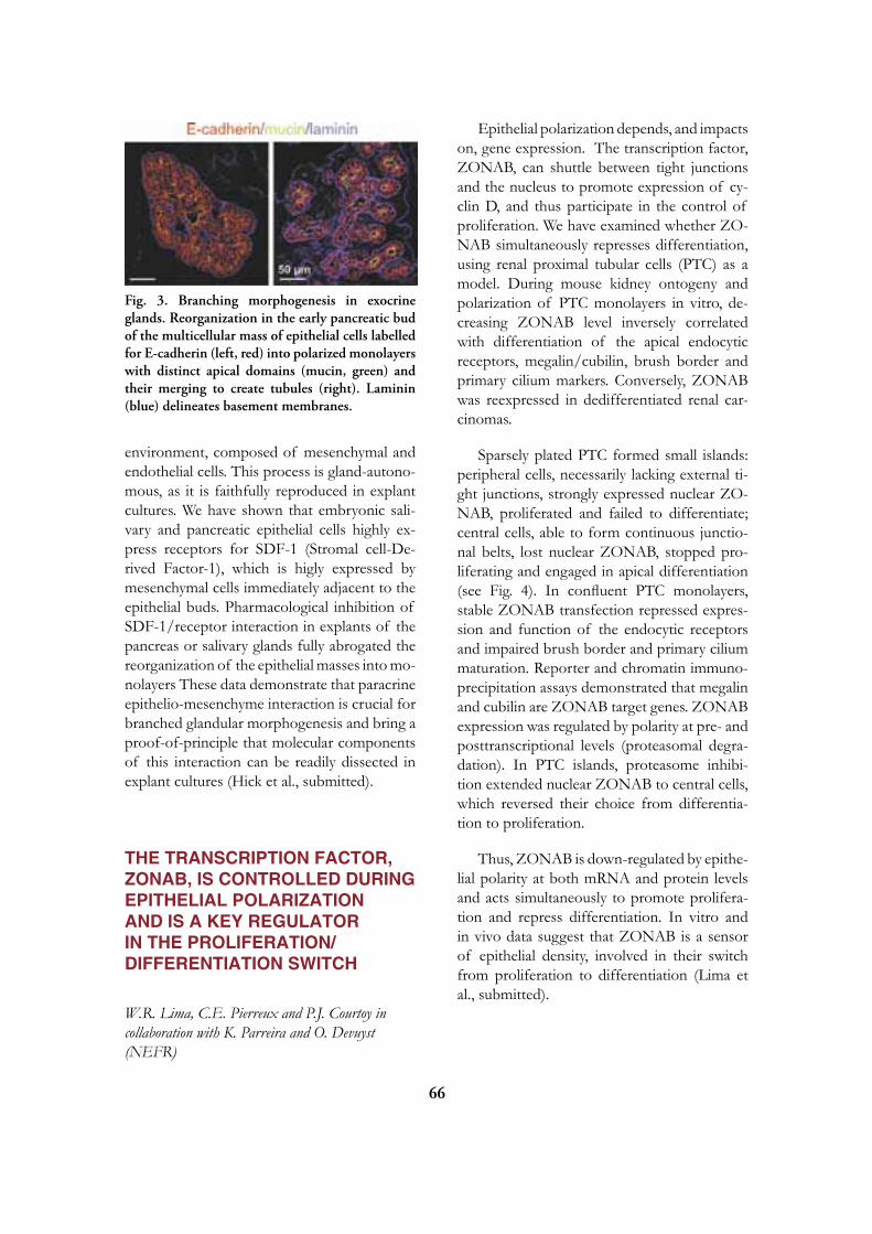
66
environment, composed of mesenchymal and endothelial cells. This process is gland-autono-mous, as it is faithfully reproduced in explant cultures. We have shown that embryonic sali-vary and pancreatic epithelial cells highly ex-press receptors for SDF-1 (Stromal cell-De-rived Factor-1), which is higly expressed by mesenchymal cells immediately adjacent to the epithelial buds. Pharmacological inhibition of SDF-1/receptor interaction in explants of the pancreas or salivary glands fully abrogated the reorganization of the epithelial masses into mo-nolayers These data demonstrate that paracrine epithelio-mesenchyme interaction is crucial for branched glandular morphogenesis and bring a proof-of-principle that molecular components of this interaction can be readily dissected in explant cultures (Hick et al., submitted).
THE TRANSCRIPTION FACTOR, zONAB, IS CONTROLLED DURING EPITHELIAL POLARIzATION AND IS A KEy REGULATOR IN THE PROLIFERATION/DIFFERENTIATION SWITCH
W.R. Lima, C.E. Pierreux and P.J. Courtoy in collaboration with K. Parreira and O. Devuyst (NEFR)
Epithelial polarization depends, and impacts on, gene expression. The transcription factor, ZONAB, can shuttle between tight junctions and the nucleus to promote expression of cy-clin D, and thus participate in the control of proliferation. We have examined whether ZO-NAB simultaneously represses differentiation, using renal proximal tubular cells (PTC) as a model. During mouse kidney ontogeny and polarization of PTC monolayers in vitro, de-creasing ZONAB level inversely correlated with differentiation of the apical endocytic receptors, megalin/cubilin, brush border and primary cilium markers. Conversely, ZONAB was reexpressed in dedifferentiated renal car-cinomas.
Sparsely plated PTC formed small islands: peripheral cells, necessarily lacking external ti-ght junctions, strongly expressed nuclear ZO-NAB, proliferated and failed to differentiate; central cells, able to form continuous junctio-nal belts, lost nuclear ZONAB, stopped pro-liferating and engaged in apical differentiation (see Fig. 4). In confluent PTC monolayers, stable ZONAB transfection repressed expres-sion and function of the endocytic receptors and impaired brush border and primary cilium maturation. Reporter and chromatin immuno-precipitation assays demonstrated that megalin and cubilin are ZONAB target genes. ZONAB expression was regulated by polarity at pre- and posttranscriptional levels (proteasomal degra-dation). In PTC islands, proteasome inhibi-tion extended nuclear ZONAB to central cells, which reversed their choice from differentia-tion to proliferation.
Thus, ZONAB is down-regulated by epithe-lial polarity at both mRNA and protein levels and acts simultaneously to promote prolifera-tion and repress differentiation. In vitro and in vivo data suggest that ZONAB is a sensor of epithelial density, involved in their switch from proliferation to differentiation (Lima et al., submitted).
Fig. 3. Branching morphogenesis in exocrine glands. Reorganization in the early pancreatic bud of the multicellular mass of epithelial cells labelled for E-cadherin (left, red) into polarized monolayers with distinct apical domains (mucin, green) and their merging to create tubules (right). Laminin (blue) delineates basement membranes.

67
PLASMA MEMBRANE LIPIDS FORM STRUCTURALLy AND KI-NETICALLy DISTINCT MICROME-TRIC DOMAINS
D. Tyteca, L. D’Auria, P. Van Der Smissen, T. Medts, S. Carpentier, P.J. Courtoy
Lipids are the most abundant constituents of biological membranes. For several decades, plasma membrane lipids were considered to form an homogenous two-dimensional barrier, acting as solvent for membrane proteins. Howe-ver, it is now well-accepted that some membra-ne lipids spontaneously cluster into discrete na-nometric “lipid rafts” (liquid-ordered domains), thereby creating small-scale lateral asymmetry. In addition, micrometric domains have been reported using fluorescent lipids incorporated into artificial liposomes, erythrocyte ghosts, and in living cells for non-natural lipids, but the na-tural occurrence of micrometric lipid domains in the plasma membrane of living cells is still questioned. Our group is addressing whether micrometric lipid domains naturally occur at the plasma membrane, using fluorescent lipid analogs, and how many distinct micrometric phases may coexist. We found that 10 different lipid analogs spontaneously formed microme-
tric domains, covering the bulk of the plasma membrane. These domains did not reflect en-docytosis nor structural surface features such as microvilli, as they could be readily observed on living erythrocytes as well. Domains with non-overlapping distributions and distinct late-ral diffusion (as assayed by fluorescence reco-very after photobleaching) could be evidenced. Perturbations of endogenous lipids selectively affected clustering of their fluorescent ana-logs, indicating that domains did not depend on substitution with the fluorophores tested. The relevance of the micrometric organiza-tion of membrane lipids to their endocytic rate and fate and to overall cell polarity is currently being investigated.
COLLABORATIONS ON MEMBRANE TRAFFICKING AND BRIEF REPORT ON THE CELL AND TISSUE IMAGING PLATFORM
P. Van Der Smissen, D. Tyteca, P.J. Courtoy
We have pursued our long-term commit-ment to promote collaborations by sharing ex-pertise in endocytosis and cellular imaging. For the recent years, we may cite our contribution to the study of endocytic trafficking of syn-decans and its role in signalling by its heparin-sulfate ligands (Zimmermann et al., 2005, Dev. Cell 9:377-88); of biogenesis of vacuolar H+-ATPase and the role of CFTR in kidney (Jouret et al., 2005, J. Am. Soc. Nephrol. 16:3235-46); of subcellular trafficking of the thrombopoie-tin receptor (Royer et al., 2005, J. Biol. Chem. 280:27251-61) and the amyloid precursor protein, APP (Feyt et al., 2005, J. Biol. Chem. 280:33220-7; 2007, Biochem. Biophys. Res. Commun. 357:1004-10); the ultrastructural analysis of differentiating hepatoblasts (Clot-man et al., 2005, Genes Dev. 19:1849-54) and the biogenesis of glycosomes in Trypanosoma brucei (Galland et al., 2007, Biochim. Biophys. Acta Mol. Cell. Res. 1773:521-35), or the mor-phological evidence by FRET of the interac-
Fig. 4. ZONAB is a key regulator in the switch between epithelial proliferation and differentiation. In colonies of kidney proximal tubular cells, ZONAB (immunolabelled in red) is absent in central differentiated cells (expressing megalin, in green, left) but is selectively detected in nuclei of peripheral proliferating cells (DNA synthesis is shown in blue at right).

68
tion between key players of CTL that is inter-rupted during anergy (Demotte et al., 2008, Immunity 28:414-24).
SELECTED PUBLICATIONS
1. Amyere M, Payrastre B, Krause U, Van Der Smissen P, Veithen A, Courtoy PJ. Consti-tutive macropinocytosis in oncogene-transformed fibroblasts depends on sequential permanent acti-vation of phosphoinositide 3-kinase and phospho-lipase C. Mol Biol Cell 2000;11:3453-67.
2. Coppens I, Courtoy PJ. The adaptative me-chanisms of Trypanosoma brucei for sterol ho-meostasis in its different life-cycle environments. Annu Rev Microbiol 2000;54:129-56.
3. Croizet-Berger K, Daumerie C, Couvreur M, Courtoy PJ, van den Hove MF. The en-docytic catalysts, Rab5a and Rab7, are tandem regulators of thyroid hormone production. Proc Natl Acad Sci USA 2002;99:8277-82.
4. Christensen EI, Devuyst O, Dom G, Niel-sen R, Van Der Smissen P, Verroust P, Leruth M, Guggino WB, Courtoy PJ. Loss of chloride channel ClC-5 impairs endocytosis by defective trafficking of megalin and cubilin in kid-ney proximal tubules. Proc Natl Acad Sci USA 2003;100:8472-7.
5. Mettlen M, Platek A, Van Der Smissen P, Carpentier S, Amyere M, Lanzetti L, de Diesbach Ph, Tyteca D, Courtoy PJ. Src triggers circular ruffling and macropinocytosis at the apical surface of polarized MDCK cells. Traffic 2006;7:589-603.
6. Nielsen R*, Courtoy PJ*, Jacobsen C, Dom G, Rezende Lima W, Jadot M, Will-now TE, Devuyst O, Christensen EI. En-docytosis provides a major alternative pathway for lysosomal biogenesis in kidney proximal tubular cells (*, equal first authors). Proc Natl Acad Sci USA 2007;104:5407-12.
7. Platek A, Vassilev VS, de Diesbach Ph, Ty-teca D, Mettlen M, Courtoy PJ. Constitutive diffuse activation of phosphoinositide 3-kinase at the plasma membrane by v-Src suppresses the chemotactic response to PDGF by abrogating the polarity of PDGF receptor signalling. Exp Cell Res 2007;313:1090-105.
8. Jouret F, Bernard A, Hermans C, Dom G, Terryn S, Leal T, Lebecque P, Cassiman J-J, Scholte BJ, de Jonge HR, Courtoy PJ, Devuyst O. Cystic fibrosis is associated with a defect in apical receptor-mediated endocytosis in mouse and human kidney. J Am Soc Neph-rol 2007;18:707-18.
9. van den Hove MF, Croizet-Berger K, Ty-teca D, Selvais C, de Diesbach Ph, Cour-toy PJ. TSH activates GDP/GTP exchange on the rate-limiting endocytic catalyst, Rab5a, in human thyrocytes in vivo and in vitro. J Clin Endocrinol Metab 2007; 92:2803-10.
10. de Diesbach Ph, Medts Th, Carpentier S, D’Auria L, Van Der Smissen P, Platek A, Mettlen M, Caplanusi A, van den Hove MF, Tyteca D*, Courtoy PJ*. Differential membrane domain recruitment of Src specifies its downstream signalling (*, equal senior authors). Exp Cell Res 2008; 314:1465-79.
Pierre Courtoyde Duve InstituteAv. Hippocrate 74-75B - 1200 Brussels
[T] +32 02 764 75 69[F] +32 02 764 75 43[E] [email protected][W] http://www.deduveinstitute.be/endocyto-sis_epithelial.php

69
Etienne MARBAIX, MemberPatrick HENRIET, Associate Member
Pierre J. COURTOY, Member Hervé EMONARD, Guest Investigator Christine GALANT, Assistant Member (part-time)Christophe CHANTRAIN, Assistant Member (part-time)Héloïse GAIDE CHEVRONNAY, Graduate Student Chrystel PRETTO, Graduate Student Charlotte SELVAIS, Graduate StudentDenis DELVAUX, Technician (until April)Céline FOREZ, Technician (from May)Pascale LEMOINE, Technician (part-time) Yves MARCHAND, Secretary
EXTRACELLULAR MATRIX BREAKDOWN
The extracellular matrix (ECM) plays a central role in the structural and functional organization of tis-sues and organs. ECM constituents, in particular fibrillar collagens, are the most abundant proteins of the human body. Physiological and pathological breakdown of ECM is predominantly achieved by a family of enzymes called matrix metalloproteinases (MMPs; see Fig. 1). Our laboratory was the first to demonstrate that menstrual bleeding is due to a dramatic change in the focal expression and/or activation of MMPs (1). This seminal observation led us to : (i) use this system as a human model to study the regulation of MMPs, in particular cellular interactions that integrate overall hormonal impregnation with local environmental changes; and (ii) explore whether this basic knowledge can lead to a better understanding and a rational treatment of abnormal uterine bleeding, a major health problem (2). We also investigate the control by individual cells of local MMP activity, which can be either increased by recruitment and retention to the plasma membrane (5), or down-regulated by receptor-mediated endocytosis and degradation.
DIFFERENTIAL CONTROL OF ENDOMETRIAL MMPS AND CyTO-KINES By OVARIAN STEROIDS, IN VIVO AND IN EXPLANTS
H. Gaide Chevronnay, C. Pretto, P.J. Courtoy, E. Marbaix, P. Henriet
Both endocrine and paracrine factors parti-cipate in controlling the expression and activity of MMPs involved in menstrual breakdown of the human endometrium. Several genes encoding MMPs and cytokines show maximal
endometrial mRNA concentrations around menstruation. However, their expression pro-files substantially diverge during the other pha-ses of the cycle. Although these variations are in part explained by differential repression of transcription by estradiol and/or progesterone receptors, there is increasing evidence of the major contribution of specific local regulators which finely adjust in time, space and ampli-tude the effect of ovarian steroids on the ex-pression of genes required for menstrual ECM breakdown (3, 4).

70
proMMPs
steroid hormones,cytokines,
growth factors,…
1. expression
TIMPsα2-macroglobulin
MMPsother proteinases active MMPs
5. cell membranefocal recruitment
and retention
3. activation
4. inhibition
6. endocytosis and degradation
Zn2+Ca2+
Zn2+Ca2+
lysosomes
Zn 2+
Zn2+Ca2+
+
?
inactive MMP:TIMP complexes
Zn 2+
Ca 2+
preproMMPs
Zn2+Ca2+
2. secretion
Figure 1. Regulation of soluble MMPs activity in the human endometrium: current model. MMPs, the major actors of extracellular proteolysis, share a common intramolecular control due to masking by a N-terminal prodomain (blue, here shown with the signal peptide in grey) of the Zn2+-dependent catalytic site (green) within the catalytic domain (orange). All but MMP-7 and -26 (matrilysins, the “mini MMPs”), contain an additional, variable C-terminal hemopexin-like domain stabilized by calcium (mauve) responsible for substrate specificity, linked by a hinge domain (black). The overall activity of MMPs can be controlled at 6 different levels: (1) expression, (2) secretion (regulated in a limited number of cell types such as neutrophils), (3) zymogen activation upon prodomain excision, (4) inhibition of active forms by physiological inhibitors such as TIMPs (represented with their tertiary structure) and a2-macroglobulin, (5) secondary membrane recruitment increasing pericellular activity, and (6) down-regulation by endocytosis.
In the human endometrium, MMPs activity is tightly regulated to remodel the extracellular matrix both for blastocyst implantation and, in its absence, for menstrual breakdown of an irreversibly specialized tissue. In particular : (1) ovarian steroids and their intracellular receptors as well as cytokines, growth factors and downstream signaling pathways interact to form an integrated system that differentially controls the focal expression of endometrial MMPs and TIMPs. (2) Neutrophils are numerous at menstruation and could contribute to an abundant secretion of MMPs. (3) MMPs can be activated by other MMPs, by plasmin, itself activated during menstruation, or by as yet unidentified proteinases. (4) TIMPs are particularly abundant in the human endometrium; like MMPs, the level of TIMPs is regulated by ovarian steroids and cytokines. (5) MMP-7 binds to membrane receptors in cholesterol-rich domains, a mechanism which enhances pericellular MMP activity. (6) Endometrial LRP-1 (brown) binds and internalizes MMP-2 and MMP-2: TIMP-2 complexes, leading to lysosomal degradation. Our research has unraveled, and is focused on, levels (1), (3), (4), (5) and (6).
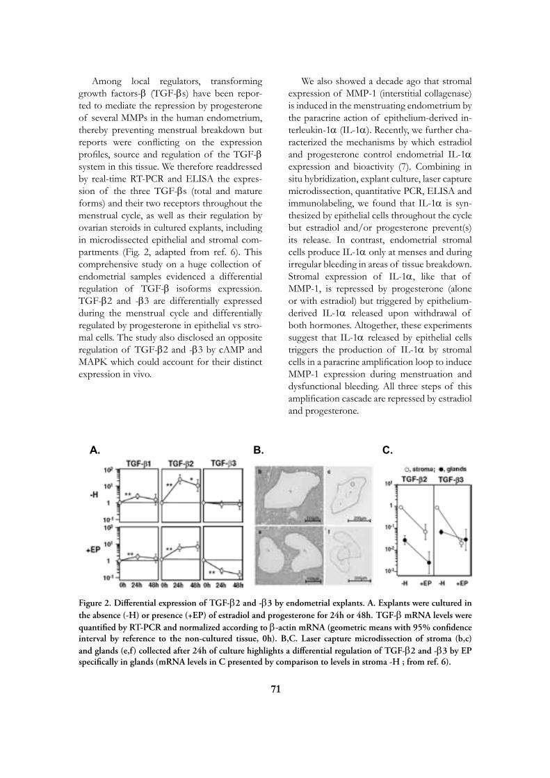
71
Among local regulators, transforming growth factors-b (TGF-bs) have been repor-ted to mediate the repression by progesterone of several MMPs in the human endometrium, thereby preventing menstrual breakdown but reports were conflicting on the expression profiles, source and regulation of the TGF-b system in this tissue. We therefore readdressed by real-time RT-PCR and ELISA the expres-sion of the three TGF-bs (total and mature forms) and their two receptors throughout the menstrual cycle, as well as their regulation by ovarian steroids in cultured explants, including in microdissected epithelial and stromal com-partments (Fig. 2, adapted from ref. 6). This comprehensive study on a huge collection of endometrial samples evidenced a differential regulation of TGF-b isoforms expression. TGF-b2 and -b3 are differentially expressed during the menstrual cycle and differentially regulated by progesterone in epithelial vs stro-mal cells. The study also disclosed an opposite regulation of TGF-b2 and -b3 by cAMP and MAPK which could account for their distinct expression in vivo.
We also showed a decade ago that stromal expression of MMP-1 (interstitial collagenase) is induced in the menstruating endometrium by the paracrine action of epithelium-derived in-terleukin-1a (IL-1a). Recently, we further cha-racterized the mechanisms by which estradiol and progesterone control endometrial IL-1a expression and bioactivity (7). Combining in situ hybridization, explant culture, laser capture microdissection, quantitative PCR, ELISA and immunolabeling, we found that IL-1a is syn-thesized by epithelial cells throughout the cycle but estradiol and/or progesterone prevent(s) its release. In contrast, endometrial stromal cells produce IL-1a only at menses and during irregular bleeding in areas of tissue breakdown. Stromal expression of IL-1a, like that of MMP-1, is repressed by progesterone (alone or with estradiol) but triggered by epithelium-derived IL-1a released upon withdrawal of both hormones. Altogether, these experiments suggest that IL-1a released by epithelial cells triggers the production of IL-1a by stromal cells in a paracrine amplification loop to induce MMP-1 expression during menstruation and dysfunctional bleeding. All three steps of this amplification cascade are repressed by estradiol and progesterone.
A. B. C.
Figure 2. Differential expression of TGF-b2 and -b3 by endometrial explants. A. Explants were cultured in the absence (-H) or presence (+EP) of estradiol and progesterone for 24h or 48h. TGF-b mRNA levels were quantified by RT-PCR and normalized according to b-actin mRNA (geometric means with 95% confidence interval by reference to the non-cultured tissue, 0h). B,C. Laser capture microdissection of stroma (b,c) and glands (e,f) collected after 24h of culture highlights a differential regulation of TGF-b2 and -b3 by EP specifically in glands (mRNA levels in C presented by comparison to levels in stroma -H ; from ref. 6).

72
METALLOPROTEINASE-DEPENDENT SHEDDING OF LRP-1 ECTODOMAIN DECREASES ENDOCyTIC CLEARANCE OF ENDOMETRIAL MMP-2 AND -9 AT MENSTRUATION
C. Selvais, P.J. Courtoy, P. Henriet, E. Marbaix, H. Emonard (in collaboration with CNRS, Reims, France)
In contrast to menstrual-restricted MMPs, e.g. interstitial collagenase (MMP-1), gelatina-ses A (MMP-2) and B (MMP-9) mRNAs are abundant throughout the cycle without detec-table tissue degradation at proliferative and secretory phases, implying a tight post-trans-lational control of both gelatinases. The re-ceptor-associated protein (RAP), used as LRP antagonist, strongly increased (pro)gelatinases accumulation in medium conditioned by endo-metrial explants, indicating a role for LRP-1 in their clearance. Although LRP-1 mRNA remai-ned constant throughout the cycle, the protein ectodomain vanished at menses. LRP-1 immu-nolabeling selectively disappeared in areas of extracellular matrix breakdown in menstrual samples. It also disappeared from explants cul-tured without estrogen and progesterone due to ectodomain shedding in the medium. The shedding was inhibited by metalloproteinase inhibitors, including a disintegrin and metallo-proteinase (ADAM) inhibitor, and by tissue in-hibitors of MMPs (TIMP)-3 and -2, but barely by TIMP-1, pointing to ADAM-12 as the puta-tive sheddase. In good agreement, ADAM-12 mRNA expression was repressed by estradiol and progesterone. In conclusion, the efficient LRP-1-mediated clearance of gelatinases acti-vity in non-bleeding endometrium is abrogated upon hormone withdrawal, due to shedding of LRP-1 ectodomain by a metalloproteinase, presumably ADAM-12, itself regulated by es-tradiol and progesterone (10).
ROLE OF MATRIX METALLOPROTEINASES IN ABNORMAL ENDOMETRIAL BLEEDING
C. Galant, J.-L. Brun, M.-L. Alvarez, A. Béliard, P.J. Courtoy, P. Henriet, E. Marbaix (in colla-boration with the Hôpital universitaire Pellegrin, Bordeaux, France, and with the University of Liège, Belgium)
Looking for a role of MMPs in triggering the occurrence of abnormal bleeding in addi-tion to normal menstrual bleeding, we investi-gated patients showing irregular dysfunctional bleeding, a spontaneous condition that cannot be accounted for by primary organic lesions of the uterus (2). As in patients on long-term pro-gesterone contraception, irregular dysfunctio-nal bleeding is associated with menstrual-like stromal breakdown, particularly in foci contai-ning low levels of ovarian steroids receptors, and correlates with increased expression and activity of several MMPs, together with de-creased production of TIMP-1 (Fig. 3). Neu-trophils were further recruited to the same foci, released abundant proMMP-8 and -9, and thereby increased ECM proteolysis.
Menorrhagia, or excessive menstrual blee-ding, is also a frequent cause of medical consul-tation and surgical treatment. Thermal ablation of the endometrium is currently largely used
Figure 3. Focal expression of MMPs cor-relates with ECM breakdown. A. MMP-3 im-munostaining. B. Silver staining. Arrows show that MMP-3 expression is restricted to the area showing disappearance of collagen-rich argy-rophilic fibers of the endometrial stroma du-ring menstruation
A B
73
as a less aggressive but efficient surgical proce-dure, despite rapid regeneration of the mucosa in most patients. We investigated whether the level of MMPs activity could be involved in the amount of menstrual bleeding in such patients and found that menstrual endometria regene-rated after thermal ablation showed smaller areas of collagen fibres lysis and contained more TIMP-1 and TIMP-2 compared with en-dometria from non-treated women with exces-sive menstrual bleeding, providing a first mole-cular explanation for the decreased amount of bleeding (8).
In collaboration with the laboratory of Dr. J.M. Foidart (ULg), we have developed a new experimental model of endometrial xenografts in immunodeficient mice (9). The model al-lowed us to investigate the alterations of endo-metrial ECM remodelling upon levonorgestrel treatment and will be used to directly address the role of MMPs in physiological and abnor-mal endometrial bleeding, endometrial angio-genesis and vessel maturation, as well as in tis-sue regeneration after menstrual shedding.
SELECTED PUBLICATIONS
1. Marbaix E, Kokorine I, Moulin P, Don-nez J, Eeckhout Y, Courtoy PJ. Menstrual breakdown of human endometrium can be mimic-ked in vitro and is selectively and reversibly bloc-ked by inhibitors of matrix metalloproteinases. Proc Natl Acad Sci USA 1996;93:9120-5.
2. Galant C, Berlière M, Dubois D, Verougs-traete J-C, Charles A, Lemoine P, Koko-rine I, Eeckhout Y, Courtoy PJ, Marbaix E. Focal expression and final activity of matrix metalloproteinases may explain irregular dys-functional endometrial bleeding. Am J Pathol 2004;165:83-94.
3. Cornet PB, Galant C, Eeckhout Y, Cour-toy PJ, Marbaix E, Henriet P. Regulation of matrix metalloproteinase-9/gelatinase B ex-pression and activation by ovarian steroids and LEFTYA/ EBAF in the human endometrium. J Clin Endocrinol Metab 2005;90:1001-11.
4. Vassilev V, Pretto CM, Cornet PB, Del-vaux D, Eeckhout Y, Courtoy PJ, Marbaix E, Henriet P. Response of matrix metallopro-teinases and tissue inhibitors of metalloproteina-ses messenger ribonucleic acids to ovarian steroids in human endometrial explants mimics their gene and phase-specific differential control in vivo. J Clin Endocrinol Metab 2005;90:5848-57.
5. Berton A, Selvais C, Lemoine P, Henriet P, Courtoy PJ, Marbaix E, Emonard H. Binding of matrilysin-1 to human epithelial cells promotes its activity. Cell Mol Life Sci 2007;64:610-20.
6. Gaide Chevronnay HP, Cornet PB, Del-vaux D, Lemoine P, Courtoy PJ, Henriet P, Marbaix E. Opposite regulation of transfor-ming growth factors-beta2 and -beta3 expression in the human endometrium. Endocrinology 2008;149:1015-25.
7. Pretto CM, Gaide Chevronnay HP, Cor-net PB, Galant C, Delvaux D, Courtoy PJ, Marbaix E, Henriet P. Production of interleu-kin-1alpha by human endometrial stromal cells is triggered during menses and dysfunctional bleeding and is induced in culture by epithelial interleukin-1alpha released upon ovarian steroids withdrawal. J Clin Endocrinol Metab 2008;93:4126-34.
8. Brun JL, Galant C, Delvaux D, Lemoine P, Henriet P, Courtoy PJ, Marbaix E. Mens-trual activity of matrix metalloproteinases is de-creased in endometrium regenerating after thermal ablation. Hum Reprod 2009;24:333-40.

74
9. Alvarez Gonzalez ML, Galant C, Fran-kenne F, Nisolle M, Labied S, Foidart JM, Marbaix E*, Béliard A*. Development of an animal experimental model to study the effects of levonorgestrel on the human endometrium. Hum Reprod 2009;24:697-704 (*, equal senior authors).
10. Selvais C, Gaide Chevronnay HP, Le-moine P, Dedieu S, Henriet P, Courtoy PJ, Marbaix E, Emonard H. Metalloprotei-nase-dependent shedding of LRP-1 ectodomain decreases endocytic clearance of endometrial ma-trix metalloproteinases-2 and -9 at menstruation. Endocrinology 2009; in press.
Etienne Marbaixde Duve InstituteAv. Hippocrate 74-75B - 1200 Brussels[T] +32 02 764 67 55 / 75 41[F] +32 02 764 75 43[E] [email protected][W] http://www.deduveinstitute.be/remodelling_of_endometrial.php

75
Jean-Baptiste DEMOULIN, Associate Member
Nicolas DIF, Postdoctoral Fellow Sandrine MEDVES, Postdoctoral FellowAmélie VELGHE, Technician (from March 2009)Catherine MARBEHANT, Technician (until November 2008)Sandrine LENGLEZ, Technician (until December 2008)Federica TOFFALINI, Graduate StudentAhmed ESSAGHIR, Graduate StudentCarmen MONTANO, Graduate StudentJean-François CLASSEN, Student (until June 2008)Alexandra COOMANS de BRACHENE, Student
GROWTH FACTOR RECEPTORS From signal transduction to human diseases
Growth factors are secreted proteins that stimulate cell proliferation via transmembrane receptors. Our re-search interests are focused on the mechanisms of activation of these receptors. We are particularly interested in receptor-tyrosine kinases that are binding to platelet-derived growth factors (PDGF) and fibroblasts growth factors (FGF). These proteins play an important role in the development of the embryo and in wound healing, as well as in cancer and in fibrosis.
Figure 1. PDGF and FGF receptors and ligands
76
REARRANGEMENTS OF THE RECEPTOR TyROSINE KINASE GENES ASSOCIATED WITH LEUKEMIA
F. Toffalini, S. Medves, C. Montano, J.B. Demoulin.
Although PDGF receptors are expressed on platelets and macrophages, PDGF receptor-deficient mice show no primary hematopoietic or immune defect. In vitro, PDGF is a poor mi-togen for hematopoietic cells. However, modi-fications of PDGF receptor genes, as a result of chromosomal translocation or deletion, are found in chronic myeloid neoplasms associa-ted with eosinophilia (formerly classified as chronic eosinophilic leukemia, atypical chronic myeloid leukemia or chronic myelomonocytic leukemia). In all cases, the rearranged gene pro-duces a hybrid protein comprising the PDGF receptor tyrosine kinase domain and an oligo-merisation domain. Similar hybrid oncogenes derive from FGF receptors.
TEL-PDGFRb (TPb, also called ETV6-PDGFRB) is a hybrid protein produced by the t(5;12) translocation. FIP1L1-PDGFRa (FPa) results from a deletion on chromosome 4q12 in patients with chronic eosinophilic leukemia. These oncogenes were studied in Ba/F3 cells, a mouse hematopoietic cell line that is easy to culture and transfect. In contrast to wild-type PDGF receptors a and b, which are quickly degraded upon activation, we observed that TPb and FPa escaped down-regulation resul-ting in stabilization of the proteins. High stabi-lity of these hybrid oncogenes was confirmed in leukocytes from two patients. Similar data were obtained in cells expressing ZNF198-FGFR1, another fusion protein associated with the 8p11 myeloproliferative syndrome. Cbl-mediated monoubiquitination of receptor lysines targets them for lysosomal degradation. Ubiquitination of TPb and FPa was much re-duced compared to wild-type receptors, despi-te marked Cbl phosphorylation in cells expres-sing hybrid receptors. Deletion of the pointed (PNT) domain, impairing TPb polymerization,
strongly destabilized the protein. In conclu-sion, chimeric receptor tyrosine kinases escape efficient ubiquitination and down-regulation through lysosomes and proteasomes (1).
In order to develop a model that is more relevant for the human disease, we introduced TPb and FPa in human CD34+ cells, which were purified from cord blood and are enriched in hematopoietic stem cells. These cells are able to differentiate in vitro into various blood cell types, depending on the cytokine cocktail that is added in the culture medium. We observed that TPb and FPa induce the proliferation and differentiation of cytokine-independent cells. We are now analyzing this process in detail.
It is particularly important to identify PDGF receptor alterations in cancer patients, as they can benefit from tyrosine kinase inhibitor the-rapy. Imatinib mesylate, for instance, is very efficient in patients with leukemia that present a PDGF receptor translocation. In collabora-tion with the hematology unit of the Saint-Luc university hospital, we identified a novel hybrid PDGF receptor in a leukemia patient harbo-ring a t(5;9) translocation. We are now looking for other mutations in tyrosine kinase genes.
ACTIVATION OF PDGF RECEPTORS IN SySTEMIC SCLEROSIS
J.-F. Classen, S. Lenglez, J.B. Demoulin in collaboration with B. Lauwerys and F. Houssiau (Rheumatology Unit).
Systemic sclerosis (also called scleroderma) is a severe connective tissue disease of unk-nown etiology, characterized by fibrosis of the skin and multiple internal organs. A recent report suggested that the disease is driven by stimulatory autoantibodies to the platelet-deri-ved growth factor receptors (PDGFR), which stimulate the production of reactive oxygen species (ROS) and collagen by fibroblasts (Ba-
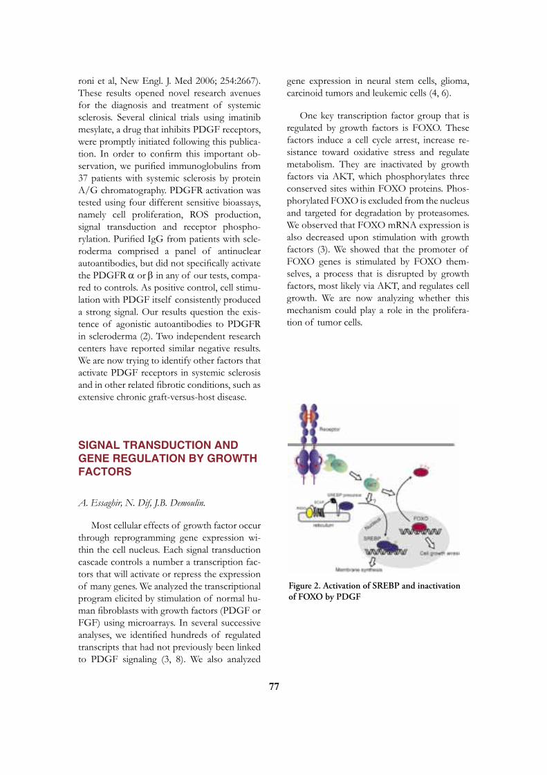
77
roni et al, New Engl. J. Med 2006; 254:2667). These results opened novel research avenues for the diagnosis and treatment of systemic sclerosis. Several clinical trials using imatinib mesylate, a drug that inhibits PDGF receptors, were promptly initiated following this publica-tion. In order to confirm this important ob-servation, we purified immunoglobulins from 37 patients with systemic sclerosis by protein A/G chromatography. PDGFR activation was tested using four different sensitive bioassays, namely cell proliferation, ROS production, signal transduction and receptor phospho-rylation. Purified IgG from patients with scle-roderma comprised a panel of antinuclear autoantibodies, but did not specifically activate the PDGFR a or b in any of our tests, compa-red to controls. As positive control, cell stimu-lation with PDGF itself consistently produced a strong signal. Our results question the exis-tence of agonistic autoantibodies to PDGFR in scleroderma (2). Two independent research centers have reported similar negative results. We are now trying to identify other factors that activate PDGF receptors in systemic sclerosis and in other related fibrotic conditions, such as extensive chronic graft-versus-host disease.
SIGNAL TRANSDUCTION AND GENE REGULATION By GROWTH FACTORS
A. Essaghir, N. Dif, J.B. Demoulin.
Most cellular effects of growth factor occur through reprogramming gene expression wi-thin the cell nucleus. Each signal transduction cascade controls a number a transcription fac-tors that will activate or repress the expression of many genes. We analyzed the transcriptional program elicited by stimulation of normal hu-man fibroblasts with growth factors (PDGF or FGF) using microarrays. In several successive analyses, we identified hundreds of regulated transcripts that had not previously been linked to PDGF signaling (3, 8). We also analyzed
gene expression in neural stem cells, glioma, carcinoid tumors and leukemic cells (4, 6).
One key transcription factor group that is regulated by growth factors is FOXO. These factors induce a cell cycle arrest, increase re-sistance toward oxidative stress and regulate metabolism. They are inactivated by growth factors via AKT, which phosphorylates three conserved sites within FOXO proteins. Phos-phorylated FOXO is excluded from the nucleus and targeted for degradation by proteasomes. We observed that FOXO mRNA expression is also decreased upon stimulation with growth factors (3). We showed that the promoter of FOXO genes is stimulated by FOXO them-selves, a process that is disrupted by growth factors, most likely via AKT, and regulates cell growth. We are now analyzing whether this mechanism could play a role in the prolifera-tion of tumor cells.
Figure 2. Activation of SREBP and inactivation of FOXO by PDGF

78
GROWTH FACTORS STIMULATE LIPID SyNTHESIS By ACTIVATING THE SREBP TRANSCRIPTION FACTORS
N. Dif, A. Coomans de Brachène, A. Essaghir, J.B. Demoulin.
In our microarray analysis, a cluster of genes involved in fatty acid and cholesterol biosynthesis, including stearoyl-CoA desatu-rase (SCD), fatty acid synthase and hydroxy-methylglutaryl-CoA synthase (HMGCS), was up-regulated by PDGF after 24 h of treatment. Their expression correlated with an increase in membrane lipid biosynthesis. All these genes are known to be controlled by sterol regulatory element-binding proteins (SREBP). PDGF in-creased the amount of mature SREBP-1, and regulated the promoters of SCD and HMGCS in a SREBP-dependent manner. In line with these results, blocking SREBP processing by addition of 25-hydroxycholesterol blunted the effects of PDGF on lipogenic enzymes and PDGF-driven proliferation. SREBP activation was dependent on the phosphatidylinositol 3-kinase (PI3K) pathway, as judged from the ef-fects of the inhibitor LY294002 and mutation of the PDGF receptor b tyrosine residues that bind the regulatory PI3K subunit p85. In con-clusion, our results suggest that PDGF indu-ces membrane lipid synthesis via PI3K and the activation of SREBP (8). The role of SREBP in PDGF and tumor development will be fur-ther analyzed. We went on by identifying novel target genes for the SREBP transcription fac-tors. We found that these transcription factors regulate p55g, a subunit of the PI3K complex and heme oxygenase, which plays an impor-tant role in stress responses (5). These results expand the list of genes regulated by SREBP to targets that are not directly involved in lipid metabolism. We are now trying to understand more precisely the role of these SREBP target genes in growth factor responses.
SELECTED PUBLICATIONS
1. Toffalini F, Kallin A, Vandenberghe P, Pierre P, Michaux L, Cools J and Demou-lin JB. The fusion proteins TEL-PDGFRb and FIP1L1-PDGFRa escape ubiquitination and degradation. Haematologica 2009; In press.
2. Classen JF, Henrohn D, Rorsman F, Len-nartsson J, Lauwerys BR, Wikström G, Rorsman C, Lenglez S, Franck-Larsson K, Tomasi JP, Kämpe O, Vanthuyne M, Houssiau FA, Demoulin JB. Lack of eviden-ce of stimulatory autoantibodies to platelet-deri-ved growth factor receptor in patients with systemic sclerosis. Arthritis Rheum 2009;60:1137-44.
3. Essaghir A, Dif N, Marbehant CY, Cof-fer PJ, Demoulin JB. The Transcription of FOXO Genes Is Stimulated by FOXO3 and Repressed by Growth Factors. J Biol Chem 2009;284:10334-42.
4. Leja J, Essaghir A, Essand M, Wester K, Oberg K, Tötterman TH, Lloyd R, Vas-matzis G, Demoulin JB, Giandomenico V. Novel markers for enterochromaffin cells and gastrointestinal neuroendocrine carcinomas. Mod Pathol 2009;22:261-72.
5. Kallin A, Johannessen LE, Cani PD, Mar-behant CY, Essaghir A, Foufelle F, Ferre P, Heldin CH, Delzenne NM and Demou-lin JB. SREBP1 regulates the expression of heme oxygenase 1 and the phosphatidylinositol-3 kinase regulatory subunit p55gamma. J Lipid Res 2007;48:1628-36.
6. Demoulin JB, Mia Enarsson M, Larsson J, Essaghir A, Adams B, Heldin CH and K Forsberg-Nilsson. The gene expression profile of PDGF-treated neural stem cells corres-ponds to partially differentiated neurons and glia. Growth Factors 2006;24:184-96.

79
7. Chiara F, Heldin CH and Demoulin JB. Autoinhibition of the PDGF b-receptor tyrosine kinase by its C-terminal tail. J Biol Chem 2004;279:19732-8.
8. Demoulin JB, Ericsson J, Kallin A, Ror-sman C, Rönnstrand L and Heldin CH. Platelet-derived growth factor stimulates mem-brane lipid synthesis through activation of phosphatidylinositol 3-kinase and sterol regula-tory element-binding proteins. J Biol Chem 2004;279:35392-402.
9. Demoulin JB, Seo J, Ekman S, Grapen-giesser E, Hellman U, Ronnstrand L, Hel-din CH. Ligand-induced recruitment of Na+/H+-exchanger regulatory factor to the PDGF (platelet-derived growth factor) receptor regula-tes actin cytoskeleton reorganization by PDGF. Biochem J 2003;376:505-10.
Jean-Baptiste Demoulinde Duve InstituteUCL 74.30Av. Hippocrate 74-75B - 1200 Brussels
[T] +32 02 764 65 29[F] +32 02 764 74 30[E] [email protected][W] http://www.deduveinstitute.be/growth_fac-tor_receptors.php
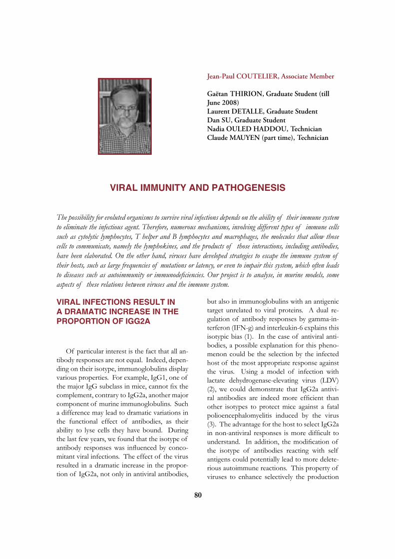
80
Jean-Paul COUTELIER, Associate Member
Gaëtan THIRION, Graduate Student (till June 2008) Laurent DETALLE, Graduate Student Dan SU, Graduate Student Nadia OULED HADDOU, Technician Claude MAUYEN (part time), Technician
VIRAL IMMUNITy AND PATHOGENESIS
The possibility for evoluted organisms to survive viral infections depends on the ability of their immune system to eliminate the infectious agent. Therefore, numerous mechanisms, involving different types of immune cells such as cytolytic lymphocytes, T helper and B lymphocytes and macrophages, the molecules that allow those cells to communicate, namely the lymphokines, and the products of those interactions, including antibodies, have been elaborated. On the other hand, viruses have developed strategies to escape the immune system of their hosts, such as large frequencies of mutations or latency, or even to impair this system, which often leads to diseases such as autoimmunity or immunodeficiencies. Our project is to analyse, in murine models, some aspects of these relations between viruses and the immune system.
VIRAL INFECTIONS RESULT IN A DRAMATIC INCREASE IN THE PROPORTION OF IGG2A
Of particular interest is the fact that all an- tibody responses are not equal. Indeed, depen- ding on their isotype, immunoglobulins display various properties. For example, IgG1, one of the major IgG subclass in mice, cannot fix the complement, contrary to IgG2a, another major component of murine immunoglobulins. Such a difference may lead to dramatic variations in the functional effect of antibodies, as their ability to lyse cells they have bound. During the last few years, we found that the isotype of antibody responses was influenced by conco-mitant viral infections. The effect of the virus resulted in a dramatic increase in the propor-tion of IgG2a, not only in antiviral antibodies,
but also in immunoglobulins with an antigenic target unrelated to viral proteins. A dual re-gulation of antibody responses by gamma-in-terferon (IFN-g) and interleukin-6 explains this isotypic bias (1). In the case of antiviral anti-bodies, a possible explanation for this pheno-menon could be the selection by the infected host of the most appropriate response against the virus. Using a model of infection with lactate dehydrogenase-elevating virus (LDV) (2), we could demonstrate that IgG2a antivi-ral antibodies are indeed more efficient than other isotypes to protect mice against a fatal polioencephalomyelitis induced by the virus (3). The advantage for the host to select IgG2a in non-antiviral responses is more difficult to understand. In addition, the modification of the isotype of antibodies reacting with self antigens could potentially lead to more delete- rious autoimmune reactions. This property of viruses to enhance selectively the production

81
of one immunoglobulin isotype could depend on the preferential activation of a subset of T helper lymphocytes. Indeed, different subpo- pulations of those cells, called Th1 and Th2, respectively, are distinguished in particular by their capability of producing selectively IFN-g or interleukin-4, which can selectively trigger B lymphocytes to produce IgG2a or IgG1, res- pectively.
ACTIVATION OF NATURAL KILLER CELLS
Many of the influences that viruses may have on diverse immune responses can be ex- plained by the production of pro-inflammato- ry cytokines, including IFN-g. Therefore, our analysis of the relationship between viruses and the immune system has focused on the activa- tion, by LDV, of cells from the innate immune system that are able to secrete this cytokine, na- mely the natural killer (NK) cells. Within a few days after infection, a strong and transient NK cell activation, characterized by accumulation of this cell population in the spleen, by enhan-ced IFN-g message expression and production, as well as by cytolysis of target cell lines was observed 4). Two pathways of IFN-g produc-tion have been observed that both involve NK cells. The first pathway, found in normal mice, is independent from type I IFN and from in-terleukin-12. The second pathway involves in-terleukin-12, but is suppressed by type I IFN. Because NK cells and IFN-g may participate in the defense against viral infection, we analyzed their possible role in the control of LDV titers, with a new agglutination assay. Our results in-dicate that neither the cytolytic activity of NK cells nor the IFN-g secretion affect the early and rapid viral replication that follows LDV inoculation.
Similarly, infection with mouse hepatitis vi-rus (MHV) is followed by NK cell activation and leads to IFN-g production by those acti-
vated cells. In contrast to LDV, MHV replica-tion is controlled by this cytokine and animals unresponsive to this molecule quickly die after infection. The protective effect of IFN-g ap-pears to target infected cells rather than lym-phocytes.
Interestingly, NK cell activation results in an increased expression of CD66a (CEACAM-1), an adhesion molecule that serves also as a receptor for MHV. However, this enhanced ex-pression, that is also found on immature NK cells, results from NK cell stimulation with IL-12 and IL-18, but not with LDV (5). Therefore, some, but not all subpopulations of activated NK cells might be susceptible to MHV infec-tion.
ACTIVATION OF MACROPHAGES AND AUTOIMMUNE DISEASES
Activation of cells of the innate immune system includes also macrophages and leads to an enhanced phagocytic activity, with potential detrimental consequences for ongoing autoim- mune diseases. Our analysis has been focused on autoantibody-mediated blood autoimmune diseases. A new experimental model of anti- platelet response was developed in the mouse (6). Immunization of CBA/Ht mice with rat platelets was followed by a transient thrombo- cytopenia and production of autoantibodies that react with epitope(s) shared by rat and mouse platelets. In contrast, BALB/C mice similarly immunized with rat platelets did not develop thrombocytopenia. We have analysed whether a viral infection could modulate such an autoantibody-mediated autoimmune disease. In mice treated with anti-platelet antibodies at a dose insufficient to induce clinical disease by themselves, infection with LDV or mouse hepatitis virus was followed by severe throm-bocytopenia (7). Similarly, administration of anti-erythrocyte monoclonal autoantibody to mice resulted in the development of a transient

82
hemolytic anemia that was dramatically enhan-ced by a simultaneous infection with LDV, lea-ding to the death of most animals. This viral infection induced an increase in the ability of macrophages to phagocytose in vitro autoanti-body-coated red cells, and an enhancement of erythrophagocytosis in the liver (8). Treatment of thrombopenic or anemic mice with clodro-nate-containing liposomes and with total IgG indicated that opsonized platelets and erythro-cytes were cleared by macrophages. Adminis-tration of clodronate-containing liposomes decreased also the in vitro phagocytosis of autoantibody-coated red cells by macrophages from LDV-infected animals. The increase of thrombocytopenia triggered by LDV after ad- ministration of anti-platelet antibodies was lar- gely suppressed in animals deficient for IFN-g receptor. Together, these results suggest that viruses may exacerbate autoantibody-media- ted thrombocytopenia and anemia by activa- ting macrophages through IFN-g production, a mechanism that may account for the patho- genic similarities of multiple infectious agents. Regulation of macrophage activation results in modulation of autoantibody-mediated cell des- truction and may be considered as a possible treatment for autoimmune diseases that invol- ve phagocytosis as a pathogenic mechanism. Together, these two models may correspond the development of some auto-immune di-seases : a first stimulus triggers the production of autoantibodies, through molecular mimicry. A second stimulus, such as a viral infection, leads to the activation of macrophages and results in the destruction of opsonized target cells (9).
Finally, the Docile strain of lymphocytic choriomeningitis virus (LCMV) induces also anemia in a number of inbred strains of mice, including C3HeB/FeJ and CBA/Ht animals. A difference in the kinetics of anemia and of compensatory reticulocytosis suggested that impaired erythropoiesis was the major patho- genic mechanism involved in CBA/Ht, but not in C3HeB/FeJ mice. In both mouse strains an anti-erythrocyte autoantibody production that depended on the presence of functional
CD4+ T lymphocytes was observed. Although depletion of T helper lymphocytes prevented anemia in C3HeB/FeJ mice, this treatment lar- gely failed to inhibit the development of the disease in CBA/Ht animals. This observation indicated that the anti-erythrocyte autoimmune response induced by the infection was at least partly responsible for the anemia of C3HeB/ FeJ, but not of CBA/Ht mice. Erythropha- gocytosis was enhanced in both mouse strains after LCMV infection, but did not appear to be a major cause of anemia. These data clearly indicate that similar disease profiles induced by the same virus in two different host strains can be the result of distinctly different mecha- nisms. Macrophage activation by LDV led also to an enhanced response to lipopolysaccharide (LPS), and to an exacerbate susceptibility to endotoxin shock (10). A synergistic effect of LDV and LPS triggered dramatic production of tumor necrosis factor (TNF) and IFN-g. Susceptibility to LPS shock was completely mediated by TNF, and partially by IFN-g.
SELECTED PUBLICATIONS
1. Markine-Goriaynoff D, van der Logt JTM, Truyens C, Nguyen TD, Heessen FWA, Bigaignon G, Carlier Y, Coutelier, J-P. IFN-g-independent IgG2a production in mice infected with viruses and parasites. Intern Immunol 2000;12:223-30.
2. Coutelier J-P, Brinton MA. Lactate dehydrogenase-elevating virus. 2007; In: The mouse in biomedical research. 2. Diseases. Fox JG, Barthold SW, Davisson MT, Newcomer CE, Quimby FW, Smith AL, eds, Academic Press, Burligton, San Diego and London, pp. 215-34.
3. Markine-Goriaynoff D, Coutelier J-P. Increased efficacy of the immunoglobulin G2a subclass in antibody-mediated protection against lactate dehydrogenase-elevating virus-induced po- lioencephalomyelitis revealed with switch mutants. J Virol 2002;76:432-35.

83
4. Markine-Goriaynoff D, Hulhoven X, Cambiaso CL, Monteyne P, Briet T, Gonzalez M-D, Coulie P, Coutelier J-P. Natural killer cell activation after infection with lactate dehydrogenase-elevating virus. J Gen Virol 2002;83:2709-16.
5. Thirion G, Agusti Feliu A, Coutelier J-P. CD66a (CEACAM1) expression by mouse natural killer cells. Immunology 2008;125:535-540.
6. Musaji A, Vanhoorelbeke K, Deckmyn H, Coutelier J-P. New model of transient strain- dependent autoimmune thrombocytopenia in mice immunized with rat platelets. Exp Hematol 2004;32:87-94.
7. Musaji A, Cormont F, Thirion G, Cambiaso CL, Coutelier J-P. Exacerbation of autoantibody-mediated thrombocytopenic purpura by infection with mouse viruses. Blood 2004;104:2102-6.
8. Meite M, Léonard S, El Azami El Idrissi M, Izui S, Masson PL, Coutelier J-P. Exa-cerbation of autoantibody-mediated hemolytic ane-mia by viral infection. J Virol 2000;74:6045-49.
9. Coutelier J-P, Detalle L, Musaji A, Meite M, Izui S. Two-step mechanism of virus-in-duced autoimmune hemolytic anemia. Ann N Y Acad Sci 2007;1109:151-7.
10. Le-Thi-Phuong T, Dumoutier L, Renauld J-C, Van Snick J, Coutelier J-P. Divergent ro-les of interferons in the sensitization to endotoxin shock by lactate dehydrogenase-elevating virus. Intern Immunol 2007;19:1303-11.
Jean-Paul Coutelierde Duve InstituteAv. Hippocrate 74-75B - 1200 Brussels
[T] +32 02 764 74 37[F] +32 02 764 74 30[E] [email protected][W] http://www.deduveinstitute.be/viral_immu-nity.php
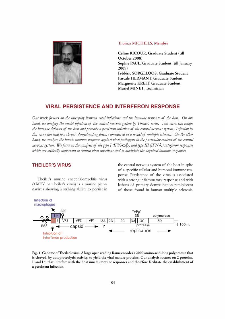
84
Thomas MICHIELS, Member
Céline RICOUR, Graduate Student (till October 2008)Sophie PAUL, Graduate Student (till January 2009)Frédéric SORGELOOS, Graduate StudentPascale HERMANT, Graduate StudentMarguerite KREIT, Graduate StudentMuriel MINET, Technician
VIRAL PERSISTENCE AND INTERFERON RESPONSE
Our work focuses on the interplay between viral infections and the immune response of the host. On one hand, we analyze the model infection of the central nervous system by Theiler’s virus. This virus can escape the immune defenses of the host and provoke a persistent infection of the central nervous system. Infection by this virus can lead to a chronic demyelinating disease considered as a model of multiple sclerosis. On the other hand, we analyze the innate immune response against viral pathogens in the particular context of the central nervous system. We focus on the analysis of the type I (IFN-a/b) and type III (IFN-l) interferon responses which are critically important to control viral infections and to modulate the acquired immune responses.
THEILER’S VIRUS
Theiler’s murine encephalomyelitis virus (TMEV or Theiler’s virus) is a murine picor-navirus showing a striking ability to persist in
the central nervous system of the host in spite of a specific cellular and humoral immune res-ponse. Persistence of the virus is associated with a strong inflammatory response and with lesions of primary demyelination reminiscent of those found in human multiple sclerosis.
Infection ofmacrophages
L VP
4 VP2 VP3 VP1 3A2A 2B 2C
"VPg"3B
3C 3DL*
8 100 nt
polymerase
proteasecapsidreplication
?IRES
CRE
Inhibition ofinterferon production
Fig. 1. Genome of Theiler’s virus. A large open reading frame encodes a 2000 amino acid-long polyprotein that is cleaved, by autoproteolytic activity, to yield the viral mature proteins. Our analysis focuses on 2 proteins, L and L*, that interfere with the host innate immune responses and therefore facilitate the establishment of a persistent infection.

85
Our work aims at understanding how a virus can persist in the central nervous system (CNS) of an immuno-competent host, thus evading the immune response.
Recently, a novel human virus closely rela-ted to Theiler’s virus was discovered and na-med Saffold virus. This virus is highly preva-lent in the human population. Further work is needed to evaluate its virulence potential and its impact on global health.
ANALySIS OF VIRAL PROTEINS INVOLVED IN THEILER’S VIRUS ESCAPE OF THE HOST IMMUNE RESPONSE.
F. Sorgeloos, C. Ricour, T. Michiels
Two viral proteins, namely L and L* were found to be dispensible for viral replication in cell culture but to be crucial for persistence of the virus in the central nervous system. Hence, these proteins are believed to interact with host factors in vivo and to counteract the host im-mune defenses.
INHIBITION OF TyPE-I IFN PRODUCTION AND ALTERATION OF NUCLEO-CyTOPLASMIC TRAFFICKING By THE L PROTEIN
The leader (L) protein encoded by Theiler’s virus is a 76 amino acid-long peptide containing a zinc-binding motif. We showed previously that the L protein could inhibit production of type-I interferons (IFNs) by infected cells (2). Mutation of the zinc-finger is sufficient to abolish the anti-IFN activity of the L protein in vitro and to dramatically impair viral persis-tence in the central nervous system of mice.
We also showed that the leader protein in-terferes with nucleo-cytoplasmic trafficking of host cell proteins (4) and with nuclear export of mRNA (10). These activities were associa-
ted with hyperphosphorylation of the Nup proteins that make up the nuclear pore com-plex (10). Perturbation of nucleo-cytoplasmic trafficking can constitute a effective way to inhibit early cell defense mechanisms. Indeed, the induction of many genes involved in early host defense, such as genes coding cytokines and chemokines, depends on the nuclear trans-location of transcription factors such as NFkB or IRF-3 into the nucleus.
Infection of mice deficient for the type-I interferon receptor (IFNAR-/-) indicates that IFN production inhibition by the L protein also occurs in vivo. However, the inhibition of IFN production is not absolute. If it were, the DA virus would cause a fatal encephalomyelitis in wild-type mice analogous to the disease it causes in IFNAR-/- mice (6, 10).
The L protein encoded by Encephalomyo-carditis (EMCV), a related Cardiovirus, sha-res about 35% amino acid identity with that of Theiler’s virus. It differs from the latter by lacking a serine/threonine-rich C-terminal do-main and by carrying phosphorylated residues not conserved in Theiler’s virus L protein. In spite of these differences, the L protein of EMCV shares, with that of Theiler’s virus, the ability to inhibit the transcription of type I in-terferon, cytokine and chemokine genes and to interfere with nucleo-cytoplasmic trafficking of host-cell proteins. Recent work performed in collaboration with S. Hato and F. van Kup-peveld (NCLMS, Nijmegen, The Netherlands) confirmed the importance of the L protein of EMCV in counteracting the IFN response in vivo.
INFLUENCE OF THE L* PROTEIN ON MACROPHAGE INFECTION AND VIRAL PERSISTENCE
Persistent strains of Theiler’s virus produce an 18 KDal protein called L*, which is encoded by an open reading frame (ORF) overlapping the ORF coding the viral polyprotein (see Fig. 1). This protein was shown to enhance the in-

86
fection of macrophage cell lines and to be cri-tical for persistence of the virus in the central nervous system of the mouse (3).
Current efforts aim at characterizing the function of the L* protein in the infection of macrophages. Macrophages are indeed key players in the demyelinating disease induced by Theiler’s virus, being simultaneously effectors of the immune response and targets of viral infection. Our recent data suggest that the L* protein antagonizes an important effector pa-thway of the interferon response. Thus Thei-ler’s virus would interfere both with the pro-duction of IFN by infected cells and with the response of cells to this cytokine. This outlines the major importance of the IFN system in the defense of the host against viral infections.
TyPE-I AND TyPE-III INTERFERONS
Interferons were the first cytokines to be identified. They were discovered about 50 years ago by Isaacs and Lindemann who ob-served that chick chorio-allantoic membranes developped resistance to viral infection after exposure to heat-inactivated influenza virus. Interferons are typically secreted by cells that are infected by a virus. They alert neighbouring cells about the presence of a viral infection and allow these cells to develop an anti-viral state. The interferon system represents a critical pro-tection mechanism of the body against viral infections. In addition, interferons have anti-cancer properties and modulate the acquired immune response of the host.
Type I IFNs
P. Hermant, F. Sorgeloos, T. Michiels
Mouse and human genomes carry more than a dozen of genes coding for closely rela-ted interferon-alpha (IFN-a) subtypes. These interferons and other interferons, like IFN-b,
IFN-k, IFN-e, IFN-w, and limitin, form the type-I IFN family. In spite of important se-quence divergences, all these IFNs bind the same receptor, raising the question of whether they possess specific functions.
We previously characterized the murine IFN-a family. 14 IFN-a genes were detected in the mouse genome, two of which, IFN-a13 and IFN-a14, were previously undescribed. In addition the mouse genome contains three IFN-a pseudogenes (5).
Ongoing work aims at understanding the specificities of the various type-I IFN subtypes.
Type-III Interferons (IFN-l)
S. Paul, M.Minet, T. Michiels
Recently, another IFN family was described and called «type-III IFN» or «IFN-l». Althou-gh type III IFNs bind a receptor distinct from the type-I IFN receptor, they activate the same signal transduction pathway and upregulate the same set of genes. Thus, they are expected to exert very similar biological activities as type I IFNs. Our work addresses the question of the redundancy of these two IFN systems in vivo.
We recently observed that the relative ex-pression of IFN-l over that of IFN-a/b exhi-bited some extent of tissue specificity and was low in the brain (9). We also used a strategy based on in vivo expression of cloned IFN genes to compare the responses of different tissues to IFN-a and IFN-l. Interestingly, res-ponse to IFN-l appeared to be specific to epi-thelial cells, unlike response to IFN-a which occurred in most cell types and was particularly prominent in endothelial cells. Accordingly, tis-sues with a high epithelial content such as in-testine, skin or lungs were the most responsive to IFN-l and expressed the higher amounts of IFN-l receptor (9).
Our data suggest that the IFN-l system evolved as a specific protection of epithelia

87
and that it might contribute to prevent viral invasion through skin and mucosal surfaces. Future work will address the physiolgical im-portance of the IFN-l system.
Production of IFN-a/b by neurons
M. Kreit, S. Paul, T. Michiels
In peripheral organs, a specialized popula-tion of dendritic cells known as «plasmacytoid» dendritic cells (pDCs) was reported to account for most of the type I IFN production. Cons-titutive expression of the IRF-7 transcription factor enables pDCs to rapidly synthesize large amounts of IFNs after viral infection. In the central nervous system, however, these cells are reportedly absent or very rare.
In collaboration with the teams of Peter Staeheli and Friedemann Weber (Univ. Frei-burg, Germany) we analyzed the cells that are responsible for type I IFN production in the central nervous system, during acute encepha-litis caused by two neurotropic viruses: Thei-ler’s virus (picornavirus) or La Crosse virus (bunyavirus).
IFN-a/b turned out to be produced by many scattered parenchymal cells and less by cells of the inflammatory foci. Interestingly, besides some macrophages and ependymal cel-
ls, neurons turned out to be important produ-cers of both IFN-a and IFN-b. However, not more than 3% of infected neurons expressed IFN, suggesting that some restriction to IFN production might occur in these cells (7).
All CNS cell types analyzed, including neu-rons, were able to respond to type I IFN by producing Mx or IRF-7. Our data show that, in vivo, in spite of the relative immune privilege of the CNS parenchyma, neurons take an ac-tive part to the antiviral defense by being both IFN producers and responders (7, 8).
SELECTED PUBLICATIONS
1. Lobert PE, Escriou N, Ruelle J, Michiels T. A coding RNA sequence acts as a replication signal in cardioviruses. Proc Natl Acad Sci USA 1999;96:11560-5.
2. van Pesch V, van Eyll O, Michiels T. The leader protein of Theiler’s virus inhibits imme-diate-early alpha/beta interferon production. J Virol 200 ;75:7811-7.
Neuron (immunostained in braun for NeuN) expressing IFN-a (detected by in situ hybridization (black dots) after La Crosse virus infection.
Fig. 3. Neurons can produce IFNa/b. Neuron (immunostained in brown for NeuN) producing IFN-b (detected by in situ hybridization: black dots) after infection with La Crosse virus.
Fig. 2. Reponse to IFN-l. Section of the urinary epithelium showing a strong response of epithelial cells to IFN-l. Shown is a fluorescent labelling of the nuclear Mx1 protein, the expression of which is typically induced in response to IFN.

88
3. van Eyll O, Michiels T. Non-AUG-initiated internal translation of the L* protein of Theiler’s virus and importance of this protein for viral per-sistence. J Virol 2002;76:10665-73.
4. Delhaye S, van Pesch V, Michiels T. The leader protein of Theiler’s virus interferes with nucleo-cytoplasmic trafficking of cellular proteins. J Virol 2004;78:4357-62.
5. van Pesch V, Lanaya H, Renauld JC, Michiels T. Characterization of the murine alpha interferon gene family. J Virol 2004;78:8219-28.
6. Brahic M, Bureau J-F, Michiels T. The gene-tics of the persistent infection and demyelinating disease caused by Theiler’s virus. Annu Rev Microbiol 2005;52:279-98.
7. Delhaye S, Paul S, Blacqori G, Weber F, Staeheli P, Michiels T. Neurons produce type I interferon in the course of viral encephalitis. Proc Natl Acad Sci USA 2006;103:7835-40.
8. Paul S, Ricour C, Sommereyns C, Sorge-loos F, Michiels T. The type I intereferon res-ponse in the central nervous system. Biochimie 2007;89:770-8.
9. Sommereyns C, Paul S, Staeheli P, Michiels T. IFN-lambda is expressed in a tissue-depen-dent fashion and primarily acts on epithelial cells in vivo. PLoS pathog 2008;4(3):e1000017.
10. Ricour C, Delhaye S, Hato S, Olenyik TO, Michel B, van Kuppeveld FJM, Gustin KE and Michiels T. Inhibition of mRNA export and IRF-3 dimerization by Theiler’s virus leader protein. J Gen Virol 2009;90:177-86.
Thomas Michielsde Duve InstituteAv. Hippocrate 74-75B - 1200 Brussels
[T] +32 02 764 74 29[F] +32 02 764 74 95[E] [email protected][W] http://www.deduveinstitute.be/viral_persis-tence.php

89
Pierre G. COULIE, Member
Sophie LUCAS, Senior Investigator Arcadi CIPPONI, Postdoctoral Fellow Pedro ALVES DE SOUSA, Postdoctoral Fellow (until April 2009)Teofila SEREMET, Graduate Student (from October 2008)Julie STOCKIS, Graduate Student Julie VANACKER, Graduate StudentGérald HAMES, Technician Catherine MULLER, Technician Maria PANAGIOTAKOPOULOS, TechnicianNathalie REMY, Technician (from September 2008)Suzanne DEPELCHIN, Secretary
HUMAN TUMOR IMMUNOLOGy
Tumor-specific antigens, such as those encoded by the MAGE genes, have been used to vaccinate melanoma patients with detectable disease. About 20 % of the vaccinated patients displayed a tumor regression, a frequency that appears well above the level reported for spontaneous melanoma regressions. Nevertheless, the treatment fails in most patients, and this can probably only be improved by a better understanding of the anti-tumor immune responses of the patients and of the mechanisms of tumor resistance to immune attack.
A first objective is to assess the immunogenicity of tumor vaccines and to compare various vaccine modalities. We have developed very sensitive methods for the detection of anti-tumor T lymphocytes, and apply them to patients included in cancer vaccination trials conducted by Dr. Jean-François Baurain at the Cliniques Uni-versitaires St Luc and by the clinical team of the Ludwig Institute.
A second objective is to understand the mechanism of the tumor regressions that occasionally occur in vacci-nated patients. Detailed analyses of a few such patients indicated that, surprisingly, the anti-vaccine T lym-phocytes are largely outnumbered by other tumor anti-T cells, which recognize tumor-specific antigens different from the vaccine antigens. These anti-tumor T cells represent most of the T cells present in a regressing tumor, and they probably play a major role in the rejection process. We wish to understand why these anti-tumor T cells become activated following vaccination. Along this line, we are setting up an in situ genetic approach to explore the functional status of T lymphocytes present within melanoma tumors.
That local immunosuppression could be involved in preventing tumor rejection is compatible with the observa-tion that the anti-tumor T cells mentioned above are often present in tumors prior to vaccination. Considering that suppressive or so-called regulatory T cells are important attenuators of immune responses, we analyze their role in vaccinated patients.

90
IMMUNE RESPONSES TO CANCER VACCINE ANTIGENS
G. Hames, V. Corbière, P.G. Coulie, in collaboration with A.-M. Feyens and J.-F. Baurain, Department of Medical Oncology, Cliniques Universitaires St Luc, and N. van Baren, Brussels branch of the Ludwig Institute for Cancer Research.
To understand why a minority of cancer pa-tients vaccinated with defined tumor antigens display an objective tumor regression while most of them do not, it is essential to know whether and how the anti-tumor immune res-ponses of the patients are either primed, am-plified, or modified by the vaccinations. We nevertheless felt that it was crucial to know whether or not low-level responses had occur-red and whether such cytolytic T lymphocytes (CTL) responses showed a correlation with tumor regression, in order to understand why most patients failed to show any evidence of regression. We therefore developed a sensi-tive approach based on in vitro restimulation of blood lymphocytes with the antigenic pep-tide over two weeks, followed by labeling with tetramers. To evaluate precursor frequencies, these mixed lymphocyte-peptide cultures were conducted under limiting dilution conditions. Cells that were labeled with the tetramer were cloned, the lytic specificity of the clones was verified, and their diversity was analyzed by T cell receptor (TCR) sequencing (1, 2).
Focusing on CD8 T cell responses to an-tigenic peptides presented by HLA-A1 or A2 molecules, we observed a surprisingly low le-vels of anti-vaccine T cell responses in several of the patients who displayed tumor regression after vaccination. Moreover we did not observe the anticipated correlation between the intensi-ties or breadth (proportions of peptides against which a response is observed) of the immune responses and the clinical impact of the vacci-nations (3, and unpublished observations). All these results suggest that the main limitation to the clinical efficacy of these therapeutic anti-cancer vaccines is not the intensity of the indu-
ced anti-vaccine T cell responses.
TUMOR REGRESSIONS OBSERVED AFTER VACCINATION: A ROLE FOR TUMOR-SPECIFIC CyTOLyTIC T LyMPHOCyTES THAT DO NOT RECOGNIzE THE VACCINE ANTIGENS
V. Corbière, T. Connerotte, T. Aerts, C. Muller, P.G. Coulie, in collaboration with C. Lurquin, B. Lethé, Brussels branch of the Ludwig Institute for Cancer Research.
Because the low levels of anti-vaccine T cells might be insufficient to produce on their own the observed tumor regressions, we examined the possibility that CTL directed against other antigens present on the tumor might contribute to the regressions. For 7 vaccinated melanoma patients, selected because it had been possible to derive a permanent cell line from their tu-mor cells, we estimated the blood frequencies of CTL directed against any antigen present on the tumor cells. For all these patients, anti-tumor CTL were found at high frequencies in the blood after vaccination. Unexpectedly, they were already present at similar high frequencies before vaccination. The frequency of anti-tu-mor CTL observed after vaccination was 12 fold to 20,000 fold higher than that of the anti-vaccine CTL (4). Because T cells directed at other tumor antigens than the vaccine antigen could make an important contribution to the tumor regressions, we felt that it was necessary to define the precise nature of their target an-tigens. We focused our effort on patient EB81, who had shown complete regression of a large number of cutaneous metastases following vaccination with ALVAC-MAGE. A majority of anti-tumor CTL clones recognized antigens encoded by MAGE-C2, a cancer-germline gene (4). Others recognized an antigen enco-ded by gp100, a melanocytic differentiation gene. In conclusion we are facing a paradoxical situation where the melanoma patients that are

91
being vaccinated, have already mounted a high spontaneous response against the types of antigens used in the vaccines. At the time of vaccination this spontaneous T cell response is clearly ineffective in halting tumor progression. The analysis of metastases of vaccinated pa-tients indicated not only the presence of anti-vaccine T cells, with a very modest enrichment compared to the blood, but mainly of much higher proportions of the other anti-tumor T cells, considerably enriched compared to the blood (5). These results suggest that the anti-vaccine CTL may not be the principal effectors that kill the bulk of the tumor cells. They may exert their effect mainly by an interaction with the tumor that creates conditions enabling the stimulation of large numbers of CTL directed against other tumor antigens, which then pro-ceed to destroy the tumor cells (6).
IN SITU ANALySIS OF TUMOR-INFILTRATING LyMPHOCyTES
A. Cipponi, T. Seremet, G. Hames, P.G. Coulie, in collaboration with N. van Baren, Brussels branch of the Ludwig Institute for Cancer Research.
Our observations that some vaccinated melanoma patients could display a clear tumor regression with a very low or sometimes un-detectable anti-vaccine T cell response led us to analyse the other anti-tumor T cells present in these patients, namely the T lymphocytes that recognized tumor-specific antigens dif-ferent from those of the vaccines. Detailed analyses carried out in two vaccinated patients clearly indicated that anti-tumor T lymphocy-tes were already present prior to vaccination, both in blood and in some tumors. It is ob-vious that there is a seemingly pacific coexis-tence between tumor cells and tumor-specific T lymphocytes that occurs in many cancer patients. The reasons for this coexistence may well be the key towards improving the clinical efficacy of cancer vaccines. Our approach is to gain information about human tumor-infiltra-
ting or tumor-associated T cells through an in situ genetic analysis. Human tumor samples are processed simultaneously for histological ana-lysis including immunochemical detection of immune cells, for complete gene profiling on a fragment of the tumor, and for laser micro-dissection on frozen material. A small piece is systematically put into culture to derive a mela-noma cell line. We use laser microdissection to isolate small numbers (1-50) of tumor cells or T lymphocytes infiltrating the tumor. A major difficulty of the approach is to maintain RNA integrity throughout the process: we measured that the RNA recovery in microdissection is usually around 1%, which is too low for the analysis of 1-50 cells. Over the last year we worked out every aspect of these experiments, and improved the recovery by a factor of 10-20, depending on whether or not an histologi-cal staining is required. We can now measure with RT-qPCR the expression of 6-8 genes on a microdissected fragment of about 50 cells. We are setting up a global cDNA amplification, in order to carry out complete gene expression profiling on fractions of the samples. We have adapted and are comparing two methods of amplification that can theoretically be used on single cells.
HUMAN REGULATORy T CELLS AND TGFb
S. Lucas, J. Stockis, N. Remy, M. Panagiotakopoulos, P.G. Coulie.
Regulatory T cells, or Tregs, are a subset of CD4+ lymphocytes specialized in the suppres-sion of immune responses. They are required to prevent the development of auto-immune diseases, but in mice they were also shown to contribute to cancer progression by inhibiting anti-tumor immune responses. Tregs could play a negative role in cancer patients, but this has remained difficult to verify due to the lack of a Treg-specific marker in humans, as well as to an incomplete understanding of the mecha-

92
nisms underlying their suppressive function.
Our objective is twofold: develop tools to quantify Tregs in human tissues, and identify mechanisms important for their suppressive function which could be specifically targeted to improve the efficiency of cancer vaccines.
Our previous work lead to the obtention of stable human Treg clones, representing long-term cultures of pure lymphocyte populations available for repeated analysis. A stable epige-netic mark unambiguously distinguished hu-man Treg clones from non regulatory CD4+ (Thelper) or CD8+ (cytolytic) clones: a con-served region in intron 1 of gene FOXP3, en-coding a transcription factor indispensable for the development and function of Tregs, was found demethylated in Treg clones only. We set up a methylation-specific real-time PCR assay to quantify demethylated FOXP3 sequences, indicative of the presence of Treg cells. We will determine whether this assay can be used to monitor the distribution of Treg cells in hu-man tissue samples such as peripheral blood or tumor fragments. Such monitoring should help to evaluate the role of human Treg cells in the inhibition of spontaneous or vaccine-induced anti-tumor immune responses.
T cell receptor (TCR) stimulation is requi-
red for the suppressive function of Tregs. We used expression microarrays to identify func-tional features that are unique to stimulated Treg clones, by comparison to stimulated Thel-per clones. This analysis revealed that a hall-mark of stimulated human Treg clones is to produce the active form of TGF-ß, a cytokine with well-known immunosuppressive actions. We are currently attempting to identify the me-chanisms by which human Tregs can produce active TGF-ß.
Many cell types, including Treg and Thel-per clones, produce the latent, inactive form of TGF-ß. In latent TGF-ß, the mature TGF-ß protein is bound to the Latency Associated Peptide, LAP, and is thereby prevented from binding to the TGF-ß receptor. We recently showed that latent TGF-ß, i.e. both LAP and mature TGF-ß, binds to GARP, a transmem-brane protein containing leucine rich repeats which is present on the surface of stimulated Treg clones but not on Th clones. Membrane localization of latent TGF-ß mediated by bin-ding to GARP may be necessary for the ability of Tregs to activate TGF-ß upon TCR stimu-lation. As illustrated in the figure above, a mo-del by which activated Tregs would accumulate latent TGF-ß on their surface and release its active form in close proximity to their target represents an interesting intermediate between
TCR stimulation
LATENT TGF-β
ACTIVE TGF-β
mature TGF-β
TGF-β receptor
LAP
GAR
P
Treg
distant activationand action
surfaceactivation
Treg
Tregtarget
Figure 1. Possible model for TGF-ß production by human Treg clones.

93
the release of a soluble active TGF-ß in the en-vironment, and that of a Treg acting by direct contact with its target. If this model proves to be relevant, it will be important to elucidate the precise mechanism which produces active TGF-ß at the surface of Tregs. Our results im-ply that binding to the GARP receptor is not sufficient, as lentiviral mediated expression of GARP in human Th cells induces binding of latent TGF-ß to the cell surface, but does not result in the production of active TGF-ß upon stimulation of these Th cells.
SELECTED PUBLICATIONS
1. Coulie PG, Karanikas V, Colau D, Lurquin C, Landry C, Marchand M, Dorval T, Brichard V, Boon T. A monoclonal cytolytic T-lymphocyte response observed in a melanoma patient vaccinated with a tumor-specific antigenic peptide encoded by gene MAGE-3. Proc Natl Acad Sci U S A 2001;98:10290-5.
2. Karanikas V, Lurquin C, Colau D, van Baren N, De Smet C, Lethé B, Connerotte T, Corbière V, Demoitié M-A, Liénard D, Dréno B, Velu T, Boon T, Coulie PG. Monoclonal anti-MAGE-3 CTL responses in melanoma patients displaying tumor regression after vaccination with a recombinant canarypox virus. J Immunol 2003;171:4898-904.
3. Lonchay C, van der Bruggen P, Connerotte T, Hanagiri T, Coulie PG, Colau D, Lucas S, Van Pel A, Thielemans K, van Baren N, Boon T. Correlation between tumor regression and T cell responses in melanoma patients vaccinated with a MAGE antigen. Proc Natl Acad Sci USA 2004;101:14631-38.
4. Germeau C, Ma W, Schiavetti F, Lur-quin C, Henry E, Vigneron N, Brasseur F, Lethé B, De Plaen E, Velu T, Coulie PG. High frequency of anti-tumor T cells in the blood of melanoma patients before and after vaccination with tumor antigens. J Exp Med 2005;201:241-8.
5. Lurquin C, Lethé B, Corbière V, Théate I, van Baren N, Coulie PG, Boon T. Contras-ting frequencies of anti-tumor and anti-vaccine T cells in metastases of a melanoma patient vaccina-ted with a MAGE tumor antigen. J Exp Med 2005;201:249-57.
6. Boon T, Coulie PG, Van den Eynde B, van der Bruggen P. Human T cell respon-ses against melanoma. Annu Rev Immunol 2006;24:6.1-6.34.
7. Stockis J, Fink W, François V, Connerotte T, De Smet C, Knoops L, van der Bruggen P, Boon T, Coulie P.G., Lucas S. Comparison of stable human Treg and TR clones by transcriptional profiling. Eur J Immunol 2009;39:869-82.
Pierre Couliede Duve Institute Av. Hippocrate 74-75B - 1200 Brussels
[T] +32 02 764 75 99[F] +32 02 764 75 90[E] [email protected][W] http://www.deduveinstitute.be/human_tu-mor_immunology.php


95
LUDWIG INSTITUTE FOR CANCER RESEARCH
BRUSSELS BRANCH

96
Cancer is a major concern in human health.
The prospects for bringing cancer under con-
trol require linked innovative basic and clinical
research. In this view, Daniel K. Ludwig crea-
ted in 1971 the Ludwig Institute for Cancer Re-
search, an international organization bringing
together scientists and clinicians from around
the world. Many Ludwig investigators are lea-
ders in many areas of science, involving gene-
tics, bioinformatics, immunology, virology, cell
biology and signal transduction.
Faithful to the organizing principles laid down by Mr Ludwig, the Institute conducts its research
through nine Branches, located in seven countries. The Branch structure allows the Institute to in-
teract with a number of different research and clinical environments. Each Branch is focused on
a research program defined by the Branch Director in relation with the overall objectives of the
Institute. The Branches are established in association with University Hospitals, to stimulate close
collaborations between research laboratories and the clinic. By organizing and controlling its own
clinical trials programs, the Institute has indeed created a continuum that integrates laboratory and
clinical research.
The biological properties of any given cancer cell constantly change, allowing tumors to spread
and become more aggressive. To overcome these obstacles, the Ludwig Institute has developed a
broad-based discovery program that seeks to understand the full complexity of cancer. Research is
organized according to the four major programmatic themes that define the Institute: genetics, cell
biology, cell signalling and immunology.
Branch staffs vary in size from 30 to over 90, and internationally the Institute employs some 600
scientists, clinicians and support personnel. The quality of the research is monitored on an ongoing
basis by the Institute’s Scientific Committee and by an external peer review process.
The Brussels Branch of the Institute was created in 1978. It is composed of 90 members and is
headed by Thierry Boon, Branch Director.

97
Administration and secretariat
Dario FLOREAN, Administrator
Aline BIEN, Accounting and Administrative AssistantNathalie KRACK, Librarian Carine LEMOINE, Scientific and Administrative Assistant Geneviève SCHOONHEYDT, Administrative AssistantPierre-Olivier SEROEN, Accounting Assistant Julie KLEIN, Scientific Secretary
Pre-clinical Research Resources
Group
Guy WARNIER, Veterinarian and Group
Head
Laurent HERMANS, Technical AssistantMohamed AHLOUMOU, Technical AssistantPhone SIRICHANTO, Technical Assistant
Technical support
Jo LEJEUNE, Head of Computer Systems Paul WAUTERS, Laboratory Manager André TONON, FACS Operator Jacques VAN CLEVE, Research Assistant in Bioinformatics Jean-Luc GERMIS, Technician Manuel ORVIZ, Senior Technical AssistantAntoinette VAN GOETSENHOVEN, Technical Assistant Jean-Claude VERHELST, Administrative and Technical Assistant Michel-Robat EVRARD-THOELEN, Laboratory Helper Samira JEBARI, Laboratory Helper Pascale MAHIEU-GAUDY, Laboratory Helper Encarnacion MARIANINI, Laboratory Hel-perRafaela REYES PRIETO, Laboratory HelperKeoprasith SOUVANNARAJ, Laboratory HelperThamma SINGHARACH, Laboratory Helper
Ludwig Institute - Brussels BranchUCL7459Avenue Hippocrate 741200 Bruxelles, Belgium
[T] +32 (2) 764 74 59 [F] +32 (2) 762 94 05
ADMINISTRATION AND GENERAL SERVICES

98
Benoît VAN DEN EYNDE, Member
Etienne DE PLAEN, Senior InvestigatorBernard LAUWERYS, Senior Investigator Vincent STROOBANT, Senior Investigator Catherine UYTTENHOVE, Senior Investigator Luc PILOTTE, Research Assistant
Ilse GUTTIEREZ-ROELENS, Postdoctoral Fellow Wenbin MA, Postdoctoral Fellow Ivan THEATE, Postdoctoral Fellow Antonia BUSSE, Postdoctoral Fellow (from June 2009)Nathalie VIGNERON, Postdoctoral FellowValérie BADOT, Guest Investigator Christophe BOURDEAUX, Graduate StudentAlexandre DALET, Graduate Student Benoît GUILLAUME, Graduate Student Olga KHOLMANSKIKH, Graduate StudentNicolas PARMENTIER, Graduate Student Céline POWIS DE TENBOSSCHE, Graduate StudentThérèse AERTS, TechnicianAline DEPASSE, TechnicianDominique DONCKERS, Technician Anne-Lise MAUDOUX, Technician Bénédicte TOLLET, Technician
TUMOR IMMUNOLOGy AND ANTIGEN PROCESSING
Building up on the molecular definition of tumor antigens recognized by T cells, our group mainly focuses on two aspects of tumor immunology, namely the processing of tumor antigens and the study of animal models to optimize cancer immunotherapy and evaluate tumor resistance mechanisms.
Tumor antigens recognized by Cytolytic T Lymphocytes (CTL) consist of peptides that are presented by MHC molecules at the cell surface and derive from intracellular proteins that are degraded by the proteasome. The intracellular pathway leading from the protein to the peptide/MHC complex is known as “antigen pro-cessing”. Our group focuses on the proteasome and recently described a new mode of production of antigenic peptides by the proteasome, based on cutting and pasting peptide fragments to form a new spliced peptide. The first example was a peptide derived from human melanocyte protein gp100. This antigenic peptide is nine-amino acid long and is produced by the splicing of two fragments that were initially non-contiguous in the parental protein. The splicing is made by the proteasome, is tightly coupled to the proteolytic reaction, and ap-pears to occur by transpeptidation involving an acyl-enzyme intermediate. We also described a second example of spliced peptide, which is a minor histocompatibility antigen, and where the two fragments are rearranged before splicing. We are currently working on additional spliced peptides. We are also studying the processing differences between the standard proteasome, which is present in most cells, and the immunoproteasome which is found in dendritic cells and in cells exposed to interferon-gamma. Several tumor antigens - including spliced peptides - were found to be processed differently by the two proteasome types, usually because of a preferential

99
cleavage made by one or the other proteasome within the antigenic peptide itself. We also study new proteasome types that are intermediate between the standard proteasome and the immunoproteasome.
Translation of knowledge on tumor antigens into efficient cancer immunotherapy requires additional studies on the various strategies that can be used. Some of these studies can be done in preclinical animal models. The study of such a model allowed us to uncover a powerful mechanism of tumor resistance, which is based on tryptophan catabolism by indoleamine-2,3 dioxygenase, an enzyme that we found to be frequently expressed in tumors. The resulting local tryptophan shortage appears to prevent the proliferation of lymphocytes at the tumor site. Inhibitors of indoleamine-2,3 dioxygenase can be used in vivo to counteract this tumor resistance mechanism. We are searching for new IDO inhibitors that could be developed clinically. We also study addi-tional tumor resistance mechanisms.
The currently available murine models are limited by the fact that they are based on transplantation of tumor cells grown in vitro into a healthy animal. This does not recapitulate the long-term host/tumor relationship that occurs in humans when a tumor slowly develops within a normal tissue. To circumvent this limitation and obtain more relevant information from such preclinical models, we have build a new mouse melanoma model where tumors expressing a given antigen can be induced, using a transgenic system based on Cre-lox recombination.
NEW PROTEASOME TyPES THAT ARE INTERMEDIATE BETWEEN THE STANDARD PROTEASOME AND THE IMMUNOPROTEASOME
B. Guillaume, V. Stroobant, W. Ma
Antigens recognized by cytolytic T lympho-cytes (CTL), such as viral or tumor antigens, usually consist of peptides of 8-10 amino acids in length, which are presented by MHC class I molecules at the cell surface. Because such peptides derive from intracellular proteins, a processing step is required before they can be exposed to the cell surface in association with MHC molecules. Firstly, the peptide is produ-ced through degradation of the parental protein by the proteasome. Secondly, it is taken up by a dedicated transporter named TAP and translo-cated inside the endoplasmic reticulum where it meets and associates with newly synthesized MHC molecules. The first step of cleavage by the proteasome is crucial in that cleavage lo-cation determines the precise sequence of the
final antigenic peptide. We have observed that this cleavage may occur differently in some cells, depending on their proteasome content. The proteasome comes in two forms: the standard proteasome, which is found in most cells, and the immunoproteasome, which is expressed by mature dendritic cells and by cells exposed to interferon-gamma (IFNg).
We previously reported that a class-I res-tricted antigenic peptide derived from an ubiquitous human protein was processed ef-ficiently by the standard proteasome but not by the immunoproteasome. As a result, the relevant epitope is not presented efficiently by mature dendritic cells, which contain immuno-proteasomes (1). We have now extended those observations to several antigenic peptides of interest for cancer immunotherapy, including HLA-A2-restricted epitopes derived from ty-rosinase, Melan-AMART1 and gp100. On the contrary, we showed that other tumor epitopes, which are derived from MAGE-3 and MAGE-C2, are processed by the immunoproteasome but not by the standard proteasome and the-

100
refore are presented to CTL only by tumor cells pre-treated with IFNg (2). By analyzing the peptidic fragments produced after in vitro digestion with the two proteasome types, we found that the differential processing can re-sult from two mechanisms. In some cases, one of the proteasome types predominantly clea-ves within the sequence of the epitope, resul-ting in its destruction (2). In other cases, the difference lies in the efficiency of cleavage at the C-terminal end of the antigenic peptide. These observations may have major implica-tions for cancer immunotherapy, as they imply that the peptide repertoire presented by tumor cells may differ from the repertoire presented by antigen-presenting cells (3). It is therefore essential to study those processing differences in detail, so as to define the most effective vac-cination strategy for each epitope. We have also observed the presence in many tumor lines of proteasome types that are intermediate between the standard proteasome and the immunopro-teasome. These intermediate proteasomes con-tain only some of the three catalytic subunits of the immunoproteasome, i.e. only ß5i or only ß1i and ß5i. In terms of production of anti-genic peptides, these intermediate proteasomes produce more or less the same peptides as the immunoproteasome, and therefore explain the recognition of tumor cells by CTL directed against these peptides in the absence of IFNg. However, we have also identified two antigenic peptides that are produced exclusively by inter-mediate proteasomes.
ANTIGENIC PEPTIDES PRODUCED By PEPTIDE SPLICING IN THE PROTEASOME
A. Dalet, V. Stroobant (in collaboration with Edus Warren, Fred Hutchinson Cancer Research Center, Seattle, USA)
By studying the antigen recognized by a CTL clone isolated from a melanoma patient, we identified an antigenic peptide composed
of two non-contiguous fragments of the same protein, namely the melanocytic protein gp100. The production of this peptide requi-res the excision of an intervening fragment of 4 amino acids and the splicing of a fragment of 3 residues with a fragment of 6 residues. We have shown that this splicing is exerted by the proteasome and can be reproduced in vitro by incubating a precursor peptide with purified proteasomes. Splicing is coupled directly to peptide bond cleavage by the proteasome and appears to occur by transpeptidation involving an acyl-enzyme intermediate (Fig. 1) (4). The splicing reaction appears not to involve a par-ticular motif, but rather to result from a low-efficiency reversal of the proteolysis reaction. Its occurrence is depending only on the occur-rence of peptide cleavage.
We have also identified a second antigenic peptide produced by peptide splicing in the proteasome (5). This peptide is recognized by CTL directed against a minor histocompati-bility antigen. The CTL was isolated from a multiple myeloma patient treated with HLA-identical bone marrow transplantation. The peptide is encoded by the polymorphic region of a gene ubiquitously expressed. Again it is made by the joining of two fragments that are initially non-contiguous in the parental protein. In addition, the two fragments are inverted in the spliced peptide, i.e. the fragment that was more N-terminal in the parental protein ends up at the C-terminal side of the spliced pep-tide, and vice-versa. We showed that splicing and transposition could be reproduced in vitro with purified proteasomes. The splicing me-chanism based on transpeptidation immedia-tely after peptide bond cleavage is compatible with a transposition of the fragments prior to splicing. Another spliced peptide was obser-ved previously by Hanada et al. We have now demonstrated that this third spliced peptide, which is derived from protein FGF-5, is also produced by transpeptidation inside the pro-teasome, despite the presence of a much longer segment between the two spliced fragments. We also showed that both the standard pro-teasome and the immunoproteasome are able

101
to splice peptides, but we noted differences in their efficiency to produce individual peptides. These differences are related to differences in the efficiency of cleavage at the relevant posi-tions to produce the fragments to splice. This observation confirms the transpeptidation mo-del, which predicts that the efficiency of spli-cing of a given peptide is mainly dependent on the efficiency of cleavage of the fragments to
splice. Altogether, these results indicate that spliced peptides are not uncommon and may represent a significant part of the peptide re-pertoire presented by MHC class I molecules.
IDENTIFICATION OF NEW ANTIGENS RECOGNIzED By AUTOLOGOUS CTL ON HUMAN MELANOMA
W. Ma, N. Vigneron (in collaboration with P. Coulie)
Melanoma patient EB81 was vaccinated with a MAGE-type antigen and showed re-gression of all cutaneous metastases. Blood lymphocytes collected after the regression were stimulated with autologous tumor cells, and CTL clones were obtained. Surprisingly, none of these clones was directed against the antigen used for vaccination. Using a cDNA expression cloning approach, we identified the antigens recognized by three of them. These antigens correspond to three distinct pepti-des all derived from MAGE-C2, a gene with a cancer-germline expression pattern, which is expressed in about 40% of melanomas and 30% of bladder carcinomas. Two peptides are presented by HLA-A2, and one by HLA-B57 (6). Because of their strict tumor-specificity and their wide expression in tumors, these new antigens represent promising targets for can-cer immunotherapy. The processing of two of these peptides is dependent on the immuno-proteasome (2).
We also started to study the intracellular processing pathway responsible for the cross-presentation of long peptides by dendritic cells.
Catalytically activethreonines
OH
NH2 RTK AWNR QLYPEWCO
NH
CO
NH
COOH
NH2 RTK QLYPEWCO
OH
NH
COOH
NH2 RTKCO
O
AWNRNH2 C O
QLYPEWNH2 COOH
OH
O
AWNRNH2 C OO
spliced peptide
transpeptidation
acyl-enzymeintermediate
Proteasome
Figure 1. Model of the peptide-splicing reaction in the proteasome. The active site of the catalytic subunits of the proteasome is made up of the side-chain of a threonine residue, which initiates proteolysis by performing a nucleophilic attack on the carbonyl group of the peptide bond. An acyl-enzyme intermediate is formed, which is then liberated by hydrolysis. In the peptide-splicing reaction, a second peptide fragment appears to compete with water molecules for performing a nucleophilic attack on the acyl-enzyme intermediate, resulting in a transpeptidation reaction producing the spliced peptide. Experimental support for this model of reverse proteolysis includes evidence that the energy required to create the new peptide bond is recovered from the peptide bond that is cleaved at the amino-terminus of the excised fragment, and that the amino-terminus of the other fragment needs to be free for transpeptidation to occur.

102
A TUMOR IMMUNE ESCAPE MECHANISM BASED ON TRyPTOPHAN DEGRADATION By INDOLEAMINE 2,3 DIOXyGENASE
L. Pilotte, I. Théate, C. Uyttenhove, D. Donckers, V. Stroobant, D. Colau, P. Larrieu
Indoleamine 2,3-dioxygenase (IDO) is an intracellular enzyme that catalyses rapid tryp-tophan degradation. Because tryptophan can freely cross the plasma membrane, IDO ex-pression results in a local depletion of trypto-phan in the extracellular medium surrounding the expressing cell. Tryptophan depletion was shown to impair T lymphocyte proliferation, and therefore IDO expression represents of powerful immunosuppressive mechanism that accounts, for example, for maternal tolerance to allogeneic fetuses, where IDO expression by placenta was found to play an essential role. Expression of IDO can be induced by inter-feron-gamma in many cellular types, including macrophages and dendritic cells, and appears to play a prominent role in immune regulation.
We have observed that many human tu-mors express IDO in a constitutive manner (7). To determine whether IDO expression provi-des tumor cells with a survival advantage by allowing their escape from immune rejection in vivo, we used the well-characterized model sys-tem of mouse tumor P815, where the antigen encoded by gene P1A is the major target of the rejection response. We observed that ex-pression of IDO by P815 tumor cells prevents their rejection by pre-immunized mice.
This effect can be partly reverted by sys-temic treatment of mice with an inhibitor of IDO, in the absence of noticeable toxicity (7). These results suggest that the efficacy of the-rapeutic vaccination of cancer patients could be improved by concomitant administration of an IDO inhibitor. We are currently searching for new IDO inhibitors that could be deve-loped at the clinical level. We have produced a novel monoclonal antibody against human
IDO (in collaboration with the group of Jean-Christophe Renauld). Using this antibody we have studied the expression of IDO in normal tissues in the steady state. Besides endothelial cells of the placenta, the only normal cells that express IDO are dendritic cells located in lym-phoid organs, including lymph nodes, spleen, gut lamina propria and thymus. We characte-rized those cells as mature myeloid dendritic cells. Contrary to published reports, we found that plasmacytoid dendritic cells do not express IDO, and that IDO-expressing dendritic cells are not enriched in tumor-draining lymph no-des. Using in vitro derived dendritic cells, we confirmed that immature dendritic cells do not express IDO, while mature dendritic cells ex-press high levels of IDO.
AN INDUCIBLE MOUSE MODEL OF MELANOMA EXPRESSING A DEFINED TUMOR ANTIGEN
C. Powis de Tenbossche, C. Uyttenhove, G. Warnier (in collaboration with A.-M. Schmitt-Verhulst, CIML, Marseille)
Cancer immunotherapy based on vaccina-tion with defined tumor antigens has not yet shown strong clinical efficacy, despite promising results in preclinical models. This discrepancy might result from the fact that available pre-clinical models rely on transplantable tumors, which do not recapitulate the long-term host-tumor interplay that occurs in patients during progressive tumor development and results in tumor tolerance. To create a faithful preclinical model for cancer immunotherapy, we genera-ted a transgenic mouse strain developing auto-logous melanomas expressing a defined tumor antigen recognized by T cells (8). We chose the antigen encoded by P1A, a well-characterized murine cancer germline gene. To transform melanocytes, we aimed at simultaneously acti-vating the Ras pathway and inactivating tumor suppressor Ink4a/Arf, thereby reproducing two genetic events frequently observed in hu-

103
man melanoma. The melanomas are induced by s.c. injection of 4-OH-tamoxifen (OHT). By activating a CreER recombinase expressed from a melanocyte-specific promoter, this treatment induces the loss of the conditional Ink4a/Arf gene in melanocytes. Because the CreER gene itself is also flanked by loxP sites, the activation of CreER also induces the dele-tion of its own coding sequence and thereby allows melanocyte-specific expression of genes H-ras and P1A, which are located downstream on the same transgene. All melanomas induced in those mice with OHT show activation of the Ras pathway and deletion of gene Ink4a/Arf. In addition, these melanomas express P1A and are recognized by P1A-specific T lymphocytes. This model will allow to characterize the inte-ractions between the immune system and natu-rally occurring tumors, and thereby to optimize immunotherapy approaches targeting a defined tumor antigen. We have now optimized the in-duction of melanoma in this model, and we reach an incidence of 70-80% tumors. In many tumor-bearing mice, we observed an accumu-lation of immature myeloid cells bearing both the CD11b and Gr1 markers. We are charac-terizing those cells, which might correspond to the myeloid-derived suppressor cells described in other models. In parallel, we have developed a strain of mice transgenic for the P1A-specific T cell receptor, which will be useful for such studies.
IMPACT OF THE THyMIC EXPRESSION OF CANCER-GERMLINE GENE P1A ON THE IMMUNOGENICITy OF P1A-DERIVED TUMOR ANTIGEN P815AB
I. Huijbers, C. Uyttenhove, D. Donckers, (in collaboration with A.-M. Schmitt-Verhulst, CIML, Marseille)
The genes encoding tumor antigens of the MAGE type are encoded by so-called cancer-
germline genes, whose expression is restricted to tumor cells and male germline cells, which do not bear MHC class I molecules. Recently, the group of Kyewski described the expression of such genes in a subset of epithelial cells lo-cated in the thymic medulla. This raised the question of the induction of a certain level of central tolerance to the antigens encoded by cancer-germline genes, which could explain the relatively poor immunogenicity of these anti-gens. To address this question, we have created a line of mice deficient for gene P1A, which is the murine prototypic cancer germline gene. Those P1AKO mice were healthy and fertile. When immunized against the P1A-derived an-tigen, they mounted slightly stronger specific immune responses than wild type mice. Simi-larly, P1AKO mice rejected P1A-expressing tu-mors more readily than wild type mice. Those results confirm a certain level of tolerance to antigens encoded by cancer-germline genes. However, this tolerance is limited, since wild type mice can develop responses to P1A-de-rived antigens, as opposed to P1A-transgenic mice that express P1A in all tissues and are fully tolerant to P1A.
TRANSCRIPTOMIC STUDIES IN SySTEMIC LUPUS ERyTHEMATOSUS (SLE) AND RHEUMATOID ARTHRITIS (RA)
B. Lauwerys, I. Gutierrez-Roelens, V. Badot, A.-L. Maudoux (in collaboration with F. Houssiau, Unité de Rhumatologie)
SLE is a systemic autoimmune disorder of unknown etiology, characterized by the acti-vation of autoreactive CD4 T and B cells di-rected against constituents of the chromatin and the production of pathogenic antinuclear antibodies. Recently, several groups identified a characteristic interferon signature in PBMC from SLE patients, i.e. the over-expression of genes induced by type I interferons. We com-pared SLE synovitis and the synovitis of os-

104
teoarthritis (OA) and RA using high-density oligonucleotide spotted microarrays. Our re-sults indicate that SLE arthritis is characterized by a very specific molecular signature that is distinct from that of OA and RA, with up-re-gulation of interferon (IFN)–inducible genes and down-regulation of genes involved in ex-tracellular matrix (ECM) homeostasis. The lat-ter observation is probably associated with the less destructive character of SLE compared to RA and OA. These results have immediate cli-nical applications for the differential diagnosis of arthritis.
We also performed global gene expression studies on synovial biopsies from RA patients treated with TNF blockers. We identified gene signatures in pre-treatment synovial tissue that predict the absence of response to TNF bloc-kade. Not surprisingly, these genes can be in-duced in synovial cells by other inflammatory cytokines (such as IL-1b or IL-17), alone or in combination with TNF-a (9). These observa-tions can be useful to guide therapeutic deci-sions.
SELECTED PUBLICATIONS
1. Morel S, Levy F, Burlet-Schiltz O, Bras-seur F, Probst-Kepper M, Peitrequin AL, Monsarrat B, Van Velthoven R, Cerottini JC, Boon T, Gairin JE, Van den Eynde BJ. Processing of some antigens by the standard pro-teasome but not by the immunoproteasome results in poor presentation by dendritic cells. Immu-nity 2000;12:107-17.
2. Chapiro J, Claverol S, Piette F, Ma W, Stroobant V, Guillaume B, Gairin JE, Mo-rel S, Burlet-Schiltz O, Monsarrat B, Boon T, Van den Eynde BJ. Destructive cleavage of antigenic peptides either by the immunopro-teasome or by the standard proteasome results in differential antigen presentation. J Immunol 2006;176:1053-61.
3. Van den Eynde BJ, Morel S. Differential processing of class-I-restricted epitopes by the standard proteasome and the immunoproteasome. Curr Opin Immunol 2001;13:147-53.
4. Vigneron N, Stroobant V, Chapiro J, Ooms A, Degiovanni G, Morel S, van der Bruggen P, Boon T, Van den Eynde B. An antigenic peptide produced by peptide splicing in the proteasome. Science 2004;304:587-90.
5. Warren EH, Vigneron N, Gavin M, Coulie P, Stroobant V, Dalet A, Tybodi S, Xue-reb S, Mito J, Riddel S, Van den Eynde BJ. An antigen produced by splicing of noncon-tiguous peptides on the reverse order. Science 2006:313,1444-48.
6. Ma W, Germeau C, Vigneron N, Maer-noudt A-S, Morel S, Boon T, Coulie PG, Van den Eynde BJ. Two new tumor-specific antigenic peptides encoded by gene MAGE-C2 and presented to cytolytic T lymphocytes by HLA-A2. Int J Cancer 2004;109:698-702.
7. Uyttenhove C, Pilotte L, Theate I, Stroo-bant V, Colau D, Parmentier N, Boon T, Van den Eynde BJ. Evidence for a tumoral immune resistance mechanism based on trypto-phan degradation by indoleamine 2,3-dioxyge-nase. Nat Med 2003;9:1269-74.
8. Huijbers IJ, Krimpenfort P, Chomez P, van der Valk MA, Song JY, Inderberg-Suso EM, Schmitt-Verhulst AM, Berns A, Van den Eynde BJ. An inducible mouse model of melanoma expressing a defined tumor antigen. Cancer Res 2006;66:3278-86.
9. Badot V, Galant C, Nzeusseu Toukap A, Theate I, Maudoux AL, Van den Eynde BJ, Durez P, Houssiau FA, Lauwerys BR. Gene expression profiling in the synovium identi-fies a predictive signature of absence of response to adalimumab therapy in rheumatoid arthritis. Arthritis Res Ther 2009; 11(2): R57.

105
10. Probst-Kepper M, Stroobant V, Kridel R, Gaugler B, Landry C, Brasseur F, Cosyns JP, Weynand B, Boon T, Van Den Eynde BJ. An alternative open reading frame of the human macrophage colony-stimulating factor gene is independently translated and codes for an an-tigenic peptide of 14 amino acids recognized by tumor-infiltrating CD8 T lymphocytes. J Exp Med 2001;193:1189-98.
Benoît Van den EyndeLudwig InstituteAv. Hippocrate 74-75B - 1200 Brussels
[T] +32 02 764 75 72[F] +32 02 764 75 90[E] [email protected][W] http://www.deduveinstitute.be/tumor_immu-nology.php

106
Pierre van der BRUGGEN, Member Thierry BOON, Member
Aline VAN PEL, Associate MemberDidier COLAU, Senior Investigator Danièle GODELAINE, Senior InvestigatorBernard LETHE, Senior InvestigatorChristophe LURqUIN, Senior InvestigatorNathalie DEMOTTE, Postdoctoral FellowIsabelle JACqUEMART, Postdoctoral FellowSabrina OTTAVIANI, Research Associate Claude WILDMANN, Research Associate Violaine FRANÇOIS, Graduate StudentEmilie GAUTHY, Graduate StudentGrégoire WIEËRS, Graduate Student Marie-Claire LETELLIER, Research AssistantMaria PANAGIOTAKOPOULOS, Research AssistantDébora PICCOLO, Research Assistant Vinh HA THI, Technician
REGULATION OF T LyMPHOCyTE FUNCTION IN TUMORS
We have recently discovered a new type of anergy of human CD8 T cells, which is observed on tumor-infiltra-ting lymphocytes. We are analyzing the mechanism of this type of anergy and we look for agents that reverse this anergy. The analysis of the T cell responses of melanoma patients vaccinated against tumor antigens has led us to consider the possibility that the limiting factor for therapeutic success is not the intensity of the anti-vaccine response but the degree of anergy presented by intratumoral lymphocytes. We therefore intend to pursue clinical trials involving the use of these agents in combination with anti-tumoral vaccination.
PREVIOUS WORK IN OUR GROUP: IDENTIFICATION OF TUMOR ANTIGENS RECOGNIzED By T CELLS
In the 1970s it became clear that T lympho-cytes, a subset of the white blood cells, were the major effectors of tumor rejection in mice. In the 1980s, human anti-tumor cytolytic T lym-phocytes (CTL) were isolated in vitro from the blood lymphocytes of cancer patients, mainly those who had melanoma. Most of these CTL were specific, i.e. they did not kill non-tumor
cells. This suggested that they target a marker, or antigen, which is expressed exclusively on tumor cells. We started to study the anti-tumor CTL response of a metastatic melanoma pa-tient and contributed to the definition of seve-ral distinct tumor antigens recognized by auto-logous CTL. In the early 1990s, we identified the gene coding for one of these antigens, and defined the antigenic peptide (1). This was the first description of a gene, MAGE-A1, coding for a human tumor antigen recognized by T lymphocytes.
Genes such as those of the MAGE family are expressed in many tumors and in male

107
germline cells, but are silent in normal tissues. They are therefore referred to as “cancer-germline genes”. They encode tumor specific antigens, which have been used in therapeutic vaccination trials of cancer patients (2). A large set of additional cancer-germline genes have now been identified by different approaches, including purely genetic approaches. As a result, a vast number of sequences are known that can code for tumor-specific shared antigens. The identification of a larger set of antigenic peptides, which are presented by HLA class I and class II molecules and recognized on tumors by T lymphocytes, could be important for therapeutic vaccination trials of cancer patients and serve as tools for a reliable monitoring of the immune response of vaccinated patients. To that purpose, we have used various approaches that we have loosely named “reverse immunology”, because they use gene sequences as starting point (3).
Human tumor antigens recognized by CD4+ or CD8+ T cells are being defined at a regular pace worldwide. Together with colleagues at the de Duve Institute, we read the new publi-cations and incorporate the newly defined an-tigens in a database accessible at http://www.cancerimmunity.org/peptidedatabase/Tcelle-pitopes.htm
ANALySIS OF THE T CELL RESPONSES OF CANCER PATIENTS
CD8+ T cells
We analyzed the T cell response in one patient who regressed upon vaccination with autologous dendritic cells pulsed with MAGE-3.A1 and MAGE-3.DP4 peptides. This patient developed a mixed tumor response, with disap-pearance/reduction of metastases and appea-rance of new metastases (Figure 1). In many MAGE-vaccinated patients who show tumor regression, the frequency of anti-vaccine T cells is very low. We wondered how such a low number of T cells could provide the main component of the specific effectors destroying the tumor cells. This led us to evaluate the fre-quency of all the T cells that recognized the autologous tumor cell line. We performed an in-depth longitudinal analysis of the anti-vacci-ne and anti-tumor responses, in the blood and in tumor sites of this patient (4). Before vacci-nation, the patient already had high frequencies of anti-tumor T cells in the blood. Skin me-tastases contained T cells but they were appa-rently inactive in destroying tumor cells. Upon vaccination, a modest anti-vaccine response was observed and this response lasted over the observation period of three years, with no evident concentration of anti-vaccine T cells
June 2004January 2003 May 2003 August 2003
Figure 1. Development of a mixed tumor response in patient LB2586. Regressing lesions on the posterior face of the right leg are encircled. The squares frame lesions that progressed over the same period of time.
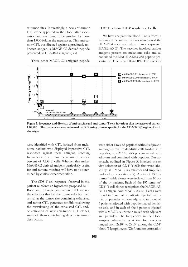
108
at tumor sites. Interestingly, a new anti-tumor CTL clone appeared in the blood after vacci-nation and was found to be enriched by more than 1,000-fold in the metastases. This anti-tu-mor CTL was directed against a previously un-known antigen, a MAGE-C2-derived peptide presented by HLA-B44 (Figure 2) (5).
Three other MAGE-C2 antigenic peptide
were identified with CTL isolated from mela-noma patients who displayed impressive CTL responses against these antigens, reaching frequencies in a tumor metastasis of several percent of CD8 T cells. Whether this makes MAGE-C2 derived antigens particularly useful for anti-tumoral vaccines will have to be deter-mined by clinical experimentation.
The CD8 T cell response observed in this patient reinforce an hypothesis proposed by T. Boon and P. Coulie: anti-vaccine CTL are not the effectors that kill the tumor cells but their arrival at the tumor site containing exhausted anti-tumor CTL, generates conditions allowing the reawakening of the exhausted CTL and/or activation of new anti-tumor CTL clones, some of them contributing directly to tumor destruction.
CD4+ T cells and CD4+ regulatory T cells
We have analyzed the blood T cells from 14 vaccinated melanoma patients who carried the HLA-DP4 allele and whose tumor expressed MAGE-A3 (6). The vaccines involved various antigens present on melanoma cells and all contained the MAGE-A3243-258 peptide pre-sented to T cells by HLA-DP4. The vaccines
were either a mix of peptides without adjuvant, autologous mature dendritic cells loaded with peptides, or a MAGE-A3 protein mixed with adjuvant and combined with peptides. Our ap-proach, outlined in Figure 3, involved the ex vivo selection of CD4+ T cells that were labe-led by DP4 MAGE-A3 tetramer and amplified under clonal conditions (7). A total of 197 te-tramer+ stable clones were isolated from 10 out of the 14 patients. Each of the 197 tetramer+ CD4+ T cell clones recognized the MAGE-A3.DP4 antigen. Anti-MAGE-A3.DP4 cells were found in 1 out of 2 patients injected with a mix of peptides without adjuvant, in 3 out of 6 patients injected with peptide-loaded dendri-tic cells, and in each of the 6 patients injected with a MAGE-A3 protein mixed with adjuvant and peptides. The frequencies in the blood samples collected after at least four vaccines ranged from 2x10⁻⁶ to 2x10⁻³ among the CD4+ blood T lymphocytes. We found no correlation
freq
uenc
y am
ong
TCR
10-6
10-5
10-4
10-3
10-2
resectedcutaneous metastases
2002 2003 2004
AA A
1 11
2 22A
1
S DNOAJ F M A M J JS DNOA AJ F M A M J J
anti-MAGE-3.DP4 clonotype 2 (PCR)
anti-MAGE-C2.B44 clonotype A (PCR)
anti-MAGE-3.A1 clonotype 1 (PCR)1
A
2
Figure 2. Frequency and diversity of anti-vaccine and anti-tumor T cells in various skin metastases of patient LB2586. The frequencies were estimated by PCR using primers specific for the CD3/TCRb region of each clonotype.
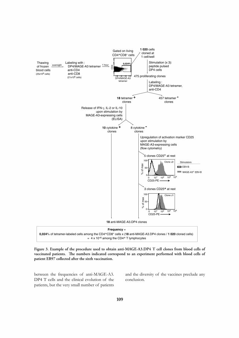
109
between the frequencies of anti-MAGE-A3.DP4 T cells and the clinical evolution of the patients, but the very small number of patients
and the diversity of the vaccines preclude any conclusion.
Frequency =0,024% of tetramer-labeled cells among the CD4+CD8- cells x (18 anti-MAGE-A3.DP4 clones / 1 020 cloned cells)
= 4 x 10⁻⁶ among the CD4+ T lymphocytes
Thawing of frozen
blood cells
Labeling with : DP4/MAGE-A3 tetramer anti-CD4 anti-CD8
1 020 cells cloned at 1 cell/well
Stimulation (x 3): peptide pulsed DP4 cells
475 proliferating clones
Labeling :DP4/MAGE-A3 tetramer,anti-CD4
18 tetramer + clones
457 tetramer -clones
CD
4
DP4/MAGE-A3tetramer
overnight 1 hour
(25x106 cells)(21x106 cells)
Gated on living CD4+CD8- cells
101 102 103 1040
EBV-B
MAGE-A3+ EBV-B
5 clones CD25- at rest
3 clones CD25+ at rest
Stimulators
0
50
100
0,024%
Release of IFN-γ, IL-2 or IL-10upon stimulation by
MAGE-A3-expressing cells(ELISA)
10 cytokine + clones
8 cytokine -clones
Upregulation of activation marker CD25upon stimulation by MAGE-A3-expressing cells(flow cytometry)
18 anti-MAGE-A3.DP4 clones
CD25-PE
% o
f max
101 102 103 1040
50
100 Clone J2
Clone J1
CD25-PE
% o
f max
0
Figure 3. Example of the procedure used to obtain anti-MAGE-A3.DP4 T cell clones from blood cells of vaccinated patients. The numbers indicated correspond to an experiment performed with blood cells of patient EB97 collected after the sixth vaccination.

110
Interestingly, twelve out of 197 clones ex-pressed CD25 in resting state. This CD4+CD25+ phenotype was evocative of T cells with sup-pressive activity, known as regulatory T cells. Because the antigen recognized by our CD4+ T cell clones was known, we designed an sup-pression assay where the potential regulatory T cell clone and an indicator T cell clone are each stimulated with their specific antigen presented by irradiated EBV-B cells. These twelve CD25+ clones had a high capacity to suppress in vitro the proliferation of another T cell clone. Ele-ven of them had an high FOXP3 expression at rest and an unmethylated FOXP3 gene. They secrete upon stimulation no IFN-g, IL-2, IL-4, IL-5 or IL-10, but they produce active TGF-b. These regulatory T cell clones represent about 5% of the anti-MAGE-A3.DP4 T cell clones that we have isolated. In addition to these clones with an unmethylated FOXP3 gene, se-veral CD25- clones had a significant but lower suppressive activity, expressed FOXP3 in the resting state but FOXP3 demethylation was not observed.
We conclude that MAGE-A3.DP4 vac-cination can produce CD4+ T cells that may influence the outcome of the treatment by exerting regulatory T cell function. The anti-MAGE-A3.DP4 potential regulatory T cells were isolated from the blood of four patients, including two patients with a mixed clinical response. This suggests at first that the occur-rence of these T cells is not sufficient to pre-vent tumor regression following vaccination. We have nevertheless no information about the presence of these cells at the tumor site. Quan-titative DNA methylation analysis of FOXP3 together with the relative frequencies of regu-latory T cells and effector T cells at the tumor site could become critical pieces of informa-tion to better establish the impact of regulatory T cells on the clinical outcome of vaccinated cancer patients.
A SCENARIO TO EXPLAIN THE LOW LEVEL OF CLINICAL RESPONSES IN VACCINATED PATIENTS
The identification of specific tumor anti-gens recognized by T lymphocytes on human cancer cells has elicited numerous clinical trials involving vaccination of tumor-bearing cancer patients with defined tumor antigens. These treatments have shown a low clinical efficacy. Among metastatic melanoma patients, about 5% show a complete or partial clinical respon-se following vaccination, whereas an additional 10% show some evidence of tumor regression without clear clinical benefit. We believe that progress depends on understanding why a small minority of patients shows tumor regres-sion following vaccination, whereas the majo-rity does not.
Recent analyses of the T cell responses of melanoma patients has led us to propose the following scenario (2, 8). Most melanoma pa-tients produce a spontaneous T cell response against melanoma tumor antigens at a relatively early stage of the disease (primary tumor or early metastatic tumor) (Figure 4).
These T cells can eliminate some tumors at an early stage, but often they do not succeed in eliminating the tumor and they become aner-gic. Thus, the tumors of the patients about to receive the vaccine, already contain anergic T cells directed against tumor antigens. Presuma-bly this anergy is maintained by immunosup-pressive factors present in the tumor. A few patients show tumor regression following vac-cination because some T cells generated by the vaccine penetrate inside the tumor, attack some tumor cells and succeed in reversing the local immunosuppression, possibly by releasing cy-tokines or chemokines. This reactivates the anergic T cells located inside the tumor and elicits new anti-tumoral T cells. These T cells then proceed to the elimination of the tumor cells. Most patients do not show tumor regres-

111
sion because the few anti-vaccine T cells that reach the tumor are immediately overwhelmed by the immunosuppressive environment.
Accordingly, our working hypothesis is that the crucial difference between the responding and the non-responding patients is not the in-tensity of their direct T cell response to the vaccine but the intensity of the immunosup-pression inside the tumor. It is therefore im-portant to know which immunosuppressive mechanisms operate in human tumors.
A MECHANISM CAUSING ANERGy OF CD8+ T LyMPHOCyTES
We have identified a novel mechanism cau-sing anergy of human CD8 cytolytic T lym-phocytes (CTL), including tumor-infiltrating lymphocytes (9-10). This type of anergy ap-pears to occur transiently during the normal stimulation cycle of T lymphocytes.
We observed that, a few days after antigen stimulation, CTL clones lose the capacity to se-crete cytokines and in some case the cytolytic activity. These functions are recovered gradual-ly and are usually completely restored after two weeks. TCRs and CD8 co-receptors were co-localized at the cell surface of functional CTL but, on the contrary, distant at the cell surface of non-functional CTL (Figure 5).
TCR and CD8 do not co-localize on tumor-infiltrating lymphocytes
Human CD8 tumor-infiltrating T lympho-cytes were isolated from tumor ascites or so-lid tumors and compared with T lymphocytes from blood donors. TCR were observed to be distant from CD8 on the cell surface of tumor-infiltrating lymphocytes, whereas TCR and CD8 co-localized on blood T lymphocy-tes. The tumor-infiltrating lymphocytes were anergic, being unable to secrete INF-g or other cytokines after non-specific stimulation with anti-CD3 and anti-CD28 antibodies.
Glycoprotein-galectin lattices restrain
Figure 4. Our working hypothesis: T cells generated by the vaccine succeed in reversing the local immunosuppression.
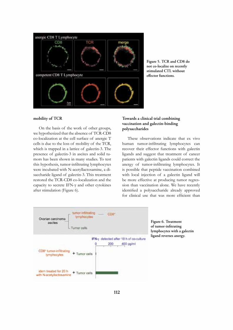
112
mobility of TCR
On the basis of the work of other groups, we hypothesized that the absence of TCR-CD8 co-localization at the cell surface of anergic T cells is due to the loss of mobility of the TCR, which is trapped in a lattice of galectin-3. The presence of galectin-3 in ascites and solid tu-mors has been shown in many studies. To test this hypothesis, tumor-infiltrating lymphocytes were incubated with N-acetyllactosamine, a di-saccharide ligand of galectin-3. This treatment restored the TCR-CD8 co-localization and the capacity to secrete IFN-g and other cytokines after stimulation (Figure 6).
Towards a clinical trial combining vaccination and galectin-binding polysaccharides
These observations indicate that ex vivo human tumor-infiltrating lymphocytes can recover their effector functions with galectin ligands and suggest that treatment of cancer patients with galectin ligands could correct the anergy of tumor-infiltrating lymphocytes. It is possible that peptide vaccination combined with local injection of a galectin ligand will be more effective at producing tumor regres-sion than vaccination alone. We have recently identified a polysaccharide already approved for clinical use that was more efficient than
Figure 6. Treatment of tumor-infitrating lymphocytes with a galectin ligand reverses anergy.
Figure 5. TCR and CD8 do not co-localize on recently stimulated CTL without effector functions.

113
N-acetyllactosamine to correct the anergy of CTL clones and human tumor-infiltrating lym-phocytes. We therefore intend to launch a clini-cal trial combining peptide vaccination and in-jections of galectin-binding polysaccharides in melanoma tumor-bearing metastatic patients.
SELECTED PUBLICATIONS
1. van der Bruggen P, Traversari C, Chomez P, Lurquin C, De Plaen E, Van den Eynde B, Knuth A, Boon T. A gene encoding an an-tigen recognized by cytolytic T lymphocytes on a human melanoma. Science 1991;254:1643-7
2. Boon T, Coulie PG, Van den Eynde B, van der Bruggen P. Human T cell responses against melanoma. Annu Rev Immunol 2006;24:175-208.
3. van der Bruggen P, Zhang Y, Chaux P, Stroobant V, Panichelli C, Schultz ES, Chapiro J, Van den Eynde BJ, Brasseur F, Boon T. Tumor-specific shared antigenic peptides recognized by human T cells. Immunol Rev 2002;188:51-64.
4. Carrasco J, Van Pel A, Neyns B, Lethé B, Brasseur F, Renkvist N, van der Bruggen P, van Baren N, Paulus R, Thielemans K, Boon T, Godelaine D. Vaccination of a melanoma patient with mature dendritic cells pulsed with MAGE-3 peptides triggers the acti-vity of nonvaccine anti-tumor cells. J Immunol 2008;180:3585-93.
5. Godelaine D, Carrasco J, Brasseur F, Neyns B, Thielemans K, Boon T, Van Pel A. A new tumor-specific antigen encoded by MAGE-C2 and presented to cytolytic T lymphocytes by HLA-B44. Cancer Immunol Immuno-ther 2007;56:753-9.
6. François V, Ottaviani S, Renkvist N, Stoc-kis J, Schuler G, Thielemans K, Colau D, Marchand M, Boon T, Lucas S and van
der Bruggen P. The CD4+ T-cell response of melanoma patients to a MAGE-A3 peptide vac-cine involves potential regulatory T cells. Cancer Res 2009;69:4335-45.
7. Zhang Y, Renkvist N, Sun Z, Schuler-Thurner B, Glaichenhaus N, Schuler G, Boon T, van der Bruggen P, Colau D. A polyclonal anti-vaccine CD4 T cell response detected with HLA-DP4 multimers in a mela-noma patient vaccinated with MAGE-3.DP4-peptide-pulsed dendritic cells. Eur J Immunol 2005;35:1066-75.
8. Lurquin C, Lethé B, De Plaen E, Corbiè-re V, Théate I, van Baren N, Coulie PG, Boon T. Contrasting frequencies of antitumor and anti-vaccine T cells in metastases of a mela-noma patient vaccinated with a MAGE tumor antigen. J Exp Med 2005;201:249-57.
9. Demotte N, Colau D, Ottaviani S, Gode-laine D, Van Pel A, Boon T, van der Brug-gen P. A reversible functional defect of CD8+ T lymphocytes involving loss of tetramer labeling. Eur J Immunol 2002;32:1688-97.
10. Demotte N, Stroobant V, Courtoy PJ, Van der Smissen P, Colau D, Luescher IF, Hi-vroz C, Nicaise J, Squifflet JL, Mourad M, Godelaine D, Boon T, van der Bruggen P. Restoring the association of the T cell receptor with CD8 reverses anergy in human tumor-infil-trating lymphocytes. Immunity 2008;28:414-24.
Pierre van der BruggenLudwig InstituteAv. Hippocrate 74-75B - 1200 Brussels
[T] +32 02 764 74 31[F] +32 02 762 94 05[E] [email protected][W] http://www.deduveinstitute.be/regulation_lymphocyte.php
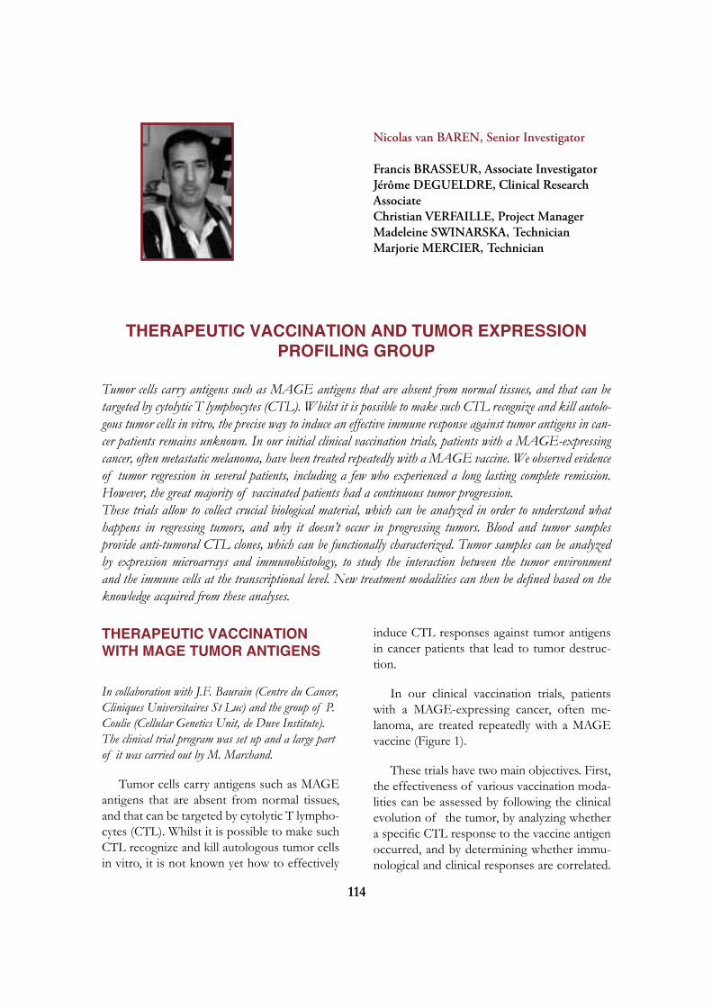
114
Nicolas van BAREN, Senior Investigator
Francis BRASSEUR, Associate Investigator Jérôme DEGUELDRE, Clinical Research Associate Christian VERFAILLE, Project Manager Madeleine SWINARSKA, Technician Marjorie MERCIER, Technician
THERAPEUTIC VACCINATION AND TUMOR EXPRESSION
PROFILING GROUP
Tumor cells carry antigens such as MAGE antigens that are absent from normal tissues, and that can be targeted by cytolytic T lymphocytes (CTL). Whilst it is possible to make such CTL recognize and kill autolo-gous tumor cells in vitro, the precise way to induce an effective immune response against tumor antigens in can-cer patients remains unknown. In our initial clinical vaccination trials, patients with a MAGE-expressing cancer, often metastatic melanoma, have been treated repeatedly with a MAGE vaccine. We observed evidence of tumor regression in several patients, including a few who experienced a long lasting complete remission. However, the great majority of vaccinated patients had a continuous tumor progression.These trials allow to collect crucial biological material, which can be analyzed in order to understand what happens in regressing tumors, and why it doesn’t occur in progressing tumors. Blood and tumor samples provide anti-tumoral CTL clones, which can be functionally characterized. Tumor samples can be analyzed by expression microarrays and immunohistology, to study the interaction between the tumor environment and the immune cells at the transcriptional level. New treatment modalities can then be defined based on the knowledge acquired from these analyses.
THERAPEUTIC VACCINATION WITH MAGE TUMOR ANTIGENS
In collaboration with J.F. Baurain (Centre du Cancer, Cliniques Universitaires St Luc) and the group of P. Coulie (Cellular Genetics Unit, de Duve Institute). The clinical trial program was set up and a large part of it was carried out by M. Marchand.
Tumor cells carry antigens such as MAGE antigens that are absent from normal tissues, and that can be targeted by cytolytic T lympho-cytes (CTL). Whilst it is possible to make such CTL recognize and kill autologous tumor cells in vitro, it is not known yet how to effectively
induce CTL responses against tumor antigens in cancer patients that lead to tumor destruc-tion.
In our clinical vaccination trials, patients with a MAGE-expressing cancer, often me-lanoma, are treated repeatedly with a MAGE vaccine (Figure 1).
These trials have two main objectives. First, the effectiveness of various vaccination moda-lities can be assessed by following the clinical evolution of the tumor, by analyzing whether a specific CTL response to the vaccine antigen occurred, and by determining whether immu-nological and clinical responses are correlated.

115
Secondly, these trials allow crucial biological material to be collected from vaccinated pa-tients. Blood samples provide anti-tumoral CTL clones, which can be functionally charac-terized.
Different immunization modalities, such as immunization with peptides, or with the MAGE-3 recombinant protein, both with or without adjuvant, or with the ALVAC recombi-nant viral vector, have already been tried, they all are devoid of significant toxicity. A minority of vaccinated melanoma patients (about 10 to 20%) showed regression of metastatic lesions. This frequency is far beyond the reported in-cidence of spontaneous regressions of mela-noma metastases, estimated at 0.2-0.3%, indi-cating that these regressions are linked to the vaccinations (Figure 2).
However, only 5% of the patients experien-ce a true clinical benefit. Some of the remis-sions have lasted for several years. There is no evidence that one of the vaccines tested is more effective against the tumors than the others. CTL responses were detected in a minority of patients vaccinated either with peptides or with the ALVAC virus. These responses were often weak, and, in the case of the MAGE-3.A1 anti-gen, were observed mostly in patients who had tumor regressions.
The most likely explanation for the poor effectiveness of cancer vaccines shown until now is the fact that tumors have acquired the ability to resist destruction by antitumoral T cells, following repetitive in vivo challenge with spontaneously occurring immune responses. Even though the molecular mechanisms of tu-mor resistance remain largely unknown, recent
Figure 1. Principle of anti-tumor vaccination with a defined antigen : The first step is to determine if the patient’s tumor cells express the tumor antigen. This can be determined by HLA typing, and by RT-PCR analysis of a tumor sample. Selected patients will receive repeated injections of a vaccine with the antigen. Usually this vaccine is a synthetic peptide, a recombinant protein, a recombinant virus coding for the antigen, or dendritic cells derived from the patient’s blood and forced to express this antigen. The effect of vaccinations on tumor progression is then assessed. Their immunogenicity is analyzed by comparing the frequency of anti-vaccine CTL in the pre and post-immune blood.

116
work in the laboratory has shown that the state of anergy that characterizes tumor-associated T cells can be reversed pharmacologically (see the contribution of Pierre van der Bruggen in this report). Inhibitors of galectin-3, a protein produced by cancer cells that is able to interfere with effective T cell activation, have been able to reactivate anergic T cells in vitro. We are cur-rently developing a new clinical trial, in which patients with advanced melanoma will receive a treatment combining a peptide vaccine and an experimental drug that inhibits galectin-3. We hope that this combined treatment will result in the induction of anti-tumoral CTL responses by the vaccine, in synergy with the inhibition of tumor resistance by the galectin-3 inhibitor.
EXPRESSION PROFILING OF TU-MOR SAMPLES FROM VACCINA-TED PATIENTS
In collaboration with the group of P. Coulie (Cellular Genetics Unit, de Duve Institute)
Using the microarray technology, we have established the gene expression profile of a series of tumor samples, mainly cutaneous metastases, obtained from melanoma patients, usually before the vaccine treatment was star-ted. These data help us to characterize the in-flammatory events that take place inside the metastases, and to understand the interaction between the tumor cells and the inflamma-tory cells at the tumor site. This approach is combined with systematic immunohistological analysis of adjacent cryosections, using anti-bodies directed against tumor cells, T and B cells, macrophages, blood vessels, and various molecules involved in inflammatory reactions (Figure 3).
Figure 2
Example of a complete regression of cutaneous metastases in a melanoma patient after repeated vaccinations with the MAGE-3.A1 peptide given without immunological adjuvant.

117
SELECTED PUBLICATIONS
1. van Baren N, Brasseur F, Godelaine D, Ha-mes G, Ferrant A, Lehmann F, André M, Ravoet C, Doyen C, Spagnoli GC, Bakkus M, Thielemans K, Boon T. Genes encoding tumor-specific antigens are expressed in human myeloma cells. Blood 1999;94:1156-64.
2. Marchand M, van Baren N, Weynants P, Brichard V, Dréno B, Tessier MH, Ran-kin E, Parmiani G, Arienti F, Humblet Y, Bourlond A, Vanwijck R, Liénard D, Beauduin M, Dietrich PY, Russo V, Ker-ger J, Masucci G, Jäger E, De Greve J, Atzpodien J, Brasseur F, Coulie PG, van der Bruggen P, Boon T. Tumor regressions observed in patients with metastatic melanoma treated with an antigenic peptide encoded by gene MAGE-3 and presented by HLA-A1. Int J Cancer 1999;80:219-30.
3. Chambost H, van Baren N, Brasseur F, Godelaine D, Xerri L, Landi SJ, Theate I, Plumas J, Spagnoli GC, Michel G, Coulie PG, Olive D. Expression of gene MAGE-A4 in Reed-Sternberg cells. Blood 2000;95:3530-3.
4. Marchand M, Brichard V, van Baren N, Coulie PG. Biological and clinical developments in melanoma vaccines. Expert Opin Biol Ther 2001;1:497-510.
5. Marchand M, Punt CJ, Aamdal S, Escu-dier B, Kruit WH, Keilholz U, Hakansson L, van Baren N, Humblet Y, Mulders P, Avril MF, Eggermont AM, Scheibenbo-gen C, Uiters J, Wanders J, Delire M, Boon T, Stoter G. Immunisation of metastatic can-cer patients with MAGE-3 protein combined with adjuvant SBAS-2: a clinical report. Eur J Cancer 2003;39:70-7.
6. Vialle-Castellano A, Laduron S, De Plaen E, Jost E, Dupont S, Ameye G, Michaux L, Coulie P, Olive D, Boon T, van Baren N. A gene expressed exclusively in acute B lym-phoblastic leukemias. Genomics 2004;83:85-94.
7. Kruit W, van Ojik H, Brichard V, Escudier B, Dorval T, Dréno B, Patel P, van Baren N, Avril M-F, Piperno S, Khammari A, Stas M, Ritter G, Lethé B, Godelaine D, Bras-seur F, Zhang Y, van der Bruggen P, Boon T, Eggermont A, Marchand M. Phase I/II study of subcutaneous and intradermal immu-nization with a recombinant MAGE-3 protein in patients with detectable non-visceral metasta-tic melanoma. Int J Cancer 2005;117:596-604.
8. van Baren N, Bonnet MC, Dréno B, Kham-mari A, Dorval T, Piperno-Neumann S, Liénard D, Speiser D, Marchand M, Bri-chard VG, Escudier B, Négrier S, Dietrich PY, Maraninchi D, Osanto S, Meyer RG, Ritter G, Moingeon P, Tartaglia J, van der Bruggen P, Coulie PG, Boon T. Tumoral
Figure 3. Cryosections obtained from a cutaneous melanoma metastasis were stained with antibodies directed against either melanoma cells (left), T lymphocytes (middle) and macrophages (right), and counterstained with hematoxylin. The corresponding cells appear in red. These images show clearly that the inflammatory cells do not infiltrate, but rather surround the tumor mass.

118
and immunological response following baccination of melanoma patients with an ALVAC virus encoding MAGE antigens recognized by T cells. J Clin Oncol 2005;23:9008-21.
9. Carrasco J, Van Pel A, Neyns B, Lethé B, Brasseur F, Renkvist N, van der Bruggen P, van Baren N, Paulus R, Thielemans K, Boon T, Godelaine D. Vaccination of a melanoma patient with mature dendritic cells pulsed with MAGE-3 peptides triggers the acti-vity of nonvaccine anti-tumor cells. J Immunol 2008;180:3585-93.
10. Baurain JF, Van der Bruggen P, Van den Eynde BJ, Coulie PG, Van Baren N. Ge-neral principles and first clinical trials of the-rapeutic vaccines against cancer. Bull Cancer 2008;95:327-35 (in French)
Nicolas van BarenLudwig InstituteAv. Hippocrate 74-75B - 1200 Brussels
[T] +32 02 764 75 08[F] +32 02 764 65 65[E] [email protected][W] http://www.deduveinstitute.be/therapeu-tic_vaccination.php

119
Jean-Christophe RENAULD, Member
Jacques VAN SNICK, Member Magali DE HEUSCH, Postdoctoral Fellow Laure DUMOUTIER, Postdoctoral FellowLaurent KNOOPS, Postdoctoral Fellow Tekla HORNAKOVA, StudentMuriel LEMAIRE, StudentIsabelle BAR, TechnicianEmilie HENDRICKX, Technician Monique STEVENS, Technician
CyTOKINES IN IMMUNITy AND INFLAMMATION
The cytokine group studies the biology of Interleukin-9 (IL-9) and IL-22, two cytokines discovered at the Branch. IL-9 is a TH2 cytokine that plays a role in immune responses against intestinal parasites and asthma. IL-22, originally identified as a gene induced by IL-9 in T lymphocytes, upregulates the production of acute phase reagents in the liver. Its activity in inflammatory responses is modulated by a specific antago-nist, the IL-22 binding protein (IL-22BP). The role of IL-9 and IL-22 in inflammation is currently being investigated using transgenic and gene-targeted mice for these cytokines and their receptors.
INTERLEUKIN 9
J.-C. Renauld, J. Van Snick, L. Knoops, V. Steenwinckel, M. Stevens
Interleukin-9 (IL-9) was discovered in our group in 1989, through its ability to sustain an-tigen-independent growth of certain murine T helper clones. We further identified human IL-9 by cross-hybridization with the mouse gene. Although IL-9 did not turn out to be a T cell growth factor for freshly isolated T cells, it was found particularly potent on T cell lymphomas, as an anti-apoptotic agent. To determine the biological activities of this factor, we generated transgenic mice overexpressing this cytokine. Analysis of these animals disclosed three es-sential properties of IL-9: its tumorigenic po-tential in T lymphocytes, its stimulatory activity on a particular subset of B lymphocytes, and its activity on mast cells and eosinophils with consecutive implications in asthma.
IL-9-transgenic mice : T cell lymphomas
IL-9 transgenic animals showed normal T cell development and T cell numbers but spon-taneously developed thymic lymphomas at low frequency (5%) when maintained in a conven-tional environment. Two lines of evidence indicate that IL-9 is not a conventional on-cogene but rather favors tumor development in response to exogenous stimuli. First, the tumor incidence was significantly lower when mice were maintained under pathogen-free conditions. Secondly, all IL-9 transgenic mice developed T cell lymphomas when exposed to subliminal doses of a chemical carcinogen or to irradiation, that were innocuous in wild type mice. The above mentioned anti-apoptotic ac-tivity of IL-9 provides an attractive explanation for these observations, namely that IL-9 could lead to increased survival of abnormal cells ge-nerated by exposure to minimal doses of on-cogenic stimuli. The potential implication of
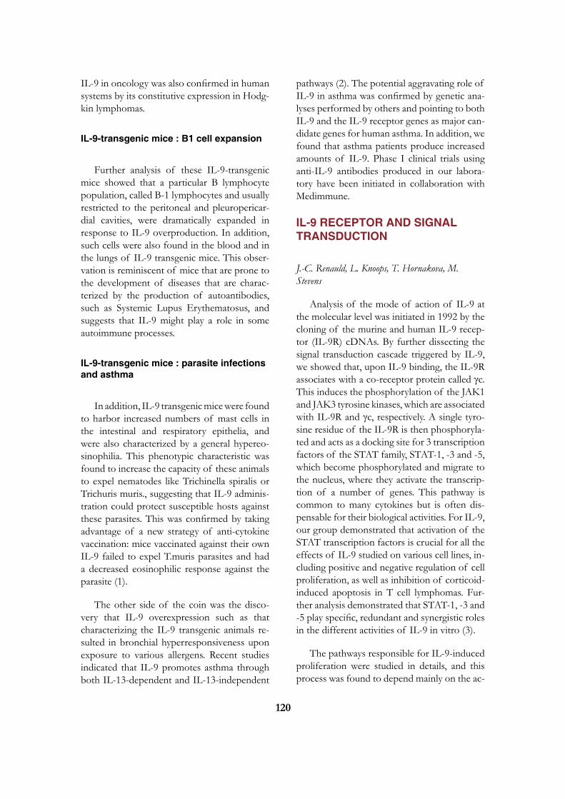
120
IL-9 in oncology was also confirmed in human systems by its constitutive expression in Hodg-kin lymphomas.
IL-9-transgenic mice : B1 cell expansion
Further analysis of these IL-9-transgenic mice showed that a particular B lymphocyte population, called B-1 lymphocytes and usually restricted to the peritoneal and pleuropericar-dial cavities, were dramatically expanded in response to IL-9 overproduction. In addition, such cells were also found in the blood and in the lungs of IL-9 transgenic mice. This obser-vation is reminiscent of mice that are prone to the development of diseases that are charac-terized by the production of autoantibodies, such as Systemic Lupus Erythematosus, and suggests that IL-9 might play a role in some autoimmune processes.
IL-9-transgenic mice : parasite infections and asthma
In addition, IL-9 transgenic mice were found to harbor increased numbers of mast cells in the intestinal and respiratory epithelia, and were also characterized by a general hypereo-sinophilia. This phenotypic characteristic was found to increase the capacity of these animals to expel nematodes like Trichinella spiralis or Trichuris muris., suggesting that IL-9 adminis-tration could protect susceptible hosts against these parasites. This was confirmed by taking advantage of a new strategy of anti-cytokine vaccination: mice vaccinated against their own IL-9 failed to expel T.muris parasites and had a decreased eosinophilic response against the parasite (1).
The other side of the coin was the disco-very that IL-9 overexpression such as that characterizing the IL-9 transgenic animals re-sulted in bronchial hyperresponsiveness upon exposure to various allergens. Recent studies indicated that IL-9 promotes asthma through both IL-13-dependent and IL-13-independent
pathways (2). The potential aggravating role of IL-9 in asthma was confirmed by genetic ana-lyses performed by others and pointing to both IL-9 and the IL-9 receptor genes as major can-didate genes for human asthma. In addition, we found that asthma patients produce increased amounts of IL-9. Phase I clinical trials using anti-IL-9 antibodies produced in our labora-tory have been initiated in collaboration with Medimmune.
IL-9 RECEPTOR AND SIGNAL TRANSDUCTION
J.-C. Renauld, L. Knoops, T. Hornakova, M. Stevens
Analysis of the mode of action of IL-9 at the molecular level was initiated in 1992 by the cloning of the murine and human IL-9 recep-tor (IL-9R) cDNAs. By further dissecting the signal transduction cascade triggered by IL-9, we showed that, upon IL-9 binding, the IL-9R associates with a co-receptor protein called gc. This induces the phosphorylation of the JAK1 and JAK3 tyrosine kinases, which are associated with IL-9R and gc, respectively. A single tyro-sine residue of the IL-9R is then phosphoryla-ted and acts as a docking site for 3 transcription factors of the STAT family, STAT-1, -3 and -5, which become phosphorylated and migrate to the nucleus, where they activate the transcrip-tion of a number of genes. This pathway is common to many cytokines but is often dis-pensable for their biological activities. For IL-9, our group demonstrated that activation of the STAT transcription factors is crucial for all the effects of IL-9 studied on various cell lines, in-cluding positive and negative regulation of cell proliferation, as well as inhibition of corticoid-induced apoptosis in T cell lymphomas. Fur-ther analysis demonstrated that STAT-1, -3 and -5 play specific, redundant and synergistic roles in the different activities of IL-9 in vitro (3).
The pathways responsible for IL-9-induced proliferation were studied in details, and this process was found to depend mainly on the ac-

121
tivation of STAT-5, on the recruitment of the IRS-1 adaptor, and on the activation of the Erk MAP-Kinase pathway.
The signal transduction pathway downs-tream the IL-9 receptor is illustrated in Fig. 1.
ANTI-APOPTOTIC ACTIVITy OF I-309RECEPTOR AND SIGNAL TRANSDUCTION
J.-C. Renauld, A. Tounsi, J. Van Snick
Incidentally, our studies of this particular model of the regulation of cell death by cy-tokines, lead them to purify another protein called I-309, originally described as a human chemotactic factor, and that turned out to exert a significant anti-apoptotic activity for thymic lymphomas (4). However, I-309 and IL-9 trig-ger completely different pathways and it was shown that the I-309 anti-apoptotic activity was dependent on the activation of G-proteins and the Ras/MAPKinase pathway, whereas the IL-9-mediated effect was not. More recently, we showed that a viral protein related to hu-man chemotactic factors (vMIP-I), and isolated from Herpes viruses that induce T cell tumors, has the same anti-apoptotic activity by binding to the I-309 receptor.
ROLE OF JAK OVEREXPRESSION IN TUMOR CELL TRANSFORMATION
J.-C. Renauld, L. Knoops, T. Hornakova, M. Stevens
Constitutive activation of the JAK-STAT pathway is frequent in cancer and contributes to oncogenesis. Some of our recent data indica-te that JAK overexpression plays a role in such processes. Using a murine proB cell line that strictly depends on IL-3 for growth in vitro, cytokine-independent and tumorigenic clones were derived from a two-step selection process. Cells transfected with a defective IL-9 receptor acquired IL-9 responsiveness during a first step of selection, and progressed after a second se-lection step to autonomously growing tumori-genic cells. Microarray analysis pointed to JAK1 overexpression as a key genetic event in this transformation. Overexpression of JAK1 not only increased the sensitivity to IL-9 but most importantly allowed a second selection step to-wards cytokine-independent growth with cons-titutive STAT activation. This progression was dependent on a functional FERM and kinase JAK1 domain. Similar results were observed after JAK2, JAK3 and TYK2 overexpression. All autonomous cell lines showed an activation of STAT5, ERK1-2 and AKT but only TYK2-overexpressing cell lines showed a constitutive activation of STAT3. Thus, JAK overexpres-sion can be considered as one of the oncoge-nic events leading to the constitutive activation of the JAK-STAT pathway (5).
IL-TIF/IL-22 : A NEW CyTOKINE STRUCTURALLy RELATED TO IL-10
L. Dumoutier, M. de Heusch, J.-C. Renauld
Searches for genes specifically regulated by IL-9 in lymphomas lead to the cloning of
Figure 1. IL-9 receptor signalling.

122
a gene that turned out to encode a 179 amino acid long protein, including a potential signal peptide, and showing a weak but significant sequence homology with IL-10. This protein, originally designated IL-TIF for IL-10-related T-cell derived Inducible Factor, was later re-named IL-22. Recombinant human IL-22 was produced (with D. Colau, LICR) and its crystal-lograhic structure solved. Despite its structural homology with IL-10, IL-22 fails to recapitu-late any of IL-10 biological activities. Biologi-cal activities of IL- 22 include the induction of acute phase proteins in liver (6) and protection against experimental hepatitis and colitis (L. Dumoutier, unpublished results). Among the different T cell subset, IL-22 was found to be preferentialy produced by TH17 cells raising some speculations about its potential role in autoimmune processes.
Analysis of genomic databases lead to the identification of a new receptor belonging to the IL-10 receptor family (7). This gene is lo-cated in the chromosome 6q24, at 24 kb from the IFNGR1 gene and at 152 kb from the IL-20R. It encodes a protein of 231 amino acid, showing 33 % and 34 % amino acid identity with the extracellular domains of the IL-22R and the IL-20R, respectively, but no cytoplas-mic nor transmembrane domains were found. IL-22BP is highly expressed in the placenta, in the breast, in the mammary gland and in the skin. A specific interaction was demonstrated between insolubilized IL-22 and an IL-22BP-Ig fusion protein. Moreover, recombinant IL-22BP could block IL-22 biological activity demonstrating that this protein can act as an IL-22 antagonist.
Although IL-22 does not share any biologi-cal activity with IL-10, these 2 cytokines share a common component of their respective re-ceptor complex, IL-10Rß. Anti-IL-10Rß anti-bodies indeed block the IL-22-induced acute phase response in HepG2 cells (6). All recep-tor complexes for IL-10-related cytokines in-clude a long chain and a short chain, based on the length of the cytoplasmic domain of these transmembrane proteins. IL-10Rß is a typical
short chain component, with only 76 amino acids in the cytoplasmic domain, whose main function seems to consist in recruiting the Tyk2 tyrosine kinase. In addition to IL-10R ß, IL-22 signalling requires the expression of a long chain protein, called IL-22R and comprising a 319 amino acid long cytoplasmic domain. This chain associates with Jak1, and is responsible for the activation of cytoplasmic signalling cascades such as the JAK/STAT, ERK, JNK and p38 MAP kinase pathways. An unexpected feature of the IL-22R chain is the fact that the C-terminal domain of this receptor is constitu-tively associated with STAT3, and that STAT3 activation by this receptor does not require the phosphorylation of the receptor, in contrast to the mechanism of STAT activation by most other cytokine receptors (Dumoutier et al., submitted for publication).
In addition to its role in IL-22 binding and signalling, the IL-22R chain also forms a func-tional heterodimeric receptor complex by asso-ciating with IL-20R ß, the second short chain member of the IL-10R-related receptor family. This complex mediates STAT-1 and –3 activa-tion by IL-20 and IL-24, but not by IL-22 (8). In addition, IL-20 and IL-24 can also bind to other complexes consisting of IL-20Ra and IL-20Rß. This promiscuity in cytokine recep-tor usage is illustrated in Fig 2 (see also ref. 9 for a review of this cytokine family).
Figure 2. Receptor complexes for IL-10, IL-20 and IL-22

123
LICR2: A NEW CyTOKINE RECEPTOR MEDIATING ANTIVIRAL ACTIVITIES
J.-C. Renauld, L. Dumoutier
Type II cytokine receptors include recep-tors for type I and Ii interferons (IFNs) and for IL-10-related cytokines. These transmembrane proteins are almost exclusively related by their extracellular part, which consists of tandem fibronectin type II domains, whereas the cy-toplasmic domain is associated with a tyrosine kinase of the Janus Kinase (JAK family). By screening genomic databases for similarity with the extracellular domain of these receptors, we identified a new receptor that we called LICR2 (Likely Interleukin or Cytokine rceptor 2). This receptor binds new cytokines designated IFN-l1-3, and mediates the same activities as those mediated by the receptors for IFN-a and ß, in-cluding antiviral and antiproliferative activities (10), raising the possibility of therapeutic appli-cations in viral infections and cancer.
SELECTED PUBLICATIONS
1. Richard M, Grencis RK, Humphreys NE, Renauld JC, Van Snick J. Anti-IL-9 vacci-nation prevents worm expulsion and blood eosi-nophilia in Trichuris muris-infected mice. Proc Natl Acad Sci USA 2000;97:767-72.
2. Steenwinckel V, Louahed J, Orabona C, Huaux F, Warnier G, McKenzie A, Li-son D, Levitt R, Renauld JC. IL-13 me-diates in vivo IL-9 activities on lung epithelial cells but not on hematopoietic cells. J Immunol 2007;178:3244-3251.
3. Demoulin JB, Van Roost E, Stevens M, Groner B, Renauld JC. Distinct roles for STAT1, STAT3, and STAT5 in differentia-tion gene induction and apoptosis inhibition by in-terleukin-9. J Biol Chem 1999; 274:25855-
25861.
4. Van Snick J, Houssiau F, Proost P, Van Damme J, Renauld JC. I-309/T cell acti-vation gene-3 chemokine protects murine T cell lymphomas against dexamethasone-induced apop-tosis. J Immunol 1996;157:2570-2576.
5. Knoops L, Hornakova T, Royer Y, Cons-tantinescu SN, Renauld JC. JAK kinases ove-rexpression promotes in vitro cell transformation. Oncogene 2008;27:1511-1519.
6. Dumoutier L, Van Roost E, Colau D, Renauld JC. Human interleukin-10-related T cell-derived inducible factor: molecular cloning and functional characterization as an hepatocyte-stimulating factor. Proc Natl Acad Sci USA 2000;97:10144-10149.
7. Dumoutier L, Lejeune D, Colau D, Re-nauld JC. Cloning and characterization of IL-22 binding protein, a natural antagonist of IL-10-related T cell-derived inducible factor/IL-22. J Immunol 2001;166:7090-7095.
8. Dumoutier L, Leemans C, Lejeune D, Kotenko SV, Renauld JC. Cutting edge: STAT activation by IL-19, IL-20 and mda-7 through IL-20 receptor complexes of two types. J Immunol 2001;167:3545-3549.
9. Renauld JC. Class II cytokine receptors and their ligands: key antiviral and inflammatory modula-tors. Nature Rev Immunol 2003;3:667-676.
10. Dumoutier L, Tounsi A, Michiels T, Som-mereyns C, Kotenko SV, Renauld JC. Role of the Interleukin-28 Receptor tyrosine residues for antiviral and antiproliferative activity of IL-29/IFN-lambda 1 : Similarities with type I Interferon signalling. J Biol Chem 2004; 279:32269-32274.

124
Jean-Christophe RenauldLudwig InstituteAv. Hippocrate 74-75B - 1200 Brussels
[T] +32 02 764 74 64[F] +32 02 762 94 05[E] [email protected]

125
Stefan N. CONSTANTINESCU, Member
Nancy CACERES, Postdoctoral FellowMichael GIRARDOT, Postdoctoral FellowJhansi KOTA, Postdoctoral FellowChristian PECqUET, Postdoctoral FellowRoxana-Irina ALBU, Graduate Student Jean-Philippe DEFOUR, Graduate StudentAlexandra DUSA, Graduate StudentJean-Michel HEINE, Graduate StudentCéline MOUTON, Research Assistant Joanne VAN HEES, Research Assistant (Lab Manager)Julie KLEIN, Administrative Assistant
STRUCTURE AND FUNCTION OF CyTOKINE RECEPTORS
Cytokines and their receptors are critical for the formation of mature blood cells and for the function of the immune system. We study the structure and function of several cytokine receptors, such as those for erythro-poietin (Epo), thrombopoietin (Tpo), Granulocyte Colony Stimulating Factor (G-CSF), and interleukins (IL) 2 and 9. Activation of these receptors is triggered by cytokine-induced changes in receptor dimerization/oligomerization, which lead to the activation of cytosolic Janus tyrosine kinases (JAK). Regulation by JAK kinases of receptor traffic, the assembly of cell-surface receptor complexes, the mechanisms of dimerization of transmembrane (TM) and cytosolic juxtamembrane (JM) domains, and mechanisms of JAK catalytic activation are major points of interest. The laboratory identified constitutively active mutants of JAK2, JAK1 and Tyk2 and of thrombopoietin receptor and is actively investigating the mechanisms by which JAK2 V617F and thrombopoietin receptor W515 mutants induce, in humans, Myeloproliferative Neoplasms, such as Polycythemia Vera or Primary Myelofibrosis.
THE MECHANISMS By WHICH A MUTANT JAK2 INDUCES POLyCyTHEMIA VERA AND OTHER MyELOPROLIFERATIVE NEOPLASMS IN HUMANS
A. Dusa, C. Pecquet, J.-M. Heine
The JAK-STAT pathway is emerging as a key player in cancer, with several mutations in genes coding for JAKs being identified in the past three years (1). Janus kinases possess two kinase domains, one active and the other, denoted as the pseudokinase domain, inactive. JAK2, one of the four known JAKs (JAK1,
JAK2, JAK3 and Tyk2) is crucial for signa-ling by several cytokine receptors, such as the erythropoietin receptor (EpoR), the thrombo-poietin receptor (TpoR), the G-CSF receptor (G-CSFR), the interleukin 3 receptor and the growth hormone receptor. JAKs are appended to the cytoplasmic juxtamembrane domains of receptors and are switched-on upon ligand bin-ding to the receptors’ extracellular domains.
Polycythemia Vera (PV), or the Vaquez di-sease, is characterized by excessive production of mature red cells and sometimes of plate-lets and granulocytes. Erythroid progenitors in PV are hypersensitive to Epo and become independent of erythropoietin (Epo) for proli-

126
feration and differentiation. Strikingly, the traf-fic of TpoR is defective in myeloid progenitors from PV. A hint that JAK2 or a JAK2-binding protein may be involved in PV came when we showed that the wild type JAK2 strongly pro-motes the maturation and cell-surface localiza-tion of TpoR, the very process that is defective in PV (2).
In collaboration with William Vainchenker at the Institut Gustave Roussy in Paris, we have been involved in the discovery of the JAK2 V617F mutation, that is responsible for >98% of Polycythemia Vera and for >50% of Essen-
tial Thrombocythemia (ET) and Primary Mye-lofibrosis (PMF) cases (3, 4). The mutation in the pseudokinase domain alters a physiologic inhibition exerted by the pseudokinase domain on the kinase domain and allows the mutated JAK2 to bind and activate EpoR, TpoR and G-CSFR in the absence of cytokines (Figure 1). Saturation mutagenesis at position V617 showed that not only Phe, but also Trp, Leu, Ile and Met can activate JAK2, although Trp is the only mutation that exhibits comparable ac-tivity with V617F (5). The homologous V617F mutations in JAK1 and Tyk2 also enable these kinases to be activated without ligand-binding
Figure 1. In the absence of cytokine ligands, cytokine receptors (left complex) are preassembled with tyrosine kinases JAK (Janus kinases) in inactive complexes. Cytokine binding to the extracellular domains of receptors (middle complex) induces a conformational change which allows the appended JAKs to cross-phosphorylate and activate each other. In turn, JAKs phosphorylate tyrosine residues (Py) on the cytosolic regions of receptors, which attract SH2- and PTB-containing signaling proteins. These proteins become themselves phosphorylated and either translocate to the nucleus to regulate gene expression (such as STATs, Signal Transducers and Activators of Transcription) or initiate kinase signaling cascades (such as Mitogen Activated Protein-Kinases, MAPK, phosphatydylinositol-3-kinase, PI3K, and Akt). The mutant JAK2 V617F binds to the cytosolic domains of receptors and can trigger signaling in the absence of any cytokine binding to the extracellular receptor domain (right complex). As a consequence, signaling is induced permanently and myeloid progenitors survive, proliferate and differentiate in an uncontrolled manner. (Jean-Michel Heine)
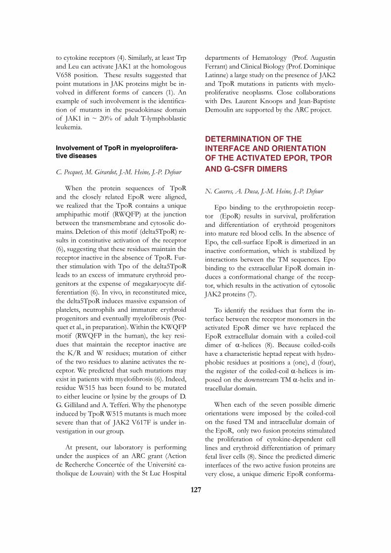
127
to cytokine receptors (4). Similarly, at least Trp and Leu can activate JAK1 at the homologous V658 position. These results suggested that point mutations in JAK proteins might be in-volved in different forms of cancers (1). An example of such involvement is the identifica-tion of mutants in the pseudokinase domain of JAK1 in ~ 20% of adult T-lymphoblastic leukemia.
Involvement of TpoR in myeloprolifera-tive diseases
C. Pecquet, M. Girardot, J.-M. Heine, J.-P. Defour
When the protein sequences of TpoR and the closely related EpoR were aligned, we realized that the TpoR contains a unique amphipathic motif (RWQFP) at the junction between the transmembrane and cytosolic do-mains. Deletion of this motif (delta5TpoR) re-sults in constitutive activation of the receptor (6), suggesting that these residues maintain the receptor inactive in the absence of TpoR. Fur-ther stimulation with Tpo of the delta5TpoR leads to an excess of immature erythroid pro-genitors at the expense of megakaryocyte dif-ferentiation (6). In vivo, in reconstituted mice, the delta5TpoR induces massive expansion of platelets, neutrophils and immature erythroid progenitors and eventually myelofibrosis (Pec-quet et al., in preparation). Within the KWQFP motif (RWQFP in the human), the key resi-dues that maintain the receptor inactive are the K/R and W residues; mutation of either of the two residues to alanine activates the re-ceptor. We predicted that such mutations may exist in patients with myelofibrosis (6). Indeed, residue W515 has been found to be mutated to either leucine or lysine by the groups of D. G. Gilliland and A. Tefferi. Why the phenotype induced by TpoR W515 mutants is much more severe than that of JAK2 V617F is under in-vestigation in our group.
At present, our laboratory is performing under the auspices of an ARC grant (Action de Recherche Concertée of the Université ca-tholique de Louvain) with the St Luc Hospital
departments of Hematology (Prof. Augustin Ferrant) and Clinical Biology (Prof. Dominique Latinne) a large study on the presence of JAK2 and TpoR mutations in patients with myelo-proliferative neoplasms. Close collaborations with Drs. Laurent Knoops and Jean-Baptiste Demoulin are supported by the ARC project.
DETERMINATION OF THE INTERFACE AND ORIENTATION OF THE ACTIVATED EPOR, TPOR AND G-CSFR DIMERS
N. Caceres, A. Dusa, J.-M. Heine, J.-P. Defour
Epo binding to the erythropoietin recep-tor (EpoR) results in survival, proliferation and differentiation of erythroid progenitors into mature red blood cells. In the absence of Epo, the cell-surface EpoR is dimerized in an inactive conformation, which is stabilized by interactions between the TM sequences. Epo binding to the extracellular EpoR domain in-duces a conformational change of the recep-tor, which results in the activation of cytosolic JAK2 proteins (7).
To identify the residues that form the in-terface between the receptor monomers in the activated EpoR dimer we have replaced the EpoR extracellular domain with a coiled-coil dimer of a-helices (8). Because coiled-coils have a characteristic heptad repeat with hydro-phobic residues at positions a (one), d (four), the register of the coiled-coil a-helices is im-posed on the downstream TM a-helix and in-tracellular domain.
When each of the seven possible dimeric orientations were imposed by the coiled-coil on the fused TM and intracellular domain of the EpoR, only two fusion proteins stimulated the proliferation of cytokine-dependent cell lines and erythroid differentiation of primary fetal liver cells (8). Since the predicted dimeric interfaces of the two active fusion proteins are very close, a unique dimeric EpoR conforma-

128
tion appears to be required for activation of signaling. In this active conformation TM resi-dues L241 and L244 and JM residue W258 are predicted to be in the interface.
Similar studies are undertaken for the re-lated TpoR and G-CSFR. Like the EpoR, the TpoR is thought to signal by activation of JAK2, of several STATs (STAT1, 3 and 5) as well as of MAP-kinase, PI-3-kinase and AktB. However, TpoR and EpoR signal quite diffe-rently since only TpoR can induce hemato-poietic differentiation of embryonic stem cells or stimulate the earliest stages of hematopoie-sis in immature hematopoietic cells.
STRUCTURE AND FUNCTION OF JUXTA-MEMBRANE AND TRANSMEMBRANE SEqUENCES OF CyTOKINE RECEPTORS
R.-I. Albu, A. Dusa, J. Van Hees, C. Mouton
We have previously shown that the EpoR exists on the cell surface as a preformed ligand-independent inactive dimer stabilized by trans-membrane domain interactions. To explore the details of inactive and active dimeric interfaces of cytokine receptors we undertake two ap-proaches: (i) cysteine scanning mutagenesis of the extracellular juxtamembrane and TM do-mains; (ii) cell surface immunofluorecence co-patching. We isolated three constitutively active
Figure 2. Bone marrow adoptive transfer in irradiated mice with hematopoietic stem cells expressing the constitutively active TpoR W515A induces severe myeloproliferative disorder, splenomegaly and fibrosis of the spleen. (A) Splenomegaly was induced by the TpoR W515A mutant at day 45 after reconstitution in contrast to the wild type TpoR or empty vector control. All mice presented >80% GFP positive cells, indicating expression of the bicistronic retroviral construct. (B) Histology of spleen sections of mice reconstituted with the indicated constructs. Silver staining (SL) for reticulin indicates extensive fibrosis of the spleen in the TpoR W515A mice. (Christian Pecquet)

129
novel mutants of the EpoR where residues L223, L226 or I227 were mutated to cysteine (9). Many other cysteine mutants formed disul-fide bonds, but only EpoR L223C, L226C and I227C dimerized in a conformation compatible with signaling. Cysteine-mediated maleimidyl crosslinking indicated that the first five TM re-sidues are not helical and that the interface of the active EpoR dimer contains residues L241 and L244. These studies led to the notion that sequences flanking the transmembrane domain might play important roles in receptor function as «switch» regions and also may regulate trans-membrane protein oligomerization.
More recently, using copatching assays, we have identified preformed homomeric com-plexes of IL2Rb, IL9Ra and g-chain, and he-teromeric IL2Rb- g-chain and IL9Ra- g-chain complexes (10). Such ligand-independent com-plexes might be important for the pre-assem-bly of signaling complexes at the cell surface. Furthermore, signaling by a constitutive active JAK3 mutant depends on preformed hetero-meric complexes that contain gamma chain (10).
TRAFFIC OF CyTOKINE RECEP-TORS TO THE CELL-SURFACE
C. Pecquet, J. Kota, R.-I. Albu
We have observed that, in hematopoietic cells, over-expression of JAK proteins leads to enhanced cell-surface localization of cytokine receptors (i.e. EpoR TpoR, IL9R, IL2R, gc). For some receptors, the effect of the cognate JAK is to promote traffic from the endoplasmic reticulum (ER) to the Golgi apparatus, while for others, such as the TpoR, JAK2 and Tyk2 also protect the mature form of the receptor from degradation by the proteasome, and thus JAKs enhance the total amount of cellular re-ceptor (2). In collaboration with Pierre Cour-toy, we are employing confocal microscopy of epitope tagged receptors in order to define the precise intracellular compartments where
receptors and JAKs interact. Our working hy-pothesis is that the N-terminus FERM domain of JAK proteins exerts a generic pro-folding effect on cytosolic domains of cytokine recep-tors. We are testing this hypothesis on several different cytokine receptors and are investiga-ting the link between proper folding in the ER and transport to the cell-surface. Using a fetal liver retroviral cDNA library cloned in pMX-IRES-CD2, we are attempting to clone novel proteins that can regulate traffic and stability of TpoR.
CONSTITUTIVE ACTIVATION OF JAK-STAT SIGNALING PATHWAyS AND GENES TARGETED By STAT5 IN TRANSFORMED HEMATOPOIETIC AND PATIENT-DERIVED LEUKEMIA CELLS
M. Girardot
Cytokine stimulation of cytokine receptors, induces transient activation of the JAK-STAT pathway. In contrast, oncogenic forms of re-ceptors or of JAKs (JAK2 V617F) transmit a continuous signal which results in constitutive activation of STAT proteins. In cultured cells this process is studied by expressing oncoge-nic forms of cytokine receptors or JAKs in cytokine-dependent cells and assaying for their transformation into cells that grow autono-mously (1). A similar picture has been noted in patient-derived leukemia cells. The critical questions we would like to answer concern: (i) the mechanisms by which the JAK-STAT pathway remains permanently activated in transformed cells; (ii) which genes are speci-fically regulated by constitutively active STAT proteins in leukemic cells. Using chromatin immunoprecipitation and sequencing of native promoters bound by STAT5 we noted that, in transformed cells, STAT5 can also bind to low affinity DNA sites, not only to the high affinity N3 sites, which are characteristic of

130
ligand-activated STAT5. We are attempting to identify the promoters actually bound by STAT proteins in living cells in physiologic and pa-thologic situations. We use a modified version of the chromatin immunoprecipitation assay pioneered by Alex Varshavsky in conjunction with DNA microarray genomic profiling. One specific target gene of constitutive active si-gnaling in megakaryocytes is the host gene for miR-28, a microRNA we identified to be pathologically induced in 30% of MPNs. Ta-gets of miR-28, such as E2F6, are critical cell cycle regulators that might influence the phe-notype of myeloproliferative disorders, thus linking spcific gene induction by constitu-tive STAT signaling to phenotype of disease.
SELECTED PUBLICATIONS
1. Constantinescu, S. N., M. Girardot, and C. Pecquet 2008. Mining for JAK–STAT mutations in cancer. Trends Biochem Sci 33(3):122-31.
2. Royer, Y., J. Staerk, M. Costuleanu, P. J. Courtoy and S. N. Constantinescu. 2005. Janus kinases affect thrombopoietin receptor cell surface localization and stability. J Biol Chem 280:27251-61.
3. James, C., V. Ugo, J. P. Le Couedic, J. Staerk, F. Delhommeau, C. Lacout, L. Garcon, H. Raslova, R. Berger, A. Bennaceur-Griscelli, J. L. Villeval, S. N. Constantinescu, N. Casadevall and W. Vainchenker. 2005. A unique clonal JAK2 mutation leading to constitutive signalling causes polycythaemia vera. Nature 434:1144-8.
4. Staerk, J., A. Kallin, J.-B. Demoulin, W. Vainchenker and S. N. Constantinescu. 2005. JAK1 and Tyk2 activation by the homo-logous Polycythemia Vera JAK2 V617F muta-tion: cross-talk with IGF1 receptor. J Biol Chem 280:41893-9.
5. Dusa A., J. Staerk, J. Elliott, C. Pecquet, H. A. Poirel, J.A. Johnston and S. N. Constan-
tinescu. 2008. Substitution of JAK2 V617 by large non-polar amino acid residues causes activa-tion of JAK2. J Biol Chem 283(19):12941-8.
6. Staerk, J., C. Lacout, S. O. Smith, W. Vain-chenker and S. N. Constantinescu. 2006. An amphipathic motif at the transmembrane-cytoplasmic junction prevents autonomous acti-vation of the thrombopoietin receptor. Blood 107:1864-71.
7. Constantinescu, S. N., L. J. Huang, H. Nam and H. F. Lodish. 2001. The erythro-poietin receptor cytosolic juxtamembrane domain contains an essential, precisely oriented, hydropho-bic motif. Mol Cell 7:377-385.
8. Seubert, N., Y. Royer, J. Staerk, K. F. Ku-batzky, V. Moucadel, S. Krishnakumar, S. O. Smith and S. N. Constantinescu. 2003. Active and inactive orientations of the transmem-brane and cytosolic domains of the erythropoietin receptor dimer. Mol Cell 12:1239-50.
9. Kubatzky, K. F., W. Liu, K. Goldgraben, C. Simmerling, S. O. Smith and S. N. Constantinescu. 2005. Structural require-ments of the extracellular to transmembrane do-main junction for erythropoietin receptor. J Biol Chem 280:14844-54.
10. Malka, Y., T. Hornakova, Y. Royer, L. Knoops, J.-C. Renauld, S. N. Constan-tinescu and Y. Henis. 2008. Ligand-inde-pendent homomeric and heteromeric complexes between interleukin-2 or -9 receptor subunits and the gamma chain. J Biol Chem 283:33569-77.
Stefan ConstantinescuLudwig InstituteAv. Hippocrate 74-75B - 1200 Brussels
[T] +32 02 764 75 40[F] +32 02 764 65 66 [E] [email protected][W] http://www.deduveinstitute.be/signal_trans-duction_and_molecular_hematology.php


www.deduveinstitute.be
Drukk. van H
oeilaart, 02 657 03 04, ww
w.serrist.com


![Dominique Bontemps arXiv:1002.1142v3 [math.ST] 8 … · ySupported by INSERM U669 and Fac. de M edecine, Universit e Paris-Sud 11 2344 arXiv:1002.1142v3 [math.ST] 8 Mar 2014](https://img.pdfslide.us/doc/110x75/5b95977509d3f29b178c7c93/dominique-bontemps-arxiv10021142v3-mathst-8-ysupported-by-inserm-u669-and.jpg)