Embed Size (px)
Citation preview
1
Title: Differential subcellular localization regulates oncogenic signaling by ROS1 kinase fusion
proteins
Authors: Dana S. Neel1,2, David V. Allegakoen1,2, Victor Olivas1,2, Manasi K. Mayekar1,2, Golzar
Hemmati1,2, Nilanjana Chatterjee1,2, Collin M. Blakely1,2, Caroline E. McCoach1,2, Julia K.
Rotow1,2, Anh Le3, Niki Karachaliou4, Rafael Rosell 4, Jonathan W. Riess5,6, Robert Nichols7,
Robert C. Doebele3, Trever G. Bivona1,2
Affiliations: 1Department of Medicine, University of California at San Francisco, San Francisco,
California, USA. 2Helen Diller Family Comprehensive Cancer Center, University of California at
San Francisco, San Francisco, California, USA. 3Department of Medicine, University of
Colorado Anschutz Medical Campus, Denver, Colorado, USA, 4Cancer Biology and Precision
Medicine Program Catalan Institute of Oncology Hospital Germans Trias i Pujol Badalona,
Barcelona, Spain. 5University of California Davis School of Medicine and 6Comprehensive
Cancer Center, Sacramento, California, USA 7Revolution Medicines, Redwood City, CA, USA.
Corresponding Author: Trever G. Bivona MD, PhD. 600 16th St, San Francisco, CA 94158.
415-476-9907 (phone), 415-514-6676 (fax). [email protected]
Running title: Localization regulates oncogenic signaling by ROS1
Keywords: RTK, oncogene, ROS1, subcellular localization, MAPK
Precis: ROS1 fusion oncoproteins exhibit differential activation of MAPK signaling according to
subcellular localization, with ROS1 fusions localized to endosomes the strongest activators of
MAPK signaling.
Research. on January 12, 2020. © 2018 American Association for Cancercancerres.aacrjournals.org Downloaded from
Author manuscripts have been peer reviewed and accepted for publication but have not yet been edited. Author Manuscript Published OnlineFirst on December 11, 2018; DOI: 10.1158/0008-5472.CAN-18-1492
2
Potential competing interests: Trever G. Bivona has received a research grant and is a
member of the SAB of Revolution Medicines. Robert Nichols is an employee of Revolution
Medicines. The other authors declare no competing interests.
Research. on January 12, 2020. © 2018 American Association for Cancercancerres.aacrjournals.org Downloaded from
Author manuscripts have been peer reviewed and accepted for publication but have not yet been edited. Author Manuscript Published OnlineFirst on December 11, 2018; DOI: 10.1158/0008-5472.CAN-18-1492
3
Abstract: Chromosomal rearrangements involving receptor tyrosine kinases (RTK) are a
clinically relevant oncogenic mechanism in human cancers. These chimeric oncoproteins often
contain the C-terminal kinase domain of the RTK joined in cis to various N-terminal, non-kinase
fusion partners. The functional role of the N-terminal fusion partner in RTK fusion oncoproteins
is poorly understood. Here we show that distinct N-terminal fusion partners drive differential
subcellular localization, which imparts distinct cell signaling and oncogenic properties of
different, clinically-relevant ROS1 RTK fusion oncoproteins. SDC4-ROS1 and SLC34A2-ROS1
fusion oncoproteins resided on endosomes and activated the MAPK pathway. CD74-ROS1
variants that localized instead to the endoplasmic reticulum (ER) showed compromised
activation of MAPK. Forced re-localization of CD74-ROS1 from the ER to endosomes restored
MAPK signaling. ROS1 fusion oncoproteins that better activate MAPK formed more aggressive
tumors. Thus, differential subcellular localization controlled by the N-terminal fusion partner
regulates the oncogenic mechanisms and output of certain RTK fusion oncoproteins.
Research. on January 12, 2020. © 2018 American Association for Cancercancerres.aacrjournals.org Downloaded from
Author manuscripts have been peer reviewed and accepted for publication but have not yet been edited. Author Manuscript Published OnlineFirst on December 11, 2018; DOI: 10.1158/0008-5472.CAN-18-1492
4
Introduction: The aberrant hyper-activation of RTKs drives the growth of many cancers. This
malignant RTK activation arises through a variety of mechanisms, including somatic genetic
alterations such as missense mutations, small insertions and deletions, copy number
alterations, and gene rearrangements (1-9). The latter class of genetic alterations comprise a
clinically important group of cancer driver genes, prominent examples of which are fusions of
the anaplastic lymphoma kinase (ALK) and ROS proto-oncogene 1 (ROS1), among other
kinases, with various fusion partners (10). These gene rearrangements often lead to the
generation of a chimeric protein with the non-kinase partner fused N-terminal to the RTK kinase
domain (e.g. EML4-ALK, CD74-ROS1, SDC4-ROS1). While targeted kinase inhibitors (TKIs)
are effective in many patients harboring cancers driven by these kinase fusions (e.g. crizotinib
targeting ALK and ROS1 fusion oncoproteins), drug resistance remains a challenge that limits
long-term patient survival (1,11-18). A better understanding of the mechanisms controlling the
oncogenic signaling properties of these kinase fusion proteins is essential to identify
complementary or alternative molecular strategies to enhance clinical outcomes.
The mechanisms by which a non-native N-terminal protein fused to the kinase domain of
an RTK such as ROS1 cause kinase hyper-activation and cancer growth are partially
understood. These mechanisms include overexpression of the C-terminal kinase as a result of
the activity of the promoter of the N-terminal gene partner, constitutive oligomerization of the
fusion kinase proteins, and release of kinase auto-inhibitory mechanisms (19). A relatively
poorly-understood aspect of the regulation of oncoprotein kinase fusions is the extent to which
the subcellular membrane localization of a particular fusion oncoprotein may contribute to its
oncogenic properties. This is a particularly relevant unanswered cell biological question, as
many oncoprotein fusion kinases present in human cancers, such as ALK and ROS1 variants,
often gain the subcellular localization signals of the N-terminal partner in the context of the
native RTK kinase domain (e.g. EML4 in EML4-ALK, CD74 in CD74-ROS1); it is plausible that
abnormal subcellular localization (versus the native full-length RTK) could be an important
Research. on January 12, 2020. © 2018 American Association for Cancercancerres.aacrjournals.org Downloaded from
Author manuscripts have been peer reviewed and accepted for publication but have not yet been edited. Author Manuscript Published OnlineFirst on December 11, 2018; DOI: 10.1158/0008-5472.CAN-18-1492
5
feature of their oncogenic properties. While evidence is emerging that in certain cases the N-
terminal fusion partner can cause aberrant subcellular localization of the fusion RTK compared
to the native RTK (e.g. EML4-ALK), whether differential subcellular localization is a more
general feature regulating oncogenesis across different oncoprotein fusions remains unclear
(20).
Fusions involving the RTK ROS1 are found in 1-2% of lung adenocarcinomas, as well as
in other tumor types (8,10). ROS1 is one of the last remaining orphan receptor tyrosine kinases,
and little is known about the wildtype function of the protein. Wildtype ROS1 contains a
substantial N-terminal extracellular domain, whose structure suggests extracellular matrix
proteins may serve as ligands (21). In cancer-driving ROS1 gene fusions this extracellular
domain is not included, leaving the transmembrane and entire kinase domain of ROS1 fused to
a variety of N-terminal fusion partners (10,22). To date, 10 distinct N-terminal fusion partners for
ROS1 kinase fusions have been identified in cancers (Figure S1A) (23). The most common
ROS1 fusion partner is CD74 (found in ~50% of ROS1 fusions) (6). Other commonly observed
ROS1 fusion partners include SDC4, SLC34A2, LRIG3, EZR, and TPM3 (22,24,25). These N-
terminal partners lack clearly unifying protein domains or functions, raising the possibility that
these fusion oncoproteins promote oncogenic signaling and cancer growth through non-identical
mechanisms. Whether the N-terminal partner in ROS1 oncoprotein fusions regulates the
subcellular localization and oncogenic properties of each kinase fusion or response to TKI
treatment is not well-understood.
We tested the hypothesis that different ROS1 oncoprotein fusions engage distinct
downstream signaling pathways and exhibit different oncogenic properties, and investigated the
mechanistic role of specific N-terminal fusion partners in regulating such differential phenotypes
through differential subcellular localization.
Research. on January 12, 2020. © 2018 American Association for Cancercancerres.aacrjournals.org Downloaded from
Author manuscripts have been peer reviewed and accepted for publication but have not yet been edited. Author Manuscript Published OnlineFirst on December 11, 2018; DOI: 10.1158/0008-5472.CAN-18-1492
6
Materials and Methods:
Cell Culture. All cell lines were maintained in a humidified incubator at 37C, 5% CO2. The
patient-derived ROS1-positive lung adenocarcinoma lines HCC78, CUTO-2, CUTO-23, and
CUTO-33, and the normal lung epithelial line BEAS2-B were all maintained in RPMI-1640
supplemented with 10% FBS and 100ug/mL of penicillin/streptomycin. HEK-293T cells and NIH-
3T3 cells were maintained in DMEM-High Glucose supplemented with 10% FBS and 100ug/mL
of penicillin/streptomycin. HCC78 cells were a kind gift from John D. Minna (University of Texas
Southwestern, Dallas, TX, USA). BEAS2-B and NIH-3T3 cells were purchased from ATCC
(Manassas, VA, USA). CUTO-2, CUTO-23, and CUTO-33 cells were a generous gift from Dr.
Robert Doebele (University of Colorado, Denver, CO, USA). All cell lines were tested for
mycoplasma every 3 months using MycoAlert Mycoplasma Detection Kit (Lonza). All cells used
were < 20 passages from thaw. Cell line authentication was performed for HCC78 using
standard STR testing.
Compounds. Crizotinib (Selleck Chemicals, Houston, TX, USA) and the SHP2 inhibitor RMC-
4550 (Revolution Medicines, Redwood City, CA, USA) were dissolved in DMSO.
Antibodies. The following Cell Signaling Technology (Danvers, MA, USA) antibodies were
used: phospho-ROS1 (Y2274, #3078), ROS1 (#3287), phospho-ALK (Y1604, #3341), ALK
(#3633), phospho-STAT3 (Y705, #9145), STAT3 (#9139), phospho-AKT (S473, #5012), AKT
(#2920), phospho-ERK (Y202/204, #4370), ERK (#4694), phospho-MEK1/2 (Ser 217/221,
#9121), MEK1 (#2352), Anti-rabbit IgG, HRP-linked Antibody (#7074), Anti-mouse IgG, HRP-
linked Antibody (#7076). The following Sigma-Aldrich (St Louis, MO, USA) antibodies were
used: Beta-Actin (#A2228). The following Santa Cruz Biotechnology (Santa Cruz, CA, USA)
antibodies were used: EEA1 (sc-6415). The following Abcam (Cambridge, UK) antibodies were
used: Calnexin-Alexa Fluor 488 (ab202574), PTP1B (ab201974). The following Life
Technologies Thermo Fisher Scientific (Waltham, MA, USA) antibodies were used: Alexa Fluor
Research. on January 12, 2020. © 2018 American Association for Cancercancerres.aacrjournals.org Downloaded from
Author manuscripts have been peer reviewed and accepted for publication but have not yet been edited. Author Manuscript Published OnlineFirst on December 11, 2018; DOI: 10.1158/0008-5472.CAN-18-1492
7
488 Donkey Anti-Mouse (#21202), Alexa Fluor 499 Donkey Anti-Goat (#11055), Alexa Fluor 594
Donkey Anti-Rabbit (#21207).
DNA transfections. 293T cells were transiently transfected using TransIt-LT1 transfection
reagent (Mirus Bio LLC, Madison, WI, USA).
Immunoblotting. For immunoblotting, cells were washed with ice-cold PBS and scraped in ice-
cold RIPA buffer [25 mM Tris⋅HCl (pH 7.6), 150 mM NaCl, 1% NP-40, 1% sodium
deoxycholate, 0.1% SDS, supplemented with 1X HALT protease inhibitor cocktail and 1X HALT
phosphatase inhibitor cocktail (Thermo Fisher Scientific, Waltham, MA)]. Lysates were clarified
with sonication and centrifiugation. Lysates were subject to SDS/PAGE followed by blotting with
the indicated antibodies. Signal was detected using Amersham ECL Prime reagent (GE
Healthcare Life Sciences, Chicago, IL, USA) and chemiluminescnce on an ImageQuant LAS
4000 (GE Healthcare Life Science, Chicago, IL, USA). 293T cells were serum starved (0%S) for
5 hours and ROS1 BEAS2-B cells were serum starved (0%S) for 24 hours prior to lysate
collection.
siRNA knockdown. Cells were seeded in 6-well plates. The following day, siRNAs were
resuspended to a final concentration of 25nM in serum-free medium with DharmaFECT
transfection reagent (Thermo Fisher Scientific), then pipetted onto cells. Lysates were harvested
55 hours later. The following ROS1 siRNAs from Sigma-Aldrich were used: Hs01_00183685
(siROS1 #1) and Hs01_00183690 (siROS1 #2). Non-targeting control siRNA was purchased
from Dharmacon (GE Life Sciences).
Constructs. Lentiviral expression constructs for SDC4-ROS1 and CD74-ROS1 were generous
gifts from Dr. Christine Lovly (Vanderbilt University, Nashville, TN, USA). Retroviral expression
construct for SLC34A2-ROS1 was a generous gift from Dr. Monika Davare (OHSU, Portland,
OR, USA). The retroviral expression constructs for MEK-DD (#15268) and CA-STAT3 (#24983)
were purchased on Addgene.
Research. on January 12, 2020. © 2018 American Association for Cancercancerres.aacrjournals.org Downloaded from
Author manuscripts have been peer reviewed and accepted for publication but have not yet been edited. Author Manuscript Published OnlineFirst on December 11, 2018; DOI: 10.1158/0008-5472.CAN-18-1492
8
Viral transduction. 293T viral packaging cells were plated in 10cm dishes the day prior to
transfection. They were transfected with lentiviral or retroviral expression constructs and the
appropriate packaging plasmids using TransIt-LT1 transfection reagent (Mirus Bio LLC,
Madison, WI, USA). Viral supernatants were collected 48-72 hours post-transfection and used
to transduce cell lines in the presence of 1X Polybrene for 24 hours. 72 hours post-infection,
media was changed to standard growth media plus the appropriate selectable marker (1ug/mL
puromycin for all lines except NIH-3T3, which were selected with 2ug/mL puromycin). CA-
STAT3-infected cells were sorted on a BD FACSAria II (BD Biosciences, San Jose, CA) for
GFP-positivity.
Crystal Violet Assays. Cells were seeded in 12-well plates at 10% confluency and treated with
drug the following day. They were grown for 6-8 days, then fixed with 4% paraformaldehyde and
stained with crystal violet. Pictures of stained cells were taking using transillumination on an
ImageQuant LAS 4000 (GE Healthcare, Chicago, IL, USA). Crystal violet was dissolved in 500ul
1% SDS and quantified based on 470nM absorbance using a SpectraMax spectrophotomer
(Molecular Devices, Sunnyvale, CA, USA). Relative cell viability was determined by normalizing
to DMSO-treated control. All crystal violet images are representative and quantification values
arise from n = 3 experiments. Statistical significance was determined by multiple t-test
analysis using Prism 6 (Graphpad Software, La Jolla, CA, USA).
Immunofluorescence. Cells were seeded in 4-well Lab Tek II Chamber Slides (Thermo Fisher
Scientific). The following day, cells were fixed for 15 minutes with 4% paraformaldehyde,
washed, and incubated in blocking buffer for 1 hour (1X PBS with 1% BSA and 0.3% Triton X-
100). Blocking buffer was aspirated and cells were incubated with primary antibody overnight in
the dark at 4C. The following day, cells were washed, incubated with fluorophore-conjugated
secondary antibody for 1 hour at room temperature in the dark, washed, then mounted using
ProLong Gold Antifade reagent with DAPI (Cell Signaling Technology, Danvers, MA). Slides
Research. on January 12, 2020. © 2018 American Association for Cancercancerres.aacrjournals.org Downloaded from
Author manuscripts have been peer reviewed and accepted for publication but have not yet been edited. Author Manuscript Published OnlineFirst on December 11, 2018; DOI: 10.1158/0008-5472.CAN-18-1492
9
were analyzed using a Nikon Ti microscope with a CSU-W1 spinning disk confocal using a
100X/1.4 Plan Apo VC objective (Nikon Imaging Center, UCSF). Images were acquired on
MicroManager software and analyzed using Fiji software(26,27).
Tumor Xenograft Studies. Xenografts were generated by injecting NIH-3T3 cells in matrigel in
flanks of 8-week old NOD/SCID mice. All animal studies were conducted under the UCSF-
approved IACUC protocol, AN107889.
Supplemental material: This manuscript contains supplemental figures and legends that
support the primary figures.
Results:
ROS1 fusion oncoproteins differentially activate the RAS/MAPK pathway
To investigate the potential differential functional properties of differential ROS1
oncoprotein fusions, first we engineered a genetically-controlled isogenic system to express
some of the most common forms of ROS1 fusion oncoproteins that are present in patient
tumors, including CD74-ROS1, SDC4-ROS1, and SLC34A2-ROS1 (Figure 1A) (23). An in silico
topological analysis using an established computational method suggested that each of these
fusions were predicted to result in a membrane-anchored cytoplasmic-facing kinase domain
(Figure 1B) (28,29). All three ROS1 fusions showed constitutive activation of the kinase in cells,
as measured by ROS1 phosphorylation (Figure 1C). While each ROS1 fusion that we tested
activated JAK/STAT and IRS1 signaling (measured by STAT3 and IRS1 phosphorylation) to an
equivalent degree, the ability of ROS1 fusions to activate the RAS/MAPK pathway (measured
by ERK phosphorylation) varied significantly across the different ROS1 fusion proteins tested
(Figure 1C). Both SDC4-ROS1 and SLC34A2-ROS1 fusions activated the MAPK pathway. In
contrast, the CD74-ROS1 fusion variant studied failed to as substantially induce RAS/MAPK
pathway signaling (Figure 1C, S2). To confirm whether this differential activation of the MAPK
pathway by different ROS1 fusions was recapitulated in patient-derived NSCLC models, we
Research. on January 12, 2020. © 2018 American Association for Cancercancerres.aacrjournals.org Downloaded from
Author manuscripts have been peer reviewed and accepted for publication but have not yet been edited. Author Manuscript Published OnlineFirst on December 11, 2018; DOI: 10.1158/0008-5472.CAN-18-1492

10
conducted short-term siRNA-mediated knockdown of ROS1 in ROS1 fusion-positive patient-
derived NSCLC cell lines that express the same fusions studied in our isogenic system. We
observed that knockdown of SDC4-ROS1 and SLC34A2-ROS1 fusion proteins resulted in
suppression of the MAPK pathway, whereas silencing CD74-ROS1 expression did not (Figure
1D-G). These findings corroborate our observations in the isogenic system.
RAS/MAPK pathway signaling is necessary and sufficient for survival of cells expressing
ROS1 fusion oncoproteins that specifically activate RAS/MAPK
Based on these findings, we hypothesized that the MAPK pathway may play a more
prominent role in controlling cell survival downstream of ROS1 fusion oncoproteins that we
found can better engage this pathway, compared to those that are less capable. Indeed, we
found that hyper-activation of the MAPK pathway by expression of a constitutively-active mutant
form of MEK (MEK-DD) was sufficient to rescue cells expressing SDC4-ROS1 and SLC34A2-
ROS1 fusions (which activate MAPK), but not the CD74-ROS1 fusion variant (that does not
substantially activate MAPK), from ROS1 inhibitor (crizotinib) sensitivity (Figure 2A-C, S3). In
contrast, hyper-activation of JAK/STAT signaling by expression of a constitutively-active mutant
form of STAT3 (CA-STAT3) was unable to rescue cells from crizotinib sensitivity across all
ROS1 fusion oncoproteins tested, suggesting a less prominent role for JAK/STAT signaling in
regulating cell survival during ROS1 inhibitor treatment in these systems (Figure S4). The
collective data suggest that IRS1 or STAT3 signaling could contribute to ROS1-fusion driven
NSCLC oncogenesis, while STAT3 activation appears to be less potent at driving ROS1
inhibitor resistance in models of tumor maintenance (i.e. models established from NSCLC
patients).
An emerging mechanism linking oncogenic RTK activation to downstream RAS/MAPK
signaling involves the protein phosphatase SHP2 (PTPN11), which is critical for augmenting
RAS-GTP levels and RAF-MEK-ERK (MAPK pathway) activation (30). Interestingly, previous
Research. on January 12, 2020. © 2018 American Association for Cancercancerres.aacrjournals.org Downloaded from
Author manuscripts have been peer reviewed and accepted for publication but have not yet been edited. Author Manuscript Published OnlineFirst on December 11, 2018; DOI: 10.1158/0008-5472.CAN-18-1492
11
work identified SHP2 as a key factor in signaling mediated by the FIG-ROS1 fusion protein in
glioblastoma, albeit not via the MAPK pathway in that context (31). Based on these collective
findings, we hypothesized that SHP2 promotes MAPK pathway activation downstream of certain
NSCLC ROS1 fusion oncoproteins. Indeed, SHP2 inhibition with an established allosteric SHP2
inhibitor, RMC-4550 (32) was highly effective in patient-derived NSCLC cell lines in which the
MAPK pathway operated downstream of the ROS1 fusion (HCC78, CUTO-2), but was less
effective in cells in which the MAPK pathway was disconnected from the ROS1 fusion
oncoprotein (CUTO-23, CUTO-33) (Figure 2D-E, S5). Interestingly, the SHP2 inhibitor was still
capable of suppressing some downstream MAPK signaling in cells driven by CD74-ROS1
(CUTO-23, CUTO-33), suggesting that MAPK signaling is still active in these cells, just not
under the control of CD74-ROS1, and thus is not required for cell survival. Additionally, mutation
of two of the major tyrosine phosphorylation sites within SDC4-ROS1 (Y591 and Y651), which
are previously characterized SH2 domain binding sites, was sufficient to abrogate binding to
SHP-2, suggesting a direct interaction between SDC4-ROS1 and SHP-2 (Figure S6). Our
collective data show that in cells harboring SDC4-ROS1 and SLC34A2-ROS1 fusions, but not in
those with the CD74-ROS1 fusion, MAPK pathway activation is necessary and sufficient for cell
survival.
What mechanism could underlie this differential signaling pathway activation operating
downstream of the different ROS1 fusion oncoproteins that we investigated? One possible
mechanism is that the different exonic breakpoints that are present in the different fusion genes
(e.g. ROS1 exon 32 versus exon 34 fused to an N-terminal partner) could contribute to
differential pathway engagement. However, we found that the differential pathway activation
observed downstream of a particular ROS1 fusion was similar whether the exonic breakpoint
was in ROS1 exon 32 or 34 (Figure S7). Furthermore, we noted that the entire ROS1 kinase
domain was retained and identical between the different fusion forms based on DNA sequence
analysis. We further found no significant differences in protein expression levels across the
Research. on January 12, 2020. © 2018 American Association for Cancercancerres.aacrjournals.org Downloaded from
Author manuscripts have been peer reviewed and accepted for publication but have not yet been edited. Author Manuscript Published OnlineFirst on December 11, 2018; DOI: 10.1158/0008-5472.CAN-18-1492
12
different fusion oncoproteins that could readily explain differential pathway engagement (Figure
1C). Together, these data suggested a potential role for the N-terminal fusion partner in driving
differential MAPK pathway activation.
ROS1 fusion oncoproteins display differential subcellular localizations
Previous studies of the FIG-ROS1 fusion, present primarily in glioblastoma, suggested
that subcellular localization to the Golgi apparatus was important for the transforming ability of
that particular fusion and that this localization was mediated by the coiled-coil domain contained
within FIG (i.e. the N-terminal fusion partner) (33). Furthermore, our study examining EML4-ALK
regulation showed that localization to cytoplasmic structures was dictated by the EML4 N-
terminal partner in the EML4-ALK fusion and was essential for downstream RAS/MAPK
pathway signaling (20). Based on these findings, we hypothesized that differential subcellular
localization of distinct ROS1 oncoprotein fusion variants may regulate the differential signaling
pathway activation that we observed. Using immunofluorescence and confocal microscopy
analysis, we examined the subcellular localization of SDC4-ROS1, SLC34A2-ROS1, and CD74-
ROS1 fusion proteins, both in isogenic BEAS2-B normal bronchial epithelial cell lines that we
engineered to express these fusions (ROS1 B2Bs) and in the available ROS1 fusion patient-
derived NSCLC cell lines (Figure 3, S8). While thought to be an integral membrane protein
localized primarily to the plasma membrane, localization of the full-length, native ROS1 protein
proved challenging due to the inability to ectopically express it given its large size (21). We
found that there was distinct subcellular distribution of the different ROS1 fusions (localization of
the full-length ROS1 protein was not possible due to difficulty with ectopic expression given its
large size). SDC4-ROS1 and SLC34A2-ROS1, which activate the MAPK pathway, were found
in punctate structures that co-localized with the established endosomal marker EEA-1 (34). The
observation of these ROS1 fusion variants localizing on endosomes is consistent with the
established principle that in mammalian cells RAS/MAPK signaling emanates exclusively from a
Research. on January 12, 2020. © 2018 American Association for Cancercancerres.aacrjournals.org Downloaded from
Author manuscripts have been peer reviewed and accepted for publication but have not yet been edited. Author Manuscript Published OnlineFirst on December 11, 2018; DOI: 10.1158/0008-5472.CAN-18-1492
13
lipid-membrane compartment, including endosomes in certain contexts (35,36). In contrast, the
CD74-ROS1 variant studied, which does not substantially activate MAPK signaling, was
localized in a different pattern that displayed perinuclear enhancement and co-localized with
calnexin and PTP1B, established markers of the ER (37,38). Thus, the ability of the different
ROS1 fusion protein variants to activate the MAPK pathway is associated with localization on
endosomes (i.e. SDC4-ROS1 and SLC34A2-ROS1); localization on the ER (i.e. CD74-ROS1) is
associated with compromised RAS/MAPK activation.
Re-localization of CD74-ROS1 to endosomes induces RAS/MAPK pathway activation
The data indicated that differential subcellular compartment localization correlated with
differential MAPK pathway activation downstream of the different ROS1 oncoprotein fusions
containing distinct N-terminal fusion partners. We next directly tested whether subcellular
localization was required for pathway activation. Wildtype CD74 encodes the invariant chain, a
type II transmembrane receptor which is involved in trafficking of MHC molecules through the
ER to the endo-lysosome. CD74 contains a 15 amino acid N-terminal cytoplasmic extension,
which anchors it into the ER and which is included in the CD74-ROS1 fusion gene used in our
studies (39,40). We created a FYVE zinc finger domain-tagged CD74-ROS1 construct to re-
localize the fusion protein to endosomes (41). Immunofluorescence analysis of ROS1 in
BEAS2-B cells expressing this construct showed re-localization of the FYVE-CD74-ROS1
protein from the ER to punctate structures where it co-localized with the endosomal marker
EEA-1, similar to SDC4-ROS1 and SLC34A2-ROS1 subcellular localization (Figure 4A).
Furthermore, in contrast to CD74-ROS1, expression of the FYVE-CD74-ROS1 protein induced
MAPK pathway activation, suggesting that the specific subcellular localization of ROS1 fusion
oncoproteins is critical in mediating RAS/MAPK pathway signaling (Figure 4B). No difference in
STAT3 phosphorylation was observed, suggesting pathway specificity in the signaling
phenotype that is regulated via differential subcellular compartment localization (Figure 4B).
Research. on January 12, 2020. © 2018 American Association for Cancercancerres.aacrjournals.org Downloaded from
Author manuscripts have been peer reviewed and accepted for publication but have not yet been edited. Author Manuscript Published OnlineFirst on December 11, 2018; DOI: 10.1158/0008-5472.CAN-18-1492
14
Thus, the differential MAPK pathway activation that is observed between different ROS1 fusions
is controlled by fusion-specific and distinct subcellular compartment localization, which is
conferred by the N-terminal fusion partner.
ROS1 fusions that activate the RAS/MAPK pathway form more aggressive tumors in vivo
We next investigated the significance of the differential ability of these ROS1 fusions to
activate the RAS/MAPK pathway. Despite multiple attempts, none of the limited number of
ROS1 fusion-positive patient-derived cell lines that are currently available grew successfully as
tumor xenografts in immunocompromised mice, and patient-derived xenograft (PDX) models
are not currently available. Thus, to examine tumor growth in vivo, we generated a genetically-
controlled isogenic system in which NIH-3T3 cells were engineered to express SDC4-ROS1 and
SLC34A2-ROS1 (that activate MAPK signaling) and CD74-ROS1 (that does not activate MAPK
signaling) (Figure 5A). While not of epithelial origin, NIH-3T3 cells are an established and
controlled system in which tumor formation induced by an oncogenic protein can be assessed
(42). Standard tumor xenograft studies in immunocompromised mice were conducted to assess
for differential tumor growth in vivo. As expected, NIH-3T3 cells expressing all three ROS1
fusions formed tumors in mice, while control NIH-3T3 cells expressing an empty vector did not
and those expressing an established lung cancer-derived oncogenic form of EGFR L858R did
(Figure 5B, S9). Interestingly, the SDC4-ROS1 and SLC34A2-ROS1 fusion expressing cells
formed more rapidly growing tumors in vivo compared to CD74-ROS1 fusion-driven tumors, and
explants from SDC4-ROS1 and SLC34A2-ROS1 tumors demonstrated higher levels of MAPK
activation (Figure 5B, S10). Additionally, we generated NIH-3T3 cells expressing the
endosomal-targeted FYVE-CD74-ROS1 fusion protein, which is able to activate the MAPK
pathway, and compared the in vivo growth rate of those cells to NIH-3T3 cells expressing
wildtype CD74-ROS1, which do not demonstrate substantial MAPK pathway activation (Figure
5C). Intriguingly, we found that the FYVE-CD74-ROS1 tumors grow at a significantly faster rate
Research. on January 12, 2020. © 2018 American Association for Cancercancerres.aacrjournals.org Downloaded from
Author manuscripts have been peer reviewed and accepted for publication but have not yet been edited. Author Manuscript Published OnlineFirst on December 11, 2018; DOI: 10.1158/0008-5472.CAN-18-1492
15
than wildtype CD74-ROS1 (Figure 5D). These data suggest that expression of a ROS1 fusion
oncoprotein that can activate the MAPK pathway as a consequence of localization to
endosomes results in tumors with an increased growth capacity, compared to tumors
expressing a ROS1 fusion oncoprotein that instead does not activate MAPK and localizes to the
ER.
Discussion: Our findings provide evidence demonstrating that the specific N-terminal fusion
partners present within gene rearrangements involving the same RTK partner can directly
control differential MAPK signaling pathway engagement by causing alternative subcellular
compartment localization. These findings have implications for the understanding of the
molecular and cell biological basis of cancer growth and establish a link between differential
subcellular localization of oncoprotein RTKs with their oncogenic mechanism and tumor growth
phenotype. The data suggest that understanding how the subcellular localization of aberrant
gene fusion oncoproteins is regulated and influences cell signaling warrants additional
investigation more broadly.
Our study demonstrates that while all ROS1 fusions examined activate the JAK/STAT
pathway to a similar degree, they vary in their ability to activate the MAPK pathway. SDC4-
ROS1 and SLC34A2-ROS1 fusions activate the MAPK pathway while the CD74-ROS1 variant
studied here does not. We found that this differential MAPK pathway activation is due to
differential ROS1 fusion oncoprotein subcellular localization, which is dictated by the specific N-
terminal fusion partner. Our data augment a growing body of evidence indicating that
RAS/MAPK signaling can occur not only from the plasma membrane but also from
endomembrane compartments such as endosomes in certain cellular contexts (35,43-47). Our
observations uncover a link between endosome-based MAPK pathway activation and human
disease.
What mediates the differential ability for pathway activation from endosomes (harboring
SDC4-ROS1 and SLC34A2-ROS1) versus the ER (harboring CD74-ROS1) remains to be
Research. on January 12, 2020. © 2018 American Association for Cancercancerres.aacrjournals.org Downloaded from
Author manuscripts have been peer reviewed and accepted for publication but have not yet been edited. Author Manuscript Published OnlineFirst on December 11, 2018; DOI: 10.1158/0008-5472.CAN-18-1492
16
elucidated in future studies. One possibility is that there is differential, compartment-specific
accessibility to a positive or a negative regulator of the MAPK pathway (e.g. a RAS guanine
nucleotide exchange factor or GTPase activating protein or a MEK/ERK phosphatase). Another
possibility is that the degree to which the ROS1 fusion oncoprotein is able to dimerize or
associate with other MAPK pathway-activating factors (e.g. KSR or SHP2) may be influenced by
subcellular localization.
The patient-derived CD74-ROS1 cDNA used in our studies contains the native ER-
targeting motif of CD74 within the CD74-ROS1 fusion protein, thus providing the ER anchor and
limiting MAPK pathway activation downstream of CD74-ROS1. Interestingly, a shorter isoform
of CD74 can exist that lacks this innate N-terminal ER-targeting motif. Thus, it is possible that a
shorter isoform of CD74-ROS1, if expressed in cells or tumors, may localize to endosomes or
the plasma membrane instead of the ER and activate MAPK signaling (40,48).
Our findings prompt a series of future investigations that aim to better understand how
subcellular localization regulates the ability of a fusion oncoprotein to activate specific signaling
pathways. Such studies may help discern unrecognized and important co-factors driving
oncogenic signaling, which could be used clinically as more precise diagnostic or therapeutic
biomarkers. Genomic advances have led to the genetic classification of tumors including
NSCLCs, with improvements in clinical outcomes through genotype-directed targeted therapy.
One prominent example is the 19-month progression-free survival observed in patients
harboring ROS1 fusion-positive NSCLC treated with ROS1 inhibitors such as crizotinib (11). In
current typical clinical practice worldwide, the diagnosis of ROS1 fusions in cancers most
commonly occurs via a break-apart FISH (Fluorescence In Situ Hybridization) assay.
Consequently, the specific N-terminal fusion partner and its genetic sequence and domain
structure are not identified typically in general clinic practice. Our findings suggest the potential
importance of altering clinical practice to more routinely employ diagnostic assays with higher
precision that detect the identity and domain features of the N-terminal fusion partner in RTK
Research. on January 12, 2020. © 2018 American Association for Cancercancerres.aacrjournals.org Downloaded from
Author manuscripts have been peer reviewed and accepted for publication but have not yet been edited. Author Manuscript Published OnlineFirst on December 11, 2018; DOI: 10.1158/0008-5472.CAN-18-1492
17
fusion oncoproteins. In our studies, the ability of individual fusion proteins to activate the MAPK
pathway is linked to tumor growth capacity. This observation provides an additional rationale to
suggest that current diagnostics identifying only the presence or absence of a fusion
oncoprotein in a binary manner may be insufficient. More precise identification of the fusion
partner may be critical to better stratify patients for prognosis or tailored treatment (either single-
agent or rational combination therapy).
While most ROS1 fusion-positive tumors initially respond to crizotinib, virtually all of
these tumors eventually develop resistance. We found that MAPK pathway activation is
necessary and sufficient for survival of cells expressing SDC4-ROS1 and SLC34A2-ROS1,
suggesting MAPK pathway reactivation may be a mechanism of resistance to crizotinib
monotherapy. Consistent with this notion, there are limited reports of RAS activating mutations
or upregulation driving resistance to crizotinib in the setting of ROS1 fusion driven cancers
(49,50). Identification of the downstream pathways activated by an oncogene could be critical to
predict what pathway a tumor may reactivate during the evolution of resistance, as we showed
in EML4-ALK NSCLC (20). This knowledge could allow for prioritization of upfront combination
therapy, such as a ROS1 inhibitor plus a MAPK pathway inhibitor (i.e. SHP2 , MEK, or ERK
inhibitor) to delay or prevent resistance from arising in tumors in which the RTK oncoprotein
fusion is coupled to activation of the pathway.
Acknowledgements: We thank C. Lovly and M. Davare for plasmids, and A. Tulpule, A.
Sabnis, and F. Haderk for helpful discussions and suggestions. D.S. Neel was supported by the
University of California, San Francisco Medical Scientist Training Program and the National
Cancer Institute (F30 CA210444-03). T.G. Bivona acknowledges support from NIH / NCI
U01CA217882, NIH / NCI U54CA224081, NIH / NCI R01CA204302, NIH / NCI R01CA211052,
NIH / NCI R01CA169338, and the Pew-Stewart Foundations.
Research. on January 12, 2020. © 2018 American Association for Cancercancerres.aacrjournals.org Downloaded from
Author manuscripts have been peer reviewed and accepted for publication but have not yet been edited. Author Manuscript Published OnlineFirst on December 11, 2018; DOI: 10.1158/0008-5472.CAN-18-1492
18
Author Contributions: Dana S Neel designed, conducted, and interpreted the experiments.
David V Allegakoen, Nilanjana Chatterjee, and Manasi K Mayekar contributed to cell line
experiments and experimental design. Golzar Hemmati and Victor Olivas contributed to the in
vivo experiments. Anh Le and Robert C Doebele contributed patient-derived cell lines and
interpretation of experiments. Collin M Blakely and Robert Nichols contributed to experimental
design and interpretation. Julia K Rotow, Caroline E McCoach, Niki Karachaliou, Rafael Rosell,
Jonathan W Riess, and Manasi K Mayekar aided in interpretation of experiments and
manuscript writing. Trever G Bivona supervised the project and contributed to the design and
interpretation of all experiments. Dana S Neel and Trever G Bivona wrote the manuscript with
input from all co-authors.
Research. on January 12, 2020. © 2018 American Association for Cancercancerres.aacrjournals.org Downloaded from
Author manuscripts have been peer reviewed and accepted for publication but have not yet been edited. Author Manuscript Published OnlineFirst on December 11, 2018; DOI: 10.1158/0008-5472.CAN-18-1492
19
References
1. Lynch TJ, Bell DW, Sordella R, Gurubhagavatula S, Okimoto RA, Brannigan BW, et al. Activating mutations in the epidermal growth factor receptor underlying responsiveness of non-small-cell lung cancer to gefitinib. N Engl J Med. Massachusetts Medical Society; 2004;350:2129–39.
2. Pao W, Hutchinson KE. Chipping away at the lung cancer genome. Nat Med. Nature Publishing Group; 2012;18:349–51.
3. Li C, Sun Y, Fang R, Han X, Luo X, Wang R, et al. Lung adenocarcinomas with HER2-activating mutations are associated with distinct clinical features and HER2/EGFR copy number gains. J Thorac Oncol. 2012;7:85–9.
4. Stephens P, Hunter C, Bignell G, Edkins S, Davies H, Teague J, et al. Lung cancer: intragenic ERBB2 kinase mutations in tumours. Nature. Nature Publishing Group; 2004;431:525–6.
5. Takeuchi K, Choi YL, Togashi Y, Soda M, Hatano S, Inamura K, et al. KIF5B-ALK, a novel fusion oncokinase identified by an immunohistochemistry-based diagnostic system for ALK-positive lung cancer. Clin Cancer Res. 2009;15:3143–9.
6. Kohno T, Nakaoku T, Tsuta K, Tsuchihara K, Matsumoto S, Yoh K, et al. Beyond ALK-RET, ROS1 and other oncogene fusions in lung cancer. Transl Lung Cancer Res. 2015;4:156–64.
7. Rikova K, Guo A, Zeng Q, Possemato A, Yu J, Haack H, et al. Global survey of phosphotyrosine signaling identifies oncogenic kinases in lung cancer. Cell. 2007;131:1190–203.
8. Bergethon K, Shaw AT, Ou S-HI, Katayama R, Lovly CM, McDonald NT, et al. ROS1 rearrangements define a unique molecular class of lung cancers. J Clin Oncol. 2012;30:863–70.
9. Vaishnavi A, Capelletti M, Le AT, Kako S, Butaney M, Ercan D, et al. Oncogenic and drug-sensitive NTRK1 rearrangements in lung cancer. Nat Med. Nature Publishing Group; 2013;19:1469–72.
10. Takeuchi K, Soda M, Togashi Y, Suzuki R, Sakata S, Hatano S, et al. RET, ROS1 and ALK fusions in lung cancer. Nat Med. Nature Publishing Group; 2012;18:378–81.
11. Shaw AT, Solomon BJ. Crizotinib in ROS1-rearranged non-small-cell lung cancer. N Engl J Med. Massachusetts Medical Society; 2015;372:683–4.
12. Shaw AT, Kim D-W, Mehra R, Tan DSW, Felip E, Chow LQM, et al. Ceritinib in ALK-rearranged non-small-cell lung cancer. N Engl J Med. Massachusetts Medical Society; 2014;370:1189–97.
13. Ou S-HI, Ahn JS, De Petris L, Govindan R, Yang JC-H, Hughes B, et al. Alectinib in Crizotinib-Refractory ALK-Rearranged Non-Small-Cell Lung Cancer: A Phase II Global Study. J Clin Oncol. 2016;34:661–8.
Research. on January 12, 2020. © 2018 American Association for Cancercancerres.aacrjournals.org Downloaded from
Author manuscripts have been peer reviewed and accepted for publication but have not yet been edited. Author Manuscript Published OnlineFirst on December 11, 2018; DOI: 10.1158/0008-5472.CAN-18-1492
20
14. Kobayashi S, Boggon TJ, Dayaram T, Jänne PA, Kocher O, Meyerson M, et al. EGFR mutation and resistance of non-small-cell lung cancer to gefitinib. N Engl J Med. Massachusetts Medical Society; 2005;352:786–92.
15. Pao W, Miller VA, Politi KA, Riely GJ, Somwar R, Zakowski MF, et al. Acquired resistance of lung adenocarcinomas to gefitinib or erlotinib is associated with a second mutation in the EGFR kinase domain. Liu ET, editor. PLoS Med. Public Library of Science; 2005;2:e73.
16. Doebele RC, Pilling AB, Aisner DL, Kutateladze TG, Le AT, Weickhardt AJ, et al. Mechanisms of resistance to crizotinib in patients with ALK gene rearranged non-small cell lung cancer. Clin Cancer Res. 2012;18:1472–82.
17. Engelman JA, Zejnullahu K, Mitsudomi T, Song Y, Hyland C, Park JO, et al. MET amplification leads to gefitinib resistance in lung cancer by activating ERBB3 signaling. Science. 2007;316:1039–43.
18. Rotow J, Bivona TG. Understanding and targeting resistance mechanisms in NSCLC. Nat Rev Cancer. Nature Publishing Group; 2017;17:637–58.
19. Medves S, Demoulin J-B. Tyrosine kinase gene fusions in cancer: translating mechanisms into targeted therapies. J Cell Mol Med. Blackwell Publishing Ltd; 2012;16:237–48.
20. Hrustanovic G, Olivas V, Pazarentzos E, Tulpule A, Asthana S, Blakely CM, et al. RAS-MAPK dependence underlies a rational polytherapy strategy in EML4-ALK-positive lung cancer. Nat Med. Nature Research; 2015;21:1038–47.
21. Acquaviva J, Wong R, Charest A. The multifaceted roles of the receptor tyrosine kinase ROS in development and cancer. Biochim Biophys Acta. 2009;1795:37–52.
22. Davies KD, Doebele RC. Molecular pathways: ROS1 fusion proteins in cancer. Clin Cancer Res. 2013;19:4040–5.
23. Forbes SA, Beare D, Boutselakis H, Bamford S, Bindal N, Tate J, et al. COSMIC: somatic cancer genetics at high-resolution. Nucleic Acids Res. 2017;45:D777–83.
24. Seo J-S, Ju YS, Lee W-C, Shin J-Y, Lee JK, Bleazard T, et al. The transcriptional landscape and mutational profile of lung adenocarcinoma. Genome Res. 2012;22:2109–19.
25. Govindan R, Ding L, Griffith M, Subramanian J, Dees ND, Kanchi KL, et al. Genomic landscape of non-small cell lung cancer in smokers and never-smokers. Cell. 2012;150:1121–34.
26. Schindelin J, Arganda-Carreras I, Frise E, Kaynig V, Longair M, Pietzsch T, et al. Fiji: an open-source platform for biological-image analysis. Nat Methods. Nature Publishing Group; 2012;9:676–82.
27. Edelstein AD, Tsuchida MA, Amodaj N, Pinkard H, Vale RD, Stuurman N. Advanced methods of microscope control using μManager software. J Biol Methods. 2014;1:10.
Research. on January 12, 2020. © 2018 American Association for Cancercancerres.aacrjournals.org Downloaded from
Author manuscripts have been peer reviewed and accepted for publication but have not yet been edited. Author Manuscript Published OnlineFirst on December 11, 2018; DOI: 10.1158/0008-5472.CAN-18-1492
21
28. Dobson L, Reményi I, Tusnády GE. CCTOP: a Consensus Constrained TOPology prediction web server. Nucleic Acids Res. 2015;43:W408–12.
29. Dobson L, Reményi I, Tusnády GE. The human transmembrane proteome. Biol Direct. BioMed Central; 2015;10:31.
30. Chen Y-NP, LaMarche MJ, Chan HM, Fekkes P, Garcia-Fortanet J, Acker MG, et al. Allosteric inhibition of SHP2 phosphatase inhibits cancers driven by receptor tyrosine kinases. Nature. Nature Publishing Group; 2016;535:148–52.
31. Charest A, Wilker EW, McLaughlin ME, Lane K, Gowda R, Coven S, et al. ROS fusion tyrosine kinase activates a SH2 domain-containing phosphatase-2/phosphatidylinositol 3-kinase/mammalian target of rapamycin signaling axis to form glioblastoma in mice. Cancer Res. 2006;66:7473–81.
32. Nichols RJ, Haderk F, Stahlhut C, Schulze CJ, Hemmati G, Wildes D, et al. RAS nucleotide cycling underlies the SHP2 phosphatase dependence of mutant BRAF-, NF1- and RAS-driven cancers. Nat Cell Biol. Nature Publishing Group; 2018;20:1064–73.
33. Charest A, Kheifets V, Park J, Lane K, McMahon K, Nutt CL, et al. Oncogenic targeting of an activated tyrosine kinase to the Golgi apparatus in a glioblastoma. Proc Natl Acad Sci USA. 2003;100:916–21.
34. Mu FT, Callaghan JM, Steele-Mortimer O, Stenmark H, Parton RG, Campbell PL, et al. EEA1, an early endosome-associated protein. EEA1 is a conserved alpha-helical peripheral membrane protein flanked by cysteine “fingers” and contains a calmodulin-binding IQ motif. J Biol Chem. 1995;270:13503–11.
35. Misaki R, Morimatsu M, Uemura T, Waguri S, Miyoshi E, Taniguchi N, et al. Palmitoylated Ras proteins traffic through recycling endosomes to the plasma membrane during exocytosis. J Cell Biol. 2010;191:23–9.
36. Hancock JF, Paterson H, Marshall CJ. A polybasic domain or palmitoylation is required in addition to the CAAX motif to localize p21ras to the plasma membrane. Cell. 1990;63:133–9.
37. Ahluwalia N, Bergeron JJ, Wada I, Degen E, Williams DB. The p88 molecular chaperone is identical to the endoplasmic reticulum membrane protein, calnexin. J Biol Chem. 1992;267:10914–8.
38. Frangioni JV, Beahm PH, Shifrin V, Jost CA, Neel BG. The nontransmembrane tyrosine phosphatase PTP-1B localizes to the endoplasmic reticulum via its 35 amino acid C-terminal sequence. Cell. 1992;68:545–60.
39. Khalil H, Brunet A, Thibodeau J. A three-amino-acid-long HLA-DRbeta cytoplasmic tail is sufficient to overcome ER retention of invariant-chain p35. J Cell Sci. The Company of Biologists Ltd; 2005;118:4679–87.
40. Schröder B. The multifaceted roles of the invariant chain CD74--More than just a chaperone. Biochim Biophys Acta. 2016;1863:1269–81.
Research. on January 12, 2020. © 2018 American Association for Cancercancerres.aacrjournals.org Downloaded from
Author manuscripts have been peer reviewed and accepted for publication but have not yet been edited. Author Manuscript Published OnlineFirst on December 11, 2018; DOI: 10.1158/0008-5472.CAN-18-1492
22
41. Hayakawa A, Hayes SJ, Lawe DC, Sudharshan E, Tuft R, Fogarty K, et al. Structural basis for endosomal targeting by FYVE domains. J Biol Chem. 2004;279:5958–66.
42. Greulich H, Chen T-H, Feng W, Jänne PA, Alvarez JV, Zappaterra M, et al. Oncogenic transformation by inhibitor-sensitive and -resistant EGFR mutants. Rosen N, editor. PLoS Med. Public Library of Science; 2005;2:e313.
43. Choy E, Chiu VK, Silletti J, Feoktistov M, Morimoto T, Michaelson D, et al. Endomembrane trafficking of ras: the CAAX motif targets proteins to the ER and Golgi. Cell. 1999;98:69–80.
44. Apolloni A, Prior IA, Lindsay M, Parton RG, Hancock JF. H-ras but not K-ras traffics to the plasma membrane through the exocytic pathway. Molecular and Cellular Biology. American Society for Microbiology (ASM); 2000;20:2475–87.
45. Chiu VK, Bivona T, Hach A, Sajous JB, Silletti J, Wiener H, et al. Ras signalling on the endoplasmic reticulum and the Golgi. Nat Cell Biol. Nature Publishing Group; 2002;4:343–50.
46. Bivona TG, Quatela SE, Bodemann BO, Ahearn IM, Soskis MJ, Mor A, et al. PKC regulates a farnesyl-electrostatic switch on K-Ras that promotes its association with Bcl-XL on mitochondria and induces apoptosis. Mol Cell. 2006;21:481–93.
47. Bivona TG, Pérez de Castro I, Ahearn IM, Grana TM, Chiu VK, Lockyer PJ, et al. Phospholipase Cgamma activates Ras on the Golgi apparatus by means of RasGRP1. Nature. Nature Publishing Group; 2003;424:694–8.
48. McCoach CE, Le A, Gowan K, Jones KL, Schubert L, Doak A, et al. Resistance mechanisms to targeted therapies in ROS1+ and ALK+ non-small cell lung cancer. Clin Cancer Res. American Association for Cancer Research; 2018;:clincanres.2452.2017.
49. Zhu Y-C, Lin X-P, Li X-F, Wu L-X, Chen H-F, Wang W-X, et al. Concurrent ROS1 gene rearrangement and KRAS mutation in lung adenocarcinoma: A case report and literature review. Thorac Cancer. 2017;30:863.
50. Cargnelutti M, Corso S, Pergolizzi M, Mévellec L, Aisner DL, Dziadziuszko R, et al. Activation of RAS family members confers resistance to ROS1 targeting drugs. Oncotarget. 2015;6:5182–94.
Research. on January 12, 2020. © 2018 American Association for Cancercancerres.aacrjournals.org Downloaded from
Author manuscripts have been peer reviewed and accepted for publication but have not yet been edited. Author Manuscript Published OnlineFirst on December 11, 2018; DOI: 10.1158/0008-5472.CAN-18-1492
23
Figure Legends
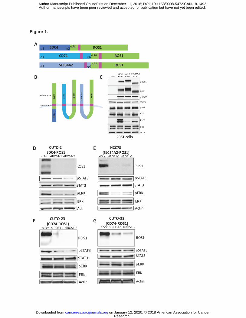
Figure 1. ROS1 fusion partners dictate differential activation of downstream signaling
pathways. (A) Diagram of the commonly occurring ROS1 fusion oncoproteins, which were
studied here. Pink denotes a transmembrane domain. (B) Topological configuration of ROS1
fusions based on CCTOP computational analysis (28). (C) Immunoblot analysis of 293T cells
transiently transfected for 48h with GFP, SDC4-ROS1, CD74-ROS1, or SLC34A2-ROS1, with
5h serum starvation. The pROS1 antibody used recognizes Y2274 of the full-length ROS1
protein. (D-G) Immunoblot analysis of patient-derived cell lines expressing (D) SDC4-ROS1, (E)
SLC34A2-ROS1, or (F-G) CD74-ROS1 with siRNA-mediated knockdown of ROS1 (55h after
transfection). Data shown in (C-G) are representative of 3 independent experiments.
Figure 2. MAPK pathway signaling is necessary and sufficient for survival of SDC4-
ROS1-positive and SLC34A2-ROS1-positive lines, but not a CD74-ROS1 positive line.
(A-C) Crystal violet quantification of ROS1 fusion-positive patient-derived cell lines (A) HCC78,
(B) CUTO-2 and (C) CUTO-23, expressing empty vector or constitutively active MEK-DD,
treated with DMSO or a dose-response of the ROS1 inhibitor crizotinib for 6 days. (D) Crystal
violet quantification of HCC78 (SLC34A2-ROS1), CUTO-2 (SDC4-ROS1), CUTO-23 (CD74-
ROS1), and CUTO-33 (CD74-ROS1) cell lines treated with DMSO or a dose-response of the
SHP2 inhibitor RMC-4550 for 6 days. (E) Half-maximal inhibitory concentration (IC50)
determination for the SHP2 inhibitor RMC-4550 in the indicated ROS1 patient-derived cell lines
based on crystal violet quantification of the experiment in (D). Data represent three independent
experiments. Data represented as mean +/- s.e.m.
Figure 3. Localization of ROS1 protein in isogenic BEAS-2B system reveals different
subcellular localization of the ROS1 fusion oncoproteins. Immunofluorescence and
confocal microscopy in BEAS-2B cells stably expressing SDC4-ROS1, SLC34A2-ROS1, and
Research. on January 12, 2020. © 2018 American Association for Cancercancerres.aacrjournals.org Downloaded from
Author manuscripts have been peer reviewed and accepted for publication but have not yet been edited. Author Manuscript Published OnlineFirst on December 11, 2018; DOI: 10.1158/0008-5472.CAN-18-1492
24
CD74-ROS1. Rows 1,2 = SDC4-ROS1; Rows 3,4 = SLC34A2-ROS1; Rows 5,6 = CD74-ROS1.
Antibodies used were specific for: (A-F) = ROS1; (G,I,K) = EEA1; (H,J,L) = Calnexin; and (M-
R) = DAPI; (S-X) = overlay image of the left 3 columns, with (right-most column) adjacent high
magnification image of representative cells (outlined by white boxes). Scale bars shown indicate
10µM. Images are representative of ≥ 10 fields and at least 2 biological replicate experiments.
Figure 4. Localization of ROS1 oncoproteins regulates engagement of downstream
signaling pathways. (A) Immunofluorescence and confocal microscopy of BEAS2-B cells
stably expressing an endosome-targeted FYVE-tagged CD74-ROS1 construct and stained with
the indicated antibodies. Far right panel = increased magnification of a representative individual
cell. Confocal images are representative of ≥ 10 fields and at least 2 independent experiments.
Scale bars shown indicate 10µM. (B) Immunoblot analysis of BEAS2-B cells transfected with
GFP, WT CD74-ROS1, or FYVE-CD74-ROS1. Immunoblot is representative of 3 independent
experiments.
Figure 5. MAPK pathway activation in ROS1 fusion oncoprotein-driven cancer models is
linked to increased tumorigenic properties in vivo. (A) Immunoblot analysis of ROS1 fusion
oncoprotein expression in isogenic NIH-3T3 cells. (B) Tumor growth rates of tumor xenografts
of 1x10⁶ NIH-3T3 ROS1 fusion oncoprotein-expressing cells described in (A) implanted into the
flanks of immunocompromised mice. (C) Tumor growth rates of tumor xenografts of 5x10⁵ cells
NIH-3T3 cells expressing CD74-ROS1 WT or FYVE-tagged CD74-ROS1. (D) Immunoblot
analysis of NIH-3T3 tumor xenograft explants expressing wild-type (WT) or FYVE-tagged CD74-
ROS1. Each lane represents an individual tumor. Data in (B-C) are shown as the mean of 6
tumors +/- s.e.m.
Research. on January 12, 2020. © 2018 American Association for Cancercancerres.aacrjournals.org Downloaded from
Author manuscripts have been peer reviewed and accepted for publication but have not yet been edited. Author Manuscript Published OnlineFirst on December 11, 2018; DOI: 10.1158/0008-5472.CAN-18-1492
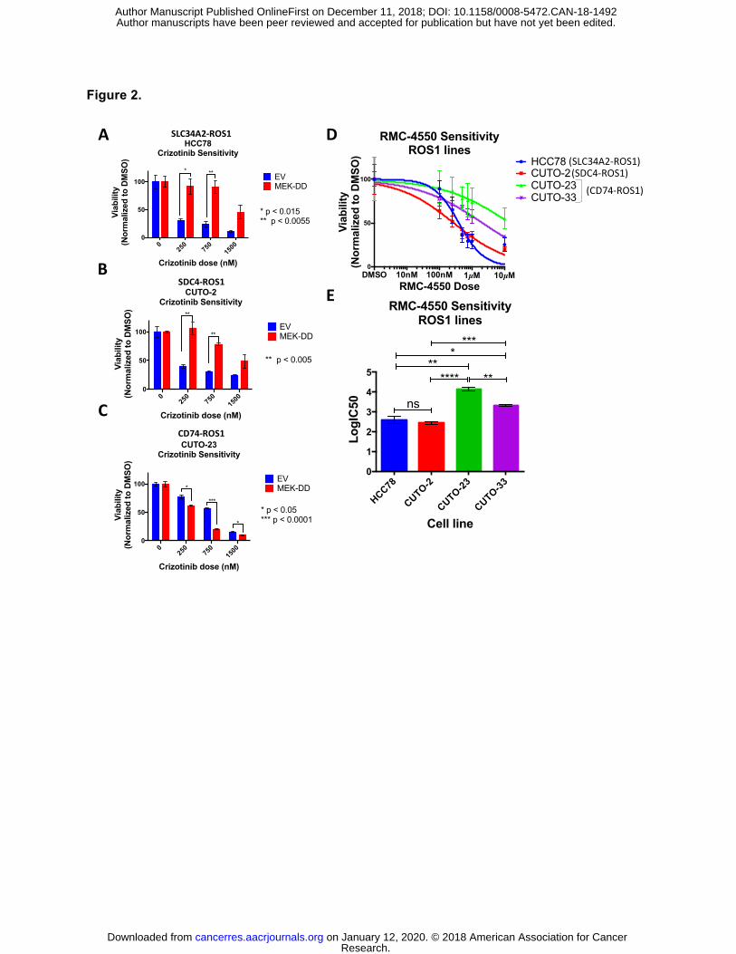
Figure 2.
025
075
015
000
50
100
Crizotinib dose (nM)
Viab
ility
(Nor
mal
ized
to D
MSO
)
CUTO-2Crizotinib Sensitivity
EVMEK-DD
**
**
** p < 0.005
025
075
015
000
50
100
Crizotinib dose (nM)
Viab
ility
(Nor
mal
ized
to D
MSO
)
CUTO-23 Crizotinib Sensitivity
EVMEK-DD*
*
**** p < 0.05*** p < 0.0001
C
025
075
015
000
50
100
Crizotinib dose (nM)
Viab
ility
(Nor
mal
ized
to D
MSO
)
HCC78 Crizotinib Sensitivity
EVMEK-DD
* **
* p < 0.015** p < 0.0055
A
B
HCC78
CUTO-2
CUTO-23
CUTO-330
1
2
3
4
5
RMC-4550 SensitivityROS1 lines
Cell line
LogI
C50 ns
****** **
****
D
E
SLC34A2-ROS1
SDC4-ROS1
CD74-ROS1
DMSO 10nM 100nM 1µM 10µM0
50
100
RMC-4550 Dose
Viab
ility
(Nor
mal
ized
to D
MSO
)
RMC-4550 SensitivityROS1 lines
HCC78CUTO-2CUTO-23CUTO-33
(SLC34A2-ROS1)(SDC4-ROS1)
(CD74-ROS1)
Research. on January 12, 2020. © 2018 American Association for Cancercancerres.aacrjournals.org Downloaded from
Author manuscripts have been peer reviewed and accepted for publication but have not yet been edited. Author Manuscript Published OnlineFirst on December 11, 2018; DOI: 10.1158/0008-5472.CAN-18-1492
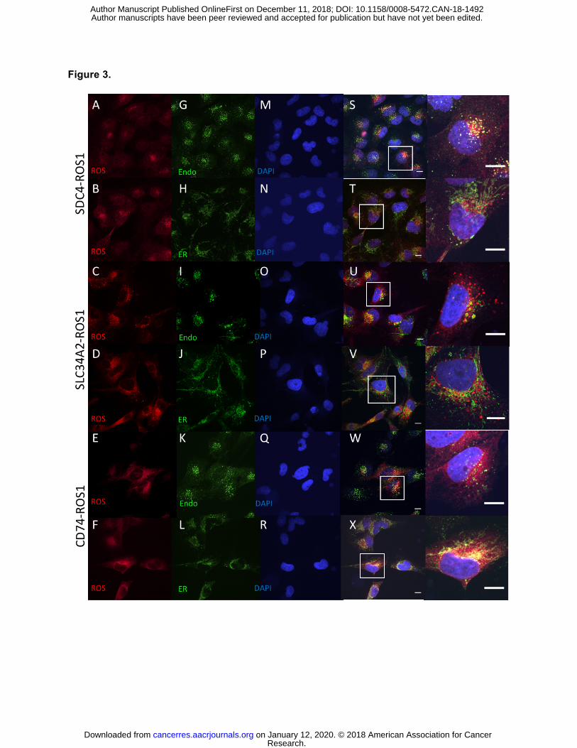
Figure 3.
Research. on January 12, 2020. © 2018 American Association for Cancercancerres.aacrjournals.org Downloaded from
Author manuscripts have been peer reviewed and accepted for publication but have not yet been edited. Author Manuscript Published OnlineFirst on December 11, 2018; DOI: 10.1158/0008-5472.CAN-18-1492
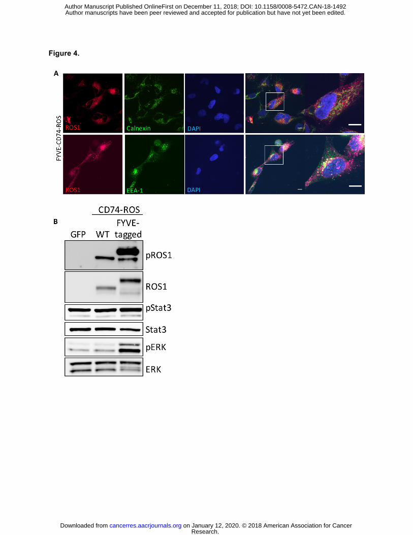
Figure 4.
Research. on January 12, 2020. © 2018 American Association for Cancercancerres.aacrjournals.org Downloaded from
Author manuscripts have been peer reviewed and accepted for publication but have not yet been edited. Author Manuscript Published OnlineFirst on December 11, 2018; DOI: 10.1158/0008-5472.CAN-18-1492
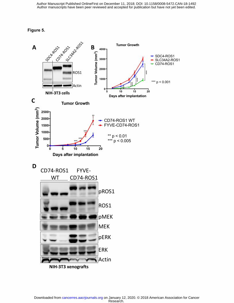
Figure 5.
NIH-3T3xenografts
CD74-ROS1WT
FYVE-CD74-ROS1
pROS1
ROS1
pERK
ERK
pMEKMEK
Actin
C
D
A
Actin
ROS1
NIH-3T3cells 5 10 15 200
1000
2000
3000
4000
Days after implantationTu
mor
Vol
ume
(mm
3 )
Tumor Growth
SDC4-ROS1SLC34A2-ROS1CD74-ROS1
***
******
***
*** p < 0.001
0 5 10 15 200
500
1000
1500
2000
2500
Days after implantation
Tum
or V
olum
e (m
m3 )
Tumor Growth
CD74-ROS1 WTFYVE-CD74-ROS1
***
***
***
**
** p < 0.01*** p < 0.005
B
Research. on January 12, 2020. © 2018 American Association for Cancercancerres.aacrjournals.org Downloaded from
Author manuscripts have been peer reviewed and accepted for publication but have not yet been edited. Author Manuscript Published OnlineFirst on December 11, 2018; DOI: 10.1158/0008-5472.CAN-18-1492
Published OnlineFirst December 11, 2018.Cancer Res Dana S. Neel, David V Allegakoen, Victor Olivas, et al. signaling by ROS1 kinase fusion proteinsDifferential subcellular localization regulates oncogenic
Updated version
10.1158/0008-5472.CAN-18-1492doi:
Access the most recent version of this article at:
Material
Supplementary
http://cancerres.aacrjournals.org/content/suppl/2018/12/11/0008-5472.CAN-18-1492.DC1
Access the most recent supplemental material at:
Manuscript
Authoredited. Author manuscripts have been peer reviewed and accepted for publication but have not yet been
E-mail alerts related to this article or journal.Sign up to receive free email-alerts
Subscriptions
Reprints and
To order reprints of this article or to subscribe to the journal, contact the AACR Publications
Permissions
Rightslink site. Click on "Request Permissions" which will take you to the Copyright Clearance Center's (CCC)
.http://cancerres.aacrjournals.org/content/early/2018/12/11/0008-5472.CAN-18-1492To request permission to re-use all or part of this article, use this link
Research. on January 12, 2020. © 2018 American Association for Cancercancerres.aacrjournals.org Downloaded from
Author manuscripts have been peer reviewed and accepted for publication but have not yet been edited. Author Manuscript Published OnlineFirst on December 11, 2018; DOI: 10.1158/0008-5472.CAN-18-1492