Embed Size (px)
Citation preview
Skin and the Integumentary
System
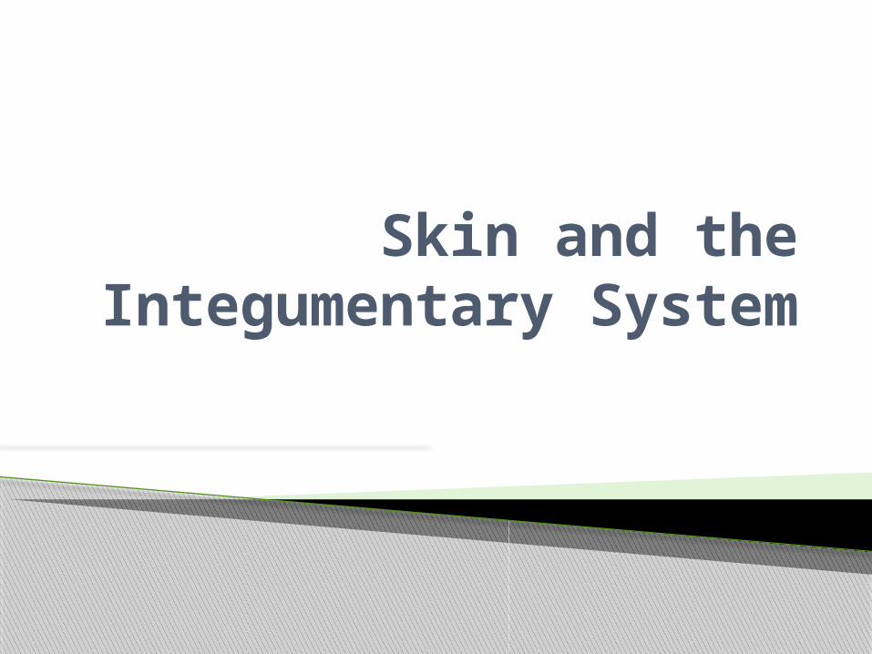
Cutaneous membrane and certain accessory organs◦ Epithelium and connective tissue
Integumentary System
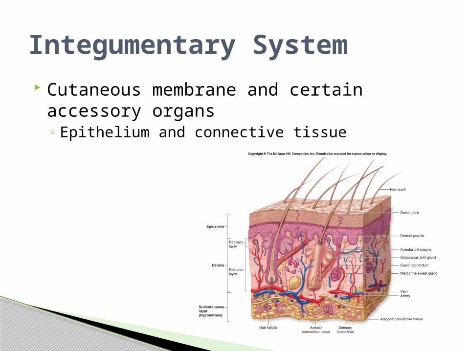
Serous: line body cavities that lack outside openings◦ Thorax, abdomen◦ Simple squamous epithelium and loose
connective tissue Secrete watery serous fluid: lubricated membrane
surface
Types of Membranes
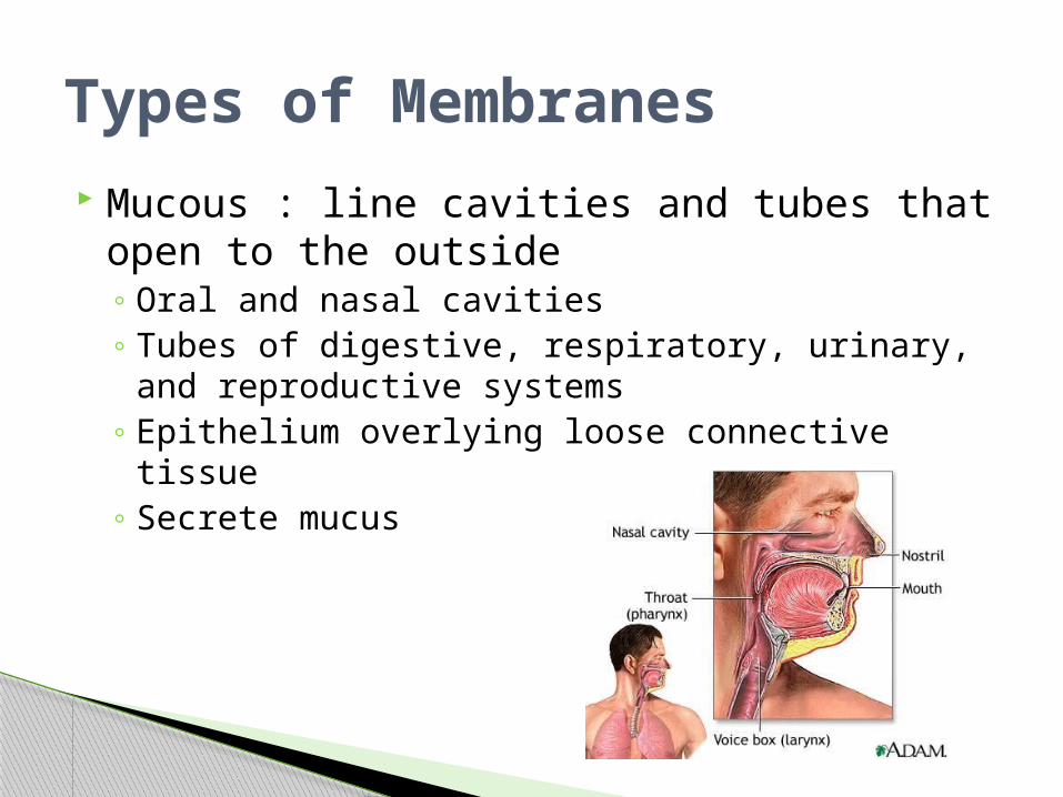
Mucous : line cavities and tubes that open to the outside◦ Oral and nasal cavities◦ Tubes of digestive, respiratory, urinary, and
reproductive systems◦ Epithelium overlying loose connective tissue◦ Secrete mucus
Types of Membranes
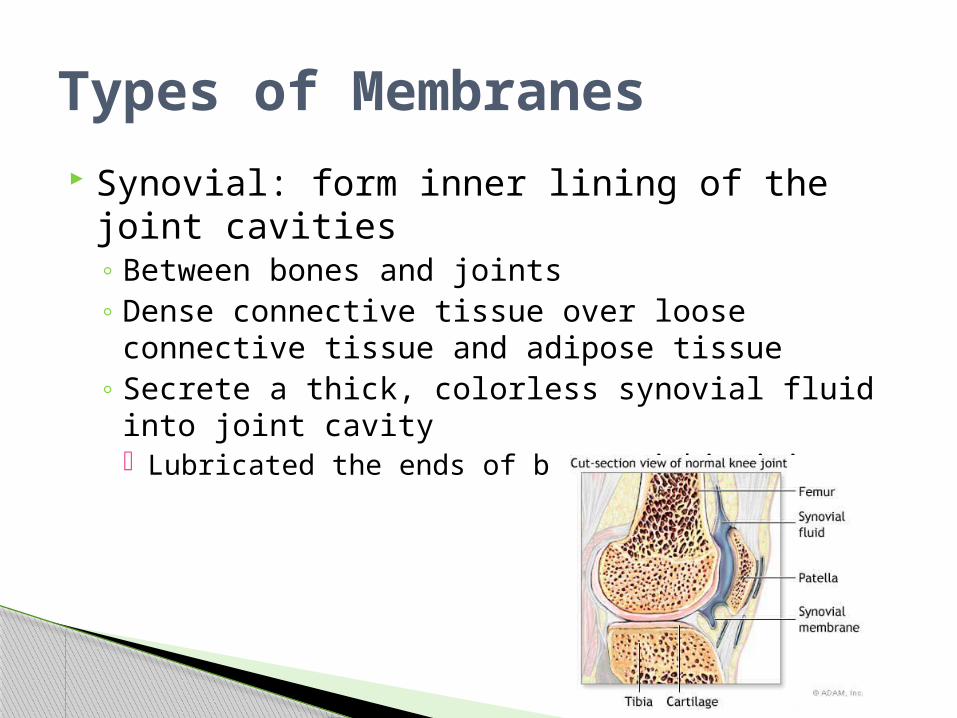
Synovial: form inner lining of the joint cavities◦ Between bones and joints◦ Dense connective tissue over loose connective
tissue and adipose tissue◦ Secrete a thick, colorless synovial fluid into joint
cavity Lubricated the ends of bones within joint
Types of Membranes

Cutaneous membrane: skin◦ One of the larger and more
versatile organs◦ Vital for homeostasis
Functions◦ Protective covering◦ Regulate body temperature◦ Retards water loss from deeper
tissue◦ Houses sensory receptors◦ Synthesizes various biochemicals◦ Excretes small quantities of
waste
Types of Membranes
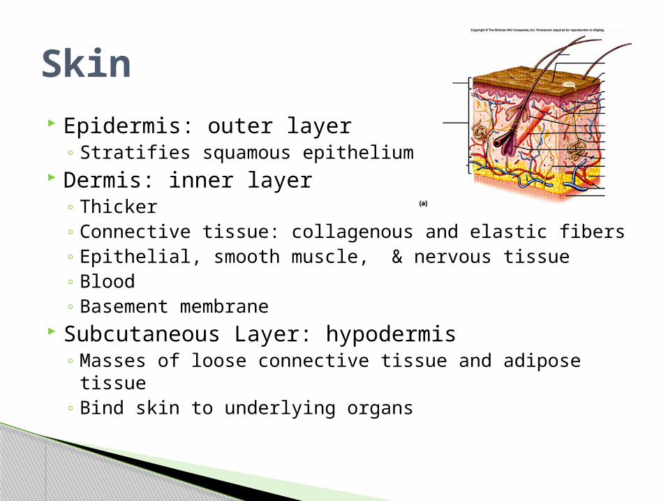
Epidermis: outer layer◦ Stratifies squamous epithelium
Dermis: inner layer◦ Thicker◦ Connective tissue: collagenous and elastic fibers◦ Epithelial, smooth muscle, & nervous tissue◦ Blood◦ Basement membrane
Subcutaneous Layer: hypodermis◦ Masses of loose connective tissue and adipose tissue◦ Bind skin to underlying organs
Skin
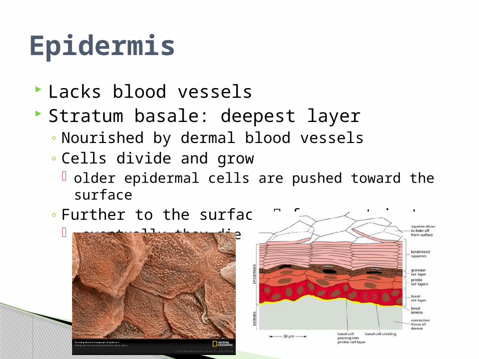
Lacks blood vessels Stratum basale: deepest layer
◦ Nourished by dermal blood vessels◦ Cells divide and grow
older epidermal cells are pushed toward the surface◦ Further to the surface fewer nutrients
eventually they die
Epidermis
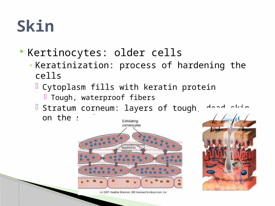
Kertinocytes: older cells◦ Keratinization: process of hardening the cells
Cytoplasm fills with keratin protein Tough, waterproof fibers
Stratum corneum: layers of tough, dead skin on the surface
Skin
Functions◦ Shields against excessive water lose◦ Mechanical injury◦ Effects of harmful chemicals◦ Protection against disease causing agents
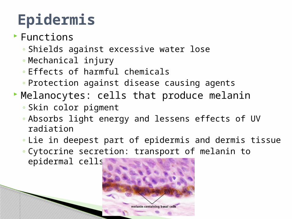
Melanocytes: cells that produce melanin◦ Skin color pigment◦ Absorbs light energy and lessens effects of UV
radiation◦ Lie in deepest part of epidermis and dermis tissue◦ Cytocrine secretion: transport of melanin to epidermal
cells
Epidermis
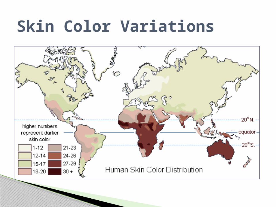
Due to melanin◦ All people have about the same number of
melanocytes◦ Genetic◦ Variations in color
Amount of melanin produce Distribution and size of pigment granules
◦ Sunlight, UV light, X-rays stimulate production of extra melanin
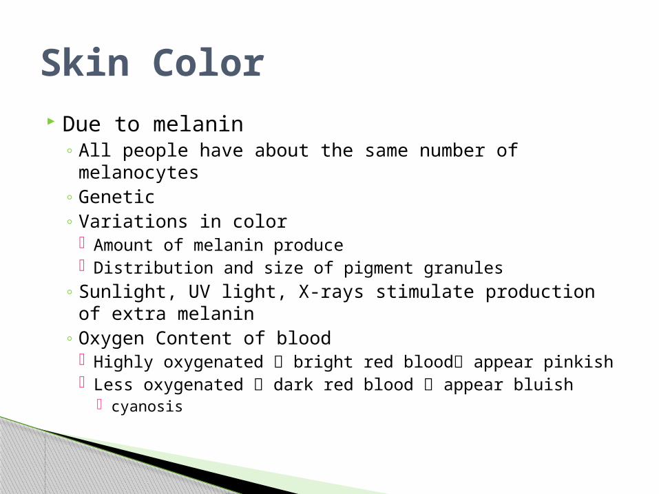
◦ Oxygen Content of blood Highly oxygenated bright red blood appear pinkish Less oxygenated dark red blood appear bluish
cyanosis
Skin Color
Skin Color Variations
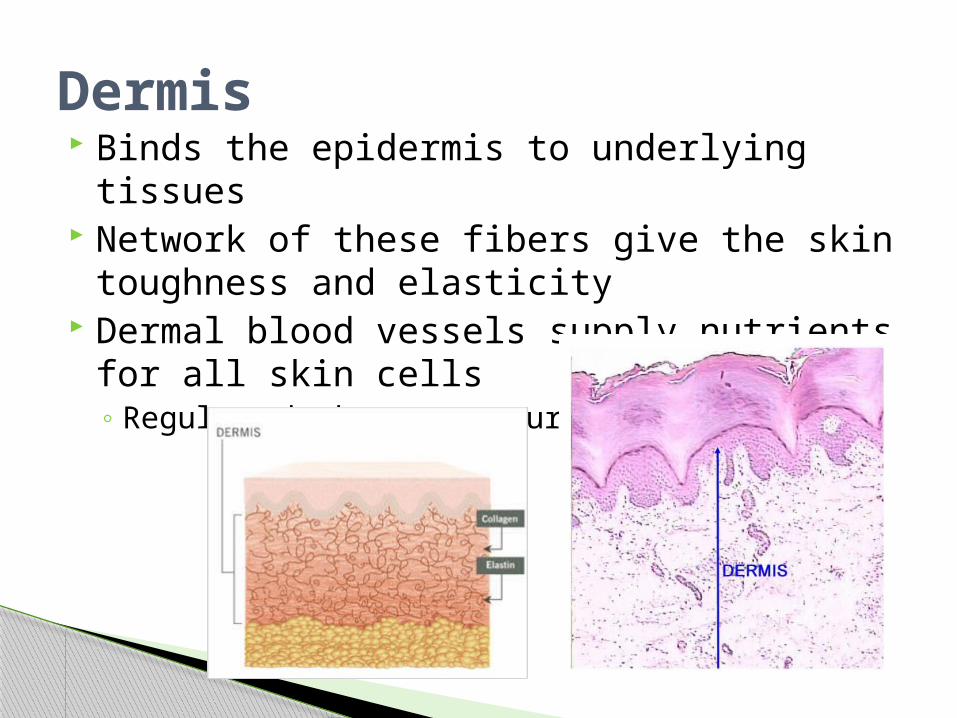
Binds the epidermis to underlying tissues Network of these fibers give the skin
toughness and elasticity Dermal blood vessels supply nutrients for all
skin cells◦ Regulate body temperature
Dermis
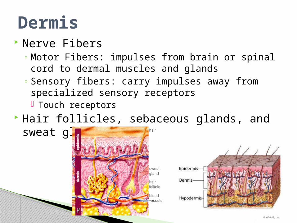
Nerve Fibers◦ Motor Fibers: impulses from brain or spinal cord to
dermal muscles and glands◦ Sensory fibers: carry impulses away from
specialized sensory receptors Touch receptors
Hair follicles, sebaceous glands, and sweat glands
Dermis
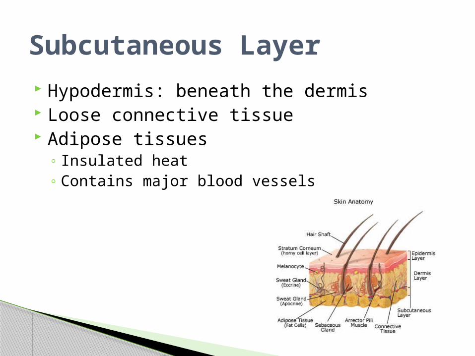
Hypodermis: beneath the dermis Loose connective tissue Adipose tissues
◦ Insulated heat◦ Contains major blood vessels
Subcutaneous Layer
Accessory Skin Organs
Present on all skin surfaces except the palm, soles, lips, nipples, and parts of external reproductive organs
Hair Follicle: tubelike depression from which the hair develops◦ Extends to the dermis and contains the hair root◦ Nourished from dermal blood vessels◦ With division and growth, older cells are pushed
toward the top◦ Become keratinized as they move up
Shaft: structure that extends from skin surface◦ Dead epidermal cells
Hair Follicles
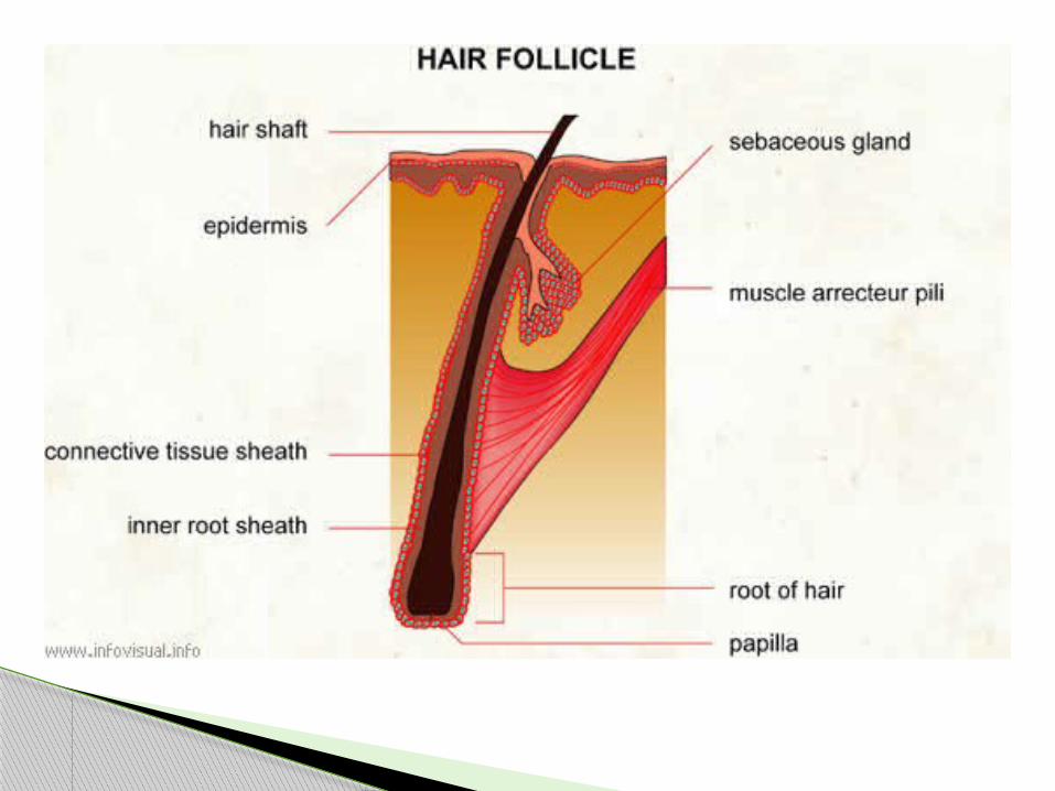
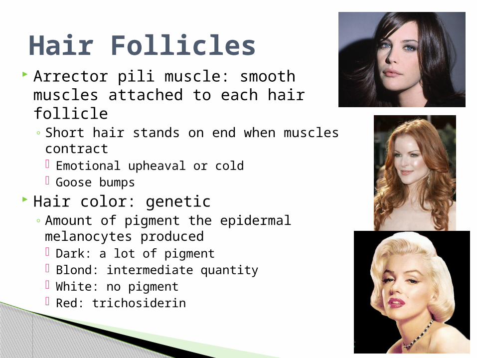
Arrector pili muscle: smooth muscles attached to each hair follicle◦ Short hair stands on end when muscles
contract Emotional upheaval or cold Goose bumps
Hair color: genetic◦ Amount of pigment the epidermal
melanocytes produced Dark: a lot of pigment Blond: intermediate quantity White: no pigment Red: trichosiderin
Hair Follicles
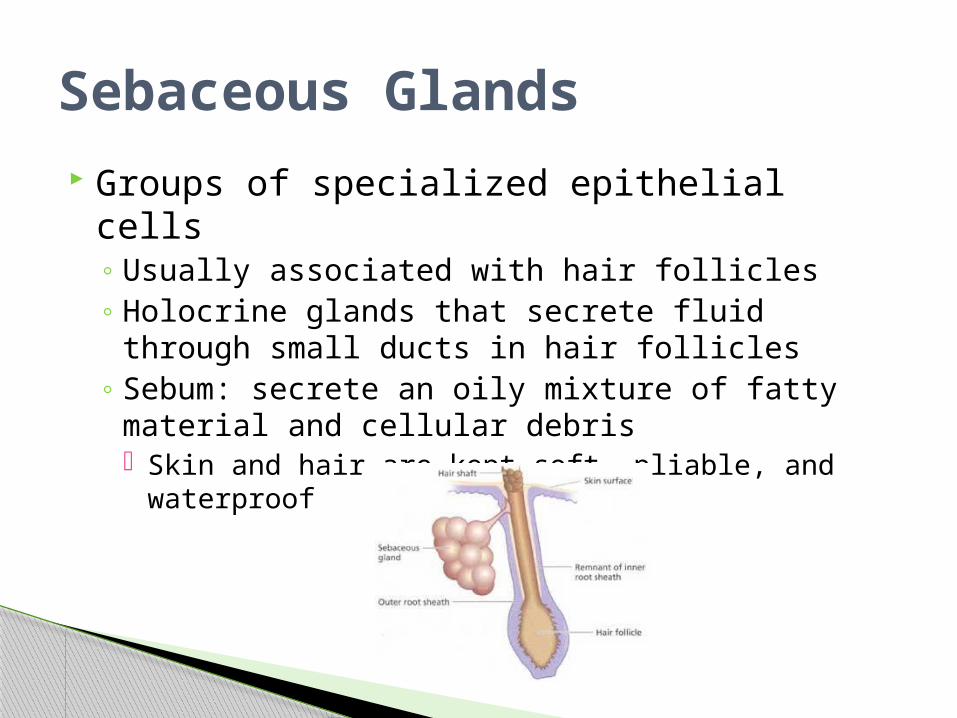
Groups of specialized epithelial cells◦ Usually associated with hair follicles◦ Holocrine glands that secrete fluid through small
ducts in hair follicles◦ Sebum: secrete an oily mixture of fatty material
and cellular debris Skin and hair are kept soft, pliable, and waterproof
Sebaceous Glands
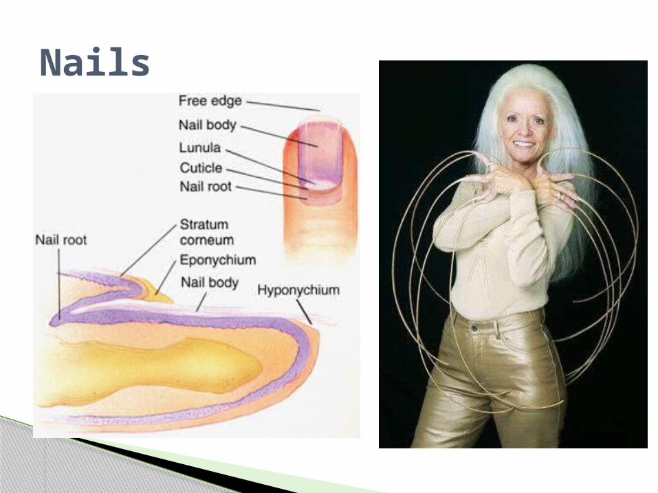
Protective coverings on the ends of fingers and toes
Keratinized stratified squamous epithelial cells
Nail Root: where nail originates◦ Near the nail’s proximal end
Lunula: whitish, half-moon shaped area◦ Most active growing region
Nail Bed: epithelium that nail slides over as it grows◦ Remains attached
Nails
Nails
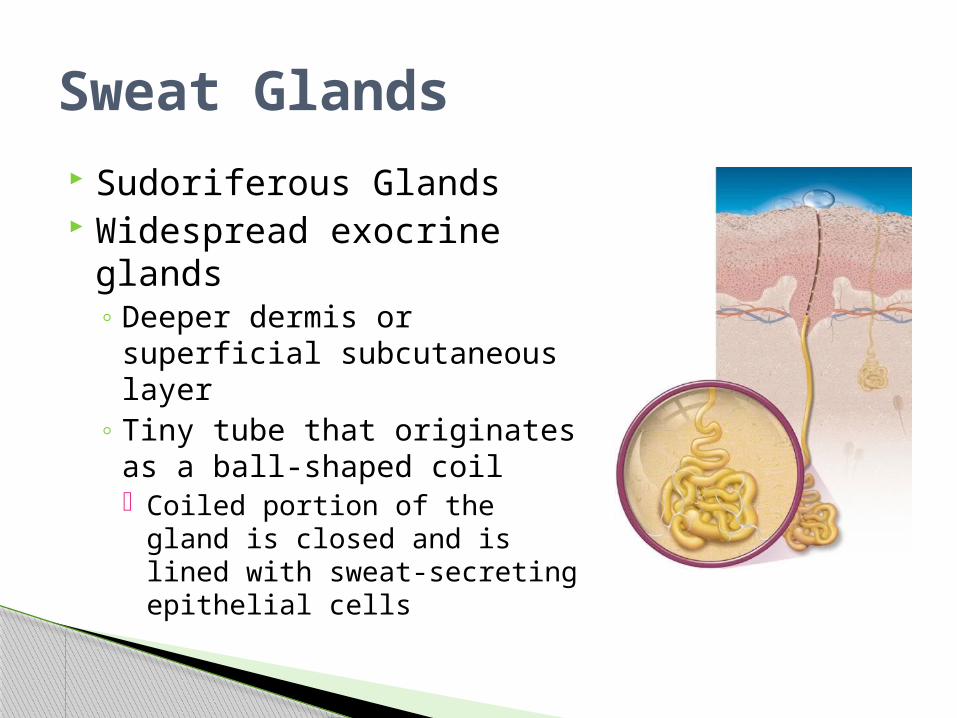
Sudoriferous Glands Widespread exocrine
glands ◦ Deeper dermis or superficial
subcutaneous layer◦ Tiny tube that originates as a
ball-shaped coil Coiled portion of the gland is
closed and is lined with sweat-secreting epithelial cells
Sweat Glands
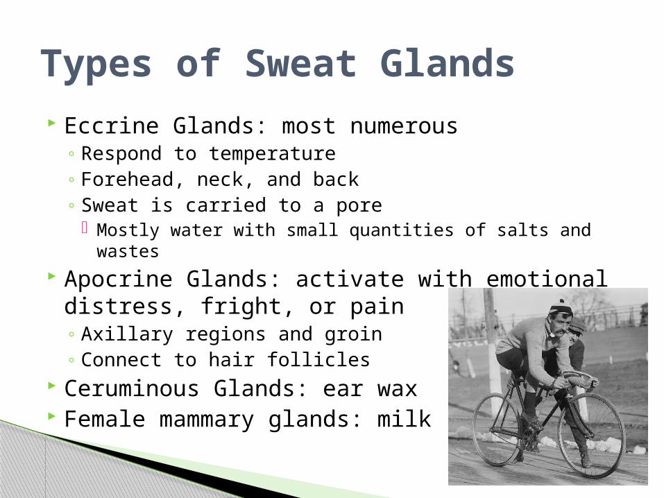
Eccrine Glands: most numerous◦ Respond to temperature◦ Forehead, neck, and back◦ Sweat is carried to a pore
Mostly water with small quantities of salts and wastes Apocrine Glands: activate with emotional
distress, fright, or pain◦ Axillary regions and groin◦ Connect to hair follicles
Ceruminous Glands: ear wax Female mammary glands: milk
Types of Sweat Glands
Regulation of Body Temperature and Wound
Healing
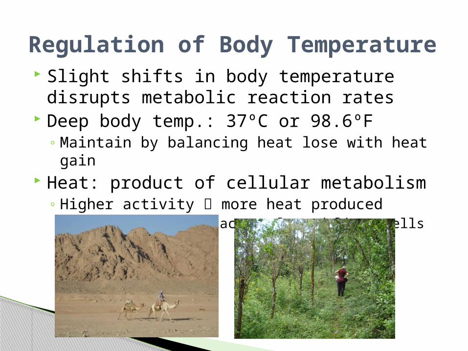
Slight shifts in body temperature disrupts metabolic reaction rates
Deep body temp.: 37ºC or 98.6ºF◦ Maintain by balancing heat lose with heat gain
Heat: product of cellular metabolism◦ Higher activity more heat produced
Skeletal and cardiac muscle and liver cells
Regulation of Body Temperature
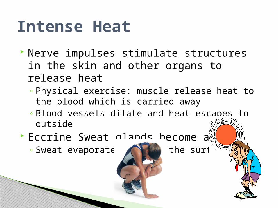
Nerve impulses stimulate structures in the skin and other organs to release heat◦ Physical exercise: muscle release heat to the
blood which is carried away◦ Blood vessels dilate and heat escapes to outside
Eccrine Sweat glands become active◦ Sweat evaporates cools the surface
Intense Heat
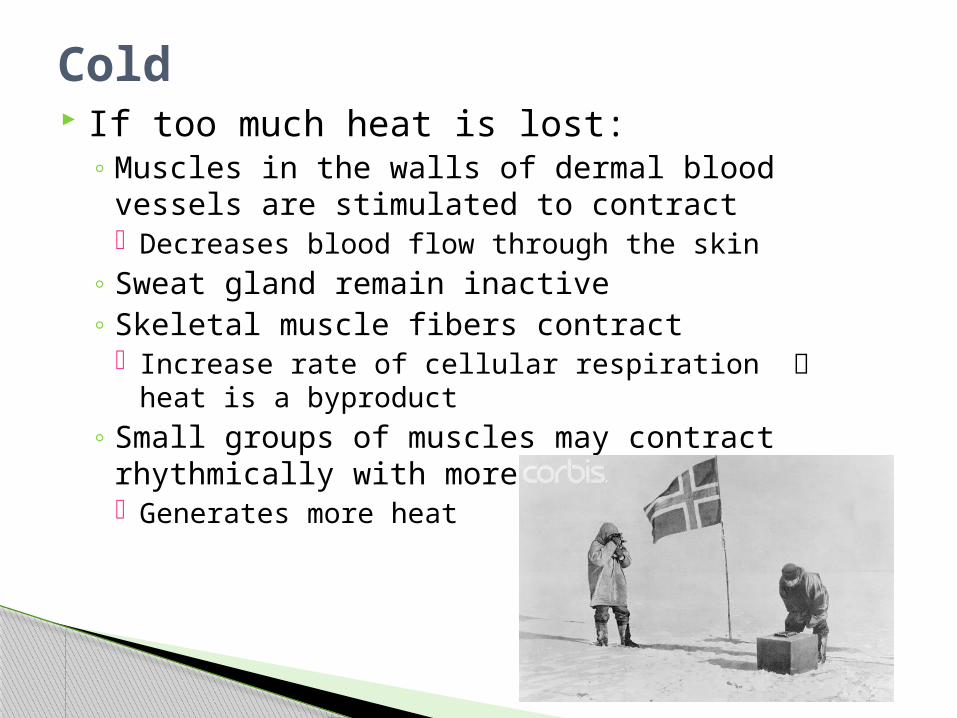
If too much heat is lost:◦ Muscles in the walls of dermal blood vessels are
stimulated to contract Decreases blood flow through the skin
◦ Sweat gland remain inactive◦ Skeletal muscle fibers contract
Increase rate of cellular respiration heat is a byproduct
◦ Small groups of muscles may contract rhythmically with more force, shiver Generates more heat
Cold
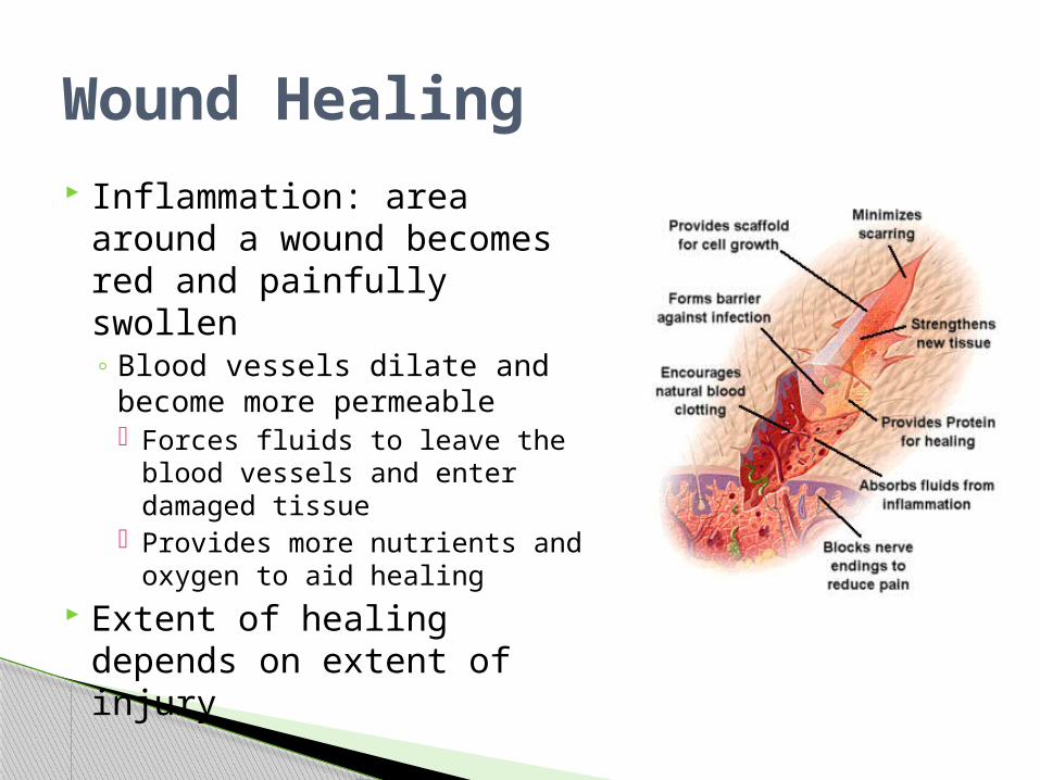
Inflammation: area around a wound becomes red and painfully swollen◦ Blood vessels dilate and
become more permeable Forces fluids to leave the
blood vessels and enter damaged tissue
Provides more nutrients and oxygen to aid healing
Extent of healing depends on extent of injury
Wound Healing
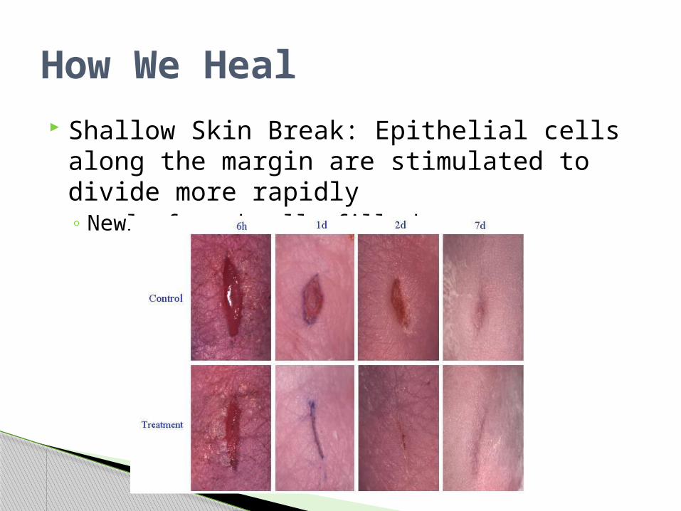
Shallow Skin Break: Epithelial cells along the margin are stimulated to divide more rapidly◦ Newly formed cells fill the gap
How We Heal
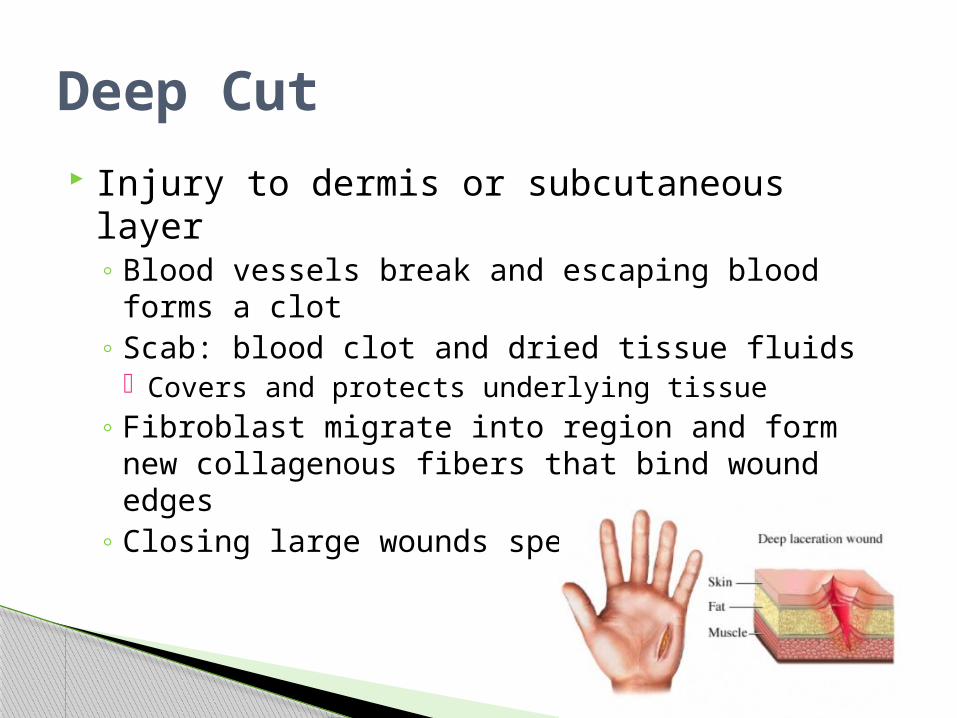
Injury to dermis or subcutaneous layer◦ Blood vessels break and escaping blood forms a
clot◦ Scab: blood clot and dried tissue fluids
Covers and protects underlying tissue◦ Fibroblast migrate into region and form new
collagenous fibers that bind wound edges◦ Closing large wounds speeds the process
Deep Cut
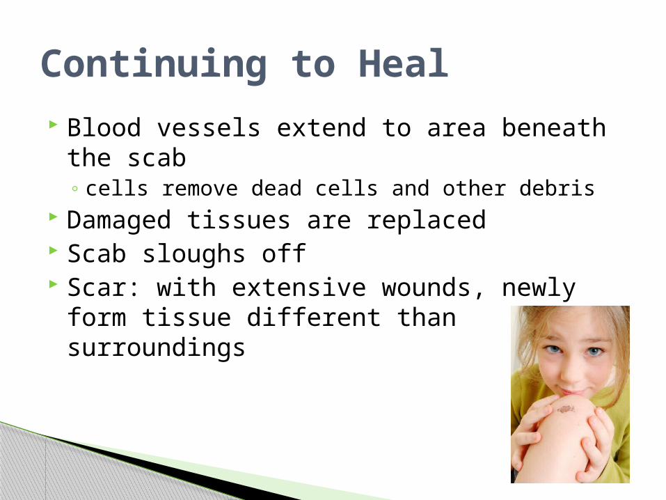
Blood vessels extend to area beneath the scab◦ cells remove dead cells and other debris
Damaged tissues are replaced Scab sloughs off Scar: with extensive wounds, newly form
tissue different than surroundings
Continuing to Heal
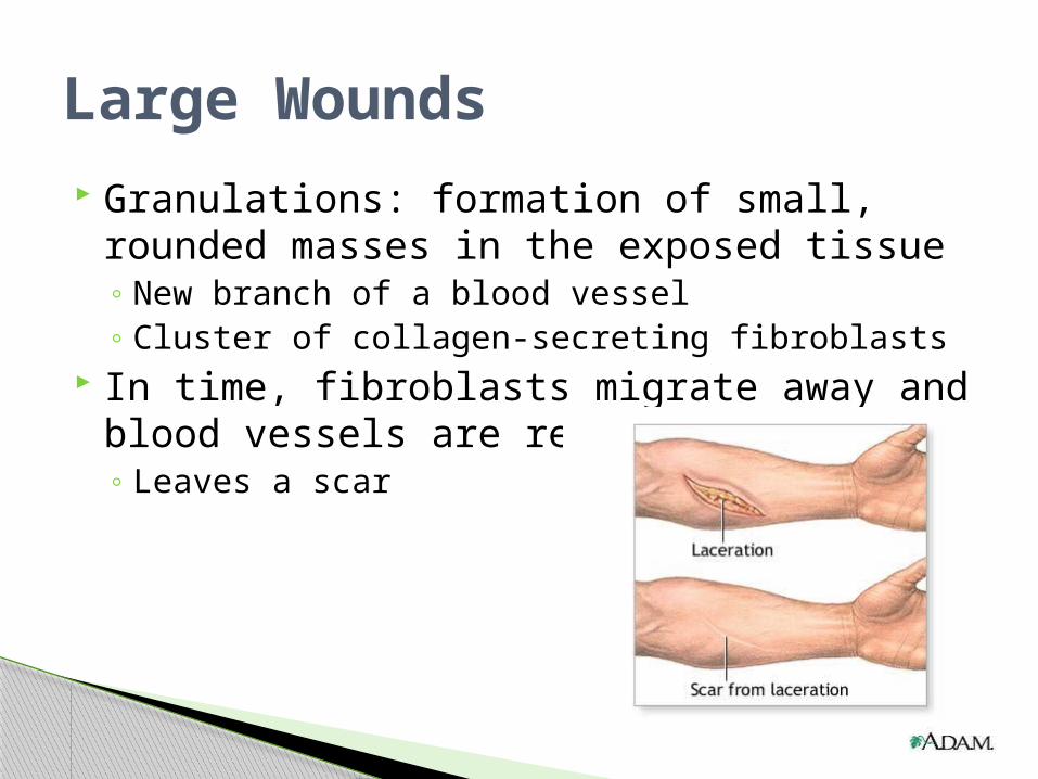
Granulations: formation of small, rounded masses in the exposed tissue◦ New branch of a blood vessel ◦ Cluster of collagen-secreting fibroblasts
In time, fibroblasts migrate away and blood vessels are reabsorbed◦ Leaves a scar
Large Wounds