Embed Size (px)
Citation preview
© copyright UEA [email protected]
Image Guided Radiation Therapy
Dr. Mark FisherSchool of Computing SciencesUEA Norwich UK
© copyright UEA [email protected]
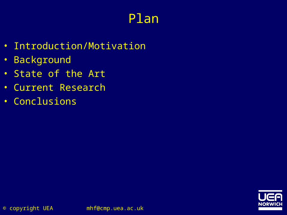
Plan
• Introduction/Motivation• Background• State of the Art• Current Research• Conclusions
© copyright UEA [email protected]
Introduction
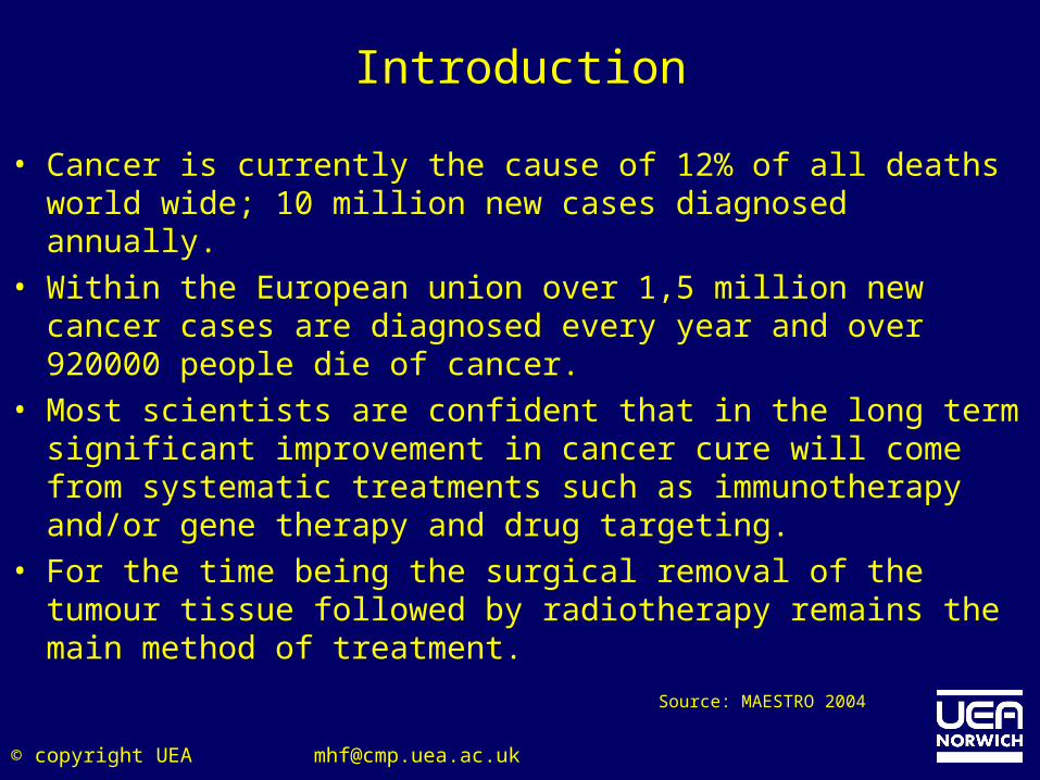
• Cancer is currently the cause of 12% of all deaths world wide; 10 million new cases diagnosed annually.
• Within the European union over 1,5 million new cancer cases are diagnosed every year and over 920000 people die of cancer.
• Most scientists are confident that in the long term significant improvement in cancer cure will come from systematic treatments such as immunotherapy and/or gene therapy and drug targeting.
• For the time being the surgical removal of the tumour tissue followed by radiotherapy remains the main method of treatment.
Source: MAESTRO 2004
© copyright UEA [email protected]
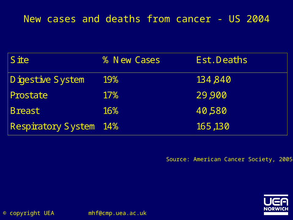
New cases and deaths from cancer - US 2004
Site % New Cases Est. Deaths
Digestive System 19% 134,840
Prostate 17% 29,900
Breast 16% 40,580
Respiratory System 14% 165,130
Source: American Cancer Society, 2005
© copyright UEA [email protected]
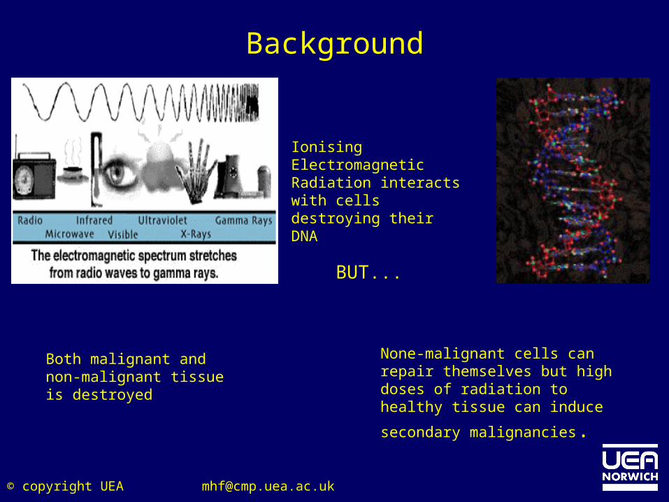
Background
Ionising Electromagnetic Radiation interacts with cells destroying their DNA
None-malignant cells can repair themselves but high doses of radiation to healthy tissue can
induce secondary malignancies.
Both malignant and non-malignant tissue is destroyed
BUT...
© copyright UEA [email protected]
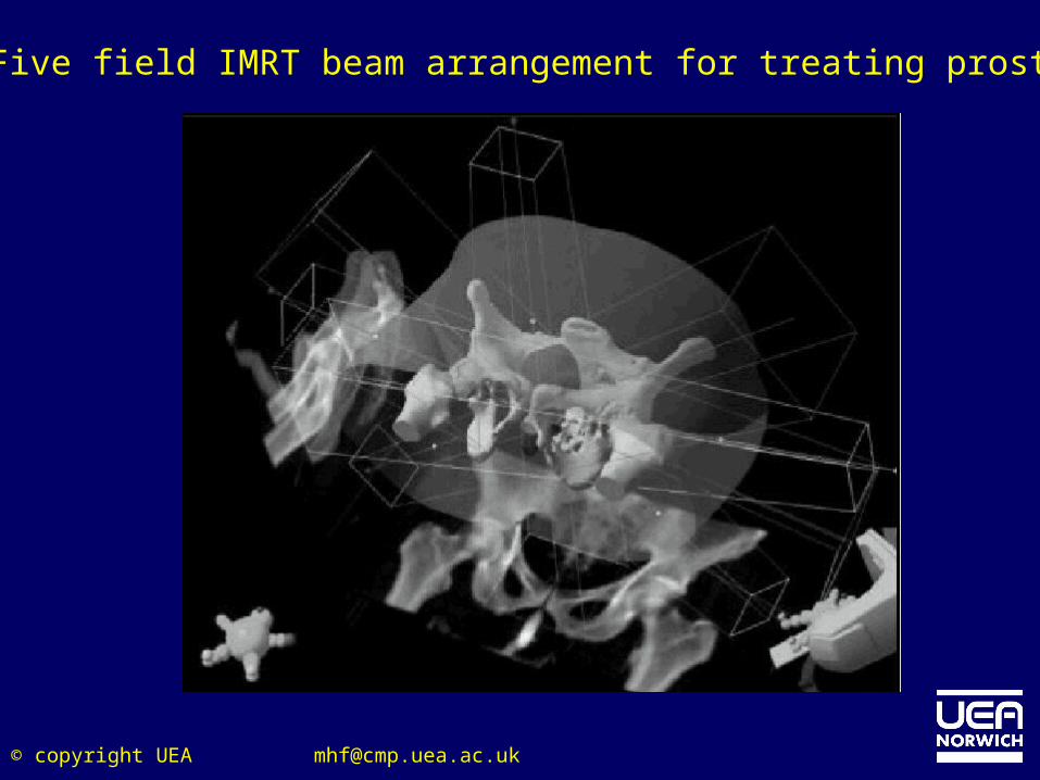
Aim of Radiotherapy Treatment I
• To deliver a high dose of Radiation to the tumour whileand a low dose to healthy tissue and organs at risk.– Possible through the use of multiple treatment fields (beams).
© copyright UEA [email protected]
Radiation Therapy Treatment Delivery
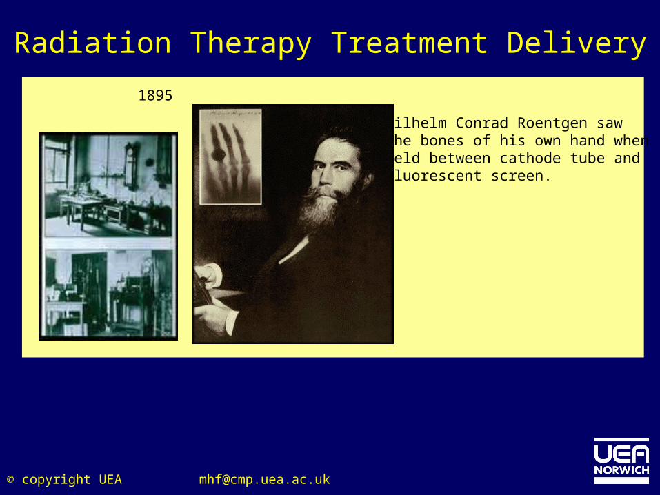
1895
Wilhelm Conrad Roentgen saw the bones of his own hand when held between cathode tube and fluorescent screen.
© copyright UEA [email protected]
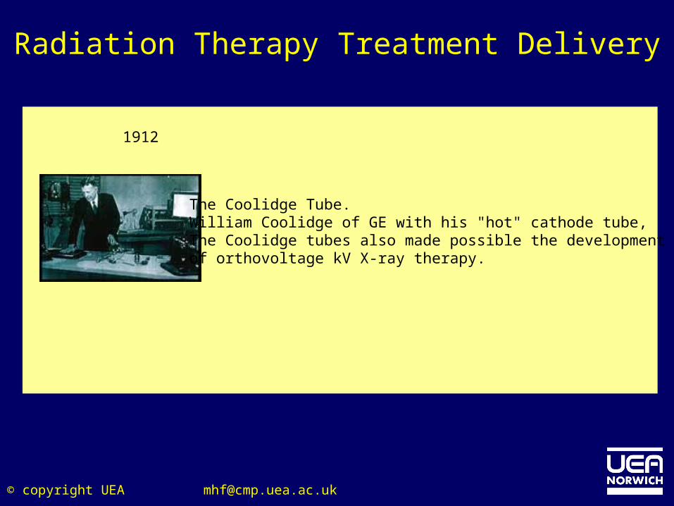
The Coolidge Tube. William Coolidge of GE with his "hot" cathode tube,The Coolidge tubes also made possible the development of orthovoltage kV X-ray therapy.
1912
Radiation Therapy Treatment Delivery
© copyright UEA [email protected]
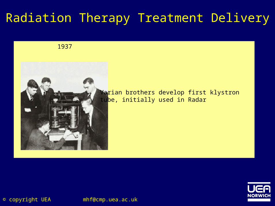
1937
Varian brothers develop first klystrontube, initially used in Radar
Radiation Therapy Treatment Delivery
© copyright UEA [email protected]
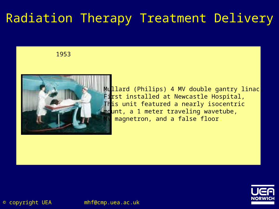
1953
Mullard (Philips) 4 MV double gantry linac. First installed at Newcastle Hospital, This unit featured a nearly isocentric mount, a 1 meter traveling wavetube, MV magnetron, and a false floor.
Radiation Therapy Treatment Delivery
© copyright UEA [email protected]
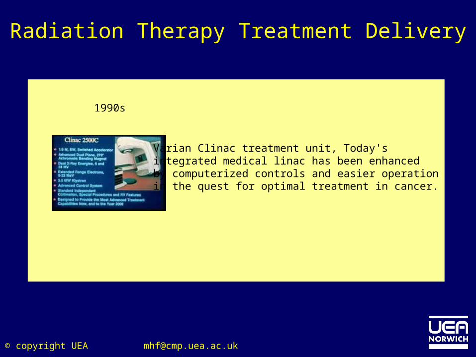
Varian Clinac treatment unit, Today's integrated medical linac has been enhanced by computerized controls and easier operation in the quest for optimal treatment in cancer.
1990s
Radiation Therapy Treatment Delivery
© copyright UEA [email protected]
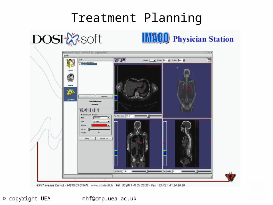
Radiation Therapy Treatment Planning
• In the early days of radiotherapy, the X-ray beams were rectangular or square in shape and were directed at the tumor from two to four different angles.– Since the dosages delivered were uniform in strength there was
some damage to healthy tissue.
• In the 1970’s conformal RT was developed. This approach used lead-alloy blocks to shape the beam.– The dose was ‘conformed’ to the shape of the tumour, healthy
tissue is spared.
© copyright UEA [email protected]
ICRU 50/62
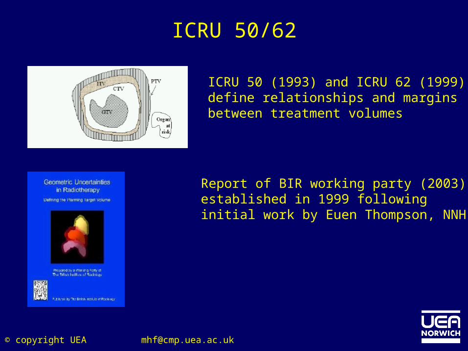
ICRU 50 (1993) and ICRU 62 (1999)define relationships and marginsbetween treatment volumes
Report of BIR working party (2003),established in 1999 followinginitial work by Euen Thompson, NNH
© copyright UEA [email protected]
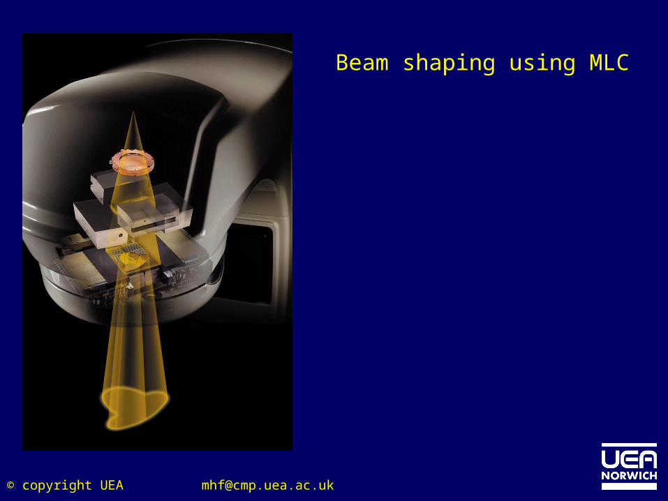
Intensity Modulated Radiotherapy Treatment
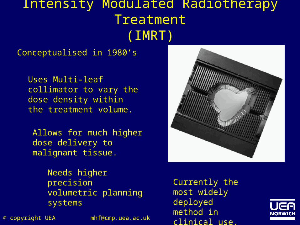
(IMRT)Conceptualised in 1980’s
Uses Multi-leaf collimator to vary the dose density within the treatment volume.
Allows for much higher dose delivery to malignant tissue.
Needs higher precision volumetric planning systems
Currently the most widely deployed method in clinical use.
© copyright UEA [email protected]
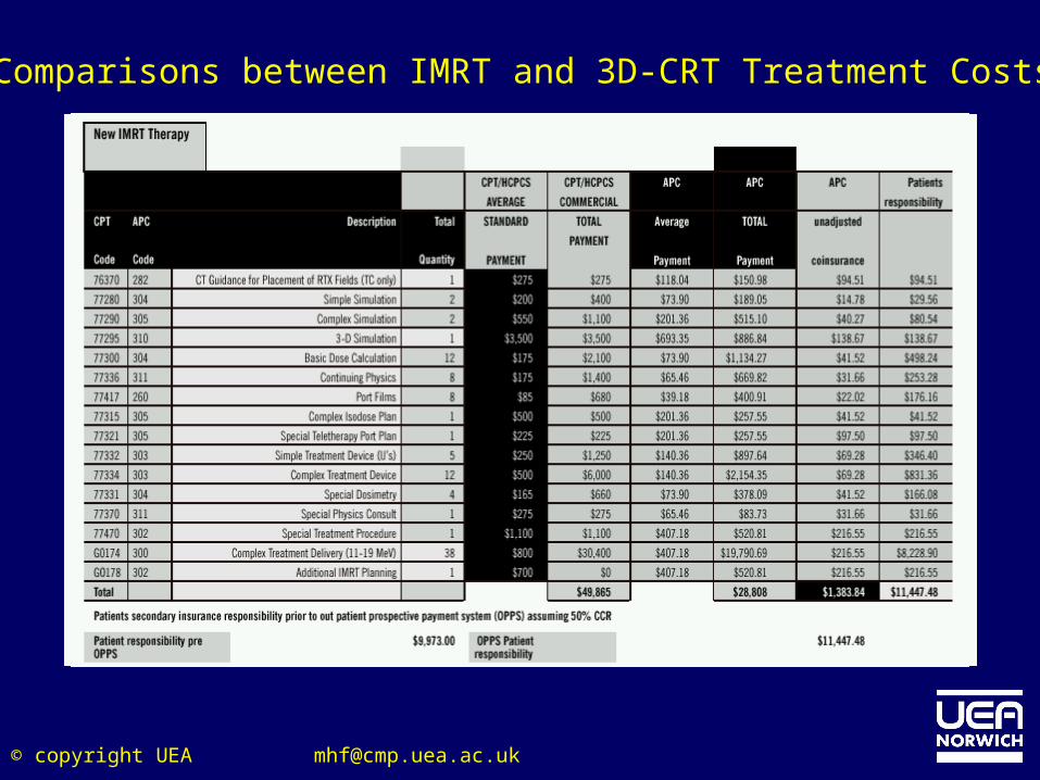
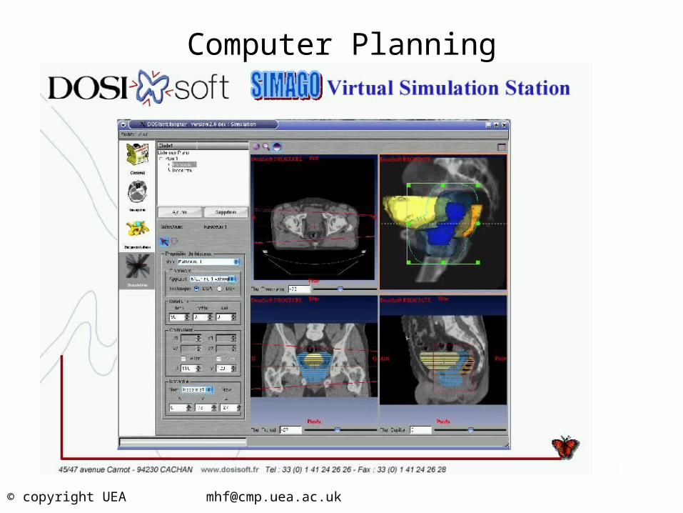
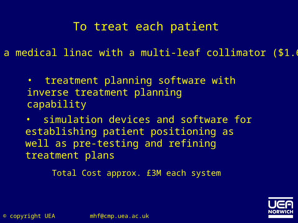
• treatment planning software with inverse treatment planning capability
Total Cost approx. £3M each system
To treat each patient
• a medical linac with a multi-leaf collimator ($1.6M)
• simulation devices and software for establishing patient positioning as well as pre-testing and refining treatment plans
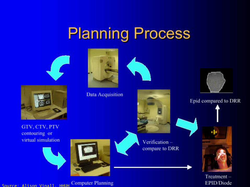
[email protected]© copyright UEASource: Alison Vinall, HHUH
© copyright UEA [email protected]
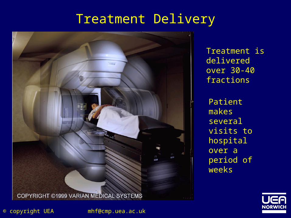
Treatment Delivery
Treatment is delivered over 30-40 fractions
Patient makes several visits to hospital over a period of weeks
© copyright UEA [email protected]
Accounting For Organ Movement
• “Most of the development of IMRT has taken place assuming that the organs don't move from fraction to fraction and are well represented by their positions determined from some pre-planning 3D imaging study, be it x-ray CT, MR or functional imaging. As the ability to conform to the target has now reached near perfection, attention is now turning to not accepting this limitation and attempting to quantitate organ movement and account for it in IMRT planning and delivery”.
• “IMRT of the moving patient is like completing a jigsaw on a jelly”
Prof. Steve Webb, Royal Marsden Hosp.
© copyright UEA [email protected]
Types of Motion
• Patient set-up errors– Position-related organ motion which can be minimised if the
patient's planning scan is performed while the patient is immobilised and in the treatment position.
• Inter-fraction motion– i.e. motion that occurs when the target volume changes from
day to day. This is a problem for organs that are close to or part of the digestive/excretory system. This work is collated under various headings: gynaecological tumours, prostate (the largest group), bladder and rectum.
• Intra-fraction– generally due to respiratory and cardiac functions which disturb
other organs. This work is collated under headings: liver, diaphragm, kidneys, pancreas, lung tumours and prostate.
© copyright UEA [email protected]
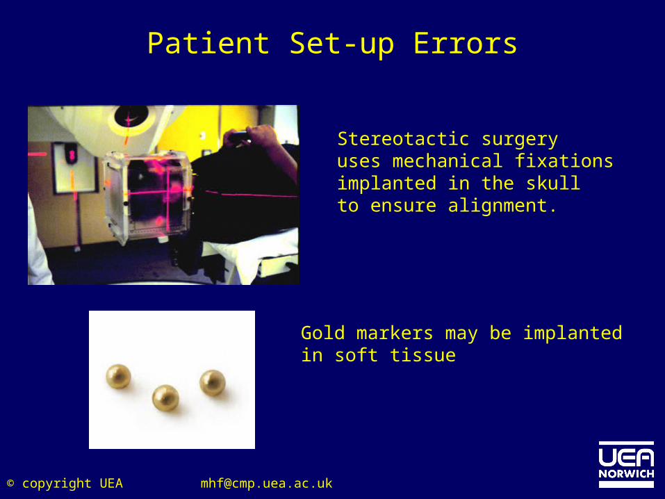
Patient Set-up Errors
Stereotactic surgeryuses mechanical fixationsimplanted in the skullto ensure alignment.
Gold markers may be implantedin soft tissue
© copyright UEA [email protected]
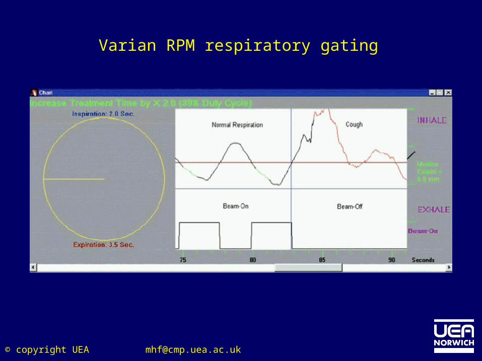
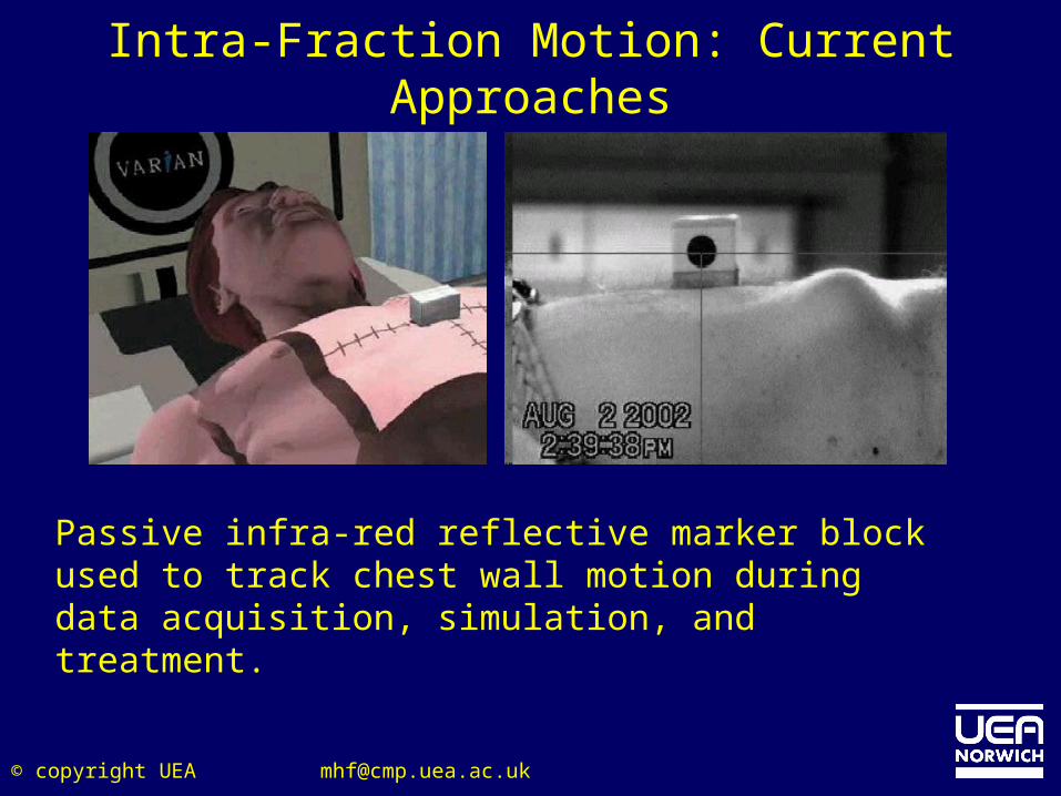
Passive infra-red reflective marker block used to track chest wall motion during data acquisition, simulation, and treatment.
Intra-Fraction Motion: Current Approaches
© copyright UEA [email protected]
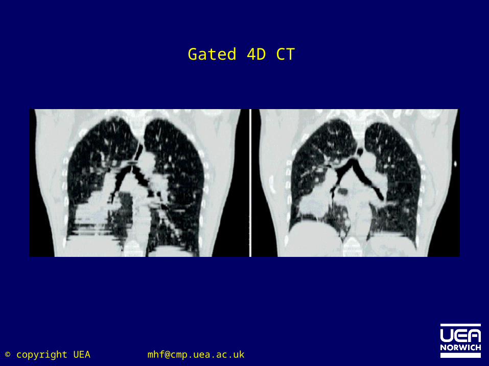
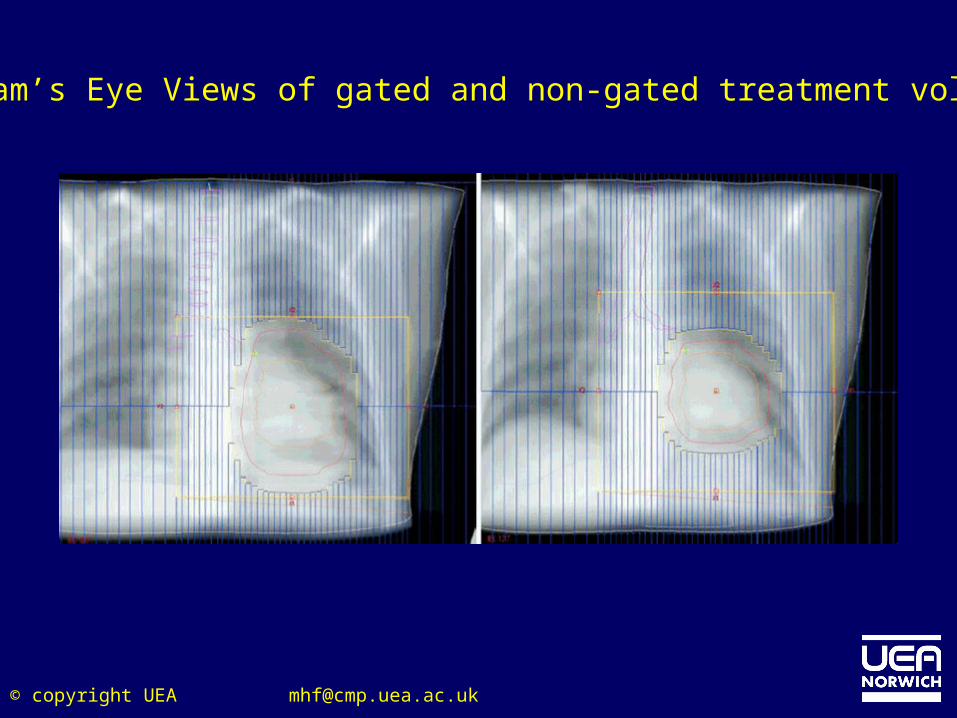
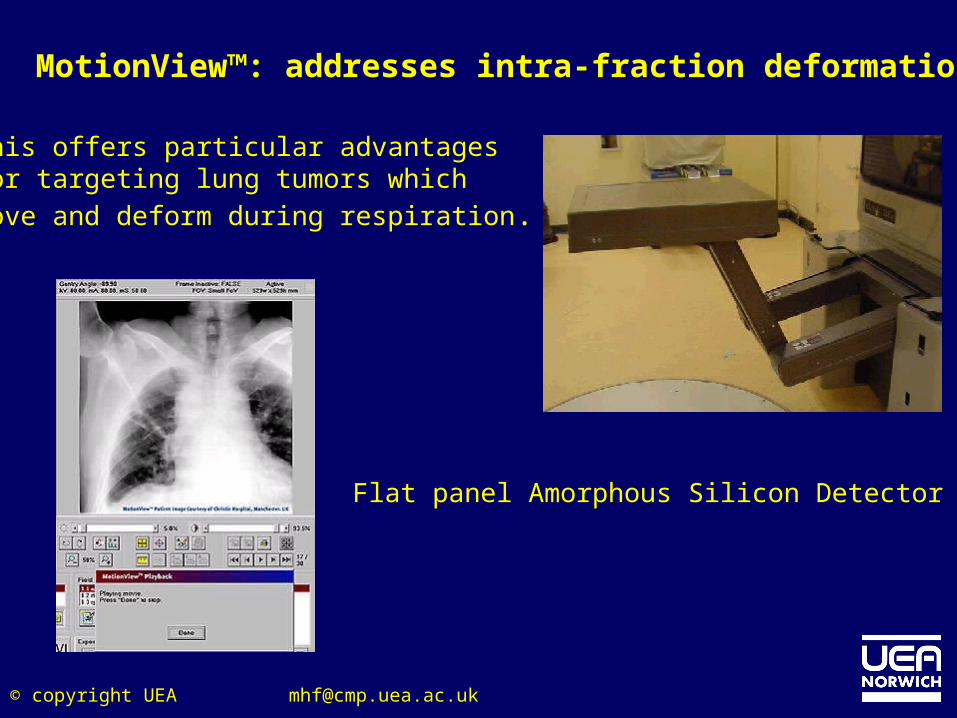
MotionView™: addresses intra-fraction deformation
This offers particular advantages for targeting lung tumors which move and deform during respiration.
Flat panel Amorphous Silicon Detector
© copyright UEA [email protected]
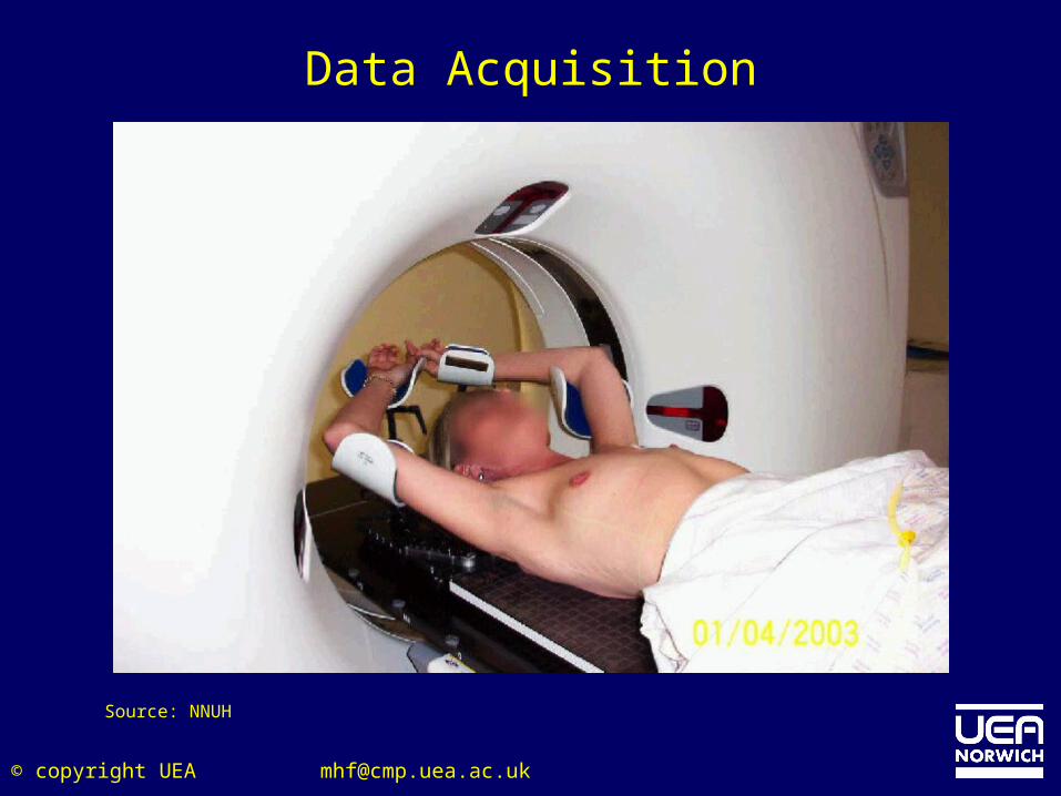
• Traditionally, imaging technology has been used to produce three-dimensional scans of the patient’s anatomy to identify the exact location of the cancer tumor prior to treatment.
• However, difficulty arises when trying to administer the radiation, since cancer tumors are constantly moving within the body • IGRT combines a new form of scanning technology, which allows planar or X-ray Volume Imaging (XVI), with IMRT. This enables physicians to adjust the radiation beam based on the position of the target tumor and critical organs, while the patient is in the treatment position.
Inter-fraction Motion: Current Approaches
Image Guided Radiation Therapy (IGRT)
© copyright UEA [email protected]
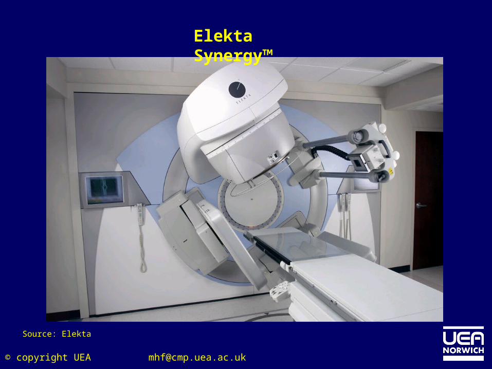
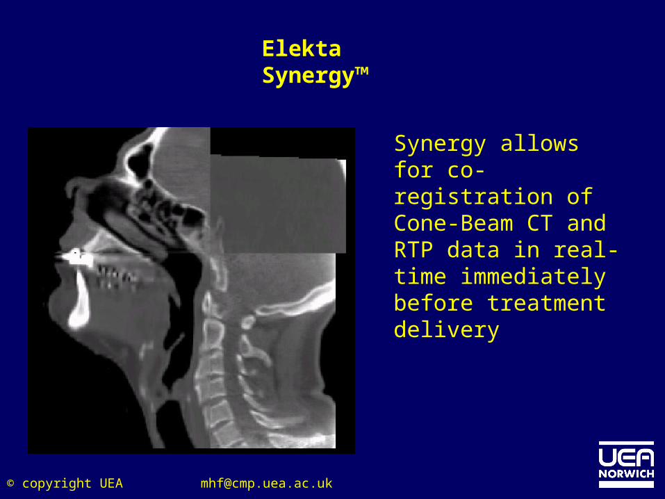
Elekta Synergy™
Synergy allows for co-registration of Cone-Beam CT and RTP data in real-time immediately before treatment delivery
© copyright UEA [email protected]
© copyright UEA [email protected]
“For the first time the cone beam system lets us see what we want to hit with our treatment by giving us a continuous set of detailed 3-D X-ray images of the patient when the patient is lyingdown on the treatment couch. This means we can even move towards better cure rates by safely increasing the doses we deliver in radiotherapy.”(Professor Chris Moore, Consultant Physicist, Christie Hospital)
Available from August 2004
© copyright UEA [email protected]
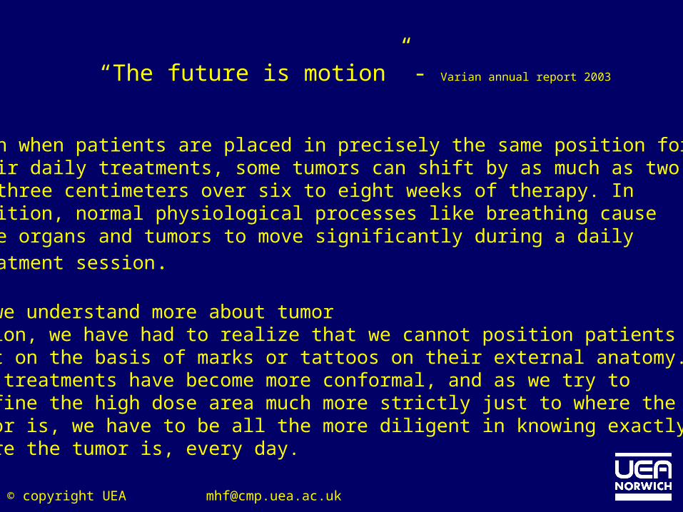
“The future is motion” - Varian annual report 2003
Even when patients are placed in precisely the same position fortheir daily treatments, some tumors can shift by as much as twoto three centimeters over six to eight weeks of therapy. Inaddition, normal physiological processes like breathing causesome organs and tumors to move significantly during a dailytreatment session.
As we understand more about tumormotion, we have had to realize that we cannot position patientsjust on the basis of marks or tattoos on their external anatomy. Asthe treatments have become more conformal, and as we try toconfine the high dose area much more strictly just to where thetumor is, we have to be all the more diligent in knowing exactlywhere the tumor is, every day.
[email protected]© copyright UEA
[email protected]© copyright UEA
© copyright UEA [email protected]
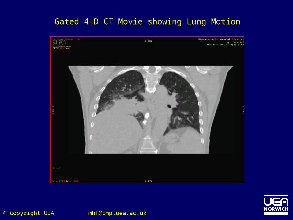
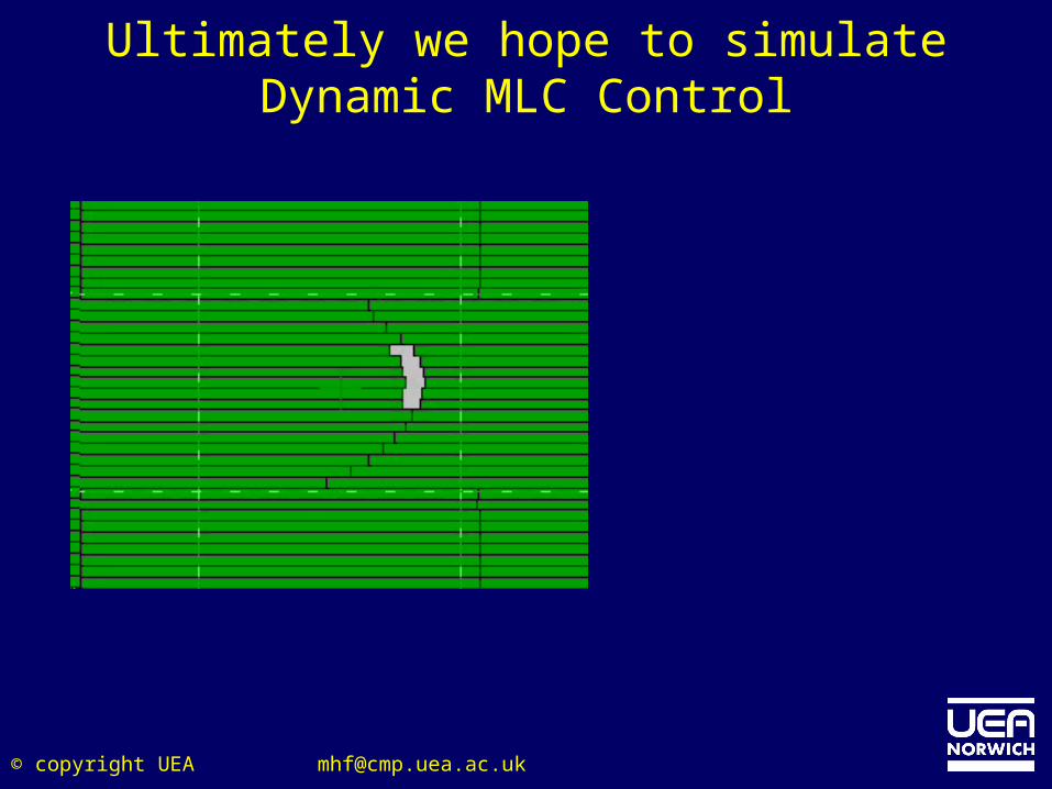
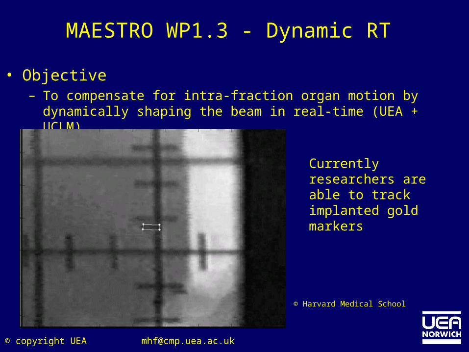
MAESTRO WP1.3 - Dynamic RT
• Objective– To compensate for intra-fraction organ motion by dynamically
shaping the beam in real-time (UEA + UCLM).
Currently researchers are able to track implanted gold markers
© Harvard Medical School
© copyright UEA [email protected]
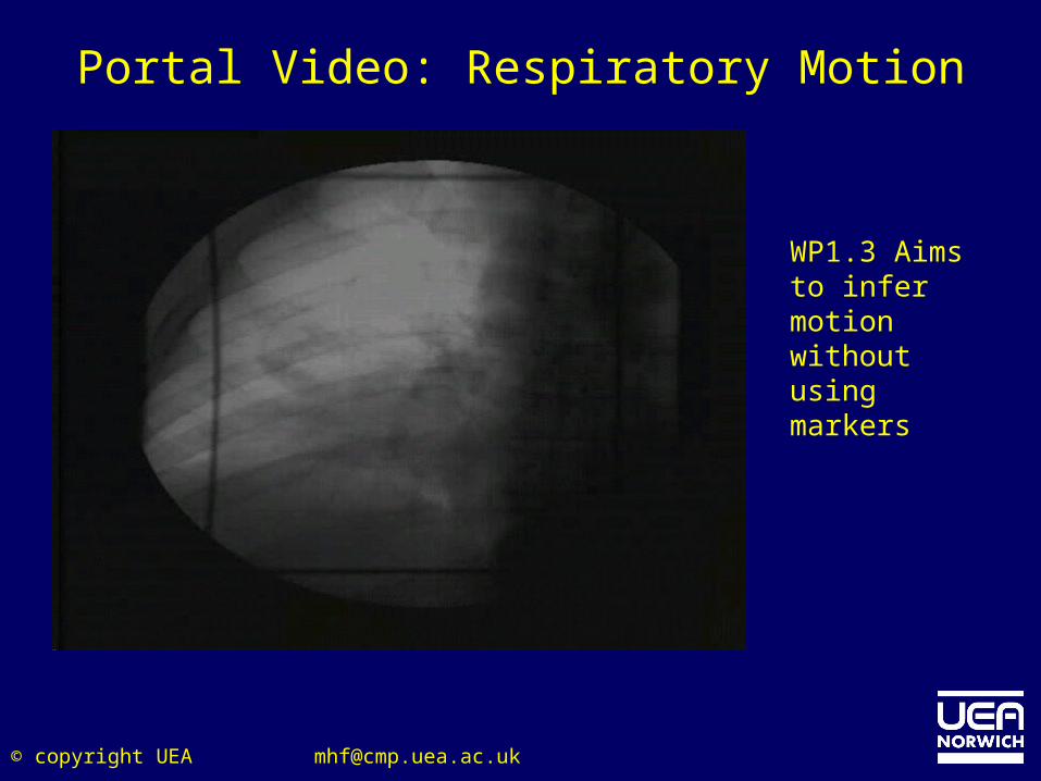
Portal Video: Respiratory Motion
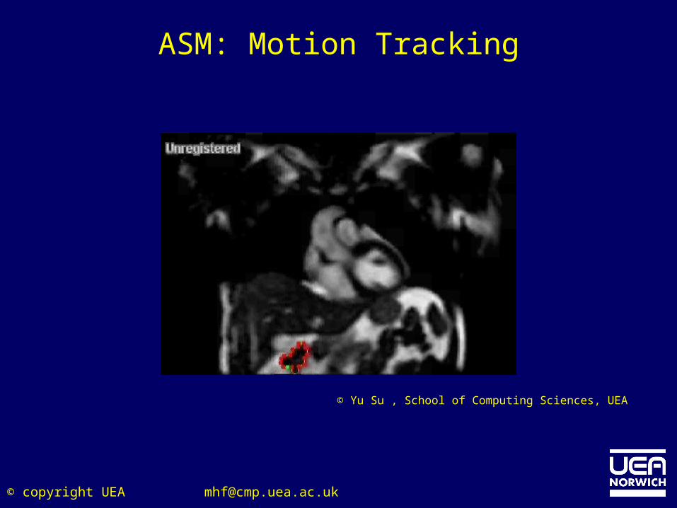
WP1.3 Aims to infer motion without using markers
© copyright UEA [email protected]
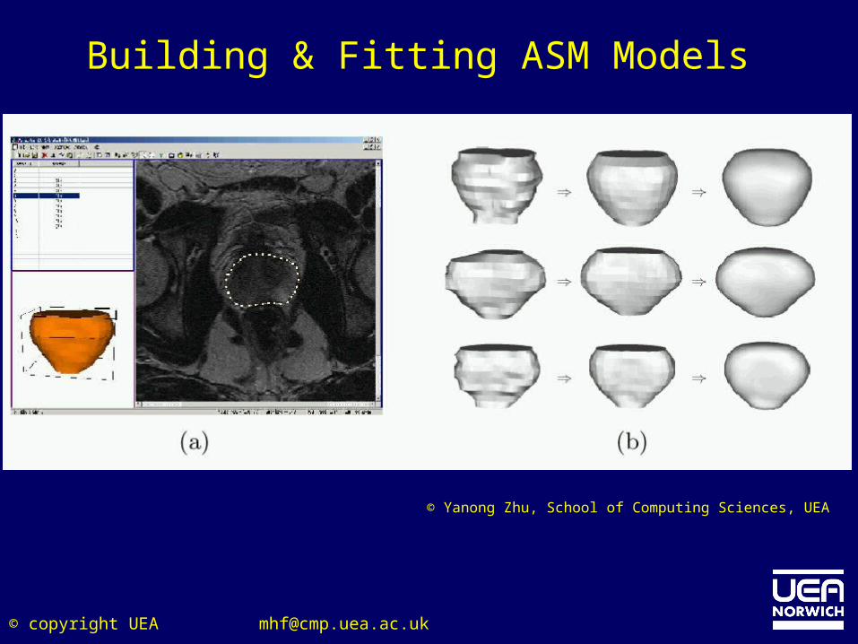
Building & Fitting ASM Models
© Yanong Zhu, School of Computing Sciences, UEA
© copyright UEA [email protected]
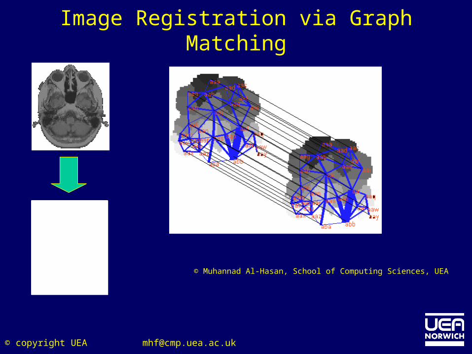
Image Registration via Graph Matching
© Muhannad Al-Hasan, School of Computing Sciences, UEA
© copyright UEA [email protected]
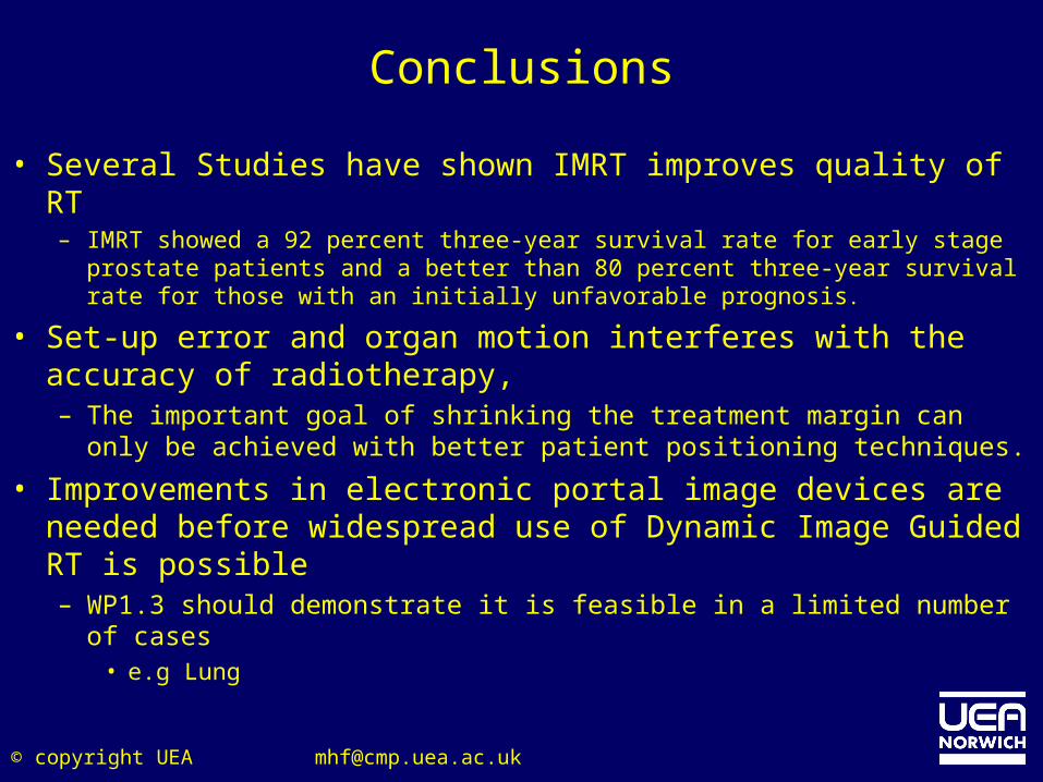
Conclusions
• Several Studies have shown IMRT improves quality of RT– IMRT showed a 92 percent three-year survival rate for early stage
prostate patients and a better than 80 percent three-year survival rate for those with an initially unfavorable prognosis.
• Set-up error and organ motion interferes with the accuracy of radiotherapy,– The important goal of shrinking the treatment margin can only
be achieved with better patient positioning techniques.
• Improvements in electronic portal image devices are needed before widespread use of Dynamic Image Guided RT is possible– WP1.3 should demonstrate it is feasible in a limited number of
cases• e.g Lung
© copyright UEA [email protected]
Acknowledgements
• Alison Vinall - Head of Radiotherapy Physics, NNUH• Dr. Yu Su, Computing Sciences, UEA• Yanong Zhu, Computing Sciences UEA• Muhannad Al-Hasan, Computing Sciences, UEA• MAESTRO